
Direct and Indirect Effects of Alloantibodies Link Neointimal...
16
Direct and Indirect Effects of Alloantibodies Link Neointimal and Medial Remodeling in Graft Arteriosclerosis Olivier Thaunat, Liliane Louedec, Jianping Dai, Florence Bellier, Emilie Groyer, Sandrine Delignat, Anh-Thu Gaston, Giuseppina Caligiuri, Etienne Joly, Didier Plissonnier, Jean-Baptiste Michel, Antonino Nicoletti Objective—Chronic vascular rejection, the main cause of allograft failure, is characterized by the destruction of smooth muscle cells (SMCs) in the media concomitantly with the proliferation of SMCs in the adjacent neointima. We hypothesized that alloantibodies might be responsible for these 2 opposite but coordinated events. Methods and Results—We used the rat aortic interposition model of chronic vascular rejection. During the rejection process, a neointima composed of proliferating SMCs from the recipient developed, whereas the SMCs in the media, all of donor origin, underwent apoptosis. Alloantibody deposition was detected only in the media. Using in vitro cultures experiments, we observed that alloantibody binding to donor SMCs exerts (1) a rapid upregulation of the transcription of growth factors genes, followed by (2) the induction of apoptosis after 24 hours. The transient production of growth factors by donor SMCs in response to the binding of alloantibodies induced the proliferation of recipient SMCs in culture supernatant transfer experiments. Additional data suggest that among the repertoire of alloantibodies, those directed against major histocompatibility complex I might carry the remodeling effect. Conclusions—Our data suggest that during chronic vascular rejection, alloantibody binding to donor medial SMCs is a crucial event that links neointimal and medial remodeling. (Arterioscler Thromb Vasc Biol. 2006;26:2359-2365.) Key Words: anti-MHC antibodies chronic rejection graft arteriosclerosis remodeling smooth muscle cells D uring the last 20 years, the half-life of transplants has remained the same as the result of chronic rejection, which represents the main cause of long-term graft failure. 1 Excluding organ-specific manifestations, the most common histopathological feature is chronic vascular rejection, also known as graft arteriosclerosis, which is estimated to affect more than 40% of recipients within the first 5 years following transplantation. 2 The animal model of aortic transplantation between histo- incompatible rat strains reproduces the main characteristics of allograft arteriosclerosis found in the arteries of rejected human grafts. 3 In previous studies, we have shown that the rejection process of the aortic graft follows 2 consecutive phases. 3 During the first phase (day 0 to 5 posttransplanta- tion), the circulating leukocytes of the recipient target the endothelial cells (ECs) of the graft that expose donor major histocompatibility complex (MHC) molecules. The cellular cytotoxic effectors rapidly destroy the endothelium of the graft but fail to reach the allogenic smooth muscle cells (SMCs) in the media because they are protected by the elastic laminas. The immune system of the recipient then switches from a cellular to a humoral response. 3 During this phase, destruction of SMCs in the media results in the shrinkage of this tunica, whereas the same cell type, ie, SMC, proliferates in the adjacent neointima leading to a widespread and diffuse narrowing of the vascular lumen. Because the events taking place in these 2 tunica are tightly coordinated, 3 we hypothe- sized that a single effector, alloantibodies, may paradoxically trigger both the destruction and the proliferation of the SMCs depending on their localization in the vessel. A seminal work by Russell et al 4 has demonstrated that the passive transfer of sera containing alloantibodies was suffi- cient to promote graft arteriosclerosis. Because MHC mole- cules are highly polymorphic, they are the main targets of the humoral alloimmune response. Several clinical studies have confirmed that the development of alloantibodies directed against donor MHC molecules is associated with an increased risk of transplant arteriosclerosis after transplantation. 5 Interestingly, in addition to their classical role in antigen presentation, MHC molecules also act as signal-transducing molecules, and it has been demonstrated that the binding of antibodies to MHC molecules could modulate the biology of cells. 6 Original received March 9, 2006; final version accepted August 6, 2006. From the Universite ´ Pierre et Marie Curie-Paris6 (O.T., F.B., E.G., S.D., A.-T.G., G.C., A.N.), INSERM UMRS 681, Centre de recherche des Cordeliers, Paris; INSERM U698 and Universite ´ Denis Diderot (L.L., D.J., J.-B.M.), Hopital Xavier Bichat, Paris; INSERM U563 (E.J.), IFR Claude de Preval, Toulouse; and Department of Vascular Surgery (D.P.), Hopital Universitaire de Rouen, France. Correspondence to Dr Olivier Thaunat, INSERM UMRS 681, Institut Biomedical des Cordeliers, 15 rue de l’e ´cole de me ´decine, 75006 Paris, France. E-mail [email protected] © 2006 American Heart Association, Inc. Arterioscler Thromb Vasc Biol. is available at http://www.atvbaha.org DOI: 10.1161/01.ATV.0000241980.09816.ac 2359 by guest on June 26, 2018 http://atvb.ahajournals.org/ Downloaded from by guest on June 26, 2018 http://atvb.ahajournals.org/ Downloaded from by guest on June 26, 2018 http://atvb.ahajournals.org/ Downloaded from by guest on June 26, 2018 http://atvb.ahajournals.org/ Downloaded from by guest on June 26, 2018 http://atvb.ahajournals.org/ Downloaded from by guest on June 26, 2018 http://atvb.ahajournals.org/ Downloaded from by guest on June 26, 2018 http://atvb.ahajournals.org/ Downloaded from by guest on June 26, 2018 http://atvb.ahajournals.org/ Downloaded from by guest on June 26, 2018 http://atvb.ahajournals.org/ Downloaded from by guest on June 26, 2018 http://atvb.ahajournals.org/ Downloaded from
Transcript of Direct and Indirect Effects of Alloantibodies Link Neointimal...
Direct and Indirect Effects of Alloantibodies LinkNeointimal and Medial Remodeling in Graft Arteriosclerosis
Olivier Thaunat, Liliane Louedec, Jianping Dai, Florence Bellier, Emilie Groyer, Sandrine Delignat,Anh-Thu Gaston, Giuseppina Caligiuri, Etienne Joly, Didier Plissonnier,
Jean-Baptiste Michel, Antonino Nicoletti
Objective—Chronic vascular rejection, the main cause of allograft failure, is characterized by the destruction of smoothmuscle cells (SMCs) in the media concomitantly with the proliferation of SMCs in the adjacent neointima. Wehypothesized that alloantibodies might be responsible for these 2 opposite but coordinated events.
Methods and Results—We used the rat aortic interposition model of chronic vascular rejection. During the rejectionprocess, a neointima composed of proliferating SMCs from the recipient developed, whereas the SMCs in the media,all of donor origin, underwent apoptosis. Alloantibody deposition was detected only in the media. Using in vitro culturesexperiments, we observed that alloantibody binding to donor SMCs exerts (1) a rapid upregulation of the transcriptionof growth factors genes, followed by (2) the induction of apoptosis after 24 hours. The transient production of growthfactors by donor SMCs in response to the binding of alloantibodies induced the proliferation of recipient SMCs inculture supernatant transfer experiments. Additional data suggest that among the repertoire of alloantibodies, thosedirected against major histocompatibility complex I might carry the remodeling effect.
Conclusions—Our data suggest that during chronic vascular rejection, alloantibody binding to donor medial SMCs is acrucial event that links neointimal and medial remodeling. (Arterioscler Thromb Vasc Biol. 2006;26:2359-2365.)
Key Words: anti-MHC antibodies � chronic rejection � graft arteriosclerosis � remodeling � smooth muscle cells
During the last 20 years, the half-life of transplants hasremained the same as the result of chronic rejection,
which represents the main cause of long-term graft failure.1
Excluding organ-specific manifestations, the most commonhistopathological feature is chronic vascular rejection, alsoknown as graft arteriosclerosis, which is estimated to affectmore than 40% of recipients within the first 5 years followingtransplantation.2
The animal model of aortic transplantation between histo-incompatible rat strains reproduces the main characteristics ofallograft arteriosclerosis found in the arteries of rejectedhuman grafts.3 In previous studies, we have shown that therejection process of the aortic graft follows 2 consecutivephases.3 During the first phase (day 0 to 5 posttransplanta-tion), the circulating leukocytes of the recipient target theendothelial cells (ECs) of the graft that expose donor majorhistocompatibility complex (MHC) molecules. The cellularcytotoxic effectors rapidly destroy the endothelium of thegraft but fail to reach the allogenic smooth muscle cells(SMCs) in the media because they are protected by the elasticlaminas. The immune system of the recipient then switches
from a cellular to a humoral response.3 During this phase,destruction of SMCs in the media results in the shrinkage ofthis tunica, whereas the same cell type, ie, SMC, proliferatesin the adjacent neointima leading to a widespread and diffusenarrowing of the vascular lumen. Because the events takingplace in these 2 tunica are tightly coordinated,3 we hypothe-sized that a single effector, alloantibodies, may paradoxicallytrigger both the destruction and the proliferation of the SMCsdepending on their localization in the vessel.
A seminal work by Russell et al4 has demonstrated that thepassive transfer of sera containing alloantibodies was suffi-cient to promote graft arteriosclerosis. Because MHC mole-cules are highly polymorphic, they are the main targets of thehumoral alloimmune response. Several clinical studies haveconfirmed that the development of alloantibodies directedagainst donor MHC molecules is associated with an increasedrisk of transplant arteriosclerosis after transplantation.5
Interestingly, in addition to their classical role in antigenpresentation, MHC molecules also act as signal-transducingmolecules, and it has been demonstrated that the binding ofantibodies to MHC molecules could modulate the biology ofcells.6
Original received March 9, 2006; final version accepted August 6, 2006.From the Universite Pierre et Marie Curie-Paris6 (O.T., F.B., E.G., S.D., A.-T.G., G.C., A.N.), INSERM UMRS 681, Centre de recherche des
Cordeliers, Paris; INSERM U698 and Universite Denis Diderot (L.L., D.J., J.-B.M.), Hopital Xavier Bichat, Paris; INSERM U563 (E.J.), IFR Claude dePreval, Toulouse; and Department of Vascular Surgery (D.P.), Hopital Universitaire de Rouen, France.
Correspondence to Dr Olivier Thaunat, INSERM UMRS 681, Institut Biomedical des Cordeliers, 15 rue de l’ecole de medecine, 75006 Paris, France.E-mail [email protected]
© 2006 American Heart Association, Inc.
Arterioscler Thromb Vasc Biol. is available at http://www.atvbaha.org DOI: 10.1161/01.ATV.0000241980.09816.ac
2359
by guest on June 26, 2018http://atvb.ahajournals.org/
Dow
nloaded from
by guest on June 26, 2018http://atvb.ahajournals.org/
Dow
nloaded from
by guest on June 26, 2018http://atvb.ahajournals.org/
Dow
nloaded from
by guest on June 26, 2018http://atvb.ahajournals.org/
Dow
nloaded from
by guest on June 26, 2018http://atvb.ahajournals.org/
Dow
nloaded from
by guest on June 26, 2018http://atvb.ahajournals.org/
Dow
nloaded from
by guest on June 26, 2018http://atvb.ahajournals.org/
Dow
nloaded from
by guest on June 26, 2018http://atvb.ahajournals.org/
Dow
nloaded from
by guest on June 26, 2018http://atvb.ahajournals.org/
Dow
nloaded from
by guest on June 26, 2018http://atvb.ahajournals.org/
Dow
nloaded from
In the present work, we have tested whether alloantibodiesbinding to MHC molecules on graft medial smooth musclecells could promote the events leading to the development ofgraft arteriosclerosis.
Materials and MethodsMurine Experimental ModelAge-matched male Brown–Norway (BN) (RT1n) and Lewis (LEW)(RT1l) rats were obtained from Charles River (l’Arbresie, France).LEW rats were used as recipients and syngeneic donors, BN rats asallogeneic donors. The aorta transplantation was performed aspreviously described.7 (Please see the online data supplement,available at http://atvb.ahajournals.org.)
Serum was collected and aortic grafts were removed after salineperfusion from the Lewis recipients under anesthesia. Fresh aorticsamples were dissected and embedded in paraffin, in Epon resin orin OCT medium (Tissue-Tek, Agar Scientific Ltd, Stansted, UK) andsnap frozen immediately in liquid nitrogen (LN2). All animalexperimentation was undertaken in compliance with the EuropeanCommunity Standards (authorization no. 75-214). Animals werekept under conventional conditions and fed a standard diet.
Titration of Alloantibody in the SerumSerum alloantibodies were titrated by flow cytometry using Lewis(recipient) fibroblasts expressing BN (donor) MHC molecules aspreviously described.7 (Please see the online data supplement.)
Immunohistochemistry AnalysisImmunohistochemistry for rat IgG (biotinylated rabbit anti rat IgG;DAKO, Trappes, France), IgM (purified mouse anti rat IgM;MARM4), rat pan MHC I (purified mouse anti-RT1.A; OX-18),BN-specific MHC I (purified mouse anti-RT1.An; OX-27), and ratpan MHC II (purified mouse anti-RT1.B; OX6) was performed on5-�m thick cryostat cross-sections of aortic allograft.
The biotinylated primary antibody was revealed with avidin-ALEXA 555. The other purified mouse anti-rat antibodies weredetected by a biotinylated anti-mouse antibody that was revealedwith avidin-fluorescein isothiocyanate or avidin-ALEXA 555 or byan anti-mouse IgG1 conjugated with ALEXA 350. The nuclei werecounterstained with 4�,6-diamidino-2-phenylindole (DAPI). Fluores-cence was examined with a microscope equipped with epifluores-cence (Leica Microsystemes SAS; France). Computer-assisted mor-phometry was used to quantify the intensity of IgG and IgMdeposition in the medial (Figure I in the online data supplement).
In Situ Apoptosis DetectionThe terminal deoxynucleotidyl transferase-mediated dUTP nick-endlabeling (TUNEL) technique was used to detect apoptosis on 5-�mthick paraffin-embedded cross-sections as previously described.7(Please see the online data supplement.)
Transmission Electron MicroscopyElectron microscopy analysis was performed on aortic grafts 5 days(5 grafts) and 2 month (5 grafts) after transplantation, as previouslydescribed.7 (Please see the online data supplement.)
In Vitro Experiments
Rat Vascular SMC Isolation and CultureThe technique of tissue processing and culture has been previouslydescribed.8 (Please see the online data supplement.) Cells were usedat passage 3 to 6. After passage 3, SMCs acquire a myofibroblasticphenotype9 and therefore represent an appropriate in vitro model ofneointima.
Generation and Purification of Polyclonal AlloantibodiesPolyclonal anti-BN alloantibodies were generated by the skin graftmethod.10 Briefly, 1-cm2 full-skin patch was removed from a BN ratand grafted orthotopically onto a LEW recipient. Four successive
skin grafts were performed every 14 days to each of the 10 sensitizedLEW rats.
Presence of anti-BN alloantibodies in sensitized LEW rats wasassessed using flow cytometry as previously described.7 (Please seethe online data supplement.)
Serum IgG were purified by chromatography on a proteinG-Sepharose column, followed by immediate size-exclusion chro-matography on a superose-12 column. (Please see the online datasupplement.)
SMC Survival AssayLEW and BN SMCs were plated (104 cells/well) in flat bottom96-well plates and cultured until confluence. After a starvationperiod of 24 hours, 0.1 mg of LEW anti-BN alloantibodies (fromsensitized LEW rats) or control IgG (from naive LEW rats) wasadded to the culture.
Cell survival was evaluated using the tetrazolium salt reduction(MTT) assay11 according to the instructions of the manufacturer(Roche Diagnostics, Indianapolis, Ind). (Please see the online datasupplement.)
Quantification of In Vitro ApoptosisApoptotic cell death of BN or LEW (control) SMCs was measuredafter 8 and 24 hours of culture with LEW anti-BN alloantibodies.The Cell Death Detection sandwich ELISAPLUS kit (Roche Diagnos-tics, Mannheim, Germany), which determines cytoplasmic histone-associated DNA fragments was used according to the instructions ofthe manufacturer.
Production of Growth Factors by SMCs IncubatedWith Alloantibodies
Transfer of Culture SupernatantSupernatants from LEW (control) or BN SMCs stimulated withLEW anti-BN alloantibodies were harvested after 8 or 24 hours ofculture. The supernatants were then added to 5.104 serum-starvedLEW SMCs for which the proliferation was assessed by the MTTassay as described above after 48 hours of culture.
Semiquantitative Polymerase Chain ReactionTranscription levels of 4 growth factors (platelet-derived growthfactor [PDGF]-A and -B, fibroblast growth factor [FGF]-2, insulin-like growth factor [IGF]-1, transforming growth factor [TGF]-�) anda housekeeping gene (GAPDH) were analyzed by semiquantitativeRT-PCR in BN SMC pellet (106 cells/ well; flat bottom 24-wellplates) after 2 or 4 hours of culture with alloantibodies. RT-PCR wasperformed as described in the online data supplement.
Statistical AnalysisData were analyzed using the Statview 5.0 software (AbacusConcept Inc). Statistical significance of results was determined by1-way ANOVA, followed by Fischer’s partial least-squares differ-ence tests and with a Mann–Whitney nonparametric test. Probabilityvalues of less than 0.05 were considered statistically significant.
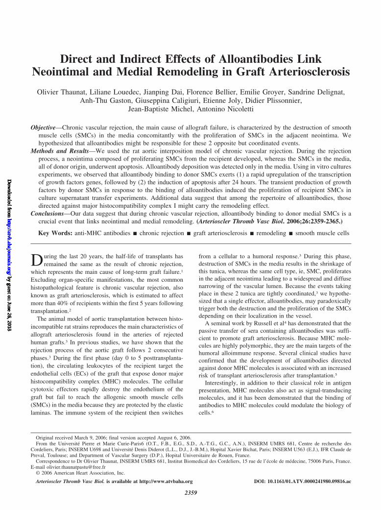
ResultsArterial Wall Is Profoundly Reshaped DuringChronic RejectionMorphological changes of EC and medial SMCs of aorticallograft were tracked during the chronic rejection process.Five days posttransplantation, transmission electron micros-copy analysis showed donor leukocytes bound to the surfaceof graft ECs (Figure 1A). This feature was associated with thenecrosis of ECs and their shedding in the lumen (Figure 1B),indicating that graft ECs are targeted early by the rejectionprocess. In contrast, no leukocyte was detectable beyond theinternal elastic lamina, suggesting that medial SMCs thatdisplayed a normal phenotype were spared by the early
2360 Arterioscler Thromb Vasc Biol. October 2006
by guest on June 26, 2018http://atvb.ahajournals.org/
Dow
nloaded from
cell-mediated injury stage of rejection. Two months post-transplantation, a neointima consisting of SMCs intermingledwith mononuclear cells had developed. Medial SMCs had losttheir normal contractile phenotype and were either apoptotic(Figure 1C) or displayed a synthetic phenotype (Figure 1D).At this time point, we were still unable to detect anyleukocyte infiltration in the media.
TUNEL staining of aortic allografts collected 2 monthsposttransplantation confirmed that medial SMCs underwentmassive apoptosis, whereas SMCs in the adjacent neointimawere spared by this process (Figure 1G and 1H). Proliferationpattern of SMCs analyzed at the same time point byproliferating-cell nuclear antigen staining revealed an in-versed situation, ie, neointimal SMCs but not medial SMCs,were stained (Figure 1E and 1F).
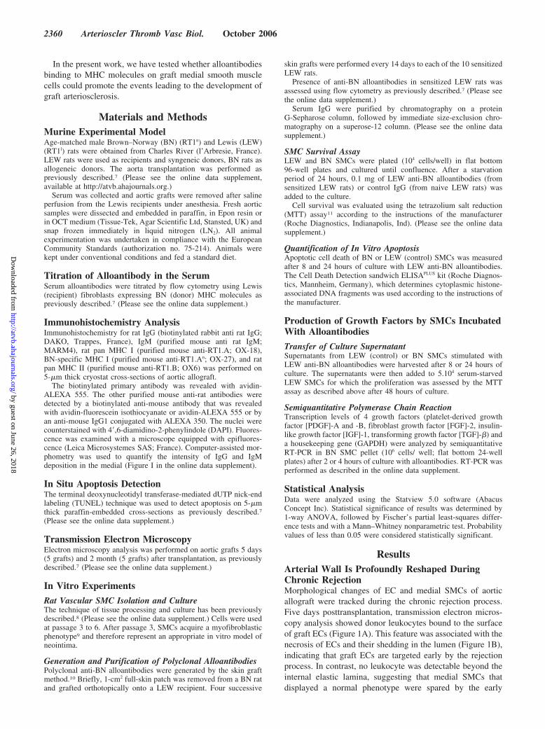
Kinetics of Alloantibody Production andDeposition in the Medial Layer of AllograftsAlloantibody levels in the serum of grafted animals peaked45 days after transplantation and dropped at 90 days (Figure2A). IgM deposits in the media of the graft were detected asearly as 15 days. Fifteen days later (at day 30), IgG were alsopresent and peaked at 45 days (Figure 2A and supplementalFigure I). Alloantibody binding was followed by a decreasedin medial thickness (Figure 2A). These kinetics suggestedthat alloantibodies may be involved in the destruction of themedial SMCs.
The pattern of immunoglobulin deposition within chroni-cally rejected aortic grafts was reproducible. Immunoglobulindeposits were bound to the remaining medial SMCs of thedonor, whereas adjacent neointima was devoid of detectableimmunoglobulin deposition (Figure 2B).
Alloantibody binding to medial SMCs is likely unable totrigger the activation of the complement cascade becauseimmunofluorescence staining for the C5b9 lytic complex wasnegative (Figure 2C).
Chronically Rejected Aortic Allografts AreChimeric for SMCsThe origin of SMCs in aortic grafts 2 months posttransplan-tation was analyzed by immunohistochemistry. We observedthat SMCs from both the media and neointima expressedMHC I molecules, as assessed by the staining with thepan-MHC I OX-18 antibody. However, only medial SMCswere stained with the OX-27 antibody (Figure 2D), which isspecific for BN MHC I molecules. This demonstrates thatchronically rejected aortic grafts are chimeras in which themedial SMCs are from the donor, whereas neointimal SMCsare from recipient origin.
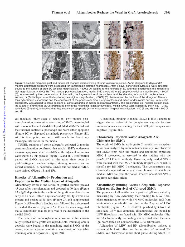
Alloantibody Binding Exerts a Sequential BiphasicEffect on the Survival of Cultured SMCsThe presence of alloantibodies in purified IgG was tested bymeasuring by flow cytometry their binding to LEW fibro-blasts transfected or not with BN MHC molecules. IgG fromnonimmune controls did not bind to the 2 types of LEWfibroblasts (Figure 3A). In contrast, purified IgG from allsensitized LEW rats contained alloantibodies that bound toLEW fibroblasts transfected with BN MHC molecules (Fig-ure 3A). Importantly, no binding was detected when the sameIgG were tested on nontransfected LEW fibroblasts.
Adjunction of LEW anti-BN alloantibodies induced asequential biphasic effect on the survival of cultured BNSMCs. We observed an initial short phase, during which BN
Figure 1. Cellular morphological and functional changes characterizing chronic vascular rejection. Aortic allografts (5 days and 2months posttransplantation) were processed for transmission electron microscopy. After 5 days, donor leukocytes (Lk) were detectedbound to the surface of graft EC (original magnification, �8000) (A), leading to the necrosis of EC and their shedding in the lumen (orig-inal magnification, �3150) (B). Two months posttransplantation, medial SMCs were either (1) apoptotic (original magnification, �8000)(C), as assessed by the condensation of chromatin, the fragmentation of the nucleus, and the shedding of apoptotic bodies (blackarrows); or (2) displayed a synthetic phenotype (original magnification, �8000) (D) characterized by the loss of the elongated filamen-tous cytoplasmic expansions and the enrichment of the perinuclear area in ergastoplasm and mitochondria (white arrows). Immunohis-tochemistry was applied to cross-sections of aortic allografts (2 month posttransplantation). The proliferating-cell nuclear antigen stain-ing (E and F) shows that SMCs proliferated only in the neointima (black arrowheads). Medial SMCs were stained by the in situ TUNELtechnique (G and H), indicating that they underwent apoptosis (white arrowheads). Original magnification, �40 (E and G) and �100 (Fand H).
Thaunat et al Alloantibodies Reshape the Vessel in Graft Arteriosclerosis 2361
by guest on June 26, 2018http://atvb.ahajournals.org/
Dow
nloaded from
SMC survival seems unaffected by alloantibodies, followedby a rapid drop in their survival (Figure 3B). As anticipated,LEW anti-BN alloantibodies had no effect on the survival ofLEW SMCs (Figure 3B).
BN SMCs cultured during 24 hours with alloantibodiesdisplayed an apoptotic phenotype (Figure 3B). Accordingly,after 8 hours, ie, during the initial phase, BN SMCs culturedwith LEW anti-BN alloantibodies displayed a level of cyto-plasmic histone-associated DNA fragments reduced by 30%as compared with BN SMCs alone (Figure 3C). In contrast,when the same measure was performed after 24 hours ofculture, ie, when BN SMCs started to undergo apoptosis, thislevel was increased by 50% (Figure 3C). No difference wasobserved at the 2 time points, when the level of cytoplasmichistone-associated DNA fragments of LEW SMCs culturedalone was compared with that of LEW SMCs cultured withLEW anti-BN alloantibodies (data not shown).
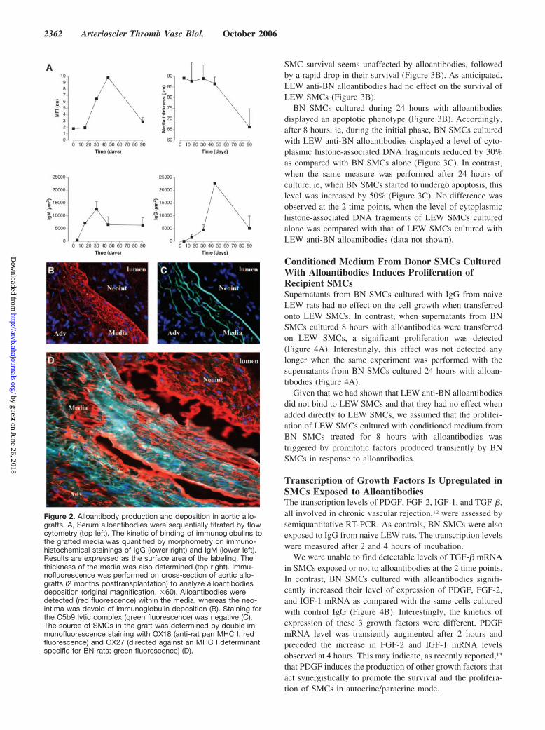
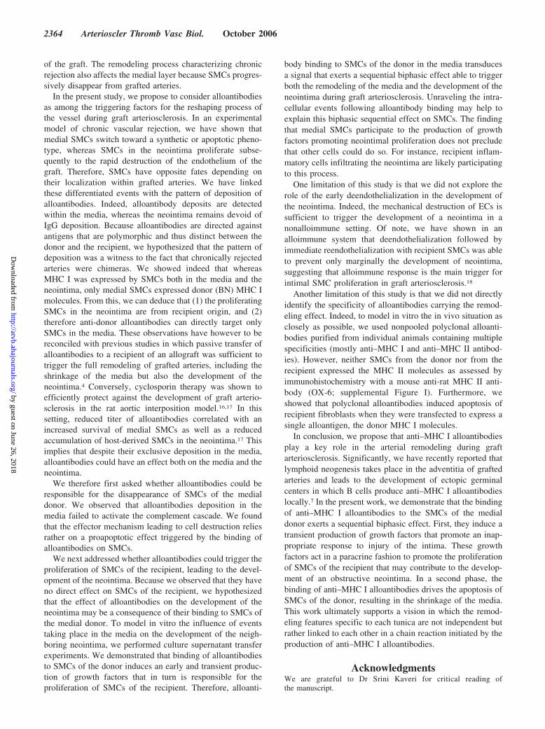
Conditioned Medium From Donor SMCs CulturedWith Alloantibodies Induces Proliferation ofRecipient SMCsSupernatants from BN SMCs cultured with IgG from naiveLEW rats had no effect on the cell growth when transferredonto LEW SMCs. In contrast, when supernatants from BNSMCs cultured 8 hours with alloantibodies were transferredon LEW SMCs, a significant proliferation was detected(Figure 4A). Interestingly, this effect was not detected anylonger when the same experiment was performed with thesupernatants from BN SMCs cultured 24 hours with alloan-tibodies (Figure 4A).
Given that we had shown that LEW anti-BN alloantibodiesdid not bind to LEW SMCs and that they had no effect whenadded directly to LEW SMCs, we assumed that the prolifer-ation of LEW SMCs cultured with conditioned medium fromBN SMCs treated for 8 hours with alloantibodies wastriggered by promitotic factors produced transiently by BNSMCs in response to alloantibodies.
Transcription of Growth Factors Is Upregulated inSMCs Exposed to AlloantibodiesThe transcription levels of PDGF, FGF-2, IGF-1, and TGF-�,all involved in chronic vascular rejection,12 were assessed bysemiquantitative RT-PCR. As controls, BN SMCs were alsoexposed to IgG from naive LEW rats. The transcription levelswere measured after 2 and 4 hours of incubation.
We were unable to find detectable levels of TGF-� mRNAin SMCs exposed or not to alloantibodies at the 2 time points.In contrast, BN SMCs cultured with alloantibodies signifi-cantly increased their level of expression of PDGF, FGF-2,and IGF-1 mRNA as compared with the same cells culturedwith control IgG (Figure 4B). Interestingly, the kinetics ofexpression of these 3 growth factors were different. PDGFmRNA level was transiently augmented after 2 hours andpreceded the increase in FGF-2 and IGF-1 mRNA levelsobserved at 4 hours. This may indicate, as recently reported,13
that PDGF induces the production of other growth factors thatact synergistically to promote the survival and the prolifera-tion of SMCs in autocrine/paracrine mode.
Figure 2. Alloantibody production and deposition in aortic allo-grafts. A, Serum alloantibodies were sequentially titrated by flowcytometry (top left). The kinetic of binding of immunoglobulins tothe grafted media was quantified by morphometry on immuno-histochemical stainings of IgG (lower right) and IgM (lower left).Results are expressed as the surface area of the labeling. Thethickness of the media was also determined (top right). Immu-nofluorescence was performed on cross-section of aortic allo-grafts (2 months posttransplantation) to analyze alloantibodiesdeposition (original magnification, �60). Alloantibodies weredetected (red fluorescence) within the media, whereas the neo-intima was devoid of immunoglobulin deposition (B). Staining forthe C5b9 lytic complex (green fluorescence) was negative (C).The source of SMCs in the graft was determined by double im-munofluorescence staining with OX18 (anti-rat pan MHC I; redfluorescence) and OX27 (directed against an MHC I determinantspecific for BN rats; green fluorescence) (D).
2362 Arterioscler Thromb Vasc Biol. October 2006
by guest on June 26, 2018http://atvb.ahajournals.org/
Dow
nloaded from
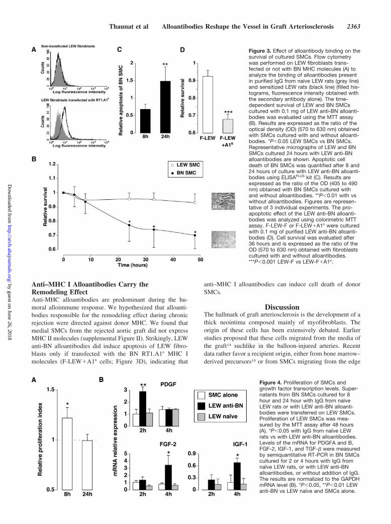
Anti–MHC I Alloantibodies Carry theRemodeling EffectAnti-MHC alloantibodies are predominant during the hu-moral alloimmune response. We hypothesized that alloanti-bodies responsible for the remodeling effect during chronicrejection were directed against donor MHC. We found thatmedial SMCs from the rejected aortic graft did not expressMHC II molecules (supplemental Figure II). Strikingly, LEWanti-BN alloantibodies did induce apoptosis of LEW fibro-blasts only if transfected with the BN RT1.A1n MHC Imolecules (F-LEW�A1n cells; Figure 3D), indicating that
anti–MHC I alloantibodies can induce cell death of donorSMCs.
DiscussionThe hallmark of graft arteriosclerosis is the development of athick neointima composed mainly of myofibroblasts. Theorigin of these cells has been extensively debated. Earlierstudies proposed that these cells migrated from the media ofthe graft14 suchlike in the balloon-injured arteries. Recentdata rather favor a recipient origin, either from bone marrow–derived precursors15 or from SMCs migrating from the edge
Figure 3. Effect of alloantibody binding on thesurvival of cultured SMCs. Flow cytometrywas performed on LEW fibroblasts trans-fected or not with BN MHC molecules (A) toanalyze the binding of alloantibodies presentin purified IgG from naive LEW rats (gray line)and sensitized LEW rats (black line) (filled his-tograms, fluorescence intensity obtained withthe secondary antibody alone). The time-dependent survival of LEW and BN SMCscultured with 0.1 mg of LEW anti-BN alloanti-bodies was evaluated using the MTT assay(B). Results are expressed as the ratio of theoptical density (OD) (570 to 630 nm) obtainedwith SMCs cultured with and without alloanti-bodies. *P�0.05 LEW SMCs vs BN SMCs.Representative micrographs of LEW and BNSMCs cultured 24 hours with LEW anti-BNalloantibodies are shown. Apoptotic celldeath of BN SMCs was quantified after 8 and24 hours of culture with LEW anti-BN alloanti-bodies using ELISAPLUS kit (C). Results areexpressed as the ratio of the OD (405 to 490nm) obtained with BN SMCs cultured withand without alloantibodies. **P�0.01 with vswithout alloantibodies. Figures are represen-tative of 3 individual experiments. The pro-apoptotic effect of the LEW anti-BN alloanti-bodies was analyzed using colorimetric MTTassay. F-LEW-F or F-LEW�A1n were culturedwith 0.1 mg of purified LEW anti-BN alloanti-bodies (D). Cell survival was evaluated after36 hours and is expressed as the ratio of theOD (570 to 630 nm) obtained with fibroblastscultured with and without alloantibodies.***P�0.001 LEW-F vs LEW-F�A1n.
Figure 4. Proliferation of SMCs andgrowth factor transcription levels. Super-natants from BN SMCs cultured for 8hour and 24 hour with IgG from naıveLEW rats or with LEW anti-BN alloanti-bodies were transferred on LEW SMCs.Proliferation of LEW SMCs was mea-sured by the MTT assay after 48 hours(A). *P�0.05 with IgG from naıve LEWrats vs with LEW anti-BN alloantibodies.Levels of the mRNA for PDGFA and B,FGF-2, IGF-1, and TGF-� were measuredby semiquantitative RT-PCR in BN SMCscultured for 2 or 4 hours with IgG fromnaıve LEW rats, or with LEW anti-BNalloantibodies, or without addition of IgG.The results are normalized to the GAPDHmRNA level (B). *P�0.05, **P�0.01 LEWanti-BN vs LEW naıve and SMCs alone.
Thaunat et al Alloantibodies Reshape the Vessel in Graft Arteriosclerosis 2363
by guest on June 26, 2018http://atvb.ahajournals.org/
Dow
nloaded from
of the graft. The remodeling process characterizing chronicrejection also affects the medial layer because SMCs progres-sively disappear from grafted arteries.
In the present study, we propose to consider alloantibodiesas among the triggering factors for the reshaping process ofthe vessel during graft arteriosclerosis. In an experimentalmodel of chronic vascular rejection, we have shown thatmedial SMCs switch toward a synthetic or apoptotic pheno-type, whereas SMCs in the neointima proliferate subse-quently to the rapid destruction of the endothelium of thegraft. Therefore, SMCs have opposite fates depending ontheir localization within grafted arteries. We have linkedthese differentiated events with the pattern of deposition ofalloantibodies. Indeed, alloantibody deposits are detectedwithin the media, whereas the neointima remains devoid ofIgG deposition. Because alloantibodies are directed againstantigens that are polymorphic and thus distinct between thedonor and the recipient, we hypothesized that the pattern ofdeposition was a witness to the fact that chronically rejectedarteries were chimeras. We showed indeed that whereasMHC I was expressed by SMCs both in the media and theneointima, only medial SMCs expressed donor (BN) MHC Imolecules. From this, we can deduce that (1) the proliferatingSMCs in the neointima are from recipient origin, and (2)therefore anti-donor alloantibodies can directly target onlySMCs in the media. These observations have however to bereconciled with previous studies in which passive transfer ofalloantibodies to a recipient of an allograft was sufficient totrigger the full remodeling of grafted arteries, including theshrinkage of the media but also the development of theneointima.4 Conversely, cyclosporin therapy was shown toefficiently protect against the development of graft arterio-sclerosis in the rat aortic interposition model.16,17 In thissetting, reduced titer of alloantibodies correlated with anincreased survival of medial SMCs as well as a reducedaccumulation of host-derived SMCs in the neointima.17 Thisimplies that despite their exclusive deposition in the media,alloantibodies could have an effect both on the media and theneointima.
We therefore first asked whether alloantibodies could beresponsible for the disappearance of SMCs of the medialdonor. We observed that alloantibodies deposition in themedia failed to activate the complement cascade. We foundthat the effector mechanism leading to cell destruction reliesrather on a proapoptotic effect triggered by the binding ofalloantibodies on SMCs.
We next addressed whether alloantibodies could trigger theproliferation of SMCs of the recipient, leading to the devel-opment of the neointima. Because we observed that they haveno direct effect on SMCs of the recipient, we hypothesizedthat the effect of alloantibodies on the development of theneointima may be a consequence of their binding to SMCs ofthe medial donor. To model in vitro the influence of eventstaking place in the media on the development of the neigh-boring neointima, we performed culture supernatant transferexperiments. We demonstrated that binding of alloantibodiesto SMCs of the donor induces an early and transient produc-tion of growth factors that in turn is responsible for theproliferation of SMCs of the recipient. Therefore, alloanti-
body binding to SMCs of the donor in the media transducesa signal that exerts a sequential biphasic effect able to triggerboth the remodeling of the media and the development of theneointima during graft arteriosclerosis. Unraveling the intra-cellular events following alloantibody binding may help toexplain this biphasic sequential effect on SMCs. The findingthat medial SMCs participate to the production of growthfactors promoting neointimal proliferation does not precludethat other cells could do so. For instance, recipient inflam-matory cells infiltrating the neointima are likely participatingto this process.
One limitation of this study is that we did not explore therole of the early deendothelialization in the development ofthe neointima. Indeed, the mechanical destruction of ECs issufficient to trigger the development of a neointima in anonalloimmune setting. Of note, we have shown in analloimmune system that deendothelialization followed byimmediate reendothelialization with recipient SMCs was ableto prevent only marginally the development of neointima,suggesting that alloimmune response is the main trigger forintimal SMC proliferation in graft arteriosclerosis.18
Another limitation of this study is that we did not directlyidentify the specificity of alloantibodies carrying the remod-eling effect. Indeed, to model in vitro the in vivo situation asclosely as possible, we used nonpooled polyclonal alloanti-bodies purified from individual animals containing multiplespecificities (mostly anti–MHC I and anti–MHC II antibod-ies). However, neither SMCs from the donor nor from therecipient expressed the MHC II molecules as assessed byimmunohistochemistry with a mouse anti-rat MHC II anti-body (OX-6; supplemental Figure I). Furthermore, weshowed that polyclonal alloantibodies induced apoptosis ofrecipient fibroblasts when they were transfected to express asingle alloantigen, the donor MHC I molecules.
In conclusion, we propose that anti–MHC I alloantibodiesplay a key role in the arterial remodeling during graftarteriosclerosis. Significantly, we have recently reported thatlymphoid neogenesis takes place in the adventitia of graftedarteries and leads to the development of ectopic germinalcenters in which B cells produce anti–MHC I alloantibodieslocally.7 In the present work, we demonstrate that the bindingof anti–MHC I alloantibodies to the SMCs of the medialdonor exerts a sequential biphasic effect. First, they induce atransient production of growth factors that promote an inap-propriate response to injury of the intima. These growthfactors act in a paracrine fashion to promote the proliferationof SMCs of the recipient that may contribute to the develop-ment of an obstructive neointima. In a second phase, thebinding of anti–MHC I alloantibodies drives the apoptosis ofSMCs of the donor, resulting in the shrinkage of the media.This work ultimately supports a vision in which the remod-eling features specific to each tunica are not independent butrather linked to each other in a chain reaction initiated by theproduction of anti–MHC I alloantibodies.
AcknowledgmentsWe are grateful to Dr Srini Kaveri for critical reading ofthe manuscript.
2364 Arterioscler Thromb Vasc Biol. October 2006
by guest on June 26, 2018http://atvb.ahajournals.org/
Dow
nloaded from
Disclosure(s)None.
References1. Sayegh MH, Carpenter CB. Transplantation 50 years later–progress,
challenges, and promises. N Engl J Med. 2004;351:2761–2766.2. Libby P, Pober JS. Chronic rejection. Immunity. 2001;14:387–397.3. Plissonnier D, Nochy D, Poncet P, Mandet C, Hinglais N, Bariety J,
Michel JB. Sequential immunological targeting of chronic experimentalarterial allograft. Transplantation. 1995;60:414–424.
4. Russell PS, Chase CM, Winn HJ, Colvin RB. Coronary atherosclerosis intransplanted mouse hearts. II. Importance of humoral immunity.J Immunol. 1994;152:5135–5141.
5. Reed EF, Hong B, Ho E, Harris PE, Weinberger J, Suciu-Foca N.Monitoring of soluble HLA alloantigens and anti-HLA antibodies iden-tifies heart allograft recipients at risk of transplant-associated coronaryartery disease. Transplantation. 1996;61:566–572.
6. Gilliland LK, Norris NA, Grosmaire LS, Ferrone S, Gladstone P,Ledbetter JA. Signal transduction in lymphocyte activation throughcrosslinking of HLA class I molecules. Hum Immunol. 1989;25:269–289.
7. Thaunat O, Field AC, Dai J, Louedec L, Patey N, Bloch MF, Mandet C,Belair MF, Bruneval P, Meilhac O, Bellon B, Joly E, Michel JB, NicolettiA. Lymphoid neogenesis in chronic rejection: evidence for a localhumoral alloimmune response. Proc Natl Acad Sci U S A. 2005;102:14723–14728.
8. Battle T, Arnal JF, Challah M, Michel JB. Selective isolation of rat aorticwall layers and their cell types in culture—application to convertingenzyme activity measurement. Tissue Cell. 1994;26:943–955.
9. Hultgardh-Nilsson A, Lovdahl C, Blomgren K, Kallin B, Thyberg J.Expression of phenotype- and proliferation-related genes in rat aorticsmooth muscle cells in primary culture. Cardiovasc Res. 1997;34:418–430.
10. Forbes RD, Kuramochi T, Guttmann RD, Klassen J, Knaack J. A con-trolled sequential morphologic study of hyperacute cardiac allograftrejection in the rat. Lab Invest. 1975;33:280–288.
11. Hussain RF, Nouri AM, Oliver RT. A new approach for measurement ofcytotoxicity using colorimetric assay. J Immunol Methods. 1993;160:89–96.
12. Vuillemin T, Lantz O, Legendre C, Meduri G, LaRue JR, Kriaa F, HiesseC, Senik A, Kreis H, Charpentier B. Potential involvement of growthfactors in the proliferation of arteriolar smooth muscle cells duringchronic graft rejection. Adv Nephrol Necker Hosp. 1996;25:39–49.
13. Millette E, Rauch BH, Defawe O, Kenagy RD, Daum G, Clowes AW.Platelet-derived growth factor-BB-induced human smooth muscle cellproliferation depends on basic FGF release and FGFR-1 activation. CircRes. 2005;96:172–179.
14. Hayry P, Paavonen T, Mennander A, Ustinov J, Raisanen A, LemstromK. Pathophysiology of allograft arteriosclerosis. Transplant Proc. 1993;25:2070.
15. Shimizu K, Sugiyama S, Aikawa M, Fukumoto Y, Rabkin E, Libby P,Mitchell RN. Host bone-marrow cells are a source of donor intimalsmooth- muscle-like cells in murine aortic transplant arteriopathy. NatMed. 2001;7:738–741.
16. Plissonnier D, Amichot G, Lecagneux J, Duriez M, Gentric D, Michel JB.Additive and synergistic effects of a low-molecular-weight, heparin-likemolecule and low doses of cyclosporin in preventing arterial graftrejection in rats. Arterioscler Thromb. 1993;13:112–119.
17. Religa P, Bojakowski K, Bojakowska M, Gaciong Z, Thyberg J, Hedin U.Allogenic immune response promotes the accumulation of host-derivedsmooth muscle cells in transplant arteriosclerosis. Cardiovasc Res. 2005;65:535–545.
18. Gomes D, Louedec L, Plissonnier D, Dauge MC, Henin D, Osborne-Pellegrin M, Michel JB. Endoluminal smooth muscle cell seeding limitsintimal hyperplasia. J Vasc Surg. 2001;34:707–715.
Thaunat et al Alloantibodies Reshape the Vessel in Graft Arteriosclerosis 2365
by guest on June 26, 2018http://atvb.ahajournals.org/
Dow
nloaded from

Michel and Antonino NicolettiJean-BaptisteDelignat, Anh-Thu Gaston, Giuseppina Caligiuri, Etienne Joly, Didier Plissonnier,
Olivier Thaunat, Liliane Louedec, Jianping Dai, Florence Bellier, Emilie Groyer, SandrineGraft Arteriosclerosis
Direct and Indirect Effects of Alloantibodies Link Neointimal and Medial Remodeling in
Print ISSN: 1079-5642. Online ISSN: 1524-4636 Copyright © 2006 American Heart Association, Inc. All rights reserved.
Greenville Avenue, Dallas, TX 75231is published by the American Heart Association, 7272Arteriosclerosis, Thrombosis, and Vascular Biology
doi: 10.1161/01.ATV.0000241980.09816.ac2006;
2006;26:2359-2365; originally published online August 17,Arterioscler Thromb Vasc Biol.
http://atvb.ahajournals.org/content/26/10/2359World Wide Web at:
The online version of this article, along with updated information and services, is located on the
http://atvb.ahajournals.org/content/suppl/2006/08/21/01.ATV.0000241980.09816.ac.DC1Data Supplement (unedited) at:
http://atvb.ahajournals.org//subscriptions/
at: is onlineArteriosclerosis, Thrombosis, and Vascular Biology Information about subscribing to Subscriptions:
http://www.lww.com/reprints
Information about reprints can be found online at: Reprints:
document. Question and AnswerPermissions and Rightspage under Services. Further information about this process is available in the
which permission is being requested is located, click Request Permissions in the middle column of the WebCopyright Clearance Center, not the Editorial Office. Once the online version of the published article for
can be obtained via RightsLink, a service of theArteriosclerosis, Thrombosis, and Vascular Biologyin Requests for permissions to reproduce figures, tables, or portions of articles originally publishedPermissions:
by guest on June 26, 2018http://atvb.ahajournals.org/
Dow
nloaded from
Thaunat et al - 1
SUPPLEMENTAL DATA – ONLINE
MATERIALS AND METHODS
Aorta transplantation
All animal experimentation was undertaken in compliance with the European Community
standards (authorization n° 75-214).
Rats were anesthetized with 50mg/Kg of pentobarbital injected intraperitoneally. Two
animals were operated simultaneously, one as the donor of aortic graft and the other as
the recipient, with the aid of an operating microscope. A 1 cm long segment of the donor
abdominal aorta was excised, perfused with normal saline and small collateral arteries
that originated from the graft were ligated. The donor aorta was transplanted in orthotopic
position by end-to-end anastomosis in the recipient aorta below the renal arteries and
above the iliac bifurcation. We used the 38 animals that survived to the surgical
procedure. No immunosuppressive or anticoagulant treatment was used. Aortic grafts
were removed at indicated time-points from the Lewis recipients under anesthesia and
perfused with saline. Fresh aortic samples were dissected and embedded in paraffin or in
OCT medium (Tissue-Tek, Agar Scientific Ltd, UK) and snap frozen immediately in
liquid nitrogen.
Titration of alloantibody in the serum
Two hundreds µl of serum harvested at various time points from grafted animals were
incubated with 200,000 Lewis (recipient) fibroblasts expressing Brown-Norway (donor)
Thaunat et al - 2
MHC molecules cells (LEW-F+A1n) for 30 min at 4°C. The binding of antibodies on the
cell surface was then determined with a FITC-conjugated rabbit anti-rat Ig secondary
antibody (PARIS; France), by measuring the mean fluorescence intensity (MFI) with a
LSR II flow cytometer (BD Biosciences; France).
Morphometry
Labeling of IgG and IgM was quantified from images acquired on the same field within
the green, red, and blue channels of a fluorescent microscope. Image analysis was
performed using a customized program (Leica Qwin). Elastin autofluorescence (green
channel) was substracted from the fluorescence detected on the blue channel while no
elastin signal was detected in the red channel in the acquisition condition used for image
capture. The external and internal elastic lamina were used to delineate the media and to
calculate its surface area and its thickness.
In-situ apoptosis detection
In situ 3-end labeling of apoptotic DNA was performed using Apotag Peroxidase Kits
(Oncor, USA) following the manufacturer's instructions. Briefly, after dewaxing,
rehydration, and blocking of endogenous peroxidase, 3-hydroxy-DNA strand breaks in
permeabilized tissue sections were enzymatically labeled with digoxigenin-nucleotides,
by using terminal deoxynucleotidyl transferase (TdT). The labeled DNA was then bound
with antidigoxigenin antibody peroxidase conjugate, and the peroxidase color reaction
was developed with a 3-amino-9-ethyl carbazole substrate.
Thaunat et al - 3
Transmission electron microscopy
Aortic graft specimens were dissected, immediately fixed in 2.5 % glutaraldehyde in PBS
buffer, post-fixed in 4% osmium tetroxide and embedded in Epon resin. Ultrathin
sections (50 to 80 nm thick) were prepared, stained with lead citrate and uranyl acetate
and observed with a Zeiss EMI transmission electron microscope.
Rat vascular SMC isolation and culture
Rat (LEW and BN) aortic SMC were isolated after microdissection of the adventitia from
the media and the endothelium. The media was cut into 1 mm-long rings and subjected to
an enzymatic digestion with a mixture of collagenase and elastase (Gibco/Invitrogen).
Cell suspensions were plated and cultured to confluence at 37°C, 5% CO2, in Dulbecco's
modified Eagle's medium (Gibco) supplemented with 10% fetal calf serum (FCS),
100U/ml penicillin and 0.1 mg/ml streptomycin (complete medium).
Polyclonal alloantibodies generation and purification
Ten µl of serum collected 10 days after the 4th skin graft were incubated 20 min at 4°C
with 2.5 105 LEW fibroblasts expressing or not BN MHC molecules. The binding of
antibodies on the cell surface was detected with a FITC-conjugated rabbit anti-rat Ig
secondary antibody (PARIS; France) by measuring the mean fluorescence intensity in a
LSRII flow cytometer (Becton Dickinson Biosciences).
A two-step procedure for IgG purification was applied to avoid the contamination of the
IgG preparation with other serum proteins. Fourteen days after the 4th skin graft, 10 ml of
blood was collected from LEW rats sensitized or not (source for control IgG) and serum
Thaunat et al - 4
was prepared. Serum IgG were purified by chromatography on a protein G-Sepharose
column, followed by immediate size-exclusion chromatography on a superose-12
column. The IgG-containing fraction was dialyzed against PBS at 4°C for 24h, adjusted
to a concentration of 10 mg/ml, aliquoted and stored at –80°C until analysis.
SMC survival assay
LEW and BN SMC were plated (104 cells/well) in flat bottom 96-well plates and cultured
at 37°C, 5% CO2, in 100 µl of complete medium (DMEM supplemented with 10% fetal
calf serum, 100U/ml penicillin, and 0.1 mg/ml streptomycin) until confluence. Complete
medium was then replaced with 90 µl of starvation medium (DMEM supplemented with
2% bovine serum albumin, 100U/ml penicillin, and 0.1 mg/ml streptomycin). After a
starvation period of 24h, 10 µl (0.1 mg) of LEW anti-BN alloantibodies (from sensitized
LEW rats) or control IgG (from naïve LEW rats) were added to the culture.
Cell survival was evaluated using the tetrazolium salt reduction (MTT) assay according
to the manufacturer’s instructions (Roche Diagnostics; IN, USA; please see
http://atvb.ahajournals.org). The MTT assay (Roche Diagnostics; IN, USA) is based on
the colorimetric measurement of a formazan derivative of 3-(4,5-dimethylthiazol-2-yl)-
2,5-diphenyltetrazolium bromide that is formed by live cells. Briefly, 10 µl of MTT
labeling solution (5 mg/ml) were added to the culture at various time-points after addition
of antibodies (4h, 8h, 24h, 36h, and 48h). After an incubation period of 4h at 37°C and
5% CO2, formazan crystals were dissolved by adding 100µl of the solubilization solution
(10%SDS in 0.01 M HCl) and the absorbance was measured using a scanning multiwell
spectrophotometer at dual wavelength 570–630 nm.
Thaunat et al - 5
All measurements were performed in duplicates and the experiments were repeated 3
times.
Semi-quantitative PCR
Messenger RNAs were extracted with 500 µl of TRIzol (Invitrogen, France) after 2 or 4 h
of culture. Reverse transcription reaction was performed with 2 µg of RNA using Oligo
dT, random hexamers, and Moloney Murine Leukemia Virus reverse transcriptase
(Invitrogen). All oligonucleotide primers were synthesized by Eurogentec (France)
(Table I). The PCR reaction was performed in capillaries with 5µl cDNA, 10µl 2x SYBR
Green master mix (Qiagen, France) and 0.5 µM of sense and antisense primer. Real time
PCR was performed in LightCycler (Roche Diagnostics, France) starting with 15 min of
pre-incubation at 95°C followed by 55 cycles of 10 sec at 95 °C, 20 sec at annealing
temperature depending on primers used (Table 1), and 30 sec at 72 °C. Amplification of
expected PCR product was confirmed by agarose gel electrophoresis (2%) and melting
curve analysis.
The threshold cycle (Ct) was determined with the maximum-second-derivative function
of the LightCycler software. Results are expressed as the ratio between the Ct of the gene
tested and the Ct of GAPDH.
Thaunat et al - 6
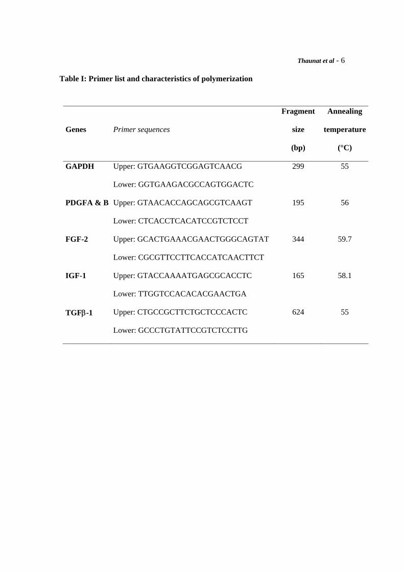
Table I: Primer list and characteristics of polymerization
Genes
Primer sequences
Fragment
size
(bp)
Annealing
temperature
(°C)
GAPDH Upper: GTGAAGGTCGGAGTCAACG
Lower: GGTGAAGACGCCAGTGGACTC
299 55
PDGFA & B Upper: GTAACACCAGCAGCGTCAAGT
Lower: CTCACCTCACATCCGTCTCCT
195 56
FGF-2 Upper: GCACTGAAACGAACTGGGCAGTAT
Lower: CGCGTTCCTTCACCATCAACTTCT
344 59.7
IGF-1 Upper: GTACCAAAATGAGCGCACCTC
Lower: TTGGTCCACACACGAACTGA
165 58.1
TGFβ-1 Upper: CTGCCGCTTCTGCTCCCACTC
Lower: GCCCTGTATTCCGTCTCCTTG
624 55
Thaunat et al - 7
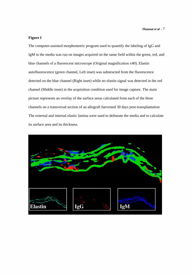
Figure I
The computer-assisted morphometric program used to quantify the labeling of IgG and
IgM in the media was run on images acquired on the same field within the green, red, and
blue channels of a fluorescent microscope (Original magnification x40). Elastin
autofluorescence (green channel, Left inset) was substracted from the fluorescence
detected on the blue channel (Right inset) while no elastin signal was detected in the red
channel (Middle inset) in the acquisition condition used for image capture. The main
picture represents an overlay of the surface areas calculated from each of the three
channels on a transversal section of an allograft harvested 30 days post-transplantation
The external and internal elastic lamina were used to delineate the media and to calculate
its surface area and its thickness.
Thaunat et al - 8
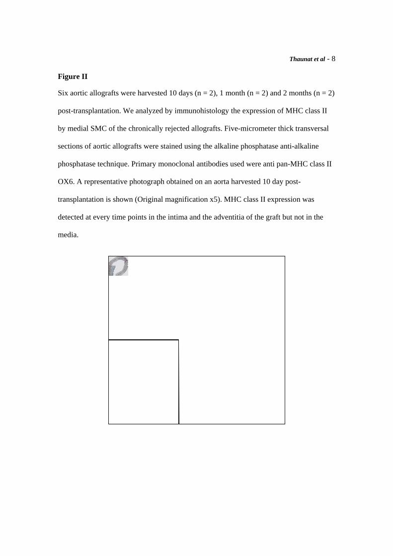
Figure II
Six aortic allografts were harvested 10 days (n = 2), 1 month (n = 2) and 2 months (n = 2)
post-transplantation. We analyzed by immunohistology the expression of MHC class II
by medial SMC of the chronically rejected allografts. Five-micrometer thick transversal
sections of aortic allografts were stained using the alkaline phosphatase anti-alkaline
phosphatase technique. Primary monoclonal antibodies used were anti pan-MHC class II
OX6. A representative photograph obtained on an aorta harvested 10 day post-
transplantation is shown (Original magnification x5). MHC class II expression was
detected at every time points in the intima and the adventitia of the graft but not in the
media.