Digital Image Processing in Veterinary Medicine Digital ... _X... · Import of any external...
23
Digital image processing in veterinary medicine 510(k) application has been cleared by the FDA No. K070618 0482 FDA dicom vet PACS Digital Image Processing in Veterinary Medicine R Digital image processing with dicom vet PACS ®
-
Upload
duongxuyen -
Category
Documents
-
view
213 -
download
0
Transcript of Digital Image Processing in Veterinary Medicine Digital ... _X... · Import of any external...

Digital image processingin veterinary medicine
510(k) application has been clearedby the FDA No. K0706180482 FDA
dicom vetPACSDigital Image Processing
in Veterinary Medicine
R
Dig
ital im
ag
e p
roce
ssin
g w
ith
dic
om
vet
PAC
S®


dicom vetPACSDigital Image Processing
in Veterinary Medicine
R
High qualityimage processingin veterinarymedicine
dicom vet
dicom vet
dicom vet
dicom vet
dicom vet
PACS
PACS
PACS
PACS
PACS
®
®
®
®
®
will make your dream of a paperless veterinary practice
come true. All images as well as any type of document (e.g. diagnostic
reports, records of healing processes, faxes) are stored by
in a digital patient file and can be accessed immediately with a simple
mouse click.
Well designed archiving and backup solutions guarantee fast access to
all data while observing the highest security standards in accordance with
the internationally recognised guidelines for human medicine. In addition,
can be integrated easily with all the popular practice
management systems.
The software includes acquisition, diagnosis, transfer
and archiving of image material. Since it has been designed and developed
in close cooperation with practising vets, you will find it easy to operate
and well suited to daily diagnosis.
Boasting more than 6,000 installed workstations locally and abroad (as of
January 2011), the system has proven itself many times over.
handles simple image processing requirements as brilliantly as complex
radiological networks.
Dig
ital im
ag
e p
roce
ssin
g w
ith
dic
om
vet
PAC
S®

of at one glancedicomPACS®
Benefits
2
Full diagnostic software for all workstations in your practice
(no 'light' versions)
User friendly and clearly arranged structure, minimal training
requirements and short familiarisation period
Individual adjustment of the user interface to your field of specialisation
and individual requirements
Flexible allocation of shortcut keys for many functions to allow fast
work without a mouse
Parallel processing (e.g. option to continue working during a CD
burning process)
Permanent online availability of all images and data in the network – no
need to store old images on CDs
“Perfect memory” – re-opening of images with all previous markings
and settings incl. zoom and orientation
Parallel diagnostic evaluation of several patients made possible by
opening any number of programme windows without loss of speed -
depending on the size of the working memory
Import of any external documents such as doctors' letters, faxes or X-ray
images – no additional module required
Installation with Windows, UNIX, LINUX or Apple Macintosh
Optimal data security, speed and compatibility by using standardised
SQL database technology
All images and documents are filed in the international DICOM
standard at all times

3
Boasting more than 6,000 installed workstations locally and abroad
(as of January 2011), the system has proven itself many times over.

4
Perfect integration of all imaging devices into your existing computer network
is an important condition for a smooth and reliable workflow. Apart from X-ray
systems, a wide range of devices including ultrasound, endoscopy, fluoroscopy,
CT and MRI systems as well as digital cameras can be connected.
In addition to imaging devices, you can also store documents such as faxes
and letters digitally in the digital patient file of your practice management system.
With , all data is immediately available and can even be easily
forwarded on request.
Continuous documentation and access to data over a period of many years
is only possible as a result of optimal integration of all information on your
animal patient.
dicom vetPACS®
Services offeredIntegrated modules and tools
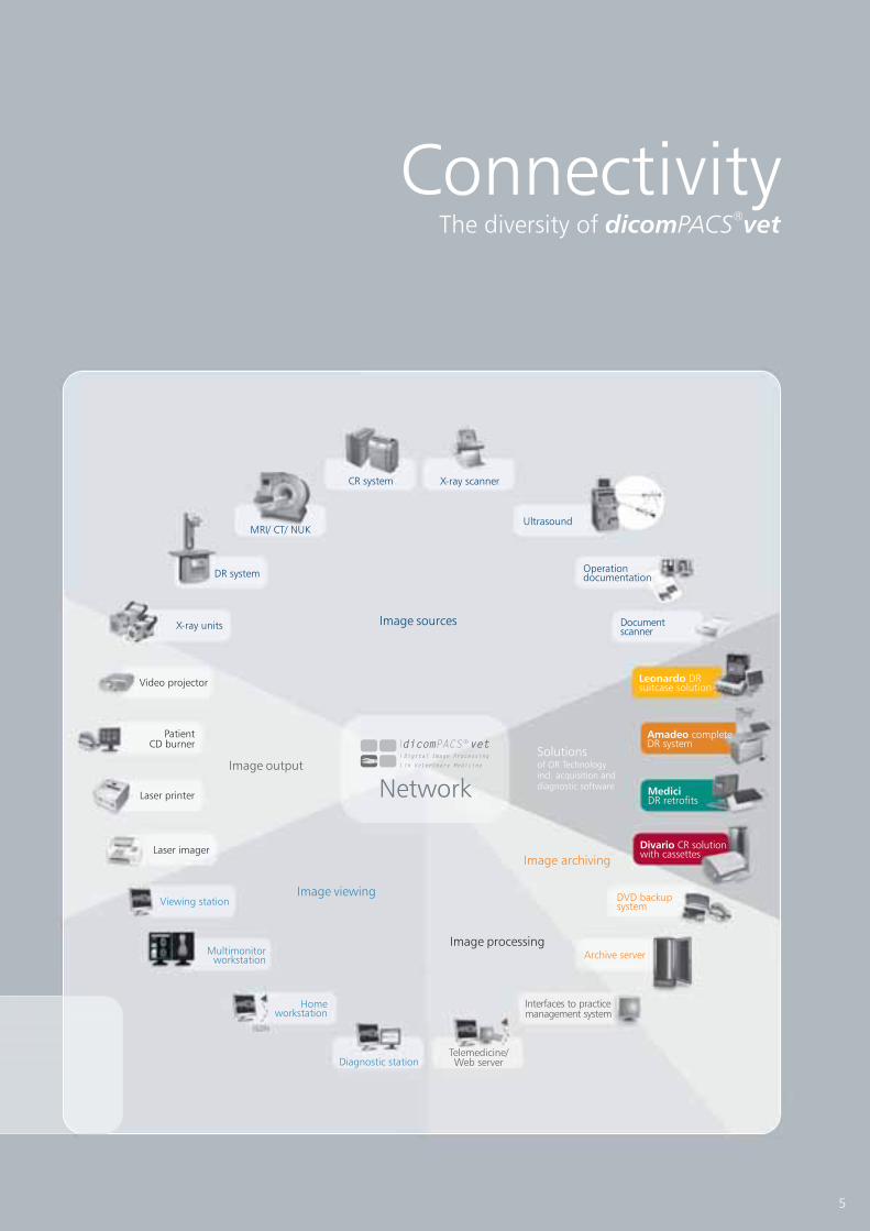
ConnectivityThe diversity of dicom vetPACS
®
Image sources
Image output
Image viewing
Image processing
Image archiving
Multimonitorworkstation
Homeworkstation
Telemedicine/Web server
Interfaces to practicemanagement system
Archive server
DVD backupsystem
MRI/ CT/ NUKUltrasound
Diagnostic station
X-ray scanner
Laser printer
Laser imager
Viewing station
X-ray units
PatientCD burner
DR system
CR system
Video projector
Operationdocumentation
Documentscanner
Amadeo completeDR system
MediciDR retrofits
Divario CR solutionwith cassettes
Solutionsof OR Technologyincl. acquisition anddiagnostic softwareNetwork
5
dicom vetPACSDigital Image Processing
in Veterinary Medicine
R
Leonardo DRsuitcase solution

6
Prosthesis documentation
Report module for X-ray services relating to equine prepurchase
examinations
Special filter for the optimization of bones and soft parts
TPLO measuring function
TTA measuring tool
HD measuring technique for dogs
Measuring the distraction index
Buchanan‘s Vertebral Heart Score
- enables the user to plan operations
with digital prosthesis templates by one or more manufacturers
see page 18/ 19
[currently only available for Germany] - enables the quick
compilation of reports by automatically assembling X-ray images. It follows the
“X-ray guideline” by the German organisations “Gesellschaft für Pferdemedizin
e.V.” (non-profit organization for equine medicine) and “Bundestierärzte-
kammer e.V.” (Federal association of veterinarians).
see page 10/ 13
-
details of interest may be made visible by means of special filter magnifiers
(Tibial Plateau Leveling Osteotomy) - it serves to
theoretically optimize the existing slope of the tibial plateau in domestic dogs
see page 18/ 19
(Tibial Tuberosity Advancement) - the TTA measuring
technique is used to apply the translated length measurements at the
tuberositas tibiae in dogs
see page 18/ 19
- provides a
special tool to guarantee very fast and reliable determination of the Norberg
angle, including documentation
see page 18/ 19
- This measuring tool serves to
determine the displacement of the femoral head from the joint socket
of the hip joint in dogs
see page 18/ 19
- This annotation is a simple and
reliable method to determine the size of the heart - it has been desiged
specifically for cats and dogs
see page 18/ 19
dicom vetPACS®
dicom vetPACS®
features
Value

7
Statistics Module
Video Modules
Web Server
Processing of CT and MRI series
Hanging protocols
Telemedicine
Special solution for multiple archives
- enables freely configurable analysis of the
complete database
- enable standard and non-standard video signals
to be recorded as single images and video sequences
- enables image distribution within the hospital or to
referring doctors via the internet and guarantees very fast image
accessibility in original quality (DICOM)
see page 14/ 15
- includes
professional tools such as MPR and MIP to evaluate cross
section series
see page 16/ 17
dicom vetPACS®
8
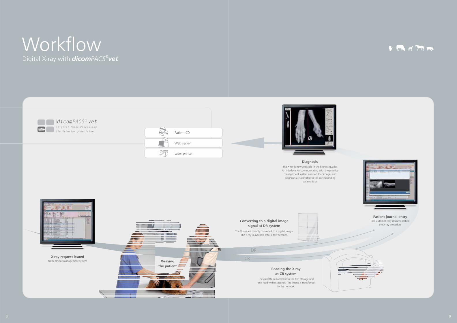
Digital X-ray with dicom vetPACS®
Workflow
X-ray request issuedfrom patient management system
Patient CD
Laser printer
Web server
X-raying
the patient
dicom vetPACSDigital Image Processing
in Veterinary Medicine
R
Reading the X-ray
at CR system
Converting to a digital image
signal at DR system
The X-rays are directly converted to a digital image.
The X-ray is available after a few seconds.
The cassette is inserted into the film storage unit
and read within seconds. The image is transferred
to the network.
DR
Diagnosis
The X-ray is now available in the highest quality.
An interface for communicating with the practice
management system ensured that images and
diagnosis are allovated to the corresponding
patient data.
Patient journal entryincl. automatically documentation
the X-ray procedure
9
CR

for X-ray services relating to equine prepurchase examinations
Report module
Presale and prepurchase examinations for horses are always particularly
challenging for veterinarians. Such specialised examinations must be
carried out swiftly yet very meticulously
documented very well, in great detail and extremely consistently.
After all, the owner of the animal justifiably expects optimal service when it
comes to undertaking the examination and presenting the results in a professional
and comprehensible fashion. Since administrative work is bothersome yet vital for
veterinarians, too, we have developed a report module specifically for X-ray services
relating to prepurchase examinations in cooperation with renowned specialists.
This module enables the quick compilation of reports by automatically assembling
X-ray images. It follows the “X-ray guideline” by the German organisations
“Gesellschaft für Pferdemedizin e.V.” (non-profit organization for equine medicine)
and “Bundestierärztekammer e.V.” (Federal association of veterinarians).
[currently only available for Germany]
Benefits:
10
Time-saving:
Easy to follow:
Reports with images:
Safe:
Presentation:
The prepurchase
examination report module allows very
fast and professional preparation of
reports on the prepurchase examination,
including perfect layout and
documentation in .
guarantees complete
implementation of the wording and
the structure of diagnostic reports in
accordance with the “Guideline for
pre-purchase X-ray examinations” by the
German “Gesellschaft für Pferdemedizin
e.V.“ and “Bundestierärztekammer e.V.”.
The texts can be easily edited and
included in the report to be compiled.
The required X-ray images, including
all modifications such as zoom,
measurements and annotations,
and are automatically added to the
respective diagnosis (e.g. fetlock joint)
for documentation in the prepurchase
report. The layout is automatically
compiled (page breaks, image
positioning etc.)
The complete report (WYSIWYG principle)
is automatically stored with the X-ray
images. Of course, these reports are also
available for patient CDs. This
guarantees that images and reports
are always kept together.
is a professional
marketing tool for referring doctors.
dicom vet
dicom vet
dicom vet
dicom vet
PACS
PACS
PACS
PACS
®
®
®
®

11
X-ray protocol
11
X-ray protocol
Left front
Left front

6. Report preparation
1. Call up the examination
Workflow for a prepurchase horse examination
12
4. Display diagnostic report options
2. Start report module
5. Diagnostic report/ evaluation
3. Allocate the projectionCurrent image assign
left front
right front
left hind
left right
Foot
Foot
Foot
Knee
Knee
Foot
Knee
Knee
Back
back
Projection add or delete
Assignment cancel
Current image assign
left front
right front
left hind
left right
Foot
Foot
Foot
Knee
Knee
Foot
Knee
Knee
Back
back
Projection add or delete
Assignment cancel
Create report Continue

By means of a single click, the completed prepurchase examination
report is stored together with the examination images in ,
where it can be accessed again in the original version at any point in time.
dicom vetPACS®
13

Making images available via the internet (or intranet) is an increasingly
important daily requirement in the medical practice. One purpose is the
distribution of images or other documents in a larger clinic. Equally
important is the integration of external referring parties (hospitals,
medical practices) or home workstations.
The intention is always the same: faster, cheaper downloading of
archived images and diagnoses via the internet or intranet (also via slow
internet connections), in diagnostic quality if possible, to every clinic or
internet PC. The use of older PCs, thin clients or terminal servers must
also be made possible.
To accommodate as many requests as possible from the medical
practice and hospital, we have developed our Web Server
in cooperation with respected doctors.
dicom vetPACS®
Web serverfor internal image sharing and external
distribution to referring doctors
Web preview Web viewer with equine prepurchase
examination images
14

Advantages of using web server
Image processing tools
(for example magnifyer)
15
Installation:
Hardware requirements:
Web server advantages at a glance
The web viewer does not require any extra investment;
Internet Explorer is all that is needed. Some minor configuration
changes regarding security settings may be necessary in
Internet Explorer.
The web server always displays the latest version of user
interface as updates take place automatically when needed.
For larger hospital installations there is the option to install several
web servers (scaling), e.g. in order to have a separate
web server available for each division.
Workstations need only a minor increase in RAM, processing
speed and possibly an update of the operating system.
A narrow band network e.g. GPRS, ISDN, internet, fixed
lines etc. is sufficient to ensure adequate download speed for images.
The installation of the web server may also be located on
the archive server itself, which means that a separate PC is
not required.
Images are available in their original quality (DICOM)
High speed availability of images even in slow networks/ with slow
internet thanks to special streaming technology and compression
procedures no compromises between image quality and
loading speed.
Automatic email notification
Extensive research options
Simple, intuitive operation
No installations costs
Extensive configuration of user and access rights
Automatic updates
The use of several web servers is possible
Only modest hardware requirements
Thin clients, terminal servers, mobile computing and
WLAN can be used
Central administration eliminates need for support to the clients
Multilingual

Administration and diagnostic
Cross section
dicom vetPACS®
includes all the necessary tools for the professional
diagnostic evaluation of slices, such as CT or MRI. Functions like hanging
protocols, cine loop, manual scrolling through series, and the visualisation of
current and delimiting outlines allow the user to work fast and professionally.
The MPR (multi-planar reconstruction) and MIP (maximum intensity
projection) functions offer the doctor increased options.
16

17
Röntgenprotokoll
Digital X-ray images have the advantage that exact measurements
can be taken at the monitor and the image quality can be improved
by a number of manipulations. now offers some
special functions.
This module allows the user to plan and document an operation.
After activating this function, the active image is displayed in its original
film-identical size. The prosthesis template is displayed in the image as an
annotation, or the existing prosthesis template films are overlaid on
the monitor.
The TTA measuring technique is used to apply the translated length
measurements at the tuberositas tibiae in dogs.
provides a special tool to guarantee very fast and
reliable determination of the Norberg angle, including documentation.
One click suffices to insert all relevant lines and angles into the image,
where they can then be positioned as required.
It serves to theoretically optimize the existing slope of the tibial plateau
in domestic dogs.
This measuring tool serves to determine the displacement of the femoral
head from the joint socket of the hip joint in dogs.
This annotation is a simple and reliable method to determine the size of
the heart. It has been designed specifically for cats and dogs. The height and
width of the heart are put into relation to the individual animal's vertebral
body width. Therefore, racial distinctions are brought to bear on the
examinations results.
dicom vet
dicom vet
PACS
PACS
®
®
Pre-operative planning with the prosthesis
documentation module
TTA (Tibial Tuberosity Advancement) measuring tool
HD measuring technique for dogs
TPLO (Tibial Plateau Leveling Osteotomy) measuring tool
Measuring the distraction index
Buchanan's Vertebral Heart Score
FeaturesSpecial functions for digital
X-ray imaging
18
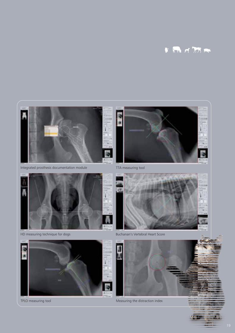
TTA measuring tool
Measuring the distraction index
Integrated prosthesis documentation module
Buchanan‘s Vertebral Heart Score
19
HD measuring technique for dogs
TPLO measuring tool
Seamless integration with the administration software
Integration
Only an optimal interface guarantees perfect networking of all systems
such as the integration of the image archive with the specific administration
software. With a single mouse click you have immediate access to patient
data to prepare an imaging request or to load archive images.
is well designed, sophisticated and flexible. It can be
integrated easily with any veterinary administration programme.
dicom vetPACS®
20
Basic functions
The way we configure an interface
in detail so that everything works perfectly
depends on the existing administration
system. We would like to present three
examples of frequently used functions
below:
Patient data is made
directly available from the index card for
the examination instruction for e.g. a
digital X-ray, MRI or similar.
The examination instruction
is allocated to the digital patient register
of that particular patient, where it is
stored and archived.
The archived data -
X-rays or documents - is called up directly
from the patient register. You can proceed
as you wish, directly choose a particular
image or document, or decide on a
specific selection, e.g. the last week's
exposures or just the ultrasound
exposures of a patient.
However you want to proceed, you
can be sure that it will work, because
we have already successfully
integrated into
many administration systems.Example 1:
Example 2:
Example 3:
dicom vetPACS®
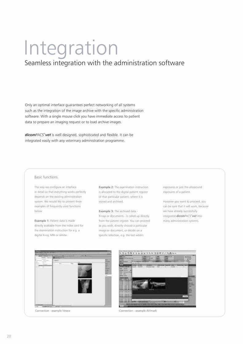
Connection - example Vetera Connection - example AVImark

Overview - products of OR Technology
Vet portfolio
Image management (PACS) - comprisesacquisition, processing, diagnosis, transfer andarchiving of image material
X-ray acquisition software [only for OEMs] -acquisition and diagnostic software for X-ray imagesfrom flat panels or CR systems
DR suitcases - compact suitcases solutions formobile and portable X-ray
DR retrofits - expanding sets for existingX-ray systems (available for U-arm and wallstand and table systems)
Conventional X-ray equipment and accessories -the latest technology for conventional X-ray systems
Complete DR systems - digital X-ray systemsincl. stand, table, generator, flat panel etc.
CR solutions - CR systems for digitalX-ray with cassettes
ConventionalX-ray EquipmentX-ray Systems for the Future
DX-RdicomPACS R
X-ray Acquisition Software
Amadeo DR Systemsvet
X-ray Systems for the Future
with dicomPACS DX-R Software®
Divario CR Systemsvet
CR Systems for the Future
with dicomPACS DX-R Software®
Leonardo DR Systemsvet
Portable X-ray Systems for the Future
with dicomPACS DX-R Software®
Medici DR Systemsvet
DR Retrofit Systems for the Future
with dicomPACS DX-R Software®
dicom vetPACSDigital Image Processing
in Veterinary Medicine
R

Ver
sion 0
01_01_2011
Equine Clinic Burg Müggenhausen,
Dr Thomas Weinberger, Germany:
Tierklinik Elversberg -Veterinary
emergency and trauma hospital,
Dr Alexander Pack and
Dr Karl Scherer, Germany:
Sudenhof horse clinic, Germany,
Dr Eberhard Mettenleiter, specialist
for horses and horse surgery:
Veterinary clinic for small
animals in Wasbek, Germany,
Dr Johannes Frahm:
Veterenary Clinic Boekelberg,
Dr Arnd Stelljes, Mönchengladbach,
Germany
“The pre-purchase examination module
of proves to be a great
help with X-ray examinations and
diagnostic...
This tool provides an enormous
reduction in work and liability risks.
Moreover, it is great that the current
X-ray Code of Practice (German X-ray
guide 07) is provided... Overall, we wish
to repeat that we are very happy with
the investment and the changeover
to ."
„HD X-ray imaging now only takes a few
minutes. Thanks to the integrated tools
for image optimisation, it is no problem
if images are not optimally exposed as a
result of animals not holding still. In that
case, I simply correct the images...
The introduction of digital X-ray
imaging has been a great success for
us. An important aspect is that we can
already say that the system will soon
have paid for itself.“
„...The system is easy and well
structured. Other users confirmed that
the system runs almost problem-free.
The first demonstration was impressive
and we could integrate the software
we use in our practice. The price-
performance ratio is excellent compared
to other systems...
We are using the web server to
comfortably transmit X-ray images to
other veterinarians, customers etc. The
whole process is relatively fast – despite
our slow Internet connection. Fast
transmission of high-quality X-ray images
and easy operation were important
criteria for us. The web server by
OR Technology fully meets these
requirements. We have also received
feedback from customers who love
the fact that the web server is efficient
and easy to operate.“
„...Now the image is available within
seconds, it can be evaluated immediately
and viewed in every treatment room. All
the images are automatically archived
and can be called up quickly and directly
from my “Vetera” patient system if they
are required again at a later stage. In an
archive system with paper envelopes it is
often the very thing you are looking for
that has gone missing. The more X-ray
images you take the more frequent these
situations are. Now all this won't happen
any more...“
„...The image processing software
by OR Technology
could be linked directly and easily to our
patient management system „Vetera“.
Now all X-rays, MRI and ultrasound
images can be easily displayed and
processed at our six workstations in the
treatment rooms. The direct interface of
the image processing software with our
patient management system offers us
preview images from all linked
modalities in our Vetera program.
Images can now also be directly called
up from the card index...“
dicom vet
dicom vet
dicom vet
PACS
PACS
PACS
You can find the extensive references
here www.or-technology.com -->
Veterinary medicine --> References
®
®
®
on from
veterinary hospital and practice
dicom vetPACS®
StatementsDr Th. Weinberger
(Oehm und Rehbein GmbH)
18057 Rostock, Germany, Waldemarstr. 20 g/h
Tel. +49 (0)381 - 20 36 126, Fax +49 (0)381 - 20 36 111
www.or-technology.com, [email protected]
OR Technology
[Stamp of distribiution partner]
Info-Hotline: +49 (0)381 - 20 36 126
R TechnologyDigital X-ray and
Imaging Solutions
O