Differential effects of PI3K and dual PI3K/mTOR...
29
Differential effects of PI3K and dual PI3K/mTOR inhibition in rat prolactin- secreting pituitary tumors Marie Chanal 1,2 , Pascale Chevallier 1,2 , Véronique Raverot 3 , Guillaume Fonteneau 1 , Kristin Lucia 4 , Jose Luis Monteserin Garcia 4 , Alexa Rachwan 1 , Emmanuel Jouanneau 1,2,5 , Jacqueline Trouillas 2,6,7 , Jérôme Honnorat 2,7 , Carole Auger 1,2 , Marily Theodoropoulou 4* , Gérald Raverot 1,2,8 * 1 INSERM U1052; CNRS UMR5286; Cancer Research Center of Lyon, Lyon, F-69372, France 2 Université Lyon 1, Lyon, F-69372, France 3 Centre de biologie Est, Groupement Hospitalier Est, Hospices Civils de Lyon, F-69677, Bron, France 4 Max-Planck Institute for Psychiatry, 80804 Munich, Germany 5 Service de Neurochirurgie, Groupement Hospitalier Est, Hospices Civils de Lyon, Bron, F- 69677, France 6 Centre de Pathologie Est, Groupement Hospitalier Est, Hospices Civils de Lyon, Bron, F- 69677, France 7 INSERM U1028; CNRS UMR5292; Lyon Neuroscience Research Center, Neuro-oncology & Neuro-inflammation team, Lyon, F-69372, France 8 Fédération d’Endocrinologie, Groupement Hospitalier Est, Hospices Civils de Lyon, Bron, F-69677, France * equal contribution Running title: Pituitary tumor treatment with PI3K/mTOR inhibitors Keywords: Pituitary tumor, pituitary adenoma, targeted therapy, PI3K/mTOR inhibitors Note: on September 9, 2018. © 2016 American Association for Cancer Research. mct.aacrjournals.org Downloaded from Author manuscripts have been peer reviewed and accepted for publication but have not yet been edited. Author Manuscript Published OnlineFirst on March 16, 2016; DOI: 10.1158/1535-7163.MCT-15-0891
Transcript of Differential effects of PI3K and dual PI3K/mTOR...

Differential effects of PI3K and dual PI3K/mTOR inhibition in rat prolactin-
secreting pituitary tumors
Marie Chanal1,2, Pascale Chevallier1,2, Véronique Raverot3, Guillaume Fonteneau1, Kristin
Lucia4, Jose Luis Monteserin Garcia4, Alexa Rachwan1, Emmanuel Jouanneau1,2,5,
Jacqueline Trouillas2,6,7, Jérôme Honnorat2,7, Carole Auger1,2, Marily Theodoropoulou4*,
Gérald Raverot1,2,8*
1 INSERM U1052; CNRS UMR5286; Cancer Research Center of Lyon, Lyon, F-69372,
France
2 Université Lyon 1, Lyon, F-69372, France
3 Centre de biologie Est, Groupement Hospitalier Est, Hospices Civils de Lyon, F-69677,
Bron, France
4 Max-Planck Institute for Psychiatry, 80804 Munich, Germany
5 Service de Neurochirurgie, Groupement Hospitalier Est, Hospices Civils de Lyon, Bron, F-
69677, France
6 Centre de Pathologie Est, Groupement Hospitalier Est, Hospices Civils de Lyon, Bron, F-
69677, France
7 INSERM U1028; CNRS UMR5292; Lyon Neuroscience Research Center, Neuro-oncology & Neuro-inflammation team, Lyon, F-69372, France
8 Fédération d’Endocrinologie, Groupement Hospitalier Est, Hospices Civils de Lyon, Bron,
F-69677, France
* equal contribution
Running title: Pituitary tumor treatment with PI3K/mTOR inhibitors
Keywords: Pituitary tumor, pituitary adenoma, targeted therapy, PI3K/mTOR inhibitors
Note:
on September 9, 2018. © 2016 American Association for Cancer Research. mct.aacrjournals.org Downloaded from
Author manuscripts have been peer reviewed and accepted for publication but have not yet been edited. Author Manuscript Published OnlineFirst on March 16, 2016; DOI: 10.1158/1535-7163.MCT-15-0891

Pituitary tumor treatment with PI3K/mTOR inhibitors
2
Grant support: G. Raverot was funded by research grants from Novartis Pharma and La
Ligue Contre le Cancer du Rhône. M.Theodoropoulou is supported by a grant from the
German Federal Ministry of Education and Research (01EX1021B, Spitzencluster M4,
Verbund Personalisierte Medizin, Teilprojekt NeoExNET (PM1)).
Corresponding author: Pr Gérald Raverot, Fédération d'Endocrinologie du Pôle Est,
Groupement Hospitalier Est, 159 Bd Pinel, F-69677 Bron, France. Phone: (33) 4 72 11 93
25; E-mail: [email protected]
Conflict of interest: The authors declare that there is no conflict of interest that could be
perceived as prejudicing the impartiality of the research reported.
Word count: 3840 for the main text; 211 for the abstract
Number of figures: 6
Number of Supplementary figures: 4
on September 9, 2018. © 2016 American Association for Cancer Research. mct.aacrjournals.org Downloaded from
Author manuscripts have been peer reviewed and accepted for publication but have not yet been edited. Author Manuscript Published OnlineFirst on March 16, 2016; DOI: 10.1158/1535-7163.MCT-15-0891

Pituitary tumor treatment with PI3K/mTOR inhibitors
3
Abstract
Aggressive pituitary tumors are rare but difficult to manage, as there is no effective
chemotherapy to restrict their growth and cause their shrinkage. Within these tumors,
growth-promoting cascades, like the PI3K/mTOR pathway, appear to be activated. We tested
the efficacy of two inhibitors of this pathway, NVP-BKM120=Buparlisib (pan-PI3K) and NVP-
BEZ235 (dual PI3K/mTOR), both in vitro on immortalized pituitary tumor cells (GH3) and on
primary cell cultures of human pituitary tumors and in vivo on a rat model of prolactin (PRL)
tumors (SMtTW3). In vitro, NVP-BEZ235 had a potent apoptotic and cytostatic effect that
was characterized by decreased cyclin D/E and Cdk4/2 protein levels and subsequent
accumulation of cells in G1. In vivo, the effect was transient, with a decrease in mitotic index
and increase in apoptosis; long term treatment had no significant inhibitory effect on tumor
growth. In contrast, while NVP-BKM120 had little effect in vitro, it dramatically limited tumor
growth in vivo. Increased Akt phosphorylation observed only in the NVP-BEZ235-treated
tumors may explain the differential response to the two inhibitors. Primary cell cultures of
human PRL pituitary tumors responded to NVP-BEZ235 with reduced cell viability and
decreased hormone secretion, while NVP-BKM120 had little effect. Altogether, these results
show a potential for PI3K inhibitors in the management of aggressive pituitary tumors.
Abbreviations: PI3K (PhosphoInositide-3-Kinase), mTOR (mammalian Target Of
Rapamycin), PRL (Prolactin), SMtTW (Spontaneous Mammotropic transplantable Tumor in
Wistar/Furth Rats), CCK-8 (Cell Counting Kit-8), FITC (Fluorescein IsoThioCyanate)
on September 9, 2018. © 2016 American Association for Cancer Research. mct.aacrjournals.org Downloaded from
Author manuscripts have been peer reviewed and accepted for publication but have not yet been edited. Author Manuscript Published OnlineFirst on March 16, 2016; DOI: 10.1158/1535-7163.MCT-15-0891
Pituitary tumor treatment with PI3K/mTOR inhibitors
4
Introduction
Aggressive pituitary tumors and carcinomas have to date shown persistent resistance
to hormonal therapy with dopamine agonists or somatostatin analogs and as such represent
a therapeutic challenge (1). While conventional chemotherapies are largely ineffective (2, 3),
recent case reports using temozolomide, an oral alkylating agent used in the management of
glioblastoma, have given some hope, especially at early stages (4-6). However, about 60%
of the published cases demonstrated only an initial response to temozolomide therapy (5, 7)
with up to 25% of these patients becoming resistant to temozolomide during follow-up (5, 7-
9). Since temozolomide treatment is not effective for all pituitary carcinomas or aggressive
tumors, the development of new therapeutic options is necessary.
The phosphoinositide 3-kinase (PI3K)/Akt/mammalian target of rapamycin (mTOR) pathway
is constitutively activated in human tumors (10) and is a key regulator of tumor cell growth,
proliferation and apoptosis. This pathway is overexpressed and/or activated in pituitary
tumors (11, 12) suggesting that pituitary adenomas would be sensitive to treatment with
mTOR inhibitors. Treatment with the allosteric mTOR inhibitor everolimus decreased viability
among pituitary tumor cell lines (13) and primary cell cultures of human nonfunctioning
pituitary tumors (14), indicating mTOR inhibition as a promising antiproliferative therapeutic
option for aggressive pituitary tumors. However, mTOR inhibitors fail to induce a response in
most human pituitary tumors in vitro (15) and to date have had no successful application in
clinical practice (16).
Resistance to the mTOR inhibitor rapamycin is in part attributed to elimination of the negative
feedback loop of the mTOR target p70 S6K onto the PI3K pathway. In an attempt to bypass
this resistance by an upstream blockade of the PI3K pathway (17), PI3K inhibitors and dual
PI3K/mTOR inhibitors were developed with favorable safety profiles (18, 19). The dual
PI3K/mTOR inhibitor NVP-BEZ235 has induced G1/S cell cycle arrest and apoptosis in
neuroendocrine tumor cell lines of various origins (20), reduced viability and activated
apoptosis among human bronchial carcinoid tumor cells in vitro (21), and inhibited cell
on September 9, 2018. © 2016 American Association for Cancer Research. mct.aacrjournals.org Downloaded from
Author manuscripts have been peer reviewed and accepted for publication but have not yet been edited. Author Manuscript Published OnlineFirst on March 16, 2016; DOI: 10.1158/1535-7163.MCT-15-0891

Pituitary tumor treatment with PI3K/mTOR inhibitors
5
viability of a rat model of pituitary adenomas in vitro (22). In xenograft models, the pan-Class
I PI3K inhibitor NVP-BKM120 (Buparlisib) has demonstrated dose-dependent inhibition of
tumor growth in vivo (19).
The aim of our present study was to investigate the potential use of PI3K/Akt/mTOR pathway
inhibitors in the treatment of aggressive pituitary tumors. We chose PRL-secreting pituitary
tumors as our test model, since while most of these tumors are easily managed with
dopamine agonists, those that acquire resistance constitute a large portion of all aggressive
pituitary tumors and carcinomas with a poor prognosis (5, 23). We compared the effects of
the pure PI3K inhibitor NVP-BKM120 with those of the dual PI3K/mTOR inhibitor NVP-
BEZ235 both in vitro on somatolactotroph pituitary tumor GH3 cells and in vivo on a rat
model of PRL pituitary tumor, SMtTW-3 (24, 25).
Material and Methods
Reagents
NVP-BEZ235 and NVP-BKM120 (Buparlisib) were kindly provided by Novartis
Pharma, Switzerland. Compounds were dissolved in DMSO (Sigma-Aldrich) for in vitro
studies. NVP-BEZ235 was prepared in a 5% (w/v) methylcellulose solution (Colorcon), and
NVP-BKM120 in the same solution with 0.5% (v/v) tween80 as vehicle for in vivo studies.
The cell counting kit assay (CCK-8) was purchased from Sigma. The Cycle Test Plus DNA
reagent kit and FITC Annexin V Apoptosis detection kit were purchased from BD
Biosciences. Primary antibodies against mTOR and p-mTOR (Ser2448) (Millipore); Akt, p-
Akt-Ser473, S6, p-S6-Ser235/236, p-Rb-Ser780, PARP, cleaved PARP (cl-PARP), cleaved
caspase-3 (cl-caspase-3), p-p44/42 MAPK (Thr202/Tyr204), p44/42 MAPK, PTEN and β-
actin were made in rabbit (Cell Signaling Technology). Primary antibodies against cyclin D3,
Cdk4 (Cell Signaling Technology), cyclin E (Santa Cruz Biotech) and Cdk2 (BD Transduction
laboratories) were made in mouse. Anti-mouse and anti-rabbit HRP-conjugated secondary
antibodies were used (purchased from Cell Signaling Technology.
on September 9, 2018. © 2016 American Association for Cancer Research. mct.aacrjournals.org Downloaded from
Author manuscripts have been peer reviewed and accepted for publication but have not yet been edited. Author Manuscript Published OnlineFirst on March 16, 2016; DOI: 10.1158/1535-7163.MCT-15-0891

Pituitary tumor treatment with PI3K/mTOR inhibitors
6
Cell line and culture conditions
GH3 cell lines obtained from the American Type Culture Collection (ATCC) were a kind gift
from Fabienne Rajas (INSERM U855) in 2009.The cells were authenticated by measuring
prolactin (PRL) and GH levels in culture media just before our experiments (February 2014).
Regular PCR tests were performed to ensure the GH3 cell lines remained mycoplasma free.
GH3 cells were cultured in DMEM supplemented with 10% (v/v) fetal bovine serum, 2% (v/v)
L-glutamine 200 mM and 2% (v/v) penicillin (10.000 U/ml)-streptomycin (10.000 µg/ml) (Life
Technologies) at 37°C and 5%CO2
SMtTW tumor model
The rats used in the experiments were 2-month-old female Wistar/Furth WF/Ico
inbred strain (Charles River laboratories). All rats were treated according to guidelines
meeting French Ethics Committee approval (agreement n°BH2011-37).
The SMtTW lineage used was one of four generated in our laboratory since 1985
from spontaneous pituitary tumors of Wistar/Furth rats. The main characteristics of the
strains produced and the grafting procedure for their generation have previously been
described in detail (25). Briefly, from each spontaneous tumor, a thin piece (2x2mm) was
slipped under the kidney capsule of female consanguineous rats (heterotopic and allogenic
graft) and produced a tumor. Each lineage was maintained by serial grafts provided from
these tumors growing under the kidney capsule. In this study, we used the SMtTW3 tumor
lineage that presented a PRL phenotype with high plasma PRL levels (1–150 μg/ml) and a
low secretion of GH (0.4μg/ml) that grows rapidly, is invasive and sometimes necrotic and
metastatic, and shares common characteristics and gene expression profile with the human
aggressive prolactinomas (23, 25).
Four weeks after grafting, tumor-bearing rats were divided into two groups of
comparable tumor size distribution before being administered via oral gavage five days a
on September 9, 2018. © 2016 American Association for Cancer Research. mct.aacrjournals.org Downloaded from
Author manuscripts have been peer reviewed and accepted for publication but have not yet been edited. Author Manuscript Published OnlineFirst on March 16, 2016; DOI: 10.1158/1535-7163.MCT-15-0891
Pituitary tumor treatment with PI3K/mTOR inhibitors
7
week, a vehicle (control) or one of either the PI3K inhibitor NVP-BKM120 or the dual
PI3K/mTOR NVP-BEZ235. NVP-BEZ235 was administered at 20mg/kg/d for 3 (control n=10,
NVP-BEZ235 n= 13) or 6 weeks (control n=9, NVP-BEZ235 n= 13). NVP-BKM120 was
administered at a reduced dose of 5mg/kg/d (n=12) for 4 weeks only and compared to
control (n=8). This reduced dose and duration was due to poor tolerance and hyperglycemia
induced by high doses initially tested. Animal weight, blood glucose (Freestyle ® blood
glucose monitor, Abbott Diabetes Care) and plasma PRL levels were regularly measured
during treatment and at autopsy. Tumors were removed, separated from kidney tissue,
measured, weighed and prepared immediately according to different analytical techniques.
Cell viability
The effect of NVP-BEZ235 and NVP-BKM120 on cell viability was established using
the CCK-8 assay according to the procedure recommended by the supplier. Cells were
plated in 96-well plates at a concentration of 5x10⁴ cells per 100 µL of medium/well,
incubated for 24h with each drug at 1, 10, 100 and 250 nM. Controls were performed in
DMSO with the same dilutions. Absorbance was measured at 450 nm using a multiplate
reader (Multiskan Ex, ThermoFisher). Three replicate wells were used for each analysis, and
at least three independent experiments were conducted.
Flow cytometry
After 24h of treatment, all cells were collected and assigned to different analytical
procedures. For cell viability analysis, 1µl propidium iodide (BD Biosciences) was added to
cells just before data acquisition on the flow cytometer (Canto II, BD Biosciences). For cell
cycle analysis, the Test Plus DNA reagent kit was used according to the recommended
procedure. For apoptosis analysis, the FITC Annexin V Apoptosis detection kit was used
according to the recommended procedures and immunolabeling was performed on 4% (v/v)
paraformaldehyde-fixed GH3 cells with anti-cleaved caspase-3 rabbit antibody and alexa488-
labeled goat anti-rabbit antibody. All experiments were repeated 3 times to ensure
on September 9, 2018. © 2016 American Association for Cancer Research. mct.aacrjournals.org Downloaded from
Author manuscripts have been peer reviewed and accepted for publication but have not yet been edited. Author Manuscript Published OnlineFirst on March 16, 2016; DOI: 10.1158/1535-7163.MCT-15-0891
Pituitary tumor treatment with PI3K/mTOR inhibitors
8
reproducibility. All data concerning viability and apoptosis were analyzed using the DIVA
software, and those relating to the cell cycle were evaluated using the ModFit software.
Western Blot
Pelleted GH3 cells or tumor fragments were extracted by sonication in lysis buffer
complemented with phosphatase and protease inhibitors (Roche Diagnostics) (26). Protein
concentrations were assessed by fluorometry using the Quant-iT Assay kit (Life
Technologies). SDS-PAGE electrophoresis (Criterion XT Precast Gel, 4-12% Bis-Tris, Bio-
Rad Laboratories) was then performed using 15 µg of total proteins for GH3 cells and 12 µg
for tumors before transfer to a nitrocellulose membrane (Protan 0.45, Whatman,
ThermoFisher). After blocking with TBS-T BSA5% (w/v), membranes were incubated with
specific primary antibodies (overnight), before their incubation for 1 hour with secondary
antibodies. Detection was achieved using an enhanced chemiluminescence system
(Immobilon Western, Millipore). Densitometric quantification of the immunoblot bands was
performed using ImageJ software.
Mitosis
For light microscopy, tumor fragments were fixed in Bouin-Hollande, embedded in
paraffin, cut into 5-µm sections and stained with hematoxylin–eosin. For mitotic index, mitotic
cells were counted at 400 × magnification in ten representative fields per tumor, with an
average count of 5000 nuclei.
Human prolactinomas in primary cell culture
This study was approved by the Max-Planck-Institute ethics committee and informed
consent was received from each patient or from their relatives. Seven prolactinomas were
included. The tumors were removed by transphenoidal surgery and a diagnosis was reached
according to clinical, biochemical, radiological and surgical findings. Post-surgical specimens
were prepared as previously described (15). Cell viability, as determined by acridine orange
on September 9, 2018. © 2016 American Association for Cancer Research. mct.aacrjournals.org Downloaded from
Author manuscripts have been peer reviewed and accepted for publication but have not yet been edited. Author Manuscript Published OnlineFirst on March 16, 2016; DOI: 10.1158/1535-7163.MCT-15-0891

Pituitary tumor treatment with PI3K/mTOR inhibitors
9
and ethidium bromide staining, was considered acceptable above 80%. Cells were seeded in
96-well plates (10,000 cells per well) and were left for 48 hours before the 24 hour treatment.
Cell proliferation was assessed using the non-radioactive colorimetric WST-1 assay (Roche
Molecular Biochemicals) according to the manufacturer’s instructions. Cells treated with
DMSO alone (in which NVP-BEZ235 and NVP-BKM120 were dissolved) were used as a
control. All treatments were carried out in quadruplicate.
Serum PRL assay
Before and after 3- and 6-week treatments with control or NVP-BEZ235 (20mg/ kg), or after a
4-week treatment with control or NVP-BKM120 (5 mg/kg), blood was drawn from the retro-
orbital sinus or collected at autopsy (24). Serum was collected for the hormone assay and
PRL concentrations were measured by double antibody radioimmunoassay with the reagents
purchased at the National Institute of Diabetes and Digestive and Kidney Diseases (PRL rat
reagents NIDDK). The intra-assay variability was less than 10%, and the inter-assay
variability was less than 12%. Results are expressed as the mean ± SD.
Human PRL was determined using a radioimmunoassay from DPC Biermann,
according to the manufacturer’s instructions. PRL values were divided by the cell viability
values and data are given in [ng/ml]/OD450nm.
Statistical analysis
Statistical analysis was performed with GraphPad Prism 5 software. The non-
parametric Mann-Whitney U Test was used to compare two groups (DMSO or control vs.
treated). In vitro data were assessed at least three times in triplicate. A p≤0.05 was
considered to be statistically significant.
Results
Effects of NVP-BEZ235 and NVP-BKM120 on GH3 cells in vitro
on September 9, 2018. © 2016 American Association for Cancer Research. mct.aacrjournals.org Downloaded from
Author manuscripts have been peer reviewed and accepted for publication but have not yet been edited. Author Manuscript Published OnlineFirst on March 16, 2016; DOI: 10.1158/1535-7163.MCT-15-0891

Pituitary tumor treatment with PI3K/mTOR inhibitors
10
In GH3 cells, treatment with either NVP-BEZ235 or NVP-BKM120 (1, 10, 100 and 250
nM) for 24 h dose-dependently reduced cell viability, reaching statistical significance for
NVP-BEZ235 only. NVP-BEZ235 had a stronger inhibitory effect on cell viability compared to
NVP-BKM120 (50 % vs. 23 % decrease, respectively, compared to control, P<0.05) (Fig.
1A), using the CCK8 assay. This difference was less prominent (17 % and 11 % reduction for
250 nM NVP-BEZ235 and NVP-BKM120, respectively; P<0.01 for NVP-BEZ235) using the
propidium iodide staining test (Fig. 1B).
NVP-BEZ235 at concentrations of 10 to 250 nM dose-dependently induced an
accumulation of GH3 cells in the G0-G1 phase, alongside a 35% decrease of those in S-
phase (P<0.01 vs. DMSO). NVP-BKM120 treatment on the other hand had limited effect on
the cell cycle (Fig. 1C). This differential effect on the cell cycle was reflected by a reduction in
Rb phosphorylation observed only after NVP-BEZ235 treatment at concentrations of 1, 100
and 250 nM; NVP-BKM120 had no effect (Fig. 1D).
Western blot for cell cycle proteins involved in G1/S revealed a strong reduction in
Cdk2 and cyclin E protein levels in the NVP-BEZ235-treated GH3 cells (10-250 nM) (Fig.
1E). NVP-BEZ235 treatment also decreased Cdk4 and cyclin D3 protein levels, though only
at the high nanomolar range (100-250 nM) with no effect at lower doses. In contrast, NVP-
BKM120 had no effect on Cdk4, cyclin D3 or Cdk2 levels, although it did suppress cyclin E
(Fig. 1E).
NVP-BEZ235 at 100-250 nM induced a significant and dose-dependent increase in
apoptosis, as shown by an increase both in cleaved caspase-3 (P<0.01) (Fig. 2A-B) and in
annexin V (P<0.01) (Fig. 2C), both hallmarks of apoptosis. Furthermore, as shown in Fig. 2D,
NVP-BEZ235 treatment increased cleaved PARP levels. NVP-BKM120 on the other hand
had no significant effect on annexin V, cleaved caspase-3 or cleaved PARP (Fig. 2).
Concerning the PI3K/Akt/mTOR pathway, NVP-BEZ235 treatment reduced levels of
Akt and pS6 phosphorylation as well as mTOR phosphorylation at Ser2448 (mediated by p70
S6K) (27) in a dose-dependent manner (Fig. 2E). While NVP-BKM120 decreased Akt
on September 9, 2018. © 2016 American Association for Cancer Research. mct.aacrjournals.org Downloaded from
Author manuscripts have been peer reviewed and accepted for publication but have not yet been edited. Author Manuscript Published OnlineFirst on March 16, 2016; DOI: 10.1158/1535-7163.MCT-15-0891

Pituitary tumor treatment with PI3K/mTOR inhibitors
11
phosphorylation, it had no effect on pS6 phosphorylation and mTOR. Neither treatment
affected total protein level at any dose.
Effect of NVP-BEZ235 and NVP-BKM120 on SMtTW3 tumors in vivo
At the doses used, neither inhibitor affected rat survival. However, while the growth
curve of the NVP-BEZ235 group of rats was similar to that of the control group (243 g vs.
262.4 g at 6 weeks) (Fig. S1A-B), the NVP-BKM120 group showed a reduction in body
weight after 4 weeks of treatment (183.7 g vs. 238.7 g, P<0.001) (Fig. S2A). Moreover, blood
glucose concentrations transiently increased after initiation of NVP-BKM120 treatment and
then normalized for the remaining 4 weeks (132.833 mg/dl vs. 73.375 mg/dl, NS) (Fig. S2B).
In vivo, NVP-BEZ235 treatment showed a weak inhibitory effect on tumor growth that
reached statistical significance after 6 weeks (10.1 ± 1.3 g vs. 11.8 ± 1.8 g at 3 weeks and
36.6 ± 5 g vs. 49.4 ± 3.9, at 6 weeks P<0.05) (Fig. 3A). In contrast, NVP-BKM120 treatment
induced a strong reduction of tumor weight relative to the control group (5.4 ± 0.5 g vs 30.6 ±
1.9 g, P<0.001) after 4 weeks of treatment, at which point the treatment was terminated in
view of the endpoint having been reached (Fig. 3B). This reduction was accompanied by
significantly reduced PRL levels in the NVP-BKM120-treated group compared to the control
group (2007 ± 231 µg/l vs. 5666 ± 1141 µg/l, P<0.001) at 4 weeks (Fig. 3D). In contrast, PRL
concentrations remained similar between the NVP-BEZ235 group and control group after 3
and 6 weeks of treatment (17530 ± 3600 µg/l vs. 23850 ±6438 µg/l at 3 weeks and 33900 ±
12780 µg/l vs. 31760 ± 14090 µg/l at 6 weeks) (Fig. 3C).
While both treatments significantly reduced the tumor mitotic index compared to
controls (NVP-BEZ235: 37 ± 3 vs. 106 ± 13, P<0.001; Fig. 4A and for NVP-BKM120: 29 ± 6
vs. 58 ± 7, P<0.05; Fig. 4B), this effect was only transient with NVP-BEZ235. Indeed, the
significant effect of NVP-BEZ235 was lost after 6 weeks (43 ± 7 vs. 58 ± 12). Moreover, while
both treatments significantly increased cleaved PARP levels, an indicator of apoptosis, NVP-
BEZ235 had only a transient significant effect observed only after 3 weeks of treatment
on September 9, 2018. © 2016 American Association for Cancer Research. mct.aacrjournals.org Downloaded from
Author manuscripts have been peer reviewed and accepted for publication but have not yet been edited. Author Manuscript Published OnlineFirst on March 16, 2016; DOI: 10.1158/1535-7163.MCT-15-0891

Pituitary tumor treatment with PI3K/mTOR inhibitors
12
(P<0.05). This NVP-BEZ235-induced effect had lost its significance after 6 weeks, while
NVP-BKM120 remained potent throughout the treatment period (P<0.05) (Fig. 4C-D).
Western blot analysis revealed increased phosphorylated Akt-Ser473 levels in the
tumors derived from the NVP-BEZ235-treated rats (6 weeks, P<0.01), while phosphorylated
S6 remained unchanged or was slightly increased in some cases (Fig. 5A-B). In contrast,
phosphorylated Akt-Ser473 levels were reduced in most NVP-BKM120-treated tumors
(P<0.01; Fig. 5C-D).
These data show that the initial efficacy of the dual PI3K/mTOR inhibitor NVP-
BEZ235 was lost during treatment, an effect that was accompanied by an inability to
decrease Akt and S6 phosphorylation. In contrast, the single PI3K inhibitor suppressed Akt
phosphorylation and displayed rapid and effective antitumor efficacy.
Effects of NVP-BEZ235 and NVP-BKM120 on primary cell cultures of human PRL
tumors
To test the efficacy of the two inhibitors in human pituitary tumors, we used primary
cell cultures of PRL-secreting pituitary tumors. NVP-BEZ235 treatment at both 10 and 100
nM significantly decreased PRL secretion (% suppression 37±6 and59±8.7 , respectively,
P<0.001; Fig. 6A) in seven human prolactinomas. In contrast, NVP-BKM120 was effective at
the 100 nM concentration only (33±23, P<0.05). Regarding cell viability, only 100 nM NVP-
BEZ235 led to a reduction (%suppression 38±12, P<0.05) with the lower doses having no
significant effect and NVP-BKM120 remaining ineffective within this nanomolar range (Fig.
6B).
Discussion
Since the recent reclassification of endocrine pituitary tumors (28, 29)not all of these
tumors are considered as benign. Indeed, around 10% of them are aggressive and
suspected of malignancy, and some progress to carcinomas with metastases.
on September 9, 2018. © 2016 American Association for Cancer Research. mct.aacrjournals.org Downloaded from
Author manuscripts have been peer reviewed and accepted for publication but have not yet been edited. Author Manuscript Published OnlineFirst on March 16, 2016; DOI: 10.1158/1535-7163.MCT-15-0891

Pituitary tumor treatment with PI3K/mTOR inhibitors
13
Aggressive pituitary tumors that are resistant to conventional treatments have a poor
prognosis. Their management requires chemotherapeutics such as temozolomide, the
success rate of which is lower than initially believed and for which long term outcomes are
questionable (30). The identification of new therapeutic options is therefore necessary (1,
31).
The PI3K/Akt/mTOR pathway is one of the most commonly overactivated pathways in
cancer and represents a promising pharmaceutical target (32). Numerous studies have
demonstrated links between aberrant PI3K/Akt/mTOR signaling and the pathogenesis of
endocrine tumors (11, 12) and in particular pituitary tumors (for review (32, 33)). However,
investigations into potential therapeutic options have been mainly based on in vitro studies
on cell lines or primary cell cultures of human pituitary tumors, while in vivo data remain
scarce. Currently, only two studies have used a xenograft model of GH3 cells implanted into
the flanks of nude mice (32): the first examined the effect on tumor growth of a combined
treatment consisting of nelfinavir and radiation (34), while the second investigated the
combination of temozolomide and XL765 (dual PI3K/mTOR inhibitor) (35). The development
of robust xenograft models must ideally consider the tissue-specific microenvironment of the
tumor entities they intend to emulate. In the case of the pituitary gland, consideration of its
dense vascular network is critical, as these vessels can be compressed during the
development of a tumor mass, ultimately providing an escape mechanism from the inhibitory
control of the hypothalamus (36). In this respect, the kidney microenvironment in which our
SMtTW3 tumor model grows allows us to study pituitary tumor growth in a context of rich
vascularization. In addition, we have previously demonstrated that the SMtTW3 tumor
grafted under the kidney capsule acquires characteristics of human aggressive PRL tumors
with activation of common proliferative pathways (37).
Using the SMtTW3 allograft rat model of aggressive PRL pituitary tumors alongside
the immortalized lactosomatotroph GH3 cells, we have shown that both the dual PI3K/mTOR
inhibitor NVP-BEZ235 and the single PI3K inhibitor NVP-BKM120 can limit pituitary tumor
on September 9, 2018. © 2016 American Association for Cancer Research. mct.aacrjournals.org Downloaded from
Author manuscripts have been peer reviewed and accepted for publication but have not yet been edited. Author Manuscript Published OnlineFirst on March 16, 2016; DOI: 10.1158/1535-7163.MCT-15-0891

Pituitary tumor treatment with PI3K/mTOR inhibitors
14
growth in vitro as well as in vivo, but to different extents. NVP-BEZ-235, but not NVP-BKM-
120, displayed potent antiproliferative action in GH3 cells, by accumulation cells in the G1
phase. The G1/S cell cycle progression is governed by the cyclin dependent kinases Cdk2, 4
and 6 and their associated cyclins D and E. Cyclin D-Cdk4/6 and cyclin E-Cdk2
phosphorylate Rb, which releases E2F transcription factors to drive the expression of genes
pivotal for the transition to the S phase (38). NVP-BEZ235 treatment at concentrations within
the high nanomolar range decreased Cdk4 and cyclin D3 in GH3 cells and at the low
nanomolar doses also Cdk2 and cyclin E. NVP-BKM120 on the other hand suppressed only
cyclin E, reflecting their different antiproliferative efficacy in vitro.
Surprisingly, our in vitro results did not reflect the in vivo situation, as NVP-BKM120
inhibited tumor growth more effectively compared to NVP-BEZ235, which had only a minimal
effect after 6 weeks of treatment. This discrepancy could be due to the bioavailability of NVP-
BEZ235 in vivo and the concentration used to treat our rats. The dose we used (20
mg/kg/day) was lower to that used in mice (40-45 mg/kg/day) (39, 40) yet similar to that
previously published in rats (41). Furthermore, NVP-BEZ235 decreased the tumor mitotic
index and increased levels of cleaved PARP, indicating a tumor cell response at least at the
beginning of the treatment.
Interestingly, neither NVP-BEZ235 nor NVP-BKM120 effectively suppressed SMtTW3
cell proliferation in vitro (supplemental data Fig. S3), yet NVP-BKM120 potently inhibited
tumor growth in vivo. No changes in MAPK phosphorylation or PTEN levels that could
explain these findings were found for either treatment in GH3 cells or in tumors
(supplementary fig. S4). This last point lends support to the anti-tumoral effect of NVP-
BKM120 in vivo being mediated, at least in part, through the tumor microenvironment. The
PI3K/Akt/mTOR pathway is a major regulator of tumor metabolism, angiogenesis and
adherence (42). Abnormal tumor vascularization has been associated with decreased
response to therapy (43), and may have rendered the somatolactotroph tumor cells resistant
to NVP-BEZ235 treatment in vivo. SMtTW-3 tumors have been described as hemorrhagic,
on September 9, 2018. © 2016 American Association for Cancer Research. mct.aacrjournals.org Downloaded from
Author manuscripts have been peer reviewed and accepted for publication but have not yet been edited. Author Manuscript Published OnlineFirst on March 16, 2016; DOI: 10.1158/1535-7163.MCT-15-0891
Pituitary tumor treatment with PI3K/mTOR inhibitors
15
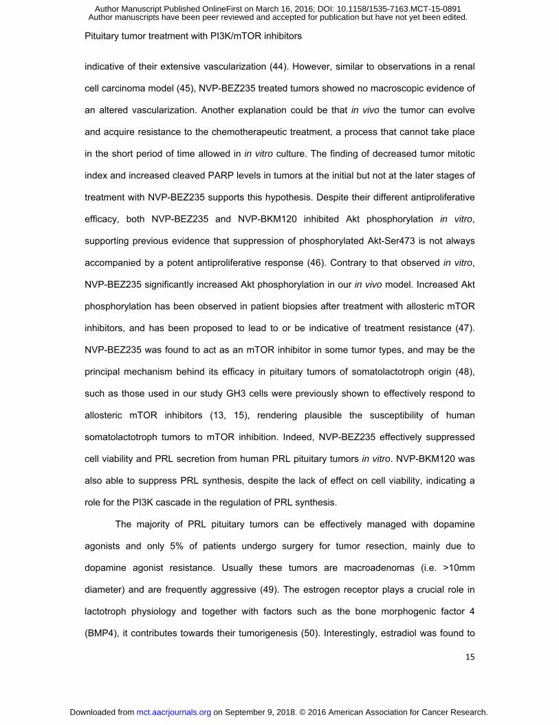
indicative of their extensive vascularization (44). However, similar to observations in a renal
cell carcinoma model (45), NVP-BEZ235 treated tumors showed no macroscopic evidence of
an altered vascularization. Another explanation could be that in vivo the tumor can evolve
and acquire resistance to the chemotherapeutic treatment, a process that cannot take place
in the short period of time allowed in in vitro culture. The finding of decreased tumor mitotic
index and increased cleaved PARP levels in tumors at the initial but not at the later stages of
treatment with NVP-BEZ235 supports this hypothesis. Despite their different antiproliferative
efficacy, both NVP-BEZ235 and NVP-BKM120 inhibited Akt phosphorylation in vitro,
supporting previous evidence that suppression of phosphorylated Akt-Ser473 is not always
accompanied by a potent antiproliferative response (46). Contrary to that observed in vitro,
NVP-BEZ235 significantly increased Akt phosphorylation in our in vivo model. Increased Akt
phosphorylation has been observed in patient biopsies after treatment with allosteric mTOR
inhibitors, and has been proposed to lead to or be indicative of treatment resistance (47).
NVP-BEZ235 was found to act as an mTOR inhibitor in some tumor types, and may be the
principal mechanism behind its efficacy in pituitary tumors of somatolactotroph origin (48),
such as those used in our study GH3 cells were previously shown to effectively respond to
allosteric mTOR inhibitors (13, 15), rendering plausible the susceptibility of human
somatolactotroph tumors to mTOR inhibition. Indeed, NVP-BEZ235 effectively suppressed
cell viability and PRL secretion from human PRL pituitary tumors in vitro. NVP-BKM120 was
also able to suppress PRL synthesis, despite the lack of effect on cell viability, indicating a
role for the PI3K cascade in the regulation of PRL synthesis.
The majority of PRL pituitary tumors can be effectively managed with dopamine
agonists and only 5% of patients undergo surgery for tumor resection, mainly due to
dopamine agonist resistance. Usually these tumors are macroadenomas (i.e. >10mm
diameter) and are frequently aggressive (49). The estrogen receptor plays a crucial role in
lactotroph physiology and together with factors such as the bone morphogenic factor 4
(BMP4), it contributes towards their tumorigenesis (50). Interestingly, estradiol was found to
on September 9, 2018. © 2016 American Association for Cancer Research. mct.aacrjournals.org Downloaded from
Author manuscripts have been peer reviewed and accepted for publication but have not yet been edited. Author Manuscript Published OnlineFirst on March 16, 2016; DOI: 10.1158/1535-7163.MCT-15-0891

Pituitary tumor treatment with PI3K/mTOR inhibitors
16
block the apoptotic action of PI3K inhibitors in breast cancer cells (51). A similar mechanism
may take place in human PRL pituitary tumors and compromise their antiproliferative
response to NVP-BEZ235 and NVP-BKM120. As our rat tumor model required the use of
females, we were not able to address the question of gender specificity in our study.
In conclusion, our data indicate that two inhibitors of the PI3K/Akt/mTOR pathway
showed various inhibitory effects on in vitro and in vivo growth of human and rat pituitary
tumors. Therapies targeting this pathway may therefore be of interest at least for treating
PRL-secreting aggressive pituitary tumors and carcinomas.
Acknowledgements
We thank the SFR platforms (ALECS and CiQLE) for their technical assistance throughout
this study and Angloscribe for scientific language editing service.
References
1. Raverot G, Jouanneau E, Trouillas J. Management of endocrine disease: clinicopathological classification and molecular markers of pituitary tumours for personalized therapeutic strategies. Eur J Endocrinol. 2014;170:R121-32. 2. Heaney A. Clinical review: Pituitary carcinoma: difficult diagnosis and treatment. J Clin Endocrinol Metab. 2011;96:3649-60. 3. Kaltsas GA, Nomikos P, Kontogeorgos G, Buchfelder M, Grossman AB. Clinical review: Diagnosis and management of pituitary carcinomas. J Clin Endocrinol Metab. 2005;90:3089-99. 4. Bush ZM, Longtine JA, Cunningham T, Schiff D, Jane JA, Jr., Vance ML, et al. Temozolomide treatment for aggressive pituitary tumors: correlation of clinical outcome with O(6)-methylguanine methyltransferase (MGMT) promoter methylation and expression. J Clin Endocrinol Metab. 2010;95:E280-90. 5. Raverot G, Castinetti F, Jouanneau E, Morange I, Figarella-Branger D, Dufour H, et al. Pituitary carcinomas and aggressive pituitary tumours: merits and pitfalls of temozolomide treatment. Clin Endocrinol (Oxf). 2012;76:769-75. 6. Losa M, Mazza E, Terreni MR, McCormack A, Gill AJ, Motta M, et al. Salvage therapy with temozolomide in patients with aggressive or metastatic pituitary adenomas: experience in six cases. Eur J Endocrinol. 2010;163:843-51. 7. McCormack AI, Wass JA, AB. G. Aggressive pituitary tumours: the role of temozolomide and the assessment of MGMT status. Eur J Clin Invest. 2011;41:1133-48. 8. Annamalai AK, Dean AF, Kandasamy N, Kovacs K, Burton H, Halsall DJ, et al. Temozolomide responsiveness in aggressive corticotroph tumours: a case report and review of the literature. Pituitary. 2012;15:276-87.
on September 9, 2018. © 2016 American Association for Cancer Research. mct.aacrjournals.org Downloaded from
Author manuscripts have been peer reviewed and accepted for publication but have not yet been edited. Author Manuscript Published OnlineFirst on March 16, 2016; DOI: 10.1158/1535-7163.MCT-15-0891

Pituitary tumor treatment with PI3K/mTOR inhibitors
17
9. Whitelaw BC, Dworakowska D, Thomas NW, Barazi S, Riordan-Eva P, King AP, et al. Temozolomide in the management of dopamine agonist-resistant prolactinomas. Clin Endocrinol (Oxf). 2012;76:877-86. 10. Shaw RJ, Cantley LC. Ras, PI(3)K and mTOR signalling controls tumour cell growth. Nature. 2006;441:424-30. 11. Dworakowska D, Wlodek E, Leontiou CA, Igreja S, Cakir M, Teng M, et al. Activation of RAF/MEK/ERK and PI3K/AKT/mTOR pathways in pituitary adenomas and their effects on downstream effectors. Endocr Relat Cancer. 2009;16:1329-38. 12. Musat M, Korbonits M, Kola B, Borboli N, Hanson MR, Nanzer AM, et al. Enhanced protein kinase B/Akt signalling in pituitary tumours. Endocr Relat Cancer. 2005;12:423-33. 13. Gorshtein A, Rubinfeld H, Kendler E, Theodoropoulou M, Cerovac V, Stalla GK, et al. Mammalian target of rapamycin inhibitors rapamycin and RAD001 (everolimus) induce anti-proliferative effects in GH-secreting pituitary tumor cells in vitro. Endocr Relat Cancer. 2009;16:1017-27. 14. Zatelli MC, Minoia M, Filieri C, Tagliati F, Buratto M, Ambrosio MR, et al. Effect of everolimus on cell viability in nonfunctioning pituitary adenomas. J Clin Endocrinol Metab. 2010;95:968-76. 15. Cerovac V, Monteserin-Garcia J, Rubinfeld H, Buchfelder M, Losa M, Florio T, et al. The somatostatin analogue octreotide confers sensitivity to rapamycin treatment on pituitary tumor cells. Cancer Res. 2010;70:666-74. 16. Jouanneau E, Wierinckx A, Ducray F, Favrel V, Borson-Chazot F, Honnorat J, et al. New targeted therapies in pituitary carcinoma resistant to temozolomide. Pituitary. 2012;15:37-43. 17. Wander SA, Hennessy BT, Slingerland JM. Next-generation mTOR inhibitors in clinical oncology: how pathway complexity informs therapeutic strategy. J Clin Invest. 2011;121:1231-41. 18. Maira SM, Stauffer F, Brueggen J, Furet P, Schnell C, Fritsch C, et al. Identification and characterization of NVP-BEZ235, a new orally available dual phosphatidylinositol 3-kinase/mammalian target of rapamycin inhibitor with potent in vivo antitumor activity. Mol Cancer Ther. 2008;7:1851-63. 19. Maira SM, Pecchi S, Huang A, Burger M, Knapp M, Sterker D, et al. Identification and characterization of NVP-BKM120, an orally available pan-class I PI3-kinase inhibitor. Mol Cancer Ther. 2012;11:317-28. 20. Zitzmann K, Ruden J, Brand S, Goke B, Lichtl J, Spottl G, et al. Compensatory activation of Akt in response to mTOR and Raf inhibitors - a rationale for dual-targeted therapy approaches in neuroendocrine tumor disease. Cancer Lett. 2010;295:100-9. 21. Gagliano T, Bellio M, Gentilin E, Mole D, Tagliati F, Schiavon M, et al. mTOR, p70S6K, AKT, and ERK1/2 levels predict sensitivity to mTOR and PI3K/mTOR inhibitors in human bronchial carcinoids. Endocr Relat Cancer. 2013;20:463-75. 22. Lee M, Theodoropoulou M, Graw J, Roncaroli F, Zatelli MC, Pellegata NS. Levels of p27 sensitize to dual PI3K/mTOR inhibition. Mol Cancer Ther. 2011;10:1450-9. 23. Raverot G, Wierinckx A, Dantony E, Auger C, Chapas G, Villeneuve L, et al. Prognostic factors in prolactin pituitary tumors: clinical, histological, and molecular data from a series of 94 patients with a long postoperative follow-up. J Clin Endocrinol Metab. 2010;95:1708-16. 24. Trouillas J, Chevallier P, Claustrat B, Hooghe-Peters E, Dubray C, Rousset B, et al. Inhibitory effects of the dopamine agonists quinagolide (CV 205-502) and bromocriptine on prolactin secretion and growth of SMtTW pituitary tumors in the rat. Endocrinology. 1994;134:401-10. 25. Trouillas J, Girod C, Claustrat B, Joly-Pharaboz M-O, Chevallier P. Spontaneous Prolactin Transplantable Tumor in the Wistar/Furth Rat (SMtTW): A New Animal Model of Human Prolactinoma. Cancer Res. 1990;50:4081-6. 26. Varrin-Doyer M, Nicolle A, Marignier R, Cavagna S, Benetollo C, Wattel E, et al. Human T lymphotropic virus type 1 increases T lymphocyte migration by recruiting the cytoskeleton organizer CRMP2. J Immunol. 2012;188:1222-33.
on September 9, 2018. © 2016 American Association for Cancer Research. mct.aacrjournals.org Downloaded from
Author manuscripts have been peer reviewed and accepted for publication but have not yet been edited. Author Manuscript Published OnlineFirst on March 16, 2016; DOI: 10.1158/1535-7163.MCT-15-0891

Pituitary tumor treatment with PI3K/mTOR inhibitors
18
27. Chiang GG, Abraham RT. Phosphorylation of mammalian target of rapamycin (mTOR) at Ser-2448 is mediated by p70S6 kinase. J Biol Chem. 2005;280:25485-90. 28. Trouillas J. In search of a prognostic classification of endocrine pituitary tumors. Endocr Pathol. 2014;25:124-32. 29. Trouillas J, Roy P, Sturm N, Dantony E, Cortet-Rudelli C, Viennet G, et al. A new prognostic clinicopathological classification of pituitary adenomas: a multicentric case-control study of 410 patients with 8 years post-operative follow-up. Acta Neuropathol. 2013;126:123-35. 30. Bengtsson D, Schroder HD, Andersen M, Maiter D, Berinder K, Feldt Rasmussen U, et al. Long-term outcome and MGMT as a predictive marker in 24 patients with atypical pituitary adenomas and pituitary carcinomas given treatment with temozolomide. J Clin Endocrinol Metab. 2015;100:1689-98. 31. Di Ieva A, Rotondo F, Syro LV, Cusimano MD, Kovacs K. Aggressive pituitary adenomas--diagnosis and emerging treatments. Nat Rev Endocrinol. 2014;10:423-35. 32. Monsalves E, Juraschka K, Tateno T, Agnihotri S, Asa SL, Ezzat S, et al. The PI3K/AKT/mTOR pathway in the pathophysiology and treatment of pituitary adenomas. Endocr Relat Cancer. 2014;21:R331-44. 33. Dworakowska D, Grossman AB. The pathophysiology of pituitary adenomas. Best Pract Res Clin Endocrinol Metab. 2009;23:525-41. 34. Zeng J, See AP, Aziz K, Thiyagarajan S, Salih T, Gajula RP, et al. Nelfinavir induces radiation sensitization in pituitary adenoma cells. Cancer Biol Ther. 2011;12:657-63. 35. Dai C, Zhang B, Liu X, Ma S, Yang Y, Yao Y, et al. Inhibition of PI3K/AKT/mTOR Pathway Enhances Temozolomide-Induced Cytotoxicity in Pituitary Adenoma Cell Lines in Vitro and Xenografted Pituitary Adenoma in Female Nude Mice. Endocrinology. 2013. 36. Perez-Castro C, Renner U, Haedo MR, Stalla GK, Arzt E. Cellular and molecular specificity of pituitary gland physiology. Physiol Rev. 2012;92:1-38. 37. Wierinckx A, Auger C, Devauchelle P, Reynaud A, Chevallier P, Jan M, et al. A diagnostic marker set for invasion, proliferation, and aggressiveness of prolactin pituitary tumors. Endocr Relat Cancer. 2007;14:887-900. 38. Sherr CJ, Roberts JM. Living with or without cyclins and cyclin-dependent kinases. Genes Dev. 2004;18:2699-711. 39. Gobin B, Battaglia S, Lanel R, Chesneau J, Amiaud J, Redini F, et al. NVP-BEZ235, a dual PI3K/mTOR inhibitor, inhibits osteosarcoma cell proliferation and tumor development in vivo with an improved survival rate. Cancer Lett. 2014;344:291-8. 40. Santiskulvong C, Konecny GE, Fekete M, Chen KY, Karam A, Mulholland D, et al. Dual targeting of phosphoinositide 3-kinase and mammalian target of rapamycin using NVP-BEZ235 as a novel therapeutic approach in human ovarian carcinoma. Clin Cancer Res. 2011;17:2373-84. 41. Schnell CR, Stauffer F, Allegrini PR, O'Reilly T, McSheehy PM, Dartois C, et al. Effects of the dual phosphatidylinositol 3-kinase/mammalian target of rapamycin inhibitor NVP-BEZ235 on the tumor vasculature: implications for clinical imaging. Cancer Res. 2008;68:6598-607. 42. Courtney KD, Corcoran RB, Engelman JA. The PI3K pathway as drug target in human cancer. J Clin Oncol. 2010;28:1075-83. 43. Carmeliet P, Jain RK. Principles and mechanisms of vessel normalization for cancer and other angiogenic diseases. Nat Rev Drug Discov. 2011;10:417-27. 44. Trouillas J, Chevallier P, Remy C, Rajas F, Cohen R, Calle A, et al. Differential Actions of the Dopamine Agonist Bromocriptine on Growth of SMtTW Tumors Exhibiting a Prolactin and/or a Somatotroph Cell Phenotype: Relation to Dopamine D2 Receptor Expression. Endocrinology. 1999;140:13-21. 45. Roulin D, Waselle L, Dormond-Meuwly A, Dufour M, Demartines N, Dormond O. Targeting renal cell carcinoma with NVP-BEZ235, a dual PI3K/mTOR inhibitor, in combination with sorafenib. Mol Cancer. 2011;10:90.
on September 9, 2018. © 2016 American Association for Cancer Research. mct.aacrjournals.org Downloaded from
Author manuscripts have been peer reviewed and accepted for publication but have not yet been edited. Author Manuscript Published OnlineFirst on March 16, 2016; DOI: 10.1158/1535-7163.MCT-15-0891

Pituitary tumor treatment with PI3K/mTOR inhibitors
19
46. Breuleux M, Klopfenstein M, Stephan C, Doughty CA, Barys L, Maira SM, et al. Increased AKT S473 phosphorylation after mTORC1 inhibition is rictor dependent and does not predict tumor cell response to PI3K/mTOR inhibition. Mol Cancer Ther. 2009;8:742-53. 47. O'Reilly KE, Rojo F, She QB, Solit D, Mills GB, Smith D, et al. mTOR inhibition induces upstream receptor tyrosine kinase signaling and activates Akt. Cancer Res. 2006;66:1500-8. 48. Serra V, Markman B, Scaltriti M, Eichhorn PJ, Valero V, Guzman M, et al. NVP-BEZ235, a dual PI3K/mTOR inhibitor, prevents PI3K signaling and inhibits the growth of cancer cells with activating PI3K mutations. Cancer Res. 2008;68:8022-30. 49. Maiter D, Delgrange E. Therapy of endocrine disease: the challenges in managing giant prolactinomas. Eur J Endocrinol. 2014;170:R213-27. 50. Paez-Pereda M, Giacomini D, Refojo D, Nagashima AC, Hopfner U, Grubler Y, et al. Involvement of bone morphogenetic protein 4 (BMP-4) in pituitary prolactinoma pathogenesis through a Smad/estrogen receptor crosstalk. Proc Natl Acad Sci U S A. 2003;100:1034-9. 51. Crowder RJ, Phommaly C, Tao Y, Hoog J, Luo J, Perou CM, et al. PIK3CA and PIK3CB inhibition produce synthetic lethality when combined with estrogen deprivation in estrogen receptor-positive breast cancer. Cancer Res. 2009;69:3955-62.
Figure legends
Figure 1.
In vitro effects of NVP-BEZ235 and NVP-BKM120 treatments on cell viability and cell cycle in
GH3 cells. Treatments consisted of DMSO, or of the inhibitors NVP-BEZ235 or NVP-
BKM120 at 1, 10, 100 and 250 nM (A-B-C-E) or all except 10 nM (D) for 24 hours. Cell
viability was measured by colorimetric assay with the CCK-8 test (A) and by flow cytometry
with propidium iodide (B). Data are expressed as a percentage of control (mean ± SEM).
Data were analyzed statistically by Mann-Whitney test, with *P< 0.05; **P< 0.01 compared
with DMSO. Cell cycle was assessed by flow cytometry (C), and S-phase data were
analyzed statistically by Mann-Whitney U test, with **P< 0.01 compared with DMSO. Protein
expression levels of phosphorylated Rb (D) were determined by western blot analysis. The
barographs represent the mean and S.E.M. Equal protein loading was examined by
detection of β-actin and data are expressed as a percentage of control. One representative
experiment out of three independent experiments at least is shown. The expression level of
G1/S cell cycle proteins was established by western blot (E) using anti-cyclin D3, -Cdk4, -
cyclin E, and -Cdk2; respective levels of β-actin are shown. Representatives of two
experiments are shown.
on September 9, 2018. © 2016 American Association for Cancer Research. mct.aacrjournals.org Downloaded from
Author manuscripts have been peer reviewed and accepted for publication but have not yet been edited. Author Manuscript Published OnlineFirst on March 16, 2016; DOI: 10.1158/1535-7163.MCT-15-0891

Pituitary tumor treatment with PI3K/mTOR inhibitors
20
Figure 2.
In vitro effects of NVP-BEZ235 and NVP-BKM120 treatments on apoptosis and on the
PI3K/Akt/mTOR pathway in GH3 cells. Treatments consisted of DMSO, or of the inhibitors
NVP-BEZ235 or NVP-BKM120 at 1, 10, 100 and 250 nM (A-B-C) or all except 10 nM (D-E)
for 24 hours. Staining of cleaved caspase-3 (A) and annexin V (C) were measured by flow
cytometry. Data were analyzed statistically by Mann-Whitney U test and values shown are
the mean ± SEM with *P<0.05 and **P<0.01 compared to DMSO. One representative
experiment out of three independent experiments at least is shown. Protein expression level
of total caspase-3 and cl-caspase-3 (B) and PARP and cl-PARP (D) was observed by
western blot analysis; respective levels of β-actin are shown. Representatives of two
experiments are shown. Expression levels and phosphorylation status of Akt, mTOR and p-
S6 were examined by western blot analysis (E). One representative blot out of three
performed is shown for NVP-BEZ235 and out of two for NVP-BKM120.
Figure 3.
In vivo effects of NVP-BEZ235 and NVP-BKM120 treatments on tumor growth and prolactin
secretion in the SMtTW3 tumor model. Rats were treated five weeks after graft placement,
(A-C) with NVP-BEZ235 20 mg/kg/d, 5 days a week, for three weeks (Control-3 n= 10 and
BEZ-3 n=13) or six weeks (Control-6 n=9 and BEZ-6 n=13) and (B-D) with NVP-BKM120 5
mg/kg/d, 5 days a week, for four weeks (Control-4 n=8 and BKM-4 n=12). Tumor weights (A-
B) were noted and different tumor sizes were illustrated by pictures, bar = 1 cm. Serum
prolactin levels (logarithmic scale; C-D) were assessed by RIA before treatment (Control-0,
BEZ-0, BKM-0) and at autopsy (Control-3, -4, -6, BEZ-3, -6 and BKM-4). Data were analyzed
statistically by Mann-Whitney test and values shown are mean ± SEM with *P<0.05,
**P<0.01 and ***P<0.001 compared to the corresponding control.
on September 9, 2018. © 2016 American Association for Cancer Research. mct.aacrjournals.org Downloaded from
Author manuscripts have been peer reviewed and accepted for publication but have not yet been edited. Author Manuscript Published OnlineFirst on March 16, 2016; DOI: 10.1158/1535-7163.MCT-15-0891

Pituitary tumor treatment with PI3K/mTOR inhibitors
21
Figure 4.
In vivo effects of NVP-BEZ235 and NVP-BKM120 treatments on cell proliferation and
apoptosis in the SMtTW3 tumor model. Rats were treated five weeks after graft placement,
(A-C) with NVP-BEZ235 20 mg/kg/d, 5 days a week, for three weeks (Control-3 n= 10 and
BEZ-3 n=13) or six weeks (Control-6 n=9 and BEZ-6 n=13) and (B-D) with NVP-BKM120 5
mg/kg/d, 5 days a week, for four weeks (Control-4 n=8 and BKM-4 n=12). Mitoses (A-B)
were assessed on hematoxylin-eosin staining and were counted at 400x magnification in 10
fields per tumor. Level of cl-PARP protein expression (C-D) was determined by western blot
analysis. The barographs represent the mean and S.E.M. Equal protein loading was
examined by detection of β-actin and data are expressed as a percentage of control. Data
were analyzed statistically by Mann-Whitney test and values shown are the mean ± SEM
with *P<0.05, **P<0.01 and ***P<0.001 compared to corresponding control.
Figure 5.
In vivo effects of NVP-BEZ235 and NVP-BKM120 treatments on Akt and S6 ribosomal
protein phosphorylation in the SMtTW3 tumor model. Rats were treated five weeks after graft
placement (A-B) with NVP-BEZ235 20 mg/kg/d, 5 days a week, for 6 weeks (control-6 n= 7
and BEZ-6 n=10) and (C-D) with NVP-BKM120 5 mg/kg/d, 5 days a week, for 4 weeks
(control-4 n=8 and BKM-4 n=9). Each western blot (A-C) for the phosphorylated protein was
followed by blotting for the total protein after stripping in Tris buffer, pH 2.0. Signal represents
mean±SEM, calculated as phosphorylated-to-total protein ratio (B-D) and presented as a
percentage of control. a.u.: arbitrary units. * P<0.05.
Figure 6.
Effects of NVP-BEZ235 and NVP-BKM120 on human PRL pituitary tumors in primary cell
culture. NVP-BEZ235 and NVP-BKM120 dose-response (1, 10, 100 nM) on (A) basal PRL
secretion and (B) cell viability from human PRL pituitary tumors in primary cell culture (n=7).
on September 9, 2018. © 2016 American Association for Cancer Research. mct.aacrjournals.org Downloaded from
Author manuscripts have been peer reviewed and accepted for publication but have not yet been edited. Author Manuscript Published OnlineFirst on March 16, 2016; DOI: 10.1158/1535-7163.MCT-15-0891

Pituitary tumor treatment with PI3K/mTOR inhibitors
22
For all cell culture experiments, each PRL RIA value was divided by cell viability counts as
determined by WST-1 at OD450nm. Data are the mean±SEM from seven cultures and are
presented as a percentage of control. * P<0.05 and ** P<0.001.
on September 9, 2018. © 2016 American Association for Cancer Research. mct.aacrjournals.org Downloaded from
Author manuscripts have been peer reviewed and accepted for publication but have not yet been edited. Author Manuscript Published OnlineFirst on March 16, 2016; DOI: 10.1158/1535-7163.MCT-15-0891

Figure 1
on September 9, 2018. © 2016 American Association for Cancer Research. mct.aacrjournals.org Downloaded from
Author manuscripts have been peer reviewed and accepted for publication but have not yet been edited. Author Manuscript Published OnlineFirst on March 16, 2016; DOI: 10.1158/1535-7163.MCT-15-0891

Figure 2
on September 9, 2018. © 2016 American Association for Cancer Research. mct.aacrjournals.org Downloaded from
Author manuscripts have been peer reviewed and accepted for publication but have not yet been edited. Author Manuscript Published OnlineFirst on March 16, 2016; DOI: 10.1158/1535-7163.MCT-15-0891
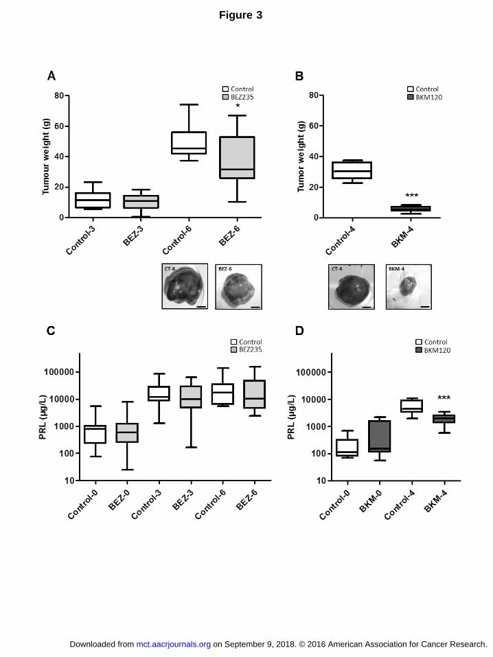
Figure 3
on September 9, 2018. © 2016 American Association for Cancer Research. mct.aacrjournals.org Downloaded from
Author manuscripts have been peer reviewed and accepted for publication but have not yet been edited. Author Manuscript Published OnlineFirst on March 16, 2016; DOI: 10.1158/1535-7163.MCT-15-0891

Figure 4
on September 9, 2018. © 2016 American Association for Cancer Research. mct.aacrjournals.org Downloaded from
Author manuscripts have been peer reviewed and accepted for publication but have not yet been edited. Author Manuscript Published OnlineFirst on March 16, 2016; DOI: 10.1158/1535-7163.MCT-15-0891

Figure 5
on September 9, 2018. © 2016 American Association for Cancer Research. mct.aacrjournals.org Downloaded from
Author manuscripts have been peer reviewed and accepted for publication but have not yet been edited. Author Manuscript Published OnlineFirst on March 16, 2016; DOI: 10.1158/1535-7163.MCT-15-0891

Figure 6
on September 9, 2018. © 2016 American Association for Cancer Research. mct.aacrjournals.org Downloaded from
Author manuscripts have been peer reviewed and accepted for publication but have not yet been edited. Author Manuscript Published OnlineFirst on March 16, 2016; DOI: 10.1158/1535-7163.MCT-15-0891

Published OnlineFirst March 16, 2016.Mol Cancer Ther Marie Chanal, Pascale Chevallier, Véronique Raverot, et al. prolactin-secreting pituitary tumorsDifferential effects of PI3K and dual PI3K/mTOR inhibition in rat
Updated version
10.1158/1535-7163.MCT-15-0891doi:
Access the most recent version of this article at:
Material
Supplementary
http://mct.aacrjournals.org/content/suppl/2016/03/16/1535-7163.MCT-15-0891.DC1
Access the most recent supplemental material at:
Manuscript
Authoredited. Author manuscripts have been peer reviewed and accepted for publication but have not yet been
E-mail alerts related to this article or journal.Sign up to receive free email-alerts
Subscriptions
Reprints and
To order reprints of this article or to subscribe to the journal, contact the AACR Publications
Permissions
Rightslink site. Click on "Request Permissions" which will take you to the Copyright Clearance Center's (CCC)
.http://mct.aacrjournals.org/content/early/2016/03/15/1535-7163.MCT-15-0891To request permission to re-use all or part of this article, use this link
on September 9, 2018. © 2016 American Association for Cancer Research. mct.aacrjournals.org Downloaded from
Author manuscripts have been peer reviewed and accepted for publication but have not yet been edited. Author Manuscript Published OnlineFirst on March 16, 2016; DOI: 10.1158/1535-7163.MCT-15-0891