
Differences in scapular kinematics and scapulohumeral rhythm during elevation and lowering of the...
6
Differences in scapular kinematics and scapulohumeral rhythm during elevation and lowering of the arm between typical children and healthy adults Fernanda A.P. Habechian a , Giovanna G. Fornasari a , Luciane S. Sacramento b , Paula R. Camargo a,b,⇑ a Methodist University of Piracicaba, Piracicaba, SP, Brazil b Federal University of São Carlos, São Carlos, SP, Brazil article info Article history: Received 14 May 2013 Received in revised form 25 July 2013 Accepted 23 October 2013 Available online xxxx Keywords: Biomechanics Childhood Rehabilitation Shoulder abstract Scapular kinematics in healthy adults is well described in the literature but little is known on typical chil- dren. This study aimed to compare the three-dimensional (3-D) scapular kinematics and scapulohumeral rhythm during the elevation and lowering of the arm in the scapular plane in typical children and healthy adults. Twenty-six healthy adults (35.34 ± 11.65 years, 1.70 ± 0.10 m, 70.00 ± 12.30 kg) and 33 typical children (9.12 ± 1.51 years, 1.40 ± 0.10 m, 35.40 ± 10.45 kg) participated in this study. 3-D scapular kine- matics were obtained using an electromagnetic tracking device. The subjects were asked to elevate and lower their arm in the scapular plane. Children showed less scapular protraction compared to adults at 120° during arm elevation, more anterior tilt than adults in the elevation and also at 60°, 90° and 120° during lowering of the arm. Children also showed higher scapulohumeral rhythm during lowering of the arm compared to adults from 90° to 60°. It was also found a low to little correlation between scapular position and age. The study showed small but significant differences in scapular kinematics and scapu- lohumeral rhythm between children and adults. These results can help clinicians to improve diagnosis and treatment protocols directed to children with dysfunction, as reference values on scapular kinemat- ics in healthy children are also provided in this study. Ó 2013 Elsevier Ltd. All rights reserved. 1. Introduction Position and control of the scapula on the thorax play an impor- tant role on normal function of the shoulder as it provides stable base for humeral motion (Kibler and McMullen, 2003). In healthy adults, it is well described that the scapula should laterally rotate and posteriorly tilt during elevation of the arm (Ludewig et al., 2009). Scapular protraction is less consistent during arm elevation, however it is accepted that some retraction will occur near end range of humeral elevation (McClure et al., 2001; Ludewig et al., 2009). The literature has supported that alterations in scapular kine- matics are associated with shoulder dysfunctions (Phadke et al., 2009; Ludewig and Braman, 2010). In the past, shoulder dysfunc- tions were mostly described in adults. The increasing number of children and adolescents athletes, especially in throwing sports, has contributed to increase the incidence of shoulder injuries in the pediatric population (Paterson and Waters, 2000; Leonard and Hutchinson, 2010; Cools et al., 2010; Eisner et al., 2013). How- ever, little is known about scapular kinematics in children. Only one study was found comparing the three-dimensional (3-D) scapular kinematics between children and adults during ele- vation of the arm (Dayanidhi et al., 2005). The authors described some differences in scapular kinematics pattern between the two groups and concluded that children have more contribution from the scapulothoracic joint than adults. This study only assessed scapular kinematics during elevation of the arm. However, lower- ing of the arm is also important to be analyzed once it is usually in this phase that scapular dyskinesia is observed in a clinical screening (Warner et al., 1992; Boublik and Hawkins, 1993; Kibler, 1998). No studies were found about lowering of the arm in chil- dren and there are few done in adults (Borstad and Ludewig, 2002; Ludewig et al., 2009; Matsuki et al., 2011; McClure et al., 2001). Thereby, investigations on scapular kinematics in children are warranted as knowing the typical motion in this population is the basis for the understanding of motor abnormalities that may be associated with shoulder dysfunctions. This can help clinicians to decide on more adequate exercise approaches for rehabilitation in children with upper extremity disorders such as rotator cuff dysfunctions, and with neurological disorders such as cerebral 1050-6411/$ - see front matter Ó 2013 Elsevier Ltd. All rights reserved. http://dx.doi.org/10.1016/j.jelekin.2013.10.013 ⇑ Corresponding author. Address: Universidade Federal de São Carlos, Rod. Washington Luis, km 235, CEP: 13565-905 São Carlos, SP, Brazil. Tel.: +55 16 3306 6696. E-mail address: [email protected] (P.R. Camargo). Journal of Electromyography and Kinesiology xxx (2013) xxx–xxx Contents lists available at ScienceDirect Journal of Electromyography and Kinesiology journal homepage: www.elsevier.com/locate/jelekin Please cite this article in press as: Habechian FAP et al. Differences in scapular kinematics and scapulohumeral rhythm during elevation and lowering of the arm between typical children and healthy adults. J Electromyogr Kinesiol (2013), http://dx.doi.org/10.1016/j.jelekin.2013.10.013
Transcript of Differences in scapular kinematics and scapulohumeral rhythm during elevation and lowering of the...

Journal of Electromyography and Kinesiology xxx (2013) xxx–xxx
Contents lists available at ScienceDirect
Journal of Electromyography and Kinesiology
journal homepage: www.elsevier .com/locate / je lek in
Differences in scapular kinematics and scapulohumeral rhythm duringelevation and lowering of the arm between typical childrenand healthy adults
1050-6411/$ - see front matter � 2013 Elsevier Ltd. All rights reserved.http://dx.doi.org/10.1016/j.jelekin.2013.10.013
⇑ Corresponding author. Address: Universidade Federal de São Carlos, Rod.Washington Luis, km 235, CEP: 13565-905 São Carlos, SP, Brazil. Tel.: +55 16 33066696.
E-mail address: [email protected] (P.R. Camargo).
Please cite this article in press as: Habechian FAP et al. Differences in scapular kinematics and scapulohumeral rhythm during elevation and lowerinarm between typical children and healthy adults. J Electromyogr Kinesiol (2013), http://dx.doi.org/10.1016/j.jelekin.2013.10.013
Fernanda A.P. Habechian a, Giovanna G. Fornasari a, Luciane S. Sacramento b, Paula R. Camargo a,b,⇑a Methodist University of Piracicaba, Piracicaba, SP, Brazilb Federal University of São Carlos, São Carlos, SP, Brazil
a r t i c l e i n f o
Article history:Received 14 May 2013Received in revised form 25 July 2013Accepted 23 October 2013Available online xxxx
Keywords:BiomechanicsChildhoodRehabilitationShoulder
a b s t r a c t
Scapular kinematics in healthy adults is well described in the literature but little is known on typical chil-dren. This study aimed to compare the three-dimensional (3-D) scapular kinematics and scapulohumeralrhythm during the elevation and lowering of the arm in the scapular plane in typical children and healthyadults. Twenty-six healthy adults (35.34 ± 11.65 years, 1.70 ± 0.10 m, 70.00 ± 12.30 kg) and 33 typicalchildren (9.12 ± 1.51 years, 1.40 ± 0.10 m, 35.40 ± 10.45 kg) participated in this study. 3-D scapular kine-matics were obtained using an electromagnetic tracking device. The subjects were asked to elevate andlower their arm in the scapular plane. Children showed less scapular protraction compared to adults at120� during arm elevation, more anterior tilt than adults in the elevation and also at 60�, 90� and 120�during lowering of the arm. Children also showed higher scapulohumeral rhythm during lowering ofthe arm compared to adults from 90� to 60�. It was also found a low to little correlation between scapularposition and age. The study showed small but significant differences in scapular kinematics and scapu-lohumeral rhythm between children and adults. These results can help clinicians to improve diagnosisand treatment protocols directed to children with dysfunction, as reference values on scapular kinemat-ics in healthy children are also provided in this study.
� 2013 Elsevier Ltd. All rights reserved.
1. Introduction
Position and control of the scapula on the thorax play an impor-tant role on normal function of the shoulder as it provides stablebase for humeral motion (Kibler and McMullen, 2003). In healthyadults, it is well described that the scapula should laterally rotateand posteriorly tilt during elevation of the arm (Ludewig et al.,2009). Scapular protraction is less consistent during arm elevation,however it is accepted that some retraction will occur near endrange of humeral elevation (McClure et al., 2001; Ludewig et al.,2009).
The literature has supported that alterations in scapular kine-matics are associated with shoulder dysfunctions (Phadke et al.,2009; Ludewig and Braman, 2010). In the past, shoulder dysfunc-tions were mostly described in adults. The increasing number ofchildren and adolescents athletes, especially in throwing sports,has contributed to increase the incidence of shoulder injuries inthe pediatric population (Paterson and Waters, 2000; Leonard
and Hutchinson, 2010; Cools et al., 2010; Eisner et al., 2013). How-ever, little is known about scapular kinematics in children.
Only one study was found comparing the three-dimensional(3-D) scapular kinematics between children and adults during ele-vation of the arm (Dayanidhi et al., 2005). The authors describedsome differences in scapular kinematics pattern between the twogroups and concluded that children have more contribution fromthe scapulothoracic joint than adults. This study only assessedscapular kinematics during elevation of the arm. However, lower-ing of the arm is also important to be analyzed once it is usuallyin this phase that scapular dyskinesia is observed in a clinicalscreening (Warner et al., 1992; Boublik and Hawkins, 1993; Kibler,1998). No studies were found about lowering of the arm in chil-dren and there are few done in adults (Borstad and Ludewig,2002; Ludewig et al., 2009; Matsuki et al., 2011; McClure et al.,2001).
Thereby, investigations on scapular kinematics in children arewarranted as knowing the typical motion in this population isthe basis for the understanding of motor abnormalities that maybe associated with shoulder dysfunctions. This can help cliniciansto decide on more adequate exercise approaches for rehabilitationin children with upper extremity disorders such as rotator cuffdysfunctions, and with neurological disorders such as cerebral
g of the

2 F.A.P. Habechian et al. / Journal of Electromyography and Kinesiology xxx (2013) xxx–xxx
palsy, brachial plexus injury and spinal cord injury (Mosquedaet al., 2004; Coluccini et al., 2007; Jaspers et al., 2011).
The hypothesis of this study is that adults and children presentdifferences in scapular kinematics in both elevation and loweringof the arm. The aim was to compare the 3-D scapular kinematicsand scapulohumeral rhythm during elevation and lowering of thearm in the scapular plane in typical children and healthy adults.
2. Methods
2.1. Subjects
This is a cross-sectional study with non-probabilistic samplingand by convenience. Twenty-six adults and 33 typical children par-ticipated in the study. The descriptive data of the participants arepresented on Table 1. The study included individuals who had nohistory of shoulder or cervical dysfunction and who had range ofmotion for shoulder elevation next to 150� as evaluated by visualobservation. The range of age accepted was between 18 and55 years for adults and 6–12 years for children.
Subjects were excluded if any of the following was found: uni-laterally dominant upper-extremity-sport/labor subjects; cervicalpain; history of surgical stabilization or repair of the rotator cuff;positive impingement or instability tests; fracture history in theclavicle, scapula or humerus; systemic diseases involving thejoints; cognitive deficits preventing the understanding of verbalcommands; brachial plexus injury; transpore tape allergy; childrenwith body mass index (BMI) higher than 1 standard deviationaccording to the growth reference for BMI in score z for youthand adolescents of the World Health Organization (De Onis et al.,2007) and adults with BMI > 28 kg/m2 (the amount of subcutane-ous tissue can compromise the quality of the motion data as sur-face sensors were used to track the bones). Children were alsoexcluded in case of delayed motor development based on parentsreport.
The study was approved by the research ethical committee ofthe University. Adults and children and their parents received ver-bal and written explanation of the objectives and methodology ofthe study and who agreed to participate signed a consent form.
2.2. Instrumentation
To evaluate 3-D kinematics, the capture and analysis were per-formed using the electromagnetic tracking device Flock of Birds�
(miniBird�) integrated with MotionMonitor™ software that is usedto collect data with a sampling frequency of 100 Hz. In a metal freeenvironment up to a 76 cm distance from the transmitter the rootmean square accuracy of the system is 0.5� for orientation and0.18 cm for position, as reported by the manufacturer.
2.3. Procedures
The dominant arm was evaluated in all children to make thetest easier for them. Dominance was determined by asking themwhich hand is used to draw or write. The adults had the tested side
Table 1Characteristics of the participants of the study.
Adults (n = 26) Children (n = 33)
Sex 14 females; 12 males 15 females; 18 malesAge (years) 35.34 ± 11.65 9.12 ± 1.51Height (m) 1.70 ± 0.10 1.40 ± 0.10Weight (kg) 70.00 ± 12.30 35.40 ± 10.45
Results are mean and standard deviation.
Please cite this article in press as: Habechian FAP et al. Differences in scapular kiarm between typical children and healthy adults. J Electromyogr Kinesiol (201
randomly chosen because it is already shown in the literature thatthere are no significant differences in scapular kinematics betweenboth sides in healthy adults (Yoshizaki et al., 2009).
The surface electromagnetic sensors were attached with doublesided tape to the sternum, the acromion of the scapula and a ther-moplastic cuff attached to the distal humerus to track humeral mo-tion. The subject stood with the arms relaxed at the side in aneutral position with the transmitter directly behind the shouldertested while bony landmarks on the thorax, scapula and humeruswere palpated and digitized with a stylus with known offsets to al-low transformation of the sensor data to local anatomically basedcoordinate systems. Local coordinate systems were establishedfor the trunk, clavicle, scapula and humerus using the digitizedlandmarks following the International Society of Biomechanics rec-ommended protocol (Wu et al., 2005). The z-axis pointed laterally,the x-axis anteriorly and the y-axis superiorly.
Kinematic data collection was done with the subjects in a re-laxed standing position. Subjects were asked to maintain light fin-gertip contact with a flat planar surface to keep positioning of thearm in the scapular plane (45� anterior to the coronal plane). Theywere also instructed to keep their hand with their thumb pointingtoward the ceiling. Three repetitions were performed. Subjectswere asked to elevate their arm from the rest position throughtheir full range of motion at a speed such that it took around 3 sto elevate their arm and 3 s to lower it.
2.4. Data reduction
MATLAB software was used to reduce the data. Scapularkinematics was analyzed at 30�, 60�, 90� and 120� of elevationand lowering of the arm. The YXZ sequence was used to describescapular motions relative to the trunk. For the scapula, the rota-tions were described in the order of protraction/retraction, lat-eral/medial rotation and anterior/posterior tilt (Wu et al., 2005).The humeral position with reference to the trunk was determinedusing the Y0XY00 sequence. The first rotation defines the plane of ele-vation, the second defines the humeral elevation angle, and thethird defines internal/external rotation. The humeral position withreference to the scapula was determined using the XZY sequence asrecommended by Phadke et al. (2011). The first rotation definesglenohumeral elevation, the second defines the plane of glenohu-meral elevation, and the third defines internal/external rotation.
For scapulohumeral rhythm, the ratio of the glenohumeralelevation relative to lateral rotation of the scapula was determinedby calculating the slope of the linear regression line using the lat-eral rotation of the scapula as X value and the glenohumeral eleva-tion as Y value as proposed by Braman et al. (2009). The ratio wascalculated from 30� to 120� of humerothoracic elevation, from120� to 30� of humerothoracic lowering, and at 30� incrementsfor elevation (30�–60�, 60�–90�, 90�–120�) and lowering(120�–90�, 90�–60�, 60�–30�) of the arm.
2.5. Statistical analysis
The data were averaged over the three repetitions of elevationand lowering of the arm and analyzed using the NCSS statisticalpackage (NCSS, Kaysville, UT). The Shapiro–Wilk test was used tocheck the normality of the data. For scapular protraction, lateralrotation and tilt, a 2-way mixed ANOVA was conducted for eachphase (elevation and lowering), in separate, with humeral angle(30�, 60�, 90� and 120�) as within factor and group (children andadults) as between factor. The primary interest was to check inter-action of group � humeral angle and secondarily the main effect ofgroup. The Bonferroni test for post hoc analysis was used whennecessary. For scapulohumeral rhythm, the same statisticalprocedure was followed but interval (30�–60�, 60�–90�, 90�–120�,
nematics and scapulohumeral rhythm during elevation and lowering of the3), http://dx.doi.org/10.1016/j.jelekin.2013.10.013
F.A.P. Habechian et al. / Journal of Electromyography and Kinesiology xxx (2013) xxx–xxx 3
30�–120�, 120�–90�, 90�–60�, 60�–30�, 30�–120�) was used insteadof humeral angle. A p value of less than 0.05 was consideredsignificant.
The relationship between scapular position and age was as-sessed by performing linear regressions at each humeral angle dur-ing elevation and lowering of the arm for each variable (scapularprotraction, lateral rotation and tilt) in separate. Age was used asindependent factor and scapular position as outcome. The coeffi-cient of determination was assessed by calculating the square ofthe correlation coefficient.
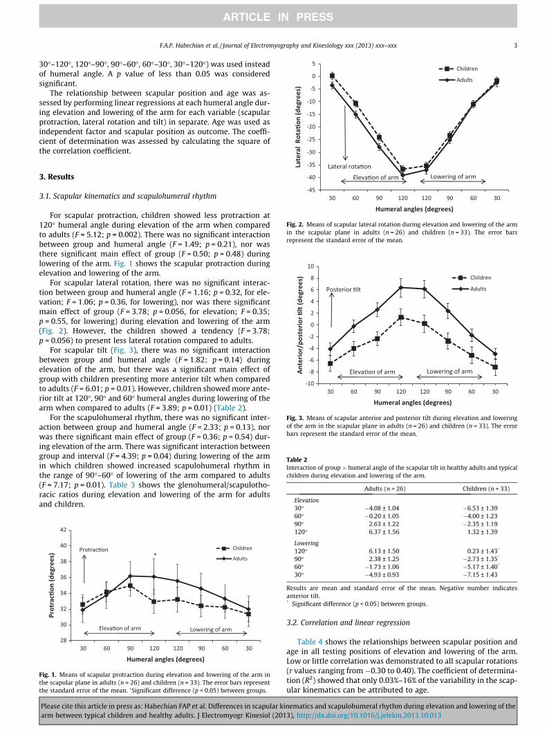
Fig. 2. Means of scapular lateral rotation during elevation and lowering of the armin the scapular plane in adults (n = 26) and children (n = 33). The error barsrepresent the standard error of the mean.
Fig. 3. Means of scapular anterior and posterior tilt during elevation and loweringof the arm in the scapular plane in adults (n = 26) and children (n = 33). The errorbars represent the standard error of the mean.
Table 2Interaction of group � humeral angle of the scapular tilt in healthy adults and typicalchildren during elevation and lowering of the arm.
Adults (n = 26) Children (n = 33)
Elevation30� �4.08 ± 1.04 �6.53 ± 1.39
3. Results
3.1. Scapular kinematics and scapulohumeral rhythm
For scapular protraction, children showed less protraction at120� humeral angle during elevation of the arm when comparedto adults (F = 5.12; p = 0.002). There was no significant interactionbetween group and humeral angle (F = 1.49; p = 0.21), nor wasthere significant main effect of group (F = 0.50; p = 0.48) duringlowering of the arm. Fig. 1 shows the scapular protraction duringelevation and lowering of the arm.
For scapular lateral rotation, there was no significant interac-tion between group and humeral angle (F = 1.16; p = 0.32, for ele-vation; F = 1.06; p = 0.36, for lowering), nor was there significantmain effect of group (F = 3.78; p = 0.056, for elevation; F = 0.35;p = 0.55, for lowering) during elevation and lowering of the arm(Fig. 2). However, the children showed a tendency (F = 3.78;p = 0.056) to present less lateral rotation compared to adults.
For scapular tilt (Fig. 3), there was no significant interactionbetween group and humeral angle (F = 1.82; p = 0.14) duringelevation of the arm, but there was a significant main effect ofgroup with children presenting more anterior tilt when comparedto adults (F = 6.01; p = 0.01). However, children showed more ante-rior tilt at 120�, 90� and 60� humeral angles during lowering of thearm when compared to adults (F = 3.89; p = 0.01) (Table 2).
For the scapulohumeral rhythm, there was no significant inter-action between group and humeral angle (F = 2.33; p = 0.13), norwas there significant main effect of group (F = 0.36; p = 0.54) dur-ing elevation of the arm. There was significant interaction betweengroup and interval (F = 4.39; p = 0.04) during lowering of the armin which children showed increased scapulohumeral rhythm inthe range of 90�–60� of lowering of the arm compared to adults(F = 7.17; p = 0.01). Table 3 shows the glenohumeral/scapulotho-racic ratios during elevation and lowering of the arm for adultsand children.
Fig. 1. Means of scapular protraction during elevation and lowering of the arm inthe scapular plane in adults (n = 26) and children (n = 33). The error bars representthe standard error of the mean. �Significant difference (p < 0.05) between groups.
60� �0.20 ± 1.05 �4.00 ± 1.2390� 2.63 ± 1.22 �2.35 ± 1.19120� 6.37 ± 1.56 1.32 ± 1.39
Lowering120� 6.13 ± 1.50 0.23 ± 1.43*
90� 2.38 ± 1.25 �2.73 ± 1.35*
60� �1.73 ± 1.06 �5.17 ± 1.40*
30� �4.93 ± 0.93 �7.15 ± 1.43
Results are mean and standard error of the mean. Negative number indicatesanterior tilt.* Significant difference (p < 0.05) between groups.
Please cite this article in press as: Habechian FAP et al. Differences in scapular kiarm between typical children and healthy adults. J Electromyogr Kinesiol (201
3.2. Correlation and linear regression
Table 4 shows the relationships between scapular position andage in all testing positions of elevation and lowering of the arm.Low or little correlation was demonstrated to all scapular rotations(r values ranging from �0.30 to 0.40). The coefficient of determina-tion (R2) showed that only 0.03%–16% of the variability in the scap-ular kinematics can be attributed to age.
nematics and scapulohumeral rhythm during elevation and lowering of the3), http://dx.doi.org/10.1016/j.jelekin.2013.10.013

Table 3Scapulohumeral rhythm during elevation and lowering of the arm in the scapularplane in healthy adults and typical children.
Adults (n = 26) Children (n = 33)
Elevation30�–60� 1.64 ± 0.13 1.88 ± 0.1160�–90� 1.44 ± 0.09 1.43 ± 0.0890�–120� 1.99 ± 0.17 1.57 ± 0.1530�–120� 1.52 ± 0.08 1.43 ± 0.07
Lowering120�–90� 1.78 ± 0.16 1.78 ± 0.1590�–60� 1.21 ± 0.15 1.75 ± 0.13*
60�–30� 2.71 ± 0.23 2.59 ± 0.21120�–30� 1.45 ± 0.10 1.67 ± 0.09
Results are mean and standard error of the mean.* Significant difference (p < 0.05) between groups.
Table 4Relationship between age and scapular kinematics in all testing positions of elevationand lowering of the arm.
Elevation of the arm Lowering of the arm
r R2 p r R2 p
Protraction30� �0.03 0.001 0.78 0.07 0.005 0.5860� �0.01 0.0003 0.90 0.09 0.01 0.4990� 0.08 0.007 0.53 0.13 0.02 0.31
120� 0.13 0.02 0.31 0.09 0.008 0.49
Lateral rotation30� �0.25 0.06 0.06 �0.09 0.01 0.4660� �0.30 0.09 0.02* �0.02 0.001 0.8490� �0.31 0.10 0.01* �0.18 0.03 0.17
120� �0.27 0.08 0.03* �0.24 0.06 0.06
Tilt30� 0.14 0.02 0.28 0.14 0.02 0.2760� 0.30 0.09 0.02* 0.28 0.08 0.03*
90� 0.39 0.16 0.002* 0.40 0.16 0.001*
120� 0.32 0.11 0.01* 0.36 0.13 0.004*
r Indicates correlation. R2 indicates coefficient of determination.* Statistically significant (p < 0.05).
4 F.A.P. Habechian et al. / Journal of Electromyography and Kinesiology xxx (2013) xxx–xxx
4. Discussion
To our knowledge this is the first study that compares scapularkinematics during both elevation and lowering of the arm betweenchildren and adults. In general, the scapula increased protractionand lateral rotation, and progressed from anterior to posterior tiltduring elevation of the arm in both groups, and returned to the ini-tial position during lowering of the arm. Despite of the similarscapular kinematics pattern, subtle differences were identified.As such, the findings of this study partially support the hypothesisthat children and adults differ in scapular kinematics during eleva-tion and lowering of the arm.
The lateral rotation was the most consistent motion of the scap-ula during elevation and lowering of the arm in both groups. It isthe scapular motion of greatest range. Although the adults had ten-dency to present more lateral rotation than the children, signifi-cance was not reached. These findings are contrary to what waspreviously demonstrated by Dayanidhi et al. (2005) who identifiedchildren to have greater lateral rotation from 25� to 125� of hum-eral elevation in the scapular plane. The difference between bothstudies can be due to methodological issues. The present studyused the posterolateral acromion, the root of the scapular spineand the inferior angle of the scapula to build the scapular localcoordinate system, while Dayanidhi et al. (2005) used the acromi-clavicular joint instead of the posterolateral acromion. Ludewiget al. (2010) have demonstrated that less lateral rotation is ob-served when the posterolateral acromion is digitized.
Please cite this article in press as: Habechian FAP et al. Differences in scapular kiarm between typical children and healthy adults. J Electromyogr Kinesiol (201
Analyzing the protraction of the scapula, children showed lessprotraction at 120� humeral angle during elevation of the armwhen compared to adults. Observing Fig. 1, it can be noticed thatchildren actually presented more retraction at 120� of arm eleva-tion. This finding is in accordance with the literature that reportsthat some retraction of the scapula occurs near end range ofmotion (Borstad and Ludewig, 2002; Ludewig and Cook, 2002;McClure et al., 2001). The pattern described in this study is alsosimilar to the pattern described by Lempereur et al. (2012) in typ-ically developing children. However, the children evaluated byDayanidhi et al. (2005) showed a retraction pattern earlier in therange of motion (beyond 60� of arm elevation). Despite of the aginginfluence, adults may also experience postural influences such asslouched posture or thoracic kyphosis, for example (Finley andLee, 2003), which contribute for increased scapular protraction.
With regards to the scapular tilt, children showed more anteriortilt than adults during elevation of the arm and at 60�, 90� and 120�humeral angles during lowering of the arm. The scapula of the chil-dren remained mostly in anterior tilt during elevation of the arm,except at 120� humeral angle where it was slightly posteriorlytilted (1.32�). This pattern differs from the adults who reached6.37� of posterior tilt at 120� humeral angle. Dayanidhi et al.(2005) reported a similar pattern of posterior tilt between childrenand adults, except for a small anterior tilt at the end of the rangedemonstrated by the children. Although we have not evaluatedclavicular kinematics, increased clavicle elevation has been alreadydemonstrated in children (Dayanidhi et al., 2005). This fact cancontribute for the more anterior tilt found in our children as theclavicle elevation seems to contribute to 75% of anterior tilt ofthe scapula relative to the trunk (Ludewig and Braman, 2010).One might argue if the length of the clavicle could also contributefor a great deal of anterior tilt. In a cadaveric study, Matsumuraet al. (2010) have demonstrated that scapular anterior tilt in-creased with 10% shortening of the clavicle when compared to anormal length clavicle. The length of the clavicle was not assessedin the current study, but the authors believe that its length can dif-fer between children and adults with children presenting shorterclavicle contributing to more anterior tilt. Investigations shouldbe done to verify if the clavicles are shorter in children and if it cor-relates with the increased anterior tilt.
The scapulothoracic muscles are very important for scapularstability. The serratus anterior contributes substantively to scapu-lar posterior tilt. It is the only scapulothoracic muscle with thecapability to both laterally rotate and posteriorly tilt the scapulaon the thorax making its contribution to normal scapular kinemat-ics very significant. Its line of action will directly approximate thescapula to the thorax, which can serve as a stable base (Phadkeet al., 2009). As children have a distinct anatomy that is in contin-uous modification to improve their systems and also to adapt totheir new environment (Schuenke et al., 2006), it is possible thatthe line of action of the scapulothoracic muscles might be differentfrom the adults causing change in the scapular kinematics.
In the current investigation, higher scapulohumeral rhythm wasseen in children from 90� to 60� of arm lowering. As the average ofscapular lateral rotation was similar between children and adults,we believe this difference may be related to less contribution of theglenohumeral joint in adults during this interval of motion.Although muscular strengthening (Wang et al., 1999) as well as fa-tigue (McQuade et al., 1995) have been shown to influence thescapulohumeral rhythm, we are not able to explain the mechanismthat may have lead to this behavior in our subjects due to the lackof literature on this topic and lack of muscle activity assessment inthis study.
Low to little correlation was demonstrated between scapularposition and age. Endo et al. (2004) have previously describedthe effects of aging on the shoulder analyzing antero-posterior
nematics and scapulohumeral rhythm during elevation and lowering of the3), http://dx.doi.org/10.1016/j.jelekin.2013.10.013

F.A.P. Habechian et al. / Journal of Electromyography and Kinesiology xxx (2013) xxx–xxx 5
radiography. The authors observed a decrease of posterior tilt andlateral rotation angle with aging. Direct comparisons cannot bedone between the present study and the Endo et al. (2004) becauseof differences in methodology and study design. It is important toconsider that the primary aim of this investigation was to comparethe scapular kinematics between children and adults. As such, thisis not the best design to determine about possible correlations onscapular position and age as a wide range of age would benecessary.
Considering that the skills to position and control the move-ments of the scapula are essential to the normal functioning ofthe upper limb (Jobe and Pink, 1993), the findings of this investiga-tion provide new knowledge about scapular kinematics during ele-vation and lowering of the arm in children. Furthermore, due to thehigh mobility of the shoulder complex, it is highly susceptible todysfunction and instability. The inability to control the scapulacan usually contribute for development of shoulder pathologies(Kamkar et al., 1993). Thus, it is relevant to understand thescapular kinematics in typical children as an attempt to improvediagnostic and treatment protocols.
As this investigation brings reference values of scapularkinematics for healthy children, these results may help cliniciansdevelop distinct treatment protocols for children and adults oncechildren cannot be considered as miniature of the adults. Thisstudy may also provide knowledge for better rehabilitation pro-grams for children with neurological problem, as for example,cerebral palsy, brachial plexus birth palsy and spinal cord injuredchildren once proximal stabilization of the upper extremity is veryimportant for them to perform functional activities. Children withupper extremity injuries such as rotator cuff tears and associatedpathologies in shoulder due to their early engagement in sports(Tarkin et al., 2005; Leonard and Hutchinson, 2010; Eisner et al.,2013), especially the throwing, can also benefit from thesefindings.
The present study also has some limitations: (1) a large range ofage in the adults group was considered; (2) scapular muscles activ-ity was not measured; and (3) clavicular motion and length werenot assessed. Further research should consider children and adultsto perform elevation of the arm with a weight in hand as this con-dition may bring out greater differences between groups.
5. Conclusion
The findings of this study indicates that there is similarity inscapular kinematics pattern between children and adults duringelevation and lowering of the arm, except for scapular protractionat 120� of humeral elevation where children present less protrac-tion. Children also present more anterior tilt than adults. Referencevalues on scapular kinematics in healthy children are also providedin this study.
Acknowledgements
The authors are deeply grateful to the volunteers who partici-pated in this study. The authors also want to thank Fundação deAmparo à Pesquisa do Estado de São Paulo (2011/23804-2) andCoordenação de Aperfeiçoamento de Pessoal de Nível Superiorfor the financial support.
References
Borstad JD, Ludewig PM. Comparison of scapular kinematics between elevation andlowering of the arm in the scapular plane. Clin Biomech 2002;17:650–9.
Boublik M, Hawkins RJ. Clinical examination of the shoulder complex. J OrthopSports Phys Therapy 1993;18:379–85.
Please cite this article in press as: Habechian FAP et al. Differences in scapular kiarm between typical children and healthy adults. J Electromyogr Kinesiol (201
Braman JP, Engel SC, LaPrade RF, Ludewig PM. In vivo assessment ofscapulohumeral rhythm during unconstrained over head reaching inasymptomatic subjects. J Shoulder Elbow Surg 2009;18(6):960e7.
Coluccini M, Maini ES, Martelloni C, Sgandurra G, Cioni G. Kinematiccharacterization of functional reach to grasp in normal and in motor disabledchildren. Gait Posture 2007;25:493–501.
Cools AM, Johansson FR, Cambier DC, Velde AV, Palmans T, Witvrouw EE.Descriptive profile of scapulothoracic position, strength and flexible variablesin adolescent elite tenis players. Br J Sports Med 2010;44:678–84.
Dayanidhi S, Orlin M, Kozin S, Duff S, Karduna A. Scapular kinematics duringhumeral elevation in adults and children. Clin Biomech 2005;20(6):600–6.
De Onis M, Onyango AW, Borghi E, Siyam A, Nishida C, Siekmann J. Development ofa WHO growth reference for school-aged children and adolescents. Bull WorldHealth Organ 2007;85:660–7.
Eisner EA, Roocroft JH, Moor MA, Edmonds EW. Partial rotator cuff tears inadolescents: factors affecting outcomes. J Pediatr Orthop 2013;33:2–7.
Endo K, Yukata K, Yasui N. Influence of age on scapulo-thoracic orientation. ClinBiomech 2004;19:1009–13.
Finley MA, Lee RY. Effect of sitting posture on 3-dimensional scapular kinematicsmeasured by skin-mounted electromagnetic tracking sensors. Arch Phys MedRehabil 2003;84:563–8.
Jaspers E, Feyes H, Bruyninckx H, Harlaar J, Molenaers G, Desloovere K. Upper limbkinematics: development and reliability of a clinical protocol for children. GaitPosture 2011;33:279–85.
Jobe FW, Pink M. Classification and treatment of shoulder dysfunction in theoverhead athlete. J Orthop Sports Phys Ther 1993;18:427–32.
Kamkar A, Irrgang JJ, Whitney SL. Nonoperative management of secondary shoulderimpingement syndrome. J Orthop Sports Phys Ther 1993;17:212–24.
Kibler WB. The role of the scapula in athletic shoulder function. Am J Sports Med1998;26(2):325–37.
Kibler WB, McMullen J. Scapular dyskinesis and its relation to shoulder pain. J AmAcad Orthop Surg 2003;11(2):142–51.
Lempereur M, Brochard S, Mao L, Rémy-Néris O. Validity and reliability of shoulderkinematics in typically developing children and children with hemiplegiccerebral palsy. J Biomech 2012;45:2028–34.
Leonard J, Hutchinson MR. Shoulder injuries in skeletally immature throwers:review and current thoughts. Br J Sports Med 2010;44:306–10.
Ludewig PM, Braman JP. Shoulder impingement: Biomechanical considerations inrehabilitation. Manual Ther 2010:1–7.
Ludewig PM, Cook TM. Translations of the humerus in persons with shoulderimpingement symptoms. J Orthop Sports Phys Ther 2002;32(6):248–59.
Ludewig PM, Phadke V, Braman JP, Hasset DR, Cieminski CJ, LaPrade RF. Motion ofthe shoulder complex during multiplanar humeral elevation. J Bone Joint SurgAm 2009;91:378–89.
Ludewig PM, Hassett DR, LaPrade RF, Camargo PR, Braman JP. Comparison ofscapular local coordinate systems. Clin Biomech 2010;25:415–21.
Matsuki K, Matsuki KO, Mu S, Yamaguchi S, Ochiai N, Sasho T, et al. In vivo 3-dimensional analysis of scapular kinematics: comparison of dominant andnondominant shoulders. J Shoulder Elbow Surg 2011;20:659–65.
Matsumura N, Ikegami H, Nakamichi N, Nakamura T, Nagura T, Imanishi N, et al.Effect of shortening deformity of the clavicle on scapular kinematics: acadaveric study. Am J Sports Med 2010;38:1000–6.
McClure PW, Michener LA, Sennett BJ, Karduna AR. Direct 3 dimensionalmeasurement of scapular kinematics during dynamic movements duringin vivo. J Shoulder Elbow Surg 2001;10:269–77.
McQuade KJ, Wei SH, Smidt GL. Effects of local muscle fatigue on three-dimensionalscapulohumeral rhythm. Clin Biomech 1995;10:144–8.
Mosqueda T, James MA, Petuskey K, Bagley A, Abdala E, Rab G. Kinematicassessment of the upper extremity in brachial plexus birth palsy. J PediatrOrthop 2004;24(6):695–9.
Paterson PD, Waters PM. Shoulder injuries in the childhood athlete. Clin Sports Med2000;19(4):681–92.
Phadke V, Camargo PR, Ludewig PM. Scapular and rotator cuff muscle activityduring arm elevation: a review of normal function and alterations withshoulder impingement. Rev Brasil Fisioterapia 2009;13(1):1–9.
Phadke V, Braman JP, LaPrade RF, Ludewig PM. Comparison of glenohumeral motionusing different rotation sequences. J Biomech 2011;44(4):700–5.
Schuenke M, Schulte E, Schumacher U, Ross LM, Lamperti ED. General Anatomy andMusculoskeletal System (Atlas of anatomy). New York: Thieme; 2006.
Tarkin IS, Morganti CM, Zilmer DA. Rotator cuff tears in adolescent athletes. Am JSports Med 2005;33(4):596–601.
Wang CH, McClure PW, Pratt NE. Stretching and strengthening exercises: theireffects on three-dimensional scapular kinematics. Arch Phys Med Rehabil1999;80:923–9.
Warner JJP, Micheli LJ, Arslanian LE, Kennedy J, Kennedy R. Scapulothoracic motionin normal shoulders and shoulders with glenohumeral instability andimpingement syndrome: a study using Moiré topographic analysis. ClinOrthop 1992;285:191–9.
Wu G, van der Helm FC, Veeger HE, Makhsous M, Van Roy P, Anglin C, et al. ISBrecommendation on definitions of joint coordinate systems of various joints forthe reporting of human joint motion – Part II: shoulder, elbow, wrist and hand. JBiomech 2005;38:981–92.
Yoshizaki K, Hamada J, Tamai K, Sahara R, Fujiwara T, Fujimoto T. Analysis of thescapulohumeral rhythm and electromyography of the shoulder muscles duringelevation and lowering: Comparison of dominant and nondominant shoulders.Shoulder Elbow Surg 2009;18:756–63.
nematics and scapulohumeral rhythm during elevation and lowering of the3), http://dx.doi.org/10.1016/j.jelekin.2013.10.013

6 F.A.P. Habechian et al. / Journal of Electromyography and Kinesiology xxx (2013) xxx–xxx
Fernanda Assis Paes Habechian received her bachelor’sdegree and a Master in Physical Therapy from Univer-sidade Metodista de Piracicaba (Brazil). She is currentlya PhD student at Universidade Federal de São Carlos(Brazil). Her research interests are shoulder biome-chanics in children and adolescents.
Giovanna Gurgel Fornasari is currently a PhysicalTherapy student at Universidade Metodista de Piraci-caba (Brazil). Her research interests focus on shoulderbiomechanics.
Please cite this article in press as: Habechian FAP et al. Differences in scapular kiarm between typical children and healthy adults. J Electromyogr Kinesiol (201
Luciane da Silva Sacramento received her bachelor’sdegree in Physical Therapy from Faculdade de Alagoas(Brazil). She is currently a Master student at Univer-sidade Federal de São Carlos (Brazil). Her researchinterest is shoulder pain.
Paula Rezende Camargo received her bachelor’sdegree,a Master and a PhD in Physical Therapy fromUniversidade Federal de São Carlos (Brazil). She is cur-rently Professor and Researcher in the Physical TherapyGraduate Program at Universidade Federal de SãoCarlos. Her research interests are shoulder biomechan-ics and evidence-based rehabilitation for shoulder dys-functions
nematics and scapulohumeral rhythm during elevation and lowering of the3), http://dx.doi.org/10.1016/j.jelekin.2013.10.013


