Effects of Scapular Stabilization Exercise Training on Scapular ... · scapulohumeral rhythm.5 As a...
12
ORIGINAL RESEARCH Effects of Scapular Stabilization Exercise Training on Scapular Kinematics, Disability, and Pain in Subacromial Impingement: A Randomized Controlled Trial Elif Turgut, PT, PhD, a Irem Duzgun, PT, PhD, a Gul Baltaci, PT, PhD b From a Hacettepe University, Ankara; and b Private Guven Hospital, Ankara, Turkey. Abstract Objective: To investigate the effects of 2 different exercise programs on 3-dimensional scapular kinematics, disability, and pain in participants with subacromial impingement syndrome (SIS). Design: Randomized controlled trial. Setting: Outpatient clinic and research laboratory. Participants: Participants who were diagnosed with SIS and who also exhibited scapular dyskinesis (NZ30). Interventions: The participants were randomized in 2 different exercise groups: (1) shoulder girdle stretching and strengthening with additional scapular stabilization exercises based on a kinetic chain approach (intervention group), and (2) shoulder girdle stretching and strengthening exercises only (control group). Main Outcome Measures: Three-dimensional scapular kinematics, self-reported shoulder pain, and disability were evaluated at baseline, after 6 weeks of training, and after 12 weeks of training. Results: Significant differences were observed between the control and intervention groups in external rotation and posterior tilt after 6 weeks of training and in external rotation, posterior tilt, and upward rotation after 12 weeks of training. All groups showed improvement in self-reported pain and disability scores; however, there were no significant differences between the groups. Conclusions: Progressive exercise training independent from specific scapular stabilization exercises provides decreased disability and pain severity in impingement syndrome. Archives of Physical Medicine and Rehabilitation 2017;98:1915-23 ª 2017 by the American Congress of Rehabilitation Medicine Shoulder pain may be a significant symptom of subacromial impingement syndrome (SIS). 1 SIS is a commonly diagnosed cause of shoulder pain whose complex etiology is not completely understood. It has been suggested that it involves mechanical compression of the subacromial structures under the cor- acoacromial arch. 2 This mechanical compression is often associ- ated with multiple causative factors, including poor scapular kinematics or scapular dyskinesis. 3,4 Common causes (eg, postural problems, dysfunction of the force couples, flexibility deficits of the pectoralis minor and posterior capsule) may particularly affect scapulohumeral rhythm. 5 As a result, because of the impaired length-tension relation of the rotator cuff muscles, a deficit in the centralization of the humeral head into the glenoid cavity may occur. 4 Although no clear relation is directly established between scapular dyskinesis and specific pathology, addressing scapular control is widely accepted as an important component of shoulder rehabilitation. 6 Exercise therapy that includes stretching and strengthening is an effective tool for controlling pain and disability in patients with SIS. 7 Although scapular stabilization exercises are commonly used as part of shoulder rehabilitation programs, the scientific rationale for the training effect of scapular stabilization exercises is less clear. McClure et al 8 reported that a 6-week course of traditional exercises had no effect on scapular kinematics. Clinical Trial Registration No.: NCT02286310. Disclosures: none. 0003-9993/17/$36 - see front matter ª 2017 by the American Congress of Rehabilitation Medicine http://dx.doi.org/10.1016/j.apmr.2017.05.023 Archives of Physical Medicine and Rehabilitation journal homepage: www.archives-pmr.org Archives of Physical Medicine and Rehabilitation 2017;98:1915-23
Transcript of Effects of Scapular Stabilization Exercise Training on Scapular ... · scapulohumeral rhythm.5 As a...

edicine and Rehabilitation
Archives of Physical M journal homepage: www.archives-pmr.orgArchives of Physical Medicine and Rehabilitation 2017;98:1915-23
ORIGINAL RESEARCH
Effects of Scapular Stabilization Exercise Training onScapular Kinematics, Disability, and Pain inSubacromial Impingement: A Randomized ControlledTrial
Elif Turgut, PT, PhD,a Irem Duzgun, PT, PhD,a Gul Baltaci, PT, PhDb
From aHacettepe University, Ankara; and bPrivate Guven Hospital, Ankara, Turkey.
Abstract
Objective: To investigate the effects of 2 different exercise programs on 3-dimensional scapular kinematics, disability, and pain in participants
with subacromial impingement syndrome (SIS).
Design: Randomized controlled trial.
Setting: Outpatient clinic and research laboratory.
Participants: Participants who were diagnosed with SIS and who also exhibited scapular dyskinesis (NZ30).
Interventions: The participants were randomized in 2 different exercise groups: (1) shoulder girdle stretching and strengthening with additional
scapular stabilization exercises based on a kinetic chain approach (intervention group), and (2) shoulder girdle stretching and strengthening
exercises only (control group).
Main Outcome Measures: Three-dimensional scapular kinematics, self-reported shoulder pain, and disability were evaluated at baseline, after 6
weeks of training, and after 12 weeks of training.
Results: Significant differences were observed between the control and intervention groups in external rotation and posterior tilt after 6 weeks of
training and in external rotation, posterior tilt, and upward rotation after 12 weeks of training. All groups showed improvement in self-reported
pain and disability scores; however, there were no significant differences between the groups.
Conclusions: Progressive exercise training independent from specific scapular stabilization exercises provides decreased disability and pain
severity in impingement syndrome.
Archives of Physical Medicine and Rehabilitation 2017;98:1915-23
ª 2017 by the American Congress of Rehabilitation Medicine
Shoulder pain may be a significant symptom of subacromialimpingement syndrome (SIS).1 SIS is a commonly diagnosedcause of shoulder pain whose complex etiology is not completelyunderstood. It has been suggested that it involves mechanicalcompression of the subacromial structures under the cor-acoacromial arch.2 This mechanical compression is often associ-ated with multiple causative factors, including poor scapularkinematics or scapular dyskinesis.3,4 Common causes (eg, posturalproblems, dysfunction of the force couples, flexibility deficits ofthe pectoralis minor and posterior capsule) may particularly affect
Clinical Trial Registration No.: NCT02286310.
Disclosures: none.
0003-9993/17/$36 - see front matter ª 2017 by the American Congress of Re
http://dx.doi.org/10.1016/j.apmr.2017.05.023
scapulohumeral rhythm.5 As a result, because of the impairedlength-tension relation of the rotator cuff muscles, a deficit in thecentralization of the humeral head into the glenoid cavity mayoccur.4 Although no clear relation is directly established betweenscapular dyskinesis and specific pathology, addressing scapularcontrol is widely accepted as an important component of shoulderrehabilitation.6
Exercise therapy that includes stretching and strengthening isan effective tool for controlling pain and disability in patients withSIS.7 Although scapular stabilization exercises are commonlyused as part of shoulder rehabilitation programs, the scientificrationale for the training effect of scapular stabilization exercisesis less clear. McClure et al8 reported that a 6-week course oftraditional exercises had no effect on scapular kinematics.
habilitation Medicine

1916 E. Turgut et al
Similarly, Struyf et al9 reported that a scapular-focused stretchingand muscular control training program had no effect on scapularupward rotation in participants with SIS. However, Worsley et al10
reported increased upward rotation and posterior tilt after a10-week scapular repositioning training program in a single-groupstudy design. Therefore, the aim of this study was to investigatethe effects of 2 different exercise programs on 3-dimensionalscapular kinematics, disability, and pain in participants with SIS.The hypothesis of this study was that a shoulder girdle stretchingand strengthening program with additional scapular stabilizationexercises would improve scapular kinematics and reducedisability and pain compared with a shoulder girdle stretching andstrengthening program without additional exercises in participantswith SIS.
Methods
A randomized trial with parallel allocation using a 1:1 ratio wascarried out in the Department of Physiotherapy and Rehabilitation,Hacettepe University, Ankara, Turkey, between November 2014and April 2015. The Hacettepe University Institutional ReviewBoard approved the protocol for this study, and all participantswere informed of the nature of the study and signed a consentform (GO14/189-35).
Participants
Patients with unilateral shoulder pain lasting >6 weeks wereincluded in the study. A consulting orthopedic surgeon diagnosedthe patients with SIS if they exhibited at least 2 of the following:(1) painful arc during flexion or abduction; (2) a positive Neer11 orHawkins-Kennedy1 test; and (3) painful resisted external rotation,abduction, or Jobe test.12 Patients were eligible for this study ifthey had type 1 (characterized by prominence of the inferiormedial scapular angle) or type 2 (characterized by prominence ofthe entire medial border) scapular dyskinesis based on observa-tional examination13 and a positive scapular assistance test14 orreposition test,15 to ensure the SIS symptoms were related toscapular dyskinesis. Patients were excluded from this study if theyhad a history of surgery, fracture, or dislocation and traumaticonset of shoulder pain; existence of type 3 acromion; massiverotator cuff tear; a long head of bicep tendon tear; or degenerativejoint disorder at the shoulder complex. Patients were alsoexcluded if they had any rheumatologic, systemic, or neurologicdisorders; any neuromusculoskeletal disorder (including cervicalradiculopathy); a body mass index >30kg/m2; or were pregnant.Those who had received steroid injections and physical therapyduring the previous 6 months were also excluded.
Sample size calculations using G*Power softwarea wereinformed by previous studies8-10,13 that were carried out withsimilar outcome measure comparisons and suggested 8� differ-ences for asymmetric motion threshold. Therefore, assuming a 5%type 1 error with statistical power of 80%, factoring in a 15% to20% dropout rate, a sample size of approximately 36 participantswere required as a study population.
List of abbreviations:
MDC minimal detectable change
SIS subacromial impingement syndrome
SPADI Shoulder Pain and Disability Index
The participants were randomly separated into one of thefollowing study groups: intervention group or control group. Anindependent researcher applied randomization by using computer-generated numbers, which were stratified based on observedscapular dyskinesis type to avoid clustering across study groups. Ablock size of 4 was used within the 2 strata.
Interventions
All exercises are listed and described in supplemental appendix S1(available online only at http://www.archives-pmr.org/). Partici-pants in the intervention group followed a supervised 12-weekexercise program consisting of a combination of closed and openkinetic chain scapular stabilization exercises followed by shouldergirdle strengthening exercises (ie, rotator cuff strengthening) andstretching exercises (ie, posterior shoulder, pectoralis minor, levatorscapula, latissimus dorsi self-stretching exercises). Scapular stabi-lization exercises based on the kinetic chain approach were chosenfrom previously published research and included wall slides withsquat, wall push-ups plus ipsilateral leg extension, lawnmower withdiagonal squat, resisted scapular retraction with contralateral 1-legsquat, and robbery with squat.16-20 Rotator cuff strengtheningexercises incorporated with kinetic chain included resisted shoulderinternal rotation at 0� abduction with ipsilateral inward step,shoulder external rotation at 0� abduction with ipsilateral sidestep,and full can with step-up.17,21
Participants in the control group followed a supervised 12-week exercise program consisting of strengthening (ie, rotator cuffstrengthening) and stretching (ie, posterior shoulder, pectoralisminor, levator scapula, latissimus dorsi self-stretching exercises).Rotator cuff strengthening exercises included resisted shoulderinternal rotation at 0� abduction, shoulder external rotation at0� abduction, and full can. All resisted exercises were performedwith elastic bandsb with red color-coded resistance levels, andprogressed through green and blue bands. The exercise programwas focused on low range (<90�), closed kinetic chain andscapular stabilization exercises, and progressed to higher range(>90�) open kinetic chain rotator cuff exercises when the patientcould perform 10 pain-free repetitions within a given resistance.The physiotherapist monitored exercise progression with weeklyvisits. To enhance compliance in both groups, participantsreceived a brochure and exercise diary.
Outcome measures
Scapular kinematics, disability status, and pain severity wererecorded at baseline, 6 weeks (midpoint), and 12 weeks(postintervention).
Three-dimensional kinematics for the scapula and humeruswere assessed using an electromagnetic tracking devicec inter-faced with the Motion Monitor software program.d Data collectedwith this electromagnetic tracking system have been found to bereliable, and this method has been validated when humerothoracicelevation is <120�.22 Previously reported between-day correlationcoefficient values range between .54 and .88, standard error ofmeasurement values range from 3.37� to 7.44�, and minimaldetectable change (MDC) values range from 7.81� to 17.27�.23
Our study found between-day reliability values that ranged from.73 to .95, standard error of measurement values that ranged from1.75� to 7.06�, and MDC values that ranged from 4.85� to 19.5�.
To collect data, 5 sensors were applied with double-sidedadhesive tape and further secured with rigid tape. The thoracic
www.archives-pmr.org

Exercise training for subacromial impingement 1917
sensor was located over the T1 spinous process. The scapularsensor was applied to each scapula over the flattest aspect of theposterolateral aspect of the acromion in an attempt to reduceartifacts produced by skin movement.4 The humeral sensor foreach arm was applied over the posterior aspect of the humerusdistal to the triceps muscle belly. Participants stood with theirarms relaxed, while bony landmarks (C7, T8, jugular notch,xiphoid process, trigonum spine scapula, inferior angle, posterioracromial angle, coracoid process, and lateral and medial epi-condyle) were digitized. The method suggested by Meskers et al24
was used to define the rotation center of the glenohumeral joint.Three-dimensional scapular and humeral kinematic data werecollected for sagittal plane shoulder elevation. Participants per-formed 3 repetitions of full overhead arm elevation (3s) andlowering (3s), using a wooden pole as a guide, at a speed matchingthe beat of a metronome (60 beats per minute). Participants wereasked to maintain full elbow extension and maintain a thumb-upposition during testing. The International Society of Biome-chanics’ standard protocol25 was followed to define the segmentalaxes and convert the local coordinate system into angular rotationsusing the Euler angle sequence. Scapular rotations were repre-sented using the Y-X0-Z00 sequence, in which the first rotationdefined the degree of internal/external rotation, the secondupward/downward rotation, and the last anterior/posterior tilt.Data for scapular orientation at 30�, 60�, 90�, and 120� ofhumerothoracic elevation and lowering were obtained for eachrepetition. The scapular orientation values at each humerothoracicelevation angle for each movement were then averaged across the3 repetitions.
Self-reported disability status was measured using the Turkishversion of the Shoulder Pain and Disability Index (SPADI), ajoint-specific validated disability scale ranging from 0 (fullyfunctional) to 100 (complete disability).26 The absolute values(SPADI-Total score) of the combined subscores, which consistedof 5 pain (SPADI pain score) and 8 disability (SPADI disabilityscore) items, were scored. Furthermore, pain severity at rest,during activity, and at night was measured on a 10-cm visualanalog scale that ranged from 0 (no pain) to 10 (unbear-able pain).27
Statistical analysis
Differences between the intervention and control groups wereanalyzed on a per protocol basis. Between-group comparisons toshow differences in continuous outcomes (eg, scapular rotations,disability and pain scores) were analyzed with a 2�3 repeated-measures analysis of variance test, using the factors group(intervention or control) and time (baseline, 6wk, or 12wk). TheGreenhouse-Geisser correction was used to adjust the degrees offreedom when the sphericity assumption was violated. When aninteraction term was significant, pairwise analyses wereperformed. When an interaction term was not significant, the maineffect for time and group was evaluated. Within-group compari-sons were performed using 1-way repeated-measures analysis ofvariance, and Bonferroni correction was used to adjust thesignificance levels.
Results
Between February 2014 and April 2015, 119 participants withSIS were recruited. Eighty-three participants were excluded
www.archives-pmr.org
because of history of surgery (nZ6), fracture (nZ4) or dislo-cation (nZ3), or traumatic onset (nZ4), existence of type 3acromion (nZ4), massive rotator cuff tear or long head of bicepstendon tear (nZ4), degenerative joint disorder at shouldercomplex (nZ6); rheumotologic, systemic, or neurologic disor-ders (nZ17); a neuromusculoskeletal disorder in the kineticchain including cervical radiculopathy (nZ15); body mass index>30kg/m2 (nZ2); applied steroid injections and physical therapyduring the prior 6 months (nZ13); or unwillingness to partici-pate (nZ5). A total of 36 participants enrolled, and 6 partici-pants withdrew after randomization (fig 1). The interventiongroup (nZ15) and control group (nZ15) shared similar baselinecharacteristics (table 1). Tables 2-5 show the scapular rotationsand disability and pain scores at baseline, 6-week follow-up, and12-week follow-up. All participants completed the exerciseprogram with a compliance rate of 91% (88.8%e93.1%) for theintervention group and 93.5% (91.7%e95.2%) for the controlgroup (P>.05).
There was a statistically significant group by time interactionat all tested humerothoracic elevation levels for scapular internal-external rotation (fig 2 and table 2). Pairwise comparisons be-tween baseline, 6-week follow-up, and 12-week follow-up at eachangle of shoulder elevation and lowering indicated that thescapula was more externally rotated in the intervention group overtime, whereas there were no differences found in the con-trol group.
There was a statistically significant group by time interactionat 30� of elevation and 60� lowering for scapular upward-downward rotation (fig 3 and table 3). Pairwise comparisons be-tween the baseline, 6-week follow-up, and 12-week follow-up at30� of elevation, and 60� of lowering, indicated that the scapulawas more upwardly rotated in intervention group at 12 weeks,whereas there were no difference found in the control group.However, there was a main effect of time at 60�, 90�, and 120� ofelevation and 90� of lowering for scapular upward-downwardrotation, indicating that, regardless of the exercise program, thescapula was more upwardly rotated for all participants.
There was statistically significant group by time interaction atall tested humerothoracic elevation levels except for 120� ofelevation for scapular anterior-posterior tilt (fig 4 and table 4)Pairwise comparisons between baseline, 6-week follow-up, and12-week follow-up at each angle of shoulder elevation andlowering indicated that the scapula was more posteriorly tilted inthe intervention group through time, whereas there were no dif-ferences found in the control group. Also, there was no statisti-cally significant main effect of time at 120� of elevation forscapular anterior-posterior tilt.
There was no statistically significant group by time interactionfor self-reported disability scores (P>.05) (see table 5). However,there was a main effect of time for self-reported disability scoresindicating that with time there was lower SPADI pain, SPADIdisability, and SPADI total scores for all study groups.
There was also no statistically significant group by timeinteraction found for pain severity (see table 5). However, therewas a main effect of time for pain during activity and pain at nightindicating that with time there was less pain for all study groups.
Discussion
In patients diagnosed with SIS and accompanying scapular dys-kinesis, exercise therapy was found to be an effective approach for
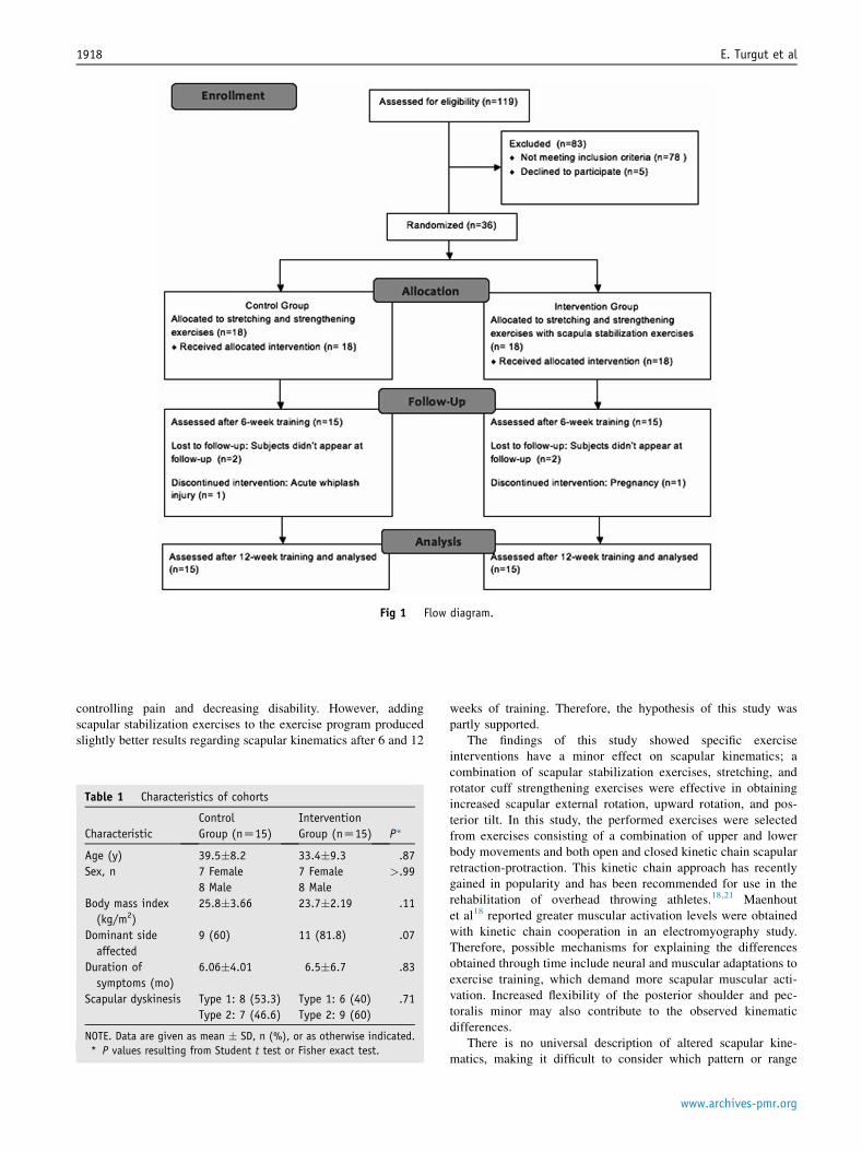
Fig 1 Flow diagram.
1918 E. Turgut et al
controlling pain and decreasing disability. However, addingscapular stabilization exercises to the exercise program producedslightly better results regarding scapular kinematics after 6 and 12
Table 1 Characteristics of cohorts
Characteristic
Control
Group (nZ15)
Intervention
Group (nZ15) P*
Age (y) 39.5�8.2 33.4�9.3 .87
Sex, n 7 Female 7 Female >.99
8 Male 8 Male
Body mass index
(kg/m2)
25.8�3.66 23.7�2.19 .11
Dominant side
affected
9 (60) 11 (81.8) .07
Duration of
symptoms (mo)
6.06�4.01 6.5�6.7 .83
Scapular dyskinesis Type 1: 8 (53.3) Type 1: 6 (40) .71
Type 2: 7 (46.6) Type 2: 9 (60)
NOTE. Data are given as mean � SD, n (%), or as otherwise indicated.
* P values resulting from Student t test or Fisher exact test.
weeks of training. Therefore, the hypothesis of this study waspartly supported.
The findings of this study showed specific exerciseinterventions have a minor effect on scapular kinematics; acombination of scapular stabilization exercises, stretching, androtator cuff strengthening exercises were effective in obtainingincreased scapular external rotation, upward rotation, and pos-terior tilt. In this study, the performed exercises were selectedfrom exercises consisting of a combination of upper and lowerbody movements and both open and closed kinetic chain scapularretraction-protraction. This kinetic chain approach has recentlygained in popularity and has been recommended for use in therehabilitation of overhead throwing athletes.18,21 Maenhoutet al18 reported greater muscular activation levels were obtainedwith kinetic chain cooperation in an electromyography study.Therefore, possible mechanisms for explaining the differencesobtained through time include neural and muscular adaptations toexercise training, which demand more scapular muscular acti-vation. Increased flexibility of the posterior shoulder and pec-toralis minor may also contribute to the observed kinematicdifferences.
There is no universal description of altered scapular kine-matics, making it difficult to consider which pattern or range
www.archives-pmr.org

Table 2 Results of scapular internal-external rotation evaluated
before and after 6 and 12 weeks of training
HTE Level
Within-Subjects Effects
Between-
Subjects Effects
Control
Group
Intervention
Group P
30� elevation
Baseline 35.69�6.15 40.61�4.23 <.001
6th week 36.13�6.30 35.86�3.88*
12th week 35.81�5.28 33.21�4.81y
P .68 <.001
60� elevation
Baseline 40.68�6.35 45.08�5.62 <.001
6th week 41.09�6.39 39.93�5.08*
12th week 41.51�6.76 37.52�6.70y
P .31 <.001
90� elevation
Baseline 42.78�6.62 47.56�5.64 <.001
6th week 43.02�5.62 40.33�5.84*
12th week 43.53�5.57 38.69�7.95y
P .35 <.001
120� elevation
Baseline 36.89�8.44 42.73�7.58 <.001
6th week 37.02�7.92 34.80�6.22*
12th week 38.62�7.16 33.9�8.63y
P .17 <.001
120� lowering
Baseline 35.81�8.72 41.01�7.14 <.001
6th week 35.98�8.88 33.56�7.32*
12th week 37.66�7.25 33.13�8.67y
P .20 <.001
90� lowering
Baseline 39.05�7.16 46.54�5.32 <.001
6th week 39.16�6.96 39.22�6.81*
12th week 39.54�6.29 36.43�7.46y
P .50 <.001
60� lowering
Baseline 38.51�6.18 44.89�5.48 <.001
6th week 38.35�6.16 37.85�5.65*
12th week 37.86�6.45 36.17�6.32y
P .51 <.001
30� lowering
Baseline 36.14�6.04 41.92�4.46 <.001
6th week 36.97�4.79 35.68�4.98*
12th week 35.29�6.05 32.77�5.31y
P .25 <.001
NOTE. Data are given as mean � SD or as otherwise indicated. Scapular
kinematics are presented in degrees.
Abbreviation: HTE, humerothoracic elevation level.
* Significant statistical difference based on pairwise comparisons
between baseline and week 6 (P<.001).y Significant statistical difference based on pairwise comparisons
between baseline and week 12 (P<.05).
Table 3 Results of scapular upward-downward rotation evalu-
ated before and after 6 and 12 weeks of training
HTE Level
Within-Subjects Effects
Between-
Subjects Effects
Control
Group
Intervention
Group P
30� elevation
Baseline �1.97�4.67 2.19�5.64 .01
6th week �1.72�5.53 �.90�6.21
12th week �1.70�5.05 �2.77�6.58*
P .88 .02
60� elevation
Baseline �10.64�5.49 �4.85�7.47 .08
6th week �10.16�6.32 �7.68�7.31
12th week �11.33�5.07 �9.18�6.81
P NT NT
90� elevation
Baseline �17.53�4.20 �12.39�6.56 .07
6th week �18.47�4.86 �15.83�6.05
12th week �18.88�3.31 �17.01�6.25
P NT NT
120� elevation
Baseline �20.09�5.12 �18.13�6.81 .31
6th week �21.26�5.28 �21.62�6.89
12th week �21.48�4.80 �20.82�8.68
P NT NT
120� lowering
Baseline �19.87�5.21 �17.51�7.82 .42
6th week �20.43�6.20 �20.11�7.82
12th week �20.67�5.12 �19.91�9.15
P NT NT
90� lowering
Baseline �17.44�5.14 �11.70�6.76 .07
6th week �17.46�6.44 �14.80�7.17
12th week �18.06�5.11 �15.95�8.03
P NT NT
60� lowering
Baseline �12.11�5.41 �5.35�6.65 .03
6th week �10.71�6.30 �8.73�6.87
12th week �12.50�5.06 �11.18�8.07*
P .11 .02
30� lowering
Baseline �2.58�5.83 2.33�5.83 .22
6th week �1.86�7.21 �.91�6.40
12th week �2.45�6.78 �1.35�8.87
P NT NT
NOTE. Data are given as mean � SD or as otherwise indicated. Scapular
kinematics are presented in degrees.
Abbreviations: HTE, humerothoracic elevation level; NT, not tested.
* Significant statistical difference based on pairwise comparisons
between baseline and week 12 (P<.05).
Exercise training for subacromial impingement 1919
should be considered normative. Increased scapular internalrotation,4 anterior tilt,3,28,29 and decreased upward rotation4,28
have been shown to be related to impingement symptoms.30 Ourfindings showed that there were increased external rotation andposterior tilt after 6 weeks of training and increased upward
www.archives-pmr.org
rotation after 12 weeks of training in the intervention group.However, independent from kinematic changes, pain severity andself-reported disability lessened to a similar degree from the sixthweek onward, compared with the baseline measurements, for bothstudy groups. Perceived results in improvement were the sameregardless of which intervention the participant received. Thistrend can be explained by the complexity of the pain being studied
Table 4 Results of scapular anterior-posterior tilt evaluated
before and after 6 and 12 weeks of training
HTE Level
Within-Subjects Effects
Between-
Subjects Effects
Control
Group
Intervention
Group P
30� elevation
Baseline �14.37�6.07 �15.30�5.46 <.001
6th week �13.25�5.51 �11.93�4.41*
12th week �13.42�5.56 �8.81�5.60y
P .20 <.001
60� elevation
Baseline �16.92�6.55 �17.38�6.75 <.001
6th week �17.39�6.08 �14.66�5.75*
12th week �16.99�6.75 �10.83�6.16y
P .75 <.001
90� elevation
Baseline �14.62�7.83 �16.43�7.66 <.001
6th week �14.62�8.16 �13.87�7.94*
12th week �14.26�7.30 �10.49�6.57y
P .85 <.001
120� elevation
Baseline �12.05�8.81 �10.78�12.22 .25
6th week �11.76�9.06 �9.85�8.64
12th week �12.09�8.81 �6.49�6.74
P - -
120� lowering
Baseline �12.28�8.50 �13.39�8.35 <.001
6th week �11.78�8.92 �9.96�8.73*
12th week �12.31�7.96 �6.61�7.08y
P .70 <.001
90� lowering
Baseline �15.81�6.47 �18.0�6.78 <.001
6th week �15.45�6.91 �15.42�8.05*
12th week �14.86�5.64 �14.86�7.59y
P .30 <.001
60� lowering
Baseline �16.80�6.80 �19.73�6.38 <.001
6th week �16.46�6.53 �17.32�6.69*
12th week �16.51�6.66 �13.11�6.01y
P .70 <.001
30� lowering
Baseline �16.10�5.93 �18.46�5.86 <.001
6th week �15.41�5.98 �14.49�5.06*
12th week �14.36�5.78 �11.71�6.16y
P .05 <.001
NOTE. Data are given as mean � SD or as otherwise indicated. Scapular
kinematics are presented in degrees.
Abbreviation: HTE, humerothoracic elevation level.
* Significant statistical difference based on pairwise comparisons
between baseline and week 6 (P<.05).y Significant statistical difference based on pairwise comparisons
between baseline and week 12 (P<.05).
Table 5 Results of self-reported disability and pain severity
evaluated before and after 6 and 12 weeks of training
HTE Level
Within-Subjects Effects
Between-
Subjects
Effects
Control
Group
Intervention
Group P
SPADI pain
Baseline 56.30�24.30 56.93�24.44 .05
6th week 33.20�16.91 26.83�22.35
12th week 27.86�21.32 13.36�12.95
SPADI disability
Baseline 41.58�22.96 36.08�22.23 .50
6th week 24.12�17.26 16.82�19.59
12th week 19.42�20.16 7.00�10.34
SPADI total
Baseline 47.25�22.94 44.07�21.66 .24
6th week 27.95�16.75 20.18�20.45
12th week 22.18�20.16 9.23�11.21
Pain at rest
Baseline .87�1.98 .62�1.24 .54
6th week 0�0 .25�.87
12th week .18�.72 0�0
Pain during activity
Baseline 5.32�2.99 4.84�2.30 .86
6th week 2.36�2.54 1.52�1.58
12th week 1.26�2.78 .38�1.01
Pain at night
Baseline 2.28�3.36 2.63�3.74 .57
6th week .62�1.49 .52�1.36
12th week .53�1.80 0�0
NOTE. Data are given as mean � SD or as otherwise indicated. SPADI
scores are presented in points, and pain severity are presented in
centimeters.
Abbreviation: HTE, humerothoracic elevation level.
1920 E. Turgut et al
(eg, perception of pain) that is regulated at the spinal and corticallevel and is often influenced by psychosocial conditions.31
Three-dimensional scapular kinematics recording with elec-tromagnetic tracking has recently been used in participants withand without impingement symptoms, but the measurement wasnot found to be highly reliable over time.23 Haik et al23 previously
reported MDC values of up to 17.27�. Standard error of mea-surement for each kinematic variable was calculated, and the re-sults suggested that the kinematic changes for some variables weregreater than the MDC value. Therefore, it can be considered truechanges mostly after 12-week training. Furthermore, kinematicdifferences in the magnitude of scapular rotations in the currentstudy reached the suggested values for symmetric scapular motionthat were described by Uhl et al.13 Our observed differences werealso similar to previously measured results that compared symp-tomatic and asymptomatic individuals.28
Appropriate scapular posterior tilt is one factor that can elevatethe anterior acromion during humeral elevation and, in turn, in-crease the subacromial space.32 The findings of this study supportprevious reports that exercise training results in increased scapularposterior tilt and decreased impingement symptoms.10,33 Royet al33 reported improved SPADI scores and increased posteriortilt at 70� of sagittal plane elevation after a 4-week progressivescapular motor control and strengthening training. Similarly,Worsley et al10 reported an improved SPADI score of approxi-mately 10 points and increased posterior tilt of approximately 3.7�
at 90� of elevation after 10 weeks of motor control retrainingexercises for the shoulder. Our study showed improved SPADIscores of up to 23.8 and 34.8 points for the intervention group, and
www.archives-pmr.org

Fig 2 Scapular internal-external rotation during shoulder elevation and lowering among study groups evaluated before and after 6 and 12
weeks of training. *Statistically significant group by time interaction.
Exercise training for subacromial impingement 1921
an increased posterior tilt at 90� of humeral elevation with amagnitude of 2.5� and 5.9� after 6 weeks and 12 weeks of training,respectively.
Study limitations
This study has some limitations. The findings of this study onlyapply to young adults diagnosed with stage 1 or 2 subacromialimpingement who had type 1 or 2 scapular dyskinesis; they are
Fig 3 Scapular upward-downward rotation during shoulder elevation an
weeks of training. *Statistically significant group by time interaction.
www.archives-pmr.org
not applicable to participants with chronic symptoms who alsoexhibit type 3 acromion or who have secondary impingementbecause of rotator cuff weakness. Additionally, the method usedto distinguish between types of scapular dyskinesis has poorreliability.13 From a methodologic perspective, the active controlgroup was preferable to a passive or placebo intervention. How-ever, previous reports have revealed that no intervention or pla-cebo application was found to have no relief on impingementssymptoms.34-36
d lowering among study groups evaluated before and after 6 and 12
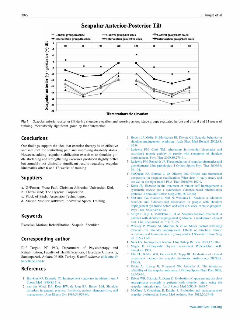
Fig 4 Scapular anterior-posterior tilt during shoulder elevation and lowering among study groups evaluated before and after 6 and 12 weeks of
training. *Statistically significant group by time interaction.
1922 E. Turgut et al
Conclusions
Our findings support the idea that exercise therapy is an effectiveand safe tool for controlling pain and improving disability status.However, adding scapular stabilization exercises to shoulder gir-dle stretching and strengthening exercises produced slightly betterbut arguably not clinically significant results regarding scapularkinematics after 6 and 12 weeks of training.
Suppliers
a. G*Power; Franz Faul, Christian-Albrechts-Universitat Kiel.b. Thera-Band; The Hygenic Corporation.c. Flock of Birds; Ascension Technologies.d. Motion Monitor software; Innovative Sports Training.
Keywords
Exercise; Motion; Rehabilitation; Scapula; Shoulder
Corresponding author
Elif Turgut, PT, PhD, Department of Physiotherapy andRehabilitation, Faculty of Health Sciences, Hacettepe University,Samanpazari, Ankara 06100, Turkey. E-mail address: [email protected].
References
1. Hawkins RJ, Kennedy JC. Impingement syndrome in athletes. Am J
Sports Med 1980;8:151-8.
2. van der Windt DA, Koes BW, de Jong BA, Bouter LM. Shoulder
disorders in general practice: Incidence, patient characteristics, and
management. Ann Rheum Dis 1995;54:959-64.
3. Hebert LJ, Moffet H, McFadyen BJ, Dionne CE. Scapular behavior in
shoulder impingement syndrome. Arch Phys Med Rehabil 2002;83:
60-9.
4. Ludewig PM, Cook TM. Alterations in shoulder kinematics and
associated muscle activity in people with symptoms of shoulder
impingement. Phys Ther 2000;80:276-91.
5. Ludewig PM, Reynolds JF. The association of scapular kinematics and
glenohumeral joint pathologies. J Orthop Sports Phys Ther 2009;39:
90-104.
6. McQuade KJ, Borstad J, de Oliveira AS. Critical and theoretical
perspective on scapular stabilization: What does it really mean, and
are we on the right track? Phys Ther 2016;96:1162-9.
7. Kuhn JE. Exercise in the treatment of rotator cuff impingement: a
systematic review and a synthesized evidence-based rehabilitation
protocol. J Shoulder Elbow Surg 2009;18:138-60.
8. McClure PW, Bialker J, Neff N, Williams G, Karduna A. Shoulder
function and 3-dimensional kinematics in people with shoulder
impingement syndrome before and after a 6-week exercise program.
Phys Ther 2004;84:832-48.
9. Struyf F, Nijs J, Mollekens S, et al. Scapular-focused treatment in
patients with shoulder impingement syndrome: a randomized clinical
trial. Clin Rheumatol 2013;32:73-85.
10. Worsley P, Warner M, Mottram S, et al. Motor control retraining
exercises for shoulder impingement: Effects on function, muscle
activation, and biomechanics in young adults. J Shoulder Elbow Surg
2013;22:e11-9.
11. Neer CS. Impingement lesions. Clin Orthop Rel Res 1983;173:70-7.
12. Magee D. Orthopaedic physical assessment. Philedelphia: W.B.
Saunders; 1997.
13. Uhl TL, Kibler WB, Gecewich B, Tripp BL. Evaluation of clinical
assessment methods for scapular dyskinesis. Arthroscopy 2009;25:
1240-8.
14. Rabin A, Irrgang JJ, Fitzgerald GK, Eubanks A. The intertester
reliability of the scapular assistance. J Orthop Sports Phys Ther 2006;
36:653-60.
15. Kibler WB, Sciascia A, Dome D. Evaluation of apparent and absolute
supraspinatus strength in patients with shoulder injury using the
scapular retraction test. Am J Sports Med 2006;34:1643-7.
16. McClure P, Greenberg E, Kareha S. Evaluation and management of
scapular dysfunction. Sports Med Arthrosc Rev 2012;20:39-48.
www.archives-pmr.org

Exercise training for subacromial impingement 1923
17. Sciascia A, Karolich D. A comprehensive approach to non-operative
rotator cuff rehabilitation. Curr Phys Med Rehabil Rep 2013;1:29-37.
18. Maenhout A, Van Praet K, Pizzi L, Van Herzeele M, Cools A. Electromyo-
graphic analysis of knee push up plus variations: what is the influence of the
kinetic chain on scapular muscle activity? Br Sports Med 2010;44:1010-5.
19. Kibler WB, Sciascia AD, Uhl TL, Tambay N, Cunningham T. Elec-
tromyographic analysis of specific exercises for scapular control in early
phases of shoulder rehabilitation. Am J Sports Med 2008;36:1789-98.
20. De Mey K, Danneels L, Cagnie B, Van den Bosch L, Flier J,
Cools AM. Kinetic chain influences on upper and lower trapezius
muscle activation during eight variations of a scapular retraction
exercise in overhead athletes. J Sci Med Sport 2013;16:65-70.
21. McMullen J, Uhl TL. A kinetic chain approach for shoulder rehabil-
itation. J Athl Train 2000;35:329-37.
22. Karduna AR, McClure PW, Michener LA, Sennett B. Dynamic
measurements of three-dimensional scapular kinematics: a validation
study. J Biomech Eng 2001;123:184-90.
23. Haik MN, Alburquerque-Sendın F, Camargo PR. Reliability and
minimal detectable change of 3-dimensional scapular orientation in
individuals with and without shoulder impingement. J Orthop Sports
Phys Ther 2014;44:341-9.
24. Meskers CG, van der Helm FC, Rozendaal LA, Rozing PM. In vivo
estimation of the glenohumeral joint rotation center from scapular
bony landmarks by linear regression. J Biomech 1998;31:93-6.
25. Wu G, van der Helm FC, Veeger HE, et al. ISB recommendation on
definitions of joint coordinate systems of various joints for the
reporting of human joint motion-Part II: shoulder, elbow, wrist and
hand. J Biomech 2005;38:981-92.
26. Bumin G, Tuzun EH, Tonga E. The Shoulder Pain and Disability
Index (SPADI): cross-cultural adaptation, reliability, and validity of
the Turkish version. J Back Musculoskel Rehabil 2008;21:57-62.
www.archives-pmr.org
27. Clark P, Lavielle P, Martinez H. Learning from pain scales: patient
perspective. J Rheumatol 2003;30:1584-8.
28. Borstad JD, Ludewig PM. Comparison of scapular kinematics be-
tween elevation and lowering of the arm in the scapular plane. Clin
Biomech 2002;17:650-9.
29. Ludewig PM, Cook TM. Translations of the humerus in persons with
shoulder impingement symptoms. J Orthop Sports Phys Ther 2002;32:
248-59.
30. McClure PW, Michener LA, Sennett BJ, Karduna AR. Direct
3-dimensional measurement of scapular kinematics during dynamic
movements in vivo. J Shoulder Elbow Surg 2001;10:269-77.
31. Price DD. Psychological and neural mechanisms of the affective
dimension of pain. Science 2000;288:1769-72.
32. Brossmann J, Preidler KW, Pedowitz RA, White LM, Trudell D,
Resnick D. Shoulder impingement syndrome: Influence of shoulder
position on rotator cuff impingement-an anatomic study. AJR Am J
Roentgenol 1996;167:1511-5.
33. Roy JS, Moffet H, Hebert LJ, Lirette R. Effect of motor control and
strengthening exercises on shoulder function in persons with
impingement syndrome: a single-subject study design. Man Ther
2009;14:180-8.
34. Blair B, Rokito AS, Cuomo F, Jarolem K, Zuckerman JD. Efficacy of
injections of corticosteroids for subacromial impingement syndrome. J
Bone Joint Surg Am 1996;78:1685-9.
35. Brox JI, Staff PH, Ljunggren AE, Brevik JI. Arthroscopic sur-
gery compared with supervised exercises in patients with rotator
cuff disease (stage II impingement syndrome). BMJ 1993;307:
899-903.
36. Ginn KA, Herbert RD, Khouw W, Lee R. A randomized, controlled
clinical trial of a treatment for shoulder pain. Phys Ther 1997;77:
802-9.

Supplemental Appendix S1 Description of exercises
Exercise Description Dosage Groups Figure
Pectoralis minor
stretching
Subject is standing, arms positioned at wall with
elbow flexion into 90�; subject performsstretching with leaning forward.
5 reps � 3 Control,
intervention
Posterior shoulder
stretching
Subject is sitting, arm positioned at flexion and
horizontal adduction; subject stretches posterior
shoulder without producing any anterior shoulder pain.
5 reps � 3 Control,
intervention
Levator scapula
stretching
Subject is sitting, hand positioned interscapular
region, and performs cervical lateral flexion.
5 reps � 3 Control,
intervention
Latissimus dorsi
stretching
Subject is sitting, hand positioned interscapular
region, other hand supports the elbow and performs
trunk lateral flexion and slight rotation and flexion.
5 reps � 3 Control,
intervention
Resisted shoulder
internal rotation
Subject is standing with 0� shoulder abduction; resistance
band is fixed laterally at waist level; subject performs
shoulder internal rotation with fixed elbow.
10 reps � 3 Control
Resisted shoulder
external rotation
Subject is standing with 0� shoulder abduction; resistance
band is fixed laterally at waist level; subject performs
shoulder external rotation with fixed elbow.
10 reps � 3 Control
Resisted full can Subject is standing, resistance band is fixed under foot;
subject performs shoulder scapular plane elevation
with elbow in extension.
10 reps � 3 Control
(continued on next page)
1923.e1 E. Turgut et al
www.archives-pmr.org

Supplemental Appendix S1 (continued )
Exercise Description Dosage Groups Figure
Wall slides with
squat
Subject is in squat position, hand positioned
on wall; while returning erect position subject
performs scapular with elbow into flexion.
10 reps � 3 Intervention
Wall pushups with
ipsilateral leg
extension
Subject standing, both hands positioned on wall;
subject performs scapular protraction with ipsilateral
leg extension.
10 reps � 3 Intervention
Lawnmower with
diagonal squat
Subject is in squat position, resistance band
is fixed under contralateral foot; subject performs
scapular retraction while returning erect and
rotated position.
10 reps � 3 Intervention
Resisted scapular
retraction with
contralateral
1-leg squat
Subject is in 1-leg squat position, resistance band is fixed
at waist level; subject performs scapular retraction.
10 reps � 3 Intervention
Robbery with squat Subject is in squat position; subject performs scapular
retraction and shoulder external rotation while
returning erect position.
10 reps � 3 Intervention
Resisted shoulder
internal rotation
with step
Subject is standing with 0� shoulder abduction;
resistance band is fixed laterally at waist level;
subject performs shoulder internal rotation with
ipsilateral inward step.
10 reps � 3 Intervention
Resisted shoulder
external rotation
with step
Subject is standing with 0� shoulder abduction;
resistance band is fixed laterally at waist level;
subject performs shoulder external rotation with
ipsilateral sidestep.
10 reps � 3 Intervention
(continued on next page)
Exercise training for subacromial impingement 1923.e2
www.archives-pmr.org
Supplemental Appendix S1 (continued )
Exercise Description Dosage Groups Figure
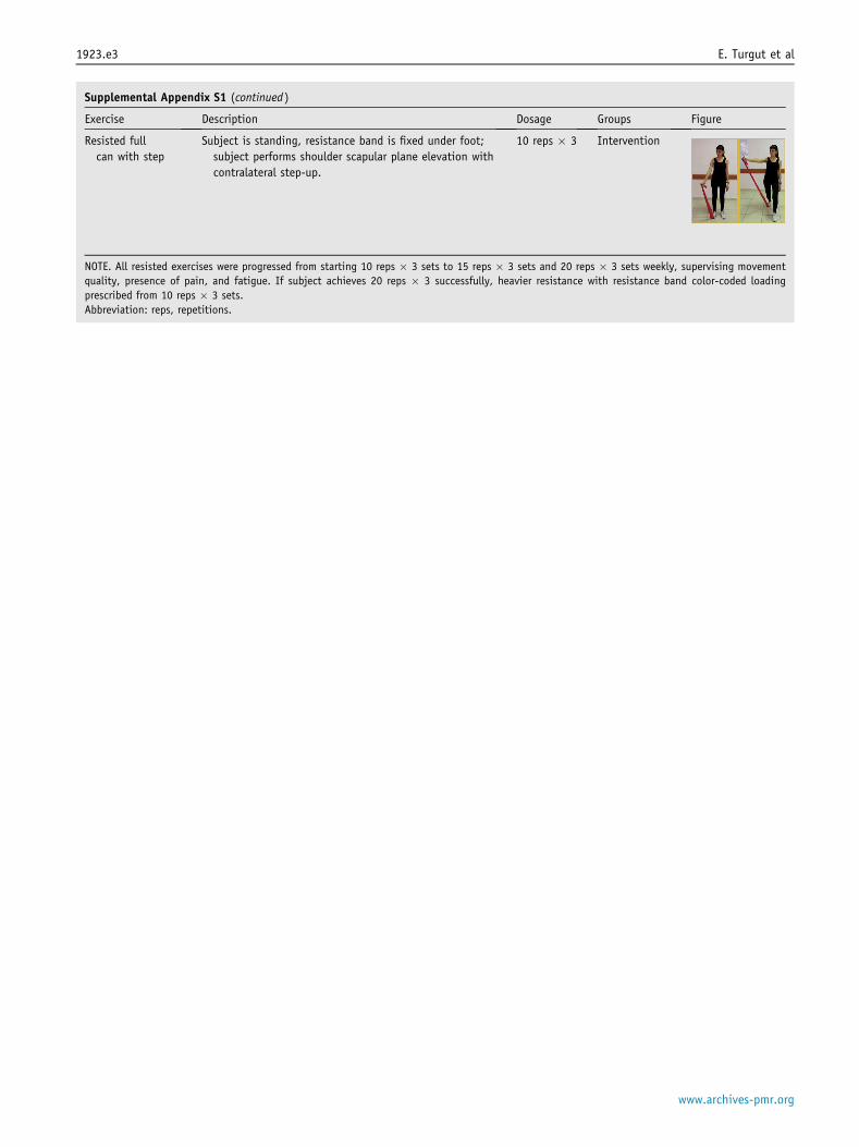
Resisted full
can with step
Subject is standing, resistance band is fixed under foot;
subject performs shoulder scapular plane elevation with
contralateral step-up.
10 reps � 3 Intervention
NOTE. All resisted exercises were progressed from starting 10 reps � 3 sets to 15 reps � 3 sets and 20 reps � 3 sets weekly, supervising movement
quality, presence of pain, and fatigue. If subject achieves 20 reps � 3 successfully, heavier resistance with resistance band color-coded loading
prescribed from 10 reps � 3 sets.
Abbreviation: reps, repetitions.
1923.e3 E. Turgut et al
www.archives-pmr.org


![]Y]989]R^ `X89ZV^U`T#RD8VP]U^9]T]PW PY]^]P8YaR]^9RT7]U`b · cluding rotator cu! tendinopathy, rotator cu! tears, traumatic, atraumatic and acquired shoulder instability, , scapular](https://static.fdocuments.net/doc/165x107/5ffa2b31c650512cba751436/y989r-x89zvutrd8vpu9tpw-pyp8yar9rt7ub-cluding-rotator-cu-tendinopathy.jpg)