Diaphragmdisease: of small non-steroidaljcp.bmj.com/content/jclinpath/41/5/516.full.pdf ·...
11
J Clin Pathol 1988;41:516-526 Diaphragm disease: pathology of disease of the small intestine induced by non-steroidal anti-inflammatory drugs J LANG,* A B PRICE,* A J LEVI,t M BURKE,§ J M GUMPEL,4 I BJARNASONt From the Departments of *Histopathology, tGastroenterology, ,Rheumatology, and §Surgery, Northwick Park Hospital and Clinical Research Centre, Harrow, Middlesex SUMMARY Operative small bowel resection specimens received over a period of 16 years were reviewed to assess whether any intestinal disease could be directly attributed to the use of non- steroidal anti-inflammatory drugs (NSAID). Seven cases of intestinal disease associated with the use of NSAID were identified, all of which occurred in the final six years of the survey, which may reflect the increasing use of these compounds. A spectrum of patterns was found from multiple pathognomonic ileal mucosal diaphragms to broad strictures similar to those seen as a complication of enteric potassium. It seems likely that the formation of diaphragm lesions requires an additional factor, but wlhat is not known as yet is whether the effects of NSAID are local or systemic. Several non-steroidal anti-inflammatory drugs (NSAID) cause characteristic small bowel lesions in animals. This is most severe in rats in which indometh- acin leads to multiple small bowel ulcers, mainly in the ileum.' 2 The ulcers often perforate, with consequent peritonitis, and death may result after as little as a single subcutaneous dose of 10 mg/kg. The pathogen- esis of these lesions is incompletely understood, but they can be prevented by treatment with prostaglan- dins and antibiotics. One hypothesis, therefore, is that a reduction in the mucosal synthesis of prostaglandins induced by non-steroidal anti-inflammatory drugs disrupts mucosal integrity and the subsequent ulcera- tion might then reflect bacterial invasion.35 Indirect evidence suggests that a similar process may occur in man. We have shown that NSAID swiftly disrupt small intestinal integrity in normal subjects assessed by a 5"chromium labelled ethylene- diaminetetra-acetate (5EDTA) absorption test, and the damage, which is likely to affect the intercellular junctions between enterocytes, seems to be propor- tional to the drugs' ability to inhibit cyclo-oxygenase. Furthermore, it is clear that long term treatment with NSAID in patients with rheumatoid and osteoarthritis is associated with distinct migratory abnormalities of "'indium-labelled leucocytes. Although these collect in the right iliac fossa, analysis of the kinetics of "'indium leucocyte accumulation suggests that the Accepted for publication 8 December 1987 whole of the small intestine may be inflamed."8 A four day estimation of faecal "'indium activity, which is a reliable objective index of intestinal inflammation, is abnormal in treated patients, while untreated ones invariably have normal results. There is now accumulating epidemiological and experimental data to show that NSAID are associated with a clinically important gastrointestinal blood and protein loss.9 The ileal dysfunction and sporadic reports of perfora- tion suggest that small intestinal inflammation associated with the use of NSAID is, in fact, a new nosological entity. ' " Histopathological confirmation of intestinal inflam- mation complementing these investigations is, however, scarce. These are accounts of potassium preparations causing small bowel ulceration, but many of these patients were also receiving NSAID.'2 There are also reports of small intestinal ulceration, perforations, and strictures induced by NSAID.'3- Our purpose was to review operative small bowel resection specimens received by the department of histopathology at Northwick Park Hospital over 16 years and to assess whether any intestinal disease could be identified, implicating NSAID as the cause of the pathology. The picture that emerged, as far as we are aware, seems to be unique to small intestinal pathology associated with NSAID, and we have termed this "diaphragm disease". 516 on 13 July 2018 by guest. Protected by copyright. http://jcp.bmj.com/ J Clin Pathol: first published as 10.1136/jcp.41.5.516 on 1 May 1988. Downloaded from
Transcript of Diaphragmdisease: of small non-steroidaljcp.bmj.com/content/jclinpath/41/5/516.full.pdf ·...

J Clin Pathol 1988;41:516-526
Diaphragm disease: pathology of disease of the smallintestine induced by non-steroidal anti-inflammatorydrugsJ LANG,* A B PRICE,* A J LEVI,t M BURKE,§ J M GUMPEL,4 I BJARNASONtFrom the Departments of *Histopathology, tGastroenterology, ,Rheumatology, and §Surgery, Northwick ParkHospital and Clinical Research Centre, Harrow, Middlesex
SUMMARY Operative small bowel resection specimens received over a period of 16 years werereviewed to assess whether any intestinal disease could be directly attributed to the use of non-steroidal anti-inflammatory drugs (NSAID). Seven cases of intestinal disease associated with the useofNSAID were identified, all ofwhich occurred in the final six years of the survey, which may reflectthe increasing use of these compounds. A spectrum of patterns was found from multiplepathognomonic ileal mucosal diaphragms to broad strictures similar to those seen as a complicationof enteric potassium. It seems likely that the formation of diaphragm lesions requires an additionalfactor, but wlhat is not known as yet is whether the effects ofNSAID are local or systemic.
Several non-steroidal anti-inflammatory drugs(NSAID) cause characteristic small bowel lesions inanimals. This is most severe in rats in which indometh-acin leads to multiple small bowel ulcers, mainly in theileum.' 2 The ulcers often perforate, with consequentperitonitis, and death may result after as little as asingle subcutaneous dose of 10 mg/kg. The pathogen-esis of these lesions is incompletely understood, butthey can be prevented by treatment with prostaglan-dins and antibiotics. One hypothesis, therefore, is thata reduction in the mucosal synthesis of prostaglandinsinduced by non-steroidal anti-inflammatory drugsdisrupts mucosal integrity and the subsequent ulcera-tion might then reflect bacterial invasion.35
Indirect evidence suggests that a similar processmay occur in man. We have shown that NSAIDswiftly disrupt small intestinal integrity in normalsubjects assessed by a 5"chromium labelled ethylene-diaminetetra-acetate (5EDTA) absorption test, andthe damage, which is likely to affect the intercellularjunctions between enterocytes, seems to be propor-tional to the drugs' ability to inhibit cyclo-oxygenase.Furthermore, it is clear that long term treatment withNSAID in patients with rheumatoid and osteoarthritisis associated with distinct migratory abnormalities of"'indium-labelled leucocytes. Although these collectin the right iliac fossa, analysis of the kinetics of"'indium leucocyte accumulation suggests that theAccepted for publication 8 December 1987
whole of the small intestine may be inflamed."8 A fourday estimation of faecal "'indium activity, which is areliable objective index of intestinal inflammation, isabnormal in treated patients, while untreated onesinvariably have normal results. There is nowaccumulating epidemiological and experimental datato show that NSAID are associated with a clinicallyimportant gastrointestinal blood and protein loss.9The ileal dysfunction and sporadic reports of perfora-tion suggest that small intestinal inflammationassociated with the use of NSAID is, in fact, a newnosological entity. ' "
Histopathological confirmation of intestinal inflam-mation complementing these investigations is,however, scarce. These are accounts of potassiumpreparations causing small bowel ulceration, butmany of these patients were also receiving NSAID.'2There are also reports of small intestinal ulceration,perforations, and strictures induced by NSAID.'3-Our purpose was to review operative small bowelresection specimens received by the department ofhistopathology at Northwick Park Hospital over 16years and to assess whether any intestinal diseasecould be identified, implicating NSAID as the cause ofthe pathology.The picture that emerged, as far as we are aware,
seems to be unique to small intestinal pathologyassociated with NSAID, and we have termed this"diaphragm disease".
516
on 13 July 2018 by guest. Protected by copyright.
http://jcp.bmj.com
/J C
lin Pathol: first published as 10.1136/jcp.41.5.516 on 1 M
ay 1988. Dow
nloaded from

Non-steroidal disease of the small intestineTable 1 Total small bowel resections 1971-86
Ischaemia (mechanical/vascular)
Surgical trauma and postoperative complications
Tumours
Congenital anomalies
Traumatic perforations
Radiation damage
Inflammatory
Miscellaneous
Normal (as part of other procedures)
Total excluded
163
48
99
71
5
9
15
28
Granulomas positive 39
(group 2) 26
19
457
Material and methods
Between January 1971 and March 1987, 576 smallbowel resections were examined in the department ofhistopathology at Northwick Park Hospital. Thepathology reports of these cases were reviewed andthose with clinical details and histological descriptionsdenoting a confident diagnosis of ischaemia, neo-plasia, operative complications, infections, congenitalanomalies and certain miscellaneous conditions, butwith pathognomonic histopathology, were excludedfrom further assessment (table 1). This accounted for457 cases.One hundred and nineteen cases remained, compris-
ing 93 diagnosed as Crohn's disease and an unclas-sified group of 26 (table 1). Study of the reports of thepatients with Crohn's disease identified 39 with gran-ulomas and 54 without. Where granulomas were notedthe diagnosis of Crohn's disease was accepted and theslides and clinical history were not reviewed.
Primary pathology due to NSAID was assumed tolie within the group of 54 cases of Crohn's diseasewithout granulomas (group 1) and the 26 cases (group2) that were unclassified. The clinical records andhistological sections of these two groups werereviewed in detail. Group 2 (unclassified) requireddivision into subgroups to encompass the range ofpathologies.
Table 2 Group 2: 26 unclassified cases
Predominant pathologyNo of
Subgroup cases Macroscopic Microscopic Drug Final diagnosisA 7 Multiple mucosal diaphragms Focal submucosal fibrosis NSAID latrogenic strictures
B 5 Single/multiple short Diffuse submucosal fibrosis 2 Crohn's disease,strictures
1 Healed tuberculosisI Recurrent hernia
Phenylbutazone 1 Iatrogenic strictureC 4 Perforation Acute inflammation Perforation ? cause
D 4 Short strictures and Apthoid ulcers, transmural inflammation Probable Crohn'sulceration disease
E 2 Hour glass deformity Full thickness inflammatory ulcers Potassium I latrogenic strictureI ? Coeliac disease
F 2 Focal areas of necrosis Ischaemia
G I Oedema Intense mucosal and submucosal inflammation ? Infection
H 1 Thick mucosa Extensive villous regenerative/dysplastic changes No diagnosis made
517
on 13 July 2018 by guest. Protected by copyright.
http://jcp.bmj.com
/J C
lin Pathol: first published as 10.1136/jcp.41.5.516 on 1 M
ay 1988. Dow
nloaded from

518Results
GROUP 1This group had been indexed Crohn's disease in theabsence of granulomas. On review all the cases hadsufficient pathological attributes for this diagnosis tobe accepted.'6 In particular, all showed a combinationof transmural inflammation, mostly as aggregates oflymphocytes, and fissuring ulceration. The clinical andradiological data supported the diagnosis. Two of thisgroup had clearly received NSAID before developingCrohn's disease, but in many cases the drug historywas inadequately documented at presentation.
GROUP 2This group comprised 26 small bowel resectionswithout a confident diagnostic label. After review ofthe macroscopic data (accompanied by photographsin many cases) the histology and the clinical historiesof these could be assigned to several broad subgroups(table 2). The drug history was then added and anattempt at a diagnostic conclusion was made. In somecases this final diagnosis was made easier by access tofollow up data not available at the time of resection.Table 2 shows that seven cases (group 2A) presented astriking macroscopic picture with characteristic his-tology which was accompanied by a convincing his-tory of recent ingestion ofNSAID. Two of these sevencases had been referred from other hospitals for apathological opinion because of their unusualfeatures. From among the other subgroups in table 2group 2B comprised five with strictures predominantlydue to submucosal fibrosis. In this respect there wassome similarity with the group 2A NSAID group (seebelow). One of this group had received NSAID. Twomore patients from other groups had regularly taken adrug with recognised gastrointestinal side effects, butno others were receiving NSAID.
Fig I Bland serosal aspect ofresected ileum (case 3)emphasises how dificult it isfor surgeons to appreciate thelesions seen (fig 2) before the bowel is opened.
Lang, Price, Levi, Burke, Gumpel, BjarnasonGROUP 2AThe macroscopic features in the five cases resected atNorthwick Park Hospital were similar and dramatic.The resections were lengths of ileum from 14 cm to 80cm which never included the terminal ileum. There wasmild or moderate dilatation of the bowel and thicken-ing of what, from the serosal aspect, were assumed tobe mucosal folds (fig 1).
This relatively bland external appearance contras-ted with the mucosal aspect. The lumen of the ileum inall cases was divided into compartments by multiplethin circumferential mucosal membranes (fig 2). Thefeatures were clearly seen because of formalin inflationof the specimens prior to opening. Each resembled thewasher of a tap or a perforated diaphragm. Theluminal orifices of these divisions varied from 3-4 mmto a width only a little less than the full intestinaldiameter (figs 3 a and b). At the latter sites theyappeared as little more than exaggerated plicae cir-culares but were circumferential and had a slightlybroader base. The distribution of these diaphragmlesions along the lengths of resected ileum was uneven,varying from every 2-3 cm to up to 10-15 cm. In thelongest resected specimen of 70 cm 19 were presentalong its length. In this instance the surgeon hadalready carried out a large number of stricturoplastiesat an initial laparotomy (table 3). Pinpoint ulcerationcould be seen around the luminal margins of thediaphragms in some specimens. Within the intestinalcompartments formed by these diaphragms focalhaemorrhagic roughened areas and occasional tinyulcers were present but the bulk of the mucosaappeared normal. The macroscopic description ofcases 6 and 7, the two referred cases (table 3) was of
Fig 2 Resected length ofileum shows multiple mucosaldiaphragms (case 2). One tablet (not an NSAID) wasfoundwithin one compartment.
on 13 July 2018 by guest. Protected by copyright.
http://jcp.bmj.com
/J C
lin Pathol: first published as 10.1136/jcp.41.5.516 on 1 M
ay 1988. Dow
nloaded from

Non-steroidal disease of the small intestine
Fig 3 (a) Direct view ofmucosal diaphragms illustratevariation in width ofluminal apertures; (b) appearancesdiffer littlefrom prominent plicae circulares.
multiple thickened ridges along the resected lengths ofileum.
Crohn's disease was the favoured initial medical andsurgical diagnosis in the earlier cases but more recentlyclinicians, aware of the condition, have suspected thediagnosis prior to laparotomy. One- patient, case 4(table 3), had received previous radiotherapy and a
stricture caused by radiation was thought likely. Onthe basis of recurrent obstruction four of the six cases
required two resections: in case 7 (table 3) these were
eight years apart.
519
MICROSCOPYThe main histological findings were limited to thevicinity of the macroscopic changes and the occasionalsmall focal lesions noted within dilated bowel seg-
ments. Two slightly different patterns were seen butboth were characterised by the pattern offibrosis in thesuperficial halfof the submucosa immediately beneaththe muscularis. The first pattern related to the thinoccluding mucosal diaphragms, the second to thebroader based ridges with less luminal occlusion.
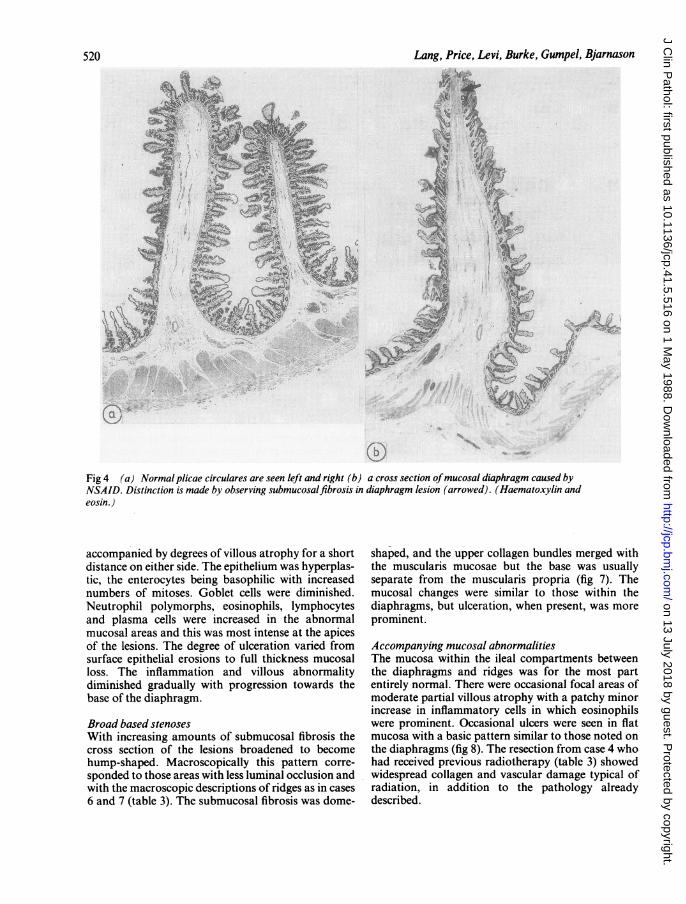
MUCOSAL DIAPHRAGMSEach had the basic structure of the normal plicaecirculares-that is, a thin fold of mucosa, muscularismucosae, and submucosa. No muscularis propria wasaffected. Characteristic and present in all the "dia-phragmatic" lesions examined was a focus of sub-mucosal fibrosis. This was best appreciated at lowpowers of magnification, which emphasised the direc-tional symmetry of collagen bundles aligned towardsthe apex of the lesion (figs 4 and 5). The muscularismucosae appeared to break up and merge with thisunderlying fibrosis. In some it was only the condensa-tion of collagen bundles seen beneath the muscularis,together with minor overlying mucosal inflammationand villous irregularity that helped distinguish thelesions from normal plicae circulares (figs 5 and 6).The mucosa at the apex was mildly but invariably
inflamed, with or without shallow ulceration. This was
Table 3 Clinical details ofpatients with small bowel disease induced by NSAID
CaseNo Age Clinicalfeatures Drugs Surgery Outcome
1 70 Osteoarthritis 2 years Piroxicam (18 months) 2 resections WellAnaemia 4 weeks apartSubacute obstruction 70 cm and 30 cm of
proximal, and mid-ileum
2* 71 Seropositive rheumatoid arthritis Long acting indomethacin; various others 2 resections Died from post-28 years over 16 years 6 months apart 27 cm operativeAnaemia and 14 cm of complicationsSubacute obstruction proximal ileum
3* 40 Seropositive rheumatoid arthritis Azapropazone 3 years; aspirin and various 1 resection 50 cm mid- Well30 years othersin past ileumAnaemiaSubacute obstruction
4 75 Osteoarthritis 15 years Piroxicam I resection Probable additionalSubacute obstruction Ketoprofen 20 cm mid-ileum radiation damageBreast and cervical cancer BrufenRadiotherapy
5* 64 Seropositive rheumatoid arthritis Piroxicam 3 years 1 resection Well15 years 15 cm distal ileumSubacute obstruction 15 cm distal ileum
6 72 Osteoarthritis? years Piroxicam 5 years and various others I resection 5 cm distal ?ileum
7 78 Osteoarthritis many years Wide variety but no details available 2 resections, 8 years ?apart, 7 cm and14 cm mid-ileum
*These patients were also receiving disease modifying drugs such as gold, penicillamine, salazopyrine.
on 13 July 2018 by guest. Protected by copyright.
http://jcp.bmj.com
/J C
lin Pathol: first published as 10.1136/jcp.41.5.516 on 1 M
ay 1988. Dow
nloaded from
z (o
;t~~~~~~~~~~~~~~~~~~~~~~~~~~~~~~~~~~~~~~~~~ ,f.
Fig 4 (a) Normal plicae circulares are seen left and right (b) a cross section ofmucosal diaphragm caused byNSAID. Distinction is made by observing submucosalfibrosis in diaphragm lesion (arrowed). (Haematoxylin andeosin.)
accompanied by degrees of villous atrophy for a shortdistance on either side. The epithelium was hyperplas-tic, the enterocytes being basophilic with increasednumbers of mitoses. Goblet cells were diminished.Neutrophil polymorphs, eosinophils, lymphocytesand plasma cells were increased in the abnormalmucosal areas and this was most intense at the apicesof the lesions. The degree of ulceration varied fromsurface epithelial erosions to full thickness mucosalloss. The inflammation and villous abnormalitydiminished gradually with progression towards thebase of the diaphragm.
Broad based stenosesWith increasing amounts of submucosal fibrosis thecross section of the lesions broadened to becomehump-shaped. Macroscopically this pattern corre-sponded to those areas with less luminal occlusion andwith the macroscopic descriptions of ridges as in cases6 and 7 (table 3). The submucosal fibrosis was dome-
shaped, and the upper collagen bundles merged withthe muscularis mucosae but the base was usuallyseparate from the muscularis propria (fig 7). Themucosal changes were similar to those within thediaphragms, but ulceration, when present, was moreprominent.
Accompanying mucosal abnormalitiesThe mucosa within the ileal compartments betweenthe diaphragms and ridges was for the most partentirely normal. There were occasional focal areas ofmoderate partial villous atrophy with a patchy minorincrease in inflammatory cells in which eosinophilswere prominent. Occasional ulcers were seen in flatmucosa with a basic pattern similar to those noted onthe diaphragms (fig 8). The resection from case 4 whohad received previous radiotherapy (table 3) showedwidespread collagen and vascular damage typical ofradiation, in addition to the pathology alreadydescribed.
Lang, Price, Levi, Burke, Gumpel, Bjarnason520
on 13 July 2018 by guest. Protected by copyright.
http://jcp.bmj.com
/J C
lin Pathol: first published as 10.1136/jcp.41.5.516 on 1 M
ay 1988. Dow
nloaded from

Non-steroidal disease of the small intestine
AT;tA
Fig 5 Higher power illustration offig 4 to emphasise intact muscularis mucosae in normal plicae circulares (a) andsubmucosalfibroticfocus at apex ofdiaphragm (b) . (Haematoxylin and eosin.)
... 4
tarFig 6 Complete cross section ofdiaphragm through itsaperture. Plicae are seen to one side. (Haematoxylin andeosin.)
PATHOLOGY OF OTHER GROUPSGroup 2BAssembled here are five cases with submucosal fibrosisas the main microscopic finding, associated with someoverlying mucosal villous distortion that suggestedprevious damage. There were no other histologicalclues to permit a confident diagnostic classification atthe time of resection: only one had received NSAID.The macroscopic pathology in all five was differentfrom that described above. The strictures were at least2-3 cm in length and never exceeded two in number.Nothing resembling the diaphragm lesions was seen.On review, and with the benefit of clinical and in
some cases biopsy follow up data, two cases werereclassified as Crohn's disease. One case, in view of thehistory, was accepted as old tuberculosis, and thechanges in another were assumed related to theaffected intestine being caught recurrently in a hernialsac (fig 9).The final case in this group is of interest, but the
521
on 13 July 2018 by guest. Protected by copyright.
http://jcp.bmj.com
/J C
lin Pathol: first published as 10.1136/jcp.41.5.516 on 1 M
ay 1988. Dow
nloaded from

Lang, Price, Levi, Burke, Gumpel, Bjarnason
.4,,
.4 '?#
Fig 9 Resected ileumfrom patient in group 2b showingsubmucosalfibrosis (arrowed) producing a stricture. Bowelwas caught in a recurrent hernia. (Haematoxylin and eosin.)
Fig 7 Ridge or humped pattern oflesion seen in severalcases, but particularly cases 6(a) and 7(b). Submucosalfibrosis is conspicuous, but a gap is seen between it and themuscularis propia. Note complete absence ofany muralinflammation and localised nature ofdeformity. There is apronounced difference in the degree of ulceration between thetwo lesions. (Haematoxylin and eosin.)
diagnosis remains equivocal. There was a long historyof phenylbutazone administration for lower backpain. An equivocal diagnosis ofankylosing spondylitiswas lacking. The resected specimen contained twoshort flat ileal strictures separated by 10 cm of mildly
tv 8 * iB+;*<O1;
-i J4
Fig 8 Ulceration andfibrosis in flat mucosa, an occasionalfinding. (Haematoxylin and eosin.)
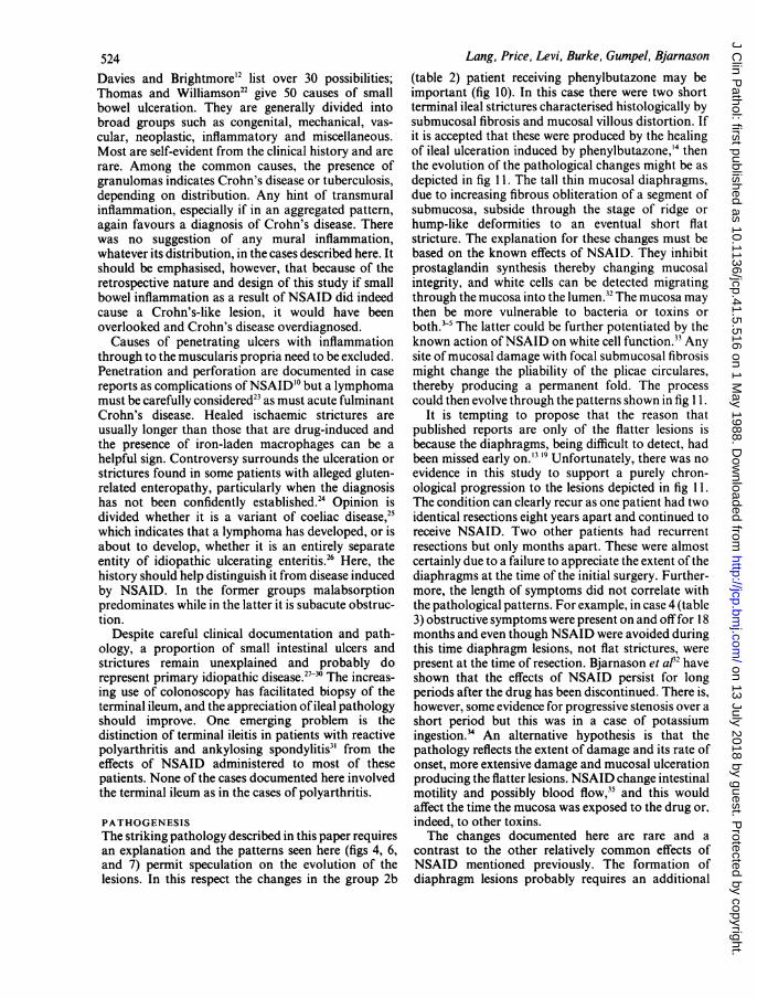
Fig 10 (a) Resected ileumfrom the patient in group 2bwho had receivedphenylbutazone. A stricture (arrowed) isseen towards the right margin; (b) strictured area seen athigher magnification (Haematoxylin and eosin.)
inflamed mucosa. Microscopy of the strictures showedonly submucosal fibrosis, break up of the muscularis,and villous irregularity suggesting old healed ulcera-tion (fig 10). The case was lost to follow up at threeyears. It was felt there were insufficient grounds toclassify this case as Crohn's disease, old tuberculosis,or ischaemia. Healed ulceration due to phenyl-butazone is a possibility.
Groups C-HThe pathology of the resected specimens in the
remainder of the unclassified cases did not resemblethose in group 2A, nor was there a history ofNSAID.In group 2E one resection showed a tight hour-glassdeformity with full thickness inflammation and ulcera-tion. The patient had been receiving potassium sup-plements. While interesting, these remaining cases arebeyond the scope of this study.
522
;a W
on 13 July 2018 by guest. Protected by copyright.
http://jcp.bmj.com
/J C
lin Pathol: first published as 10.1136/jcp.41.5.516 on 1 M
ay 1988. Dow
nloaded from

Non-steroidal disease of the small intestine
0 00000°°0°oc:ooFig 1 I Possible evolution ofmucosal and submucosal changes seen after NSAID (a) thin mucosal diaphragm which byprogressive submucosalfibrosis (arrows) broadens to b and ultimately to aflat stricture c.
Discussion
This investigation identified a small group of patientstaking NSAID who develop what seems to be apathognomonic macroscopic picture of ileal diseaseaccompanied by typical, though probably not specific,histological changes. The pattern was of segmentationmainly of the mid-ileum brought about by the forma-tion of incomplete diaphragms of mucosa. At oneextreme, they appeared as grossly exaggerated plicaecirculares reducing the lumen to a few millimetres (figs4-6): at the other, the mucosal and submucosalabnormalities were blunter forming a ridge or hump(fig 7). There was often circumferential ulceration. Thelength of the bowel compartment formed varied from3-4 cm up to 10-15 cm. The dramatic mucosal aspectcontrasted sharply with a misleadingly blandappearance seen by the surgeon at laparotomy (figs 1and 2).The important histological features relate to the
different gross appearances of the thin diaphragmsand the broader based lesions. What was striking andthe best clue to the diagnosis in both, was the patternof submucosal fibrosis. In the thin diaphragms it waslimited to a tiny submucosal focus at the luminalborder (fig Sb) but became dome-shaped in the lesionswith a broader base (fig 7). In the thinnest lesions thecollagen bundles were aligned at right angles to themucosa (figs 4 and 5). It is this fibrosis that distingui-shes these very thin diaphragms from plicae circulares.The mucosa, when ulcerated, remained flat or slightlyraised rather than excavated. This also applied to theoccasional ulcerative foci in flat areas of mucosa (fig8). The inflammatory changes and villous distortion
accompanying the lesion seemed to be secondary andnon-specific.
DIFFERENTIAL DIAGNOSISThe multiple thin diaphragms produced a pattern that,at the time ofwriting, seemed a unique complication ofNSAID ingestion. The broader based stenoses,although also highly suggestive, cannot be consideredto be specific, and open up several diagnoses for thepathologist and clinician to consider. For example,they conform more to the pattern of iatrogenicstrictures and ulceration described by other authors inrelation to potassium salts.'2" 18 It is interesting thatseveral reported cases of patients with alleged ulcera-tion induced by potassium were also receivingNSAID.'2 It is not clear, however, why other auth-ors'3 '4' 9 reporting ulceration and perforation due toNSAID have not observed the striking diaphragmpatterns described here.20 It may be relevant that theclinical diagnosis of the diaphragm lesions is difficult,requiring a high degree of suspicion and awareness.Because they are thin and do not distort the bowel wallthey are not, or only rarely, shown by conventionalradiological techniques. Even at surgery they are easilymissed unless specifically sought. The ridged lesionswith broader ulceration, however, pose no diagnosticchallenge for the experienced radiologist. The status ofthe "mucosal diaphragms" illustrated by Thompson2'in a review of the pathology of coeliac disease, is notclear. He thought that they were probably congenitaland a non-steroidal aetiology was not considered.Furthermore, the lesions were in the duodenum andjejunum.The differential diagnosis of ileal ulceration is wide.
523
on 13 July 2018 by guest. Protected by copyright.
http://jcp.bmj.com
/J C
lin Pathol: first published as 10.1136/jcp.41.5.516 on 1 M
ay 1988. Dow
nloaded from
524Davies and Brightmore'2 list over 30 possibilities;Thomas and Williamson22 give 50 causes of smallbowel ulceration. They are generally divided intobroad groups such as congenital, mechanical, vas-
cular, neoplastic, inflammatory and miscellaneous.Most are self-evident from the clinical history and arerare. Among the common causes, the presence ofgranulomas indicates Crohn's disease or tuberculosis,depending on distribution. Any hint of transmuralinflammation, especially if in an aggregated pattern,again favours a diagnosis of Crohn's disease. Therewas no suggestion of any mural inflammation,whatever its distribution, in the cases described here. Itshould be emphasised, however, that because of theretrospective nature and design of this study if smallbowel inflammation as a result of NSAID did indeedcause a Crohn's-like lesion, it would have beenoverlooked and Crohn's disease overdiagnosed.
Causes of penetrating ulcers with inflammationthrough to the muscularis propria need to be excluded.Penetration and perforation are documented in case
reports as complications ofNSAID'° but a lymphomamust be carefully considered23 as must acute fulminantCrohn's disease. Healed ischaemic strictures areusually longer than those that are drug-induced andthe presence of iron-laden macrophages can be ahelpful sign. Controversy surrounds the ulceration or
strictures found in some patients with alleged gluten-related enteropathy, particularly when the diagnosishas not been confidently established.24 Opinion isdivided whether it is a variant of coeliac disease,25which indicates that a lymphoma has developed, or isabout to develop, whether it is an entirely separateentity of idiopathic ulcerating enteritis.26 Here, thehistory should help distinguish it from disease inducedby NSAID. In the former groups malabsorptionpredominates while in the latter it is subacute obstruc-tion.
Despite careful clinical documentation and path-ology, a proportion of small intestinal ulcers andstrictures remain unexplained and probably dorepresent primary idiopathic disease.27`30 The increas-ing use of colonoscopy has facilitated biopsy of theterminal ileum, and the appreciation of ileal pathologyshould improve. One emerging problem is thedistinction of terminal ileitis in patients with reactivepolyarthritis and ankylosing spondylitis3' from theeffects of NSAID administered to most of thesepatients. None of the cases documented here involvedthe terminal ileum as in the cases of polyarthritis.
PATHOGENESISThe striking pathology described in this paper requiresan explanation and the patterns seen here (figs 4, 6,and 7) permit speculation on the evolution of thelesions. In this respect the changes in the group 2b
Lang, Price, Levi, Burke, Gumpel, Bjarnason
(table 2) patient receiving phenylbutazone may beimportant (fig 10). In this case there were two shortterminal ileal strictures characterised histologically bysubmucosal fibrosis and mucosal villous distortion. Ifit is accepted that these were produced by the healingof ileal ulceration induced by phenylbutazone,'4 thenthe evolution of the pathological changes might be asdepicted in fig 11. The tall thin mucosal diaphragms,due to increasing fibrous obliteration of a segment ofsubmucosa, subside through the stage of ridge orhump-like deformities to an eventual short flatstricture. The explanation for these changes must bebased on the known effects of NSAID. They inhibitprostaglandin synthesis thereby changing mucosalintegrity, and white cells can be detected migratingthrough the mucosa into the lumen.32 The mucosa maythen be more vulnerable to bacteria or toxins orboth.5 The latter could be further potentiated by theknown action ofNSAID on white cell function.33 Anysite of mucosal damage with focal submucosal fibrosismight change the pliability of the plicae circulares,thereby producing a permanent fold. The processcould then evolve through the patterns shown in fig 1 1.
It is tempting to propose that the reason thatpublished reports are only of the flatter lesions isbecause the diaphragms, being difficult to detect, hadbeen missed early on.'3 '9 Unfortunately, there was noevidence in this study to support a purely chron-ological progression to the lesions depicted in fig 11.The condition can clearly recur as one patient had twoidentical resections eight years apart and continued toreceive NSAID. Two other patients had recurrentresections but only months apart. These were almostcertainly due to a failure to appreciate the extent of thediaphragms at the time of the initial surgery. Further-more, the length of symptoms did not correlate withthe pathological patterns. For example, in case 4 (table3) obstructive symptoms were present on and off for 18months and even though NSAID were avoided duringthis time diaphragm lesions, not flat strictures, werepresent at the time of resection. Bjarnason et at32 haveshown that the effects of NSAID persist for longperiods after the drug has been discontinued. There is,however, some evidence for progressive stenosis over ashort period but this was in a case of potassiumingestion.34 An alternative hypothesis is that thepathology reflects the extent of damage and its rate ofonset, more extensive damage and mucosal ulcerationproducing the flatter lesions. NSAID change intestinalmotility and possibly blood flow,35 and this wouldaffect the time the mucosa was exposed to the drug or,indeed, to other toxins.The changes documented here are rare and a
contrast to the other relatively common effects ofNSAID mentioned previously. The formation ofdiaphragm lesions probably requires an additional
on 13 July 2018 by guest. Protected by copyright.
http://jcp.bmj.com
/J C
lin Pathol: first published as 10.1136/jcp.41.5.516 on 1 M
ay 1988. Dow
nloaded from

Non-steroidal disease of the small intestine 525trigger. Support for this comes from the detailedclinical analysis of some of the cases.36 In one thepatient was exceeding the maximum recommendeddose of NSAID three to four fold, but perhaps morerelevant was that in three an attack of "gastroenterit-is", with weight loss and protein-losing enteropathy,heralded the onset of the intestinal problems. The boutof severe gastroenteritis might be the additional triggerrequired to set the process off, with subsequent repairbeing faulty in the presence of NSAID.A key question and one without an answer at
present, is whether the effects of NSAID in causingthese diaphragm lesions are local or systemic. Amongthe drugs known to cause strictures the action ofpotassium is believed to be one of local mucosalirritation and subsequent ischaemic damage.37 IfNSAID work in a similar local manner the observa-tion that several patients' presentations wereprecipitated by a severe gastroenteritic illness meansthat there may have been a period of disturbedintestinal motility with a decreased transit time. Thisexposes the ileum to high concentrations ofthe drug orits metabolites, as well as bacteria and their toxins,which are normally removed higher up in the gut. If,on the other hand, the mechanism is systemic it wouldrequire an abnormal and preferential ileal reaction tochanged prostaglandin metabolism-this perhapsinteracting across a defective mucosal barrier withluminal bacteria or toxins. It is therefore interestingthat four of the patients had been prescribed pirox-icam, a drug with a prolonged half-life. Even with thisspeculation, there is clearly a wide gap between what isknown about the action of NSAID and an adequateexplanation for the striking symmetrical ileal dia-phragms illustrated in this study. The problem is mademore poignant when it is appreciated that in case 1(table 3) 19 such lesions were seen in the resection andthe surgeon had carried out at least this number ofstricturoplasties at the first laparotomy.
We thank Drs Hasleton and Gostelow for referringcases six and seven. We also thank Janet MacKenzieand medical illustration, the department of histopath-ology, and Lisa Rhodes, all of Northwick ParkHospital, for their help with this paper.
References
I Kent TH, Cardelli RM, Stammler FW. Small intestinal ulcers andintestinal flora in rats given indomethacin. Am J Pathol1 969;54:237-45.
2 Fang W-F, Broughton A, Jacobsen ED. Indomethacin-inducedintestinal inflammation. Am J Dig Dis 1977;22:749-60.
3 Robert A, Ansano T. Resistance of germ free rats toindomethacin-induced intestinal lesions. Prostaglandins1977;14:331-41.
4 Satoh H, Guth PH, Grossman MI. Role of bacteria in gastriculceration produced by indomethacin in the rat: cytoprotective
action of antibodies. Gastroenterology, 1983;84:483-9.5 Whittle BJR. Temporal relationship between cyclooxygenase
inhibition, as measured by prostacyclin biosynthesis, and thegastrointestinal damage induced by indomethacin in the rat.Gastroenterologi 1981;80:94-8.
6 Bjarnason I, Williams P, So A, et al. Intestinal permeability andinflammation in rheumatoid arthritis: effects of non-steroidalanti-inflammatory drugs. Lancet 19841ii:1171-4.
7 Bjarnason I, Peters TJ, Levi AJ. Intestinal permeability: clinicalcorrelates. Dig Dis Sci 1986;4:83-92.
8 Bjarnason I, Williams P, Smethurst P, Peters TJ, Levi AJ. Theeffect of non-steroidal anti-inflammatory drugs and prostaglan-dins on the permeability of the human small intestine. Gut1 986;27: 1292-7.
9 Bjarnason 1, Zanelli G, Prouse P, et al. Blood and protein loss viasmall-intestinal inflammation induced by non-steroidal anti-inflammatory drugs. Lancet 1987;ii:71 1-4.
10 Langman MJS, Morgan L. Worrall A. Use of anti-inflammatorydrugs by patients admitted with small or large bowel perfora-tions and haemorrhage. Br MedJ 1985;290:347-9.
11 Sturge HF, Krone CL. Ulceration and stricture of the jejunum in apatient on long-term indomethacin therapy. Am J Gastroenterol1 973;59:162-9.
12 Davies DR, Brightmore T. Idiopathic and drug-induced ulcera-tion of the small intestine. Br J Surg 1970;57:134-9.
13 Saverymuttu SH, Thomas A, Grundy A, Maxwell JD. Ilealstricturing after long-term indomethacin treatment. PostgradMedJ 1986;62:967-8.
14 Neoptolemos JP. Locke TJ. Recurrent small bowel obstructionassociated with phenylbutazone. Br J Surg 1983;70:244-5.
15 Day TK. Intestinal perforation associated with osmotic slowrelease indomethacin capsules. Br Med J 1983;287:167-2.
16 Price AB, Morson BC. Inflammatory Bowel Disease. The surgicalpathology of Crohn's disease and ulcerative colitis. Hum Pathol1975;6:7-29.
17 Morgenstern L, Freilich M, Panish JF. The circumferential small-bowel ulcer JAMA 1965:191:101-4.
18 Lawrason FD, Alpert E, Moht FI, McMahon FG. Ulcerative-obstructive lesions of the small intestine. JAMA 1965;191:105-8.
19 Madhok R, MacKenzie JA, Lee FD, Bruckner FE, Terry TR,Sturrock RD. Small bowel ulceration in patients receivingnon-steroidal anti-inflammatory drugs for rheumatoid arthritis.Q J Med 1986;255:53-8.
20 Lang J, Bjarnason I, Levi AJ, Price AB. Pathology of iatrogenicileal strictures caused by non-steroidal anti-inflammatorydrugs. J Pathol 1986;149:221 A.
21 Thompson H. Pathology of Coeliac Disease. In: Morson BC, ed.Patholog ofthe gastro-intestinal tract. Berlin: Springer-Verlag,1976:49-75.
22 Thomas WEG, Williamson RCN. Enteric ulceration and itscomplications. World J Surg 1985;9:876-86.
23 Isaacson P, Wright DM. Malignant histiocytosis of the intestine:its relationship to malabsorption and ulcerative jejunitis. HumPathol 1978;9:661-77.
24 Robertson DAF, Dixon MF, Scott BB, Simpson FG,Losowsky MS. Small intestinal ulceration: diagnosticdifficulties in relation to coeliac disease. Gut 1983;24:565-74.
25 Bayless TM, Kapelowitz RF, Shelley WM, Ballinger WF,Hendrix TR. Intestinal ulceration: a complication of coeliacdisease. N Engi J Med 1967;276:996-1002.
26 Mills PR, Brown IL, Watkinson G. Idiopathic chronic ulcerativeenteritis. Q J Med 1980:49:133-49.
27 Thomas WEG, Williamson RCN. Nonspecific small bowel ulcera-tion. Postgrad Med J 1985;61:587-91.
28 Reid J, Gilmour HM, Holt S. Primary non-specific ulcer of thesmall intestine. J R Coll Surg Edinb 1982;27:228-32.
29 Ballantyne KC, Morris DC, Hawkey CJ. Hardcastle JD.Haemorrhage from idiopathic annular ulcers of the smallintestine. Ann R Coil Surg Engl 1986;68:168-9.
on 13 July 2018 by guest. Protected by copyright.
http://jcp.bmj.com
/J C
lin Pathol: first published as 10.1136/jcp.41.5.516 on 1 M
ay 1988. Dow
nloaded from

526 Lang, Price, Levi, Burke, Gumpel, Bjarnason30 Boydstun JS, Gaffey TA, Bartholomew LG. Clinico-pathologic
study of non-specific ulcers of the small intestine. Dig Dis Sci1981;26:91 1-6.
31 Cuvelier C, Barbatis C, Mielants H, De Vos M, Roels H, Veys E.Histopathology of intestinal inflammation related to reactivearthritis. Gut 1987;28:394-401.
32 Bjarnason I, Zanelli G, Smith T, et al. Nonsteroidal anti-inflammatory drug-induced intestinal inflammation in humans.Gastroenterology 1987;93:480-9.
33 Warne PJ, West GB. Inhibition of leucocyte migration bysalicylates and indomethacin. J Pharm Pharmacol 1978;30:783-5.
34 Tresaderm J, Rickwood AMK, Spitz L. Multiple small bowelstrictures in a child and accidental potassium chloride ingestion.Br Med J 1977;ii: 1 124-5.
35 Nowak J, Wennmalm A. Influence of indomethacin and ofprostaglandin El on total and regional blood flow in man. ActaPhysiol Scand 1978;102:484-9.
36 Bjarnason I, Price AB, Zanelli G, et al. Clinico-pathologicalfeatures of non-steroidal anti-inflammatory drug induced smallintestinal strictures. Gastroenterology 1988:(in press).
37 Boley SJ, Schultz L, Krieger H, Schwartz S, Elguezabel A,Allen AC. Experimental evaluation of thiazides and potassiumas a cause of small bowel ulcer. JAMA 1965;192:763-8.
Requests for reprints to: Dr A B Price, Department ofPathology, Northwick Park Hospital and Clinical ResearchCentre, Watford Road, Harrow, Middlesex HA 1 3UJ,England.
Magpie Mine, Sheldon W R TIMPERLEY
Many pathologists are also photographers. To lighten the occasional space at the end of articles the Journal isanxious to acknowledge this, and is pleased to publish various examples of their work as above. Please submitunmounted black and white prints, 14 cm x 12 cm, with an appropriate legend. Those accepted will beattributed. Within the bounds of good taste, subject matter is immaterial-quality is everything.
on 13 July 2018 by guest. Protected by copyright.
http://jcp.bmj.com
/J C
lin Pathol: first published as 10.1136/jcp.41.5.516 on 1 M
ay 1988. Dow
nloaded from