Diamond Beamline Proposal 056- S A Hard X-ray · PDF fileDiamond Beamline Proposal 056- S . A...
36
Science Division Doc No: SCI-BLP-056-0100 Issue: 3 Date: 21 January 2011 Page: 1 of 36 1 Diamond Beamline Proposal 056- S A Hard X-ray Nanoscale Probe for Complex Systems A proposal prepared for the SAC March 2011
Transcript of Diamond Beamline Proposal 056- S A Hard X-ray · PDF fileDiamond Beamline Proposal 056- S . A...

Science
Division
Doc No: SCI-BLP-056-0100
Issue: 3
Date: 21 January 2011
Page: 1 of 36
1
Diamond Beamline Proposal 056- S
A Hard X-ray Nanoscale Probe for Complex Systems
A proposal prepared for the SAC March 2011
1. Acknowledgements
The proposal was presented to the SAC in March 2011 by Professor Chris Binns and produced by the working groups for beamline P056:
User Working Group Chris Binns (chair) Dept of Physics and Astronomy University of Leicester Charles Cockell Geomicrobiology Research Group Open University Steve Eichhorn Materials Science Centre Manchester University Peter Fratzl Department of Biomaterials MPI for Colloids and Interfaces Himadri Gupta School of Engineering & Materials Queen Mary University of London Angus Kirkland Dept of Materials University of Oxford Graeme Morrison Dept of Physics King’s College, London Ian Robinson London Centre for Nanoscience University College, London Sam Shaw School of Earth and Environment University of Leeds Rudolph Winter Institute of Mathematics and Physics University of Aberystwyth Technical Working Group Lucia Alianelli Science Division Diamond Light Source Ltd Marc Malfois Science Division Diamond Light Source Ltd Fred Mosselmans Science Division Diamond Light Source Ltd Paul Quinn Science Division Diamond Light Source Ltd Christoph Rau Science Division Diamond Light Source Ltd Kawal Sawhney Science Division Diamond Light Source Ltd Nick Terrill Science Division Diamond Light Source Ltd with contributions and support from: Owen Addison School of Dentistry University of Birmingham Maisoon Al-Jawad Institute of Dentistry Queen Mary University of London Clair Baldock Wellcome Trust Centre for Matrix
Research University of Manchester
Asa H. Barber School of Engineering and Materials Science
Queen Mary University of London
Liane Benning School of Earth and Environment University of Leeds Guiallaume Beutier Laboratory SIMaP Grenoble Craig Boote School of Optometry and Vision
Sciences Cardiff University
David Bradley Faculty of Engineering & Physical Sciences
University of Surrey
Nigel Brandon Dept of Earth Science and Engineering Imperial College, London John Bridges Dept of Physics and Astronomy University of Leicester Rik Brydson Inst for Materials Research University of Leeds Andy Bushby School of Engineering and Materials
Science Queen Mary University of London
Markys Cain National Physical Laboratory National Physical Laboratory Joanna Collingwood School of Engineering University of Warwick Fred Currell School of Maths and Physics Queen’s University, Belfast Alison Davenport School of Metallurgy and Materials University of Birmingham
2

David Dunbar Department of Chemical & Biological
Engineering University of Sheffield
David Dye Department of Materials Imperial College London Karen Edler Department of Chemistry University of Bath Stephen Eichhorn Materials Science Centre University of Manchester Patrick Fairclough Department of Chemistry University of Sheffield Michael Farquharson Dept of Medical Physics McMaster University, Ontario Michael A. Ferenczi Molecular Medicine Section
National Heart and Lung Institute Imperial College London
George Fern Wolfson Centre for Materials Processing
Brunel University
Adrian Finch Dept of Earth Sciences University of St Andrews George Fraser Dept of Physics and Astronomy University of Leicester Finn Giuliani Dept of Materials Imperial College London Guenter Goerigk Outstation JCNS-FRMII c/o
Technische Universitaet Muenchen Forschungszentrum Jülich
Chris Gourlay Dept of Materials Imperial College London Ian Hamley Department of Chemistry University of Reading Alister Hart Dept of Surgery & Cancer Imperial College, London Mark Hodson Dept of Geography and Environmental
Science University of Reading
Armin Hoell Institute für angewandte Materialforschung
Helmholtz Zentrum Berlin
Simon Hogg Dept of Materials University of Loughborough Felix Hoffman Dept of Materials University of Oxford Neil Hyatt Dept of Materials Science &
Engineering University of Sheffield
Matthew Johnson Stevenage Laboratory GlaxoSmithKline Ltd. Craig Kennedy Conservation Group Historic Scotland Carlo Knupp School of Optometry and Vision
Sciences Cardiff University
Peter Lee Dept of Materials Imperial College, London Rob Mairs Dept of Radiation Oncology Cancer Research UK Beatson
Laboratories Richard Martin Aston Research Centre for Healthy
Aging Aston University
Keith Meek School of Optometry and Vision Sciences
Cardiff University
Moreton Moore Dept of Physics Royal Holloway College, London Kevin O’Donnell SUPA Dept of Physics University Of Strathclyde Sandro Olivo Dept of Medical Physics and
Bioengineering University College, London
Richard Pattrick Schoo of Earth, Atmospheric and Environmental Sciences
University of Manchester
Ton Peijs School of Engineering and Materials Science
Queen Mary University of London
Nicola Poccia Physics Department University "La Sapienza" of Rome Andrew Quantock School of Optometry and Vision
Sciences Cardiff University
Pierre Rizkallah School of Medicine Cardiff University Giuseppe Schettino Centre for Cancer Research and Cell
Biology Queen’s University Belfast
3

Paul F Schofield Department of Mineralogy Natural History Museum, London Alexandre Simionovici Laboratiore de Géodynamique des
Chaines Alpines Universite Joseph Fourier, Grenoble
Stephen Skinner Dept of Materials Imperial College, London Jeremy Sloan Science Division Diamond Light Source Ltd Tony Stead School of Biological Sciences Royal Holloway College, London Manoj Tiwari Science Division Diamond Light Source Ltd Pankaj Vadgama Interdisciplinary Research Centre in
Biomedical Materials Queen Mary University of London
G van Der Laan Science Division Diamond Light Source Ltd Joachim Wagner Institut fuer Chemie Universitaet Rostock, Rostock Tim Wess School of Optometry and Vision
Sciences Cardiff University
Phil Withers School of Materials University of Manchester Yanqui Zhu Dept of Engineering University of Exeter
4

2. Summary It is proposed to build a hard X-ray beamline that will provide a suite of techniques to study nanostructures and the complex systems they build, including artificial nanostructured materials, biological systems and naturally occurring nanomaterials on Earth and from space. It will provide a powerful multi-scale probe able to provide a detailed understanding from the fundamental properties of the nanoscale building blocks through to the functioning of complex large-scale assemblies. The beamline will form part of the Phase III construction programme for the Diamond Light Source and will be operational in 2016. This facility will present new opportunities for UK users in the strategically important and technically challenging areas of nanoscience and nanotechnology; an area recognised by the UK government as one of the ‘Grand Challenges’. It will have particular application to embedded systems, which cannot be studied by other techniques and will complement the other imaging beamlines on Diamond. It will extend the reach of Diamond to new scientific areas in a wide range of disciplines, including materials and engineering science, life science, Earth and environmental science, and space science.
The photon energy range, 3.5 -30 keV, will provide access to the K or L edges of the entire periodic table above Ar and also mm-scale transmission through glass and water to enables in vitro studies. The beamline will serve 2 end-stations. One will be a nanoprobe for which the design priority will be to achieve the smallest possible focus, with a development goal of 10 nm and initial aim of 30 nm. It will thus be able to probe individual nanoscale inclusions within inhomogenous natural, artificial or biological materials. The optical design will be optimised for scanning X-ray fluorescence, X-ray spectroscopy and diffraction. The other station will be optimised to carry out small and wide angle X-ray scattering studies as well as scanning fluorescence mapping with a variable focus beam in the range 5µm – 100 nm. This size range is chosen large enough to average (collective) over nanoscale inclusions to provide X-ray scattering data, but small enough that sub-micron structural inhomogeneities can be imaged. The combination of techniques, spanning length scales from a few nm to the macroscopic will facilitate a deep understanding of the behaviour of complex ‘real’ systems in real environments whose function depends not only on the special properties of nanostructures but also how they interact in large-scale assemblies.
To maximise the distance from the focusing optic to the sample, the beamline will extend beyond the main building and up to 250m from the source. Measurements will be made in the presence of electric and magnetic fields, and in different environments at controlled temperatures and pressures. The vibration level will be a key parameter and will require highly stable positioning stages for the sample and the focusing optics. Location in a separate building will minimise disturbance from neighbouring beamlines. Efficient detection with large area multielement detectors will be required to fully exploit the high flux and to minimise the exposure of delicate materials to the beam.
The beamline will compete well with similar beamlines planned at other synchrotron radiation centres. It will complement more conventional imaging technologies such as electron and optical microscopy, and the other imaging beamlines in operation, or in construction at Diamond. It will fill the gap that currently exists in the UK for a multi-modal beamline operating at sub micron to nanometre resolution in the hard X-ray region. It will be an important resource and will attract many users from HEIs and industry. More than 60 expressions of interest, involving more than 130 researchers, have been received in support of the proposal.
New areas include the study of the micro-structure of electrodes used in fuel cell technology; local chemistry of corrosion sites at alloy-electrolyte interfaces; mechanisms of biosensing of proteins; distribution of trace elements in human tissue and their role in the growth of tumours and the development of neuron-degenerative diseases; structural defects in fibres of connective tissues causing cardiac or pulmonary disorders; the determination of the composition of biominerals in coral skeletons to reconstruct past environments; and the analysis of interstellar dust, cometary particles and Martian rock returned from space missions.
5

3. Science Case The response to the call for Expressions of Interest from the Diamond Science directors on 6/8/2010 was strong and included contributions from researchers who have not previously used synchrotron radiation. The responses are listed in Appendix I. They demonstrate that the beamline will have an impact on a wide area of science, and will make possible new measurements of complex materials of fundamental interest, which have relevance to health, electronics and engineering, the environment and our understanding of the origins of the solar system The examples given below are taken from the EOIs and are subdivided into the four relevant areas of science: life, materials, environmental and space. The expected use of the beamline will be wider than shown by these examples.
3.1 Introduction Nanostructures, with a dimension between 1 and 100 nm, have properties that differ significantly from those of the same materials in the bulk form. This arises principally from the quantum confinement of electrons and the consequent perturbation of the electronic structure, and the enhanced proportion of atoms at the surface. The lower coordination of surface atoms can increase the chemical reactivity and lead to increased strain within the particle. In consequence, nanoscale particles can exhibit markedly different structural, electronic, magnetic, optical and chemical behaviour. Similar considerations can be extended to fibrous materials in which the enhanced proportion of surface in nanofibres, relative to micro- or macroscopic fibres produces effects such as increased surface tension, modified phonon modes and molecular confinement at interfaces.
This highlights the importance of developing techniques that can probe individual nanostructures. It is also important to determine how nanostructures interact and are organised into larger systems in order to understand how complex systems function. In this context the system can be a biological organism, an inhomogeneous natural material or an artificial nanostructured material. The basic philosophy driving this proposal is to have a hard X-ray beamline that can probe individual nanostructures that are buried within real systems at a very high spatial resolution (10 - 30 nm - nanoprobe) and in addition to understand the multi-scale organisation and functioning of the systems by using X-ray scattering to probe the behaviour of assemblies (mesoprobe).
Nanoparticles are ubiquitous in the environment occurring as aerosols, hydrosols, inclusions in solids and space dusts. Till recently naturally occurring nanoparticles have been relatively overlooked because their mass fraction (or volume fraction) is a small proportion of the whole distribution. However as illustrated in Fig. 1, for urban atmospheric aerosol[1], the number density (which is the most important parameter for most processes) peaks in the nanometre range. This appears to be the case generally for hydrosols, solid inclusions and space dusts as well. The same size scale is important to understand artificial materials whose structure is controlled at the nanometre level. Man-made nanomaterials are unceasingly being tailored for specific applications including further miniaturisation of electronics, higher performance fuel cells and batteries, stronger materials and high performance nanovectors for therapy. In biology the structures of interest (proteins, viruses, nanoparticles within cells) are also on the scale of 10 – 50 nm). In all these areas therefore, the probe size required to study individual nanostructures is ~10 nm.
The fundamental interest in nanostructures as a distinct class of materials together with the huge potential for technological advance and the need to understand their impact in the natural environment and on health has led to a massive surge in activity world-wide. Nanotechnology was one of the Grand Challenges identified by the Department for Innovation, Universities and Skills in the UK, where future research effort should be directed [2]. Two key factors determine the progress in this area: the production or availability of material, and the development of non-destructive probes that can reveal information on the size, the form, the elemental constituents and their chemical state, and the crystalline phase of the particles.
6
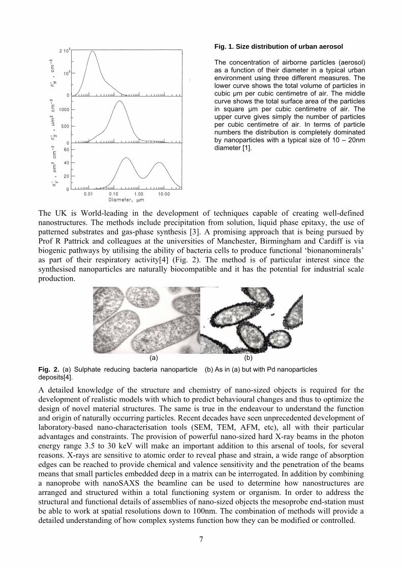
Fig. 1. Size distribution of urban aerosol
The concentration of airborne particles (aerosol) as a function of their diameter in a typical urban environment using three different measures. The lower curve shows the total volume of particles in cubic µm per cubic centimetre of air. The middle curve shows the total surface area of the particles in square µm per cubic centimetre of air. The upper curve gives simply the number of particles per cubic centimetre of air. In terms of particle numbers the distribution is completely dominated by nanoparticles with a typical size of 10 – 20nm diameter [1].
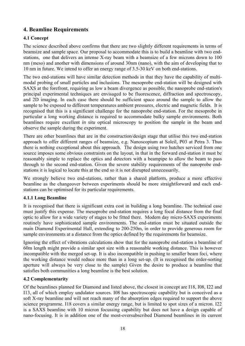
The UK is World-leading in the development of techniques capable of creating well-defined nanostructures. The methods include precipitation from solution, liquid phase epitaxy, the use of patterned substrates and gas-phase synthesis [3]. A promising approach that is being pursued by Prof R Pattrick and colleagues at the universities of Manchester, Birmingham and Cardiff is via biogenic pathways by utilising the ability of bacteria cells to produce functional ‘bionanominerals’ as part of their respiratory activity[4] (Fig. 2). The method is of particular interest since the synthesised nanoparticles are naturally biocompatible and it has the potential for industrial scale production.
(a) (b)
Fig. 2. (a) Sulphate reducing bacteria nanoparticle deposits[4].
(b) As in (a) but with Pd nanoparticles
A detailed knowledge of the structure and chemistry of nano-sized objects is required for the development of realistic models with which to predict behavioural changes and thus to optimize the design of novel material structures. The same is true in the endeavour to understand the function and origin of naturally occurring particles. Recent decades have seen unprecedented development of laboratory-based nano-characterisation tools (SEM, TEM, AFM, etc), all with their particular advantages and constraints. The provision of powerful nano-sized hard X-ray beams in the photon energy range 3.5 to 30 keV will make an important addition to this arsenal of tools, for several reasons. X-rays are sensitive to atomic order to reveal phase and strain, a wide range of absorption edges can be reached to provide chemical and valence sensitivity and the penetration of the beams means that small particles embedded deep in a matrix can be interrogated. In addition by combining a nanoprobe with nanoSAXS the beamline can be used to determine how nanostructures are arranged and structured within a total functioning system or organism. In order to address the structural and functional details of assemblies of nano-sized objects the mesoprobe end-station must be able to work at spatial resolutions down to 100nm. The combination of methods will provide a detailed understanding of how complex systems function how they can be modified or controlled.
7

The fundamental specifications of the beam to be delivered to the end-stations on the beamline are thus clear. The X-ray energy needs to cover the range 3.5-30 keV to provide access to the K or L edges of the entire periodic table above Ar and mm-scale transmission through glass and water. The ultimate spatial resolution of the nanoprobe station should eventually reach 10 nm while the mesoprobe end-station has to operate with a variable spatial resolution covering 100 nm – 5µm. Note that the day 1 specification for the nanoprobe is 30 nm; this is a conservative value achievable with zone plates today. While zone plates continue to improve, other focusing technologies, for example, KB mirrors and refractive X-ray lenses are achieving similar resolution. During the detailed design phase a decision will be taken on the best technology to achieve the ultimate target of 10 nm resolution.
Station 1 Mesoprobe X-ray Scattering SAXS to reveal structure, phase and or morphology
X-ray fluorescence XRF to reveal the spatial distribution of the elements;
X-ray absorption Spectroscopy XAS, to identify the chemical state and speciation (XANES)
X-ray diffraction XRD to reveal structure, phase, strain and texture;
Station 2 Nanoprobe X-ray fluorescence XRF to reveal the spatial distribution of the elements;
X-ray diffraction XRD to reveal structure, phase, strain and texture;
X-ray absorption Spectroscopy XAS, to identify the chemical state (XANES);
Below are presented some examples of the science to be carried out on the new beamline. It is clear that many areas will benefit from the combined nanoprobe/nanoSAXS methods applied to the same systems. 3.2 Life Science The major contribution that the beamline will make to life science research arises from its ability to provide elemental maps using hard X-rays at a resolution much smaller than a cell (nanoprobe) and to determine the hierarchical structure within biological tissue (nanoSAXS). The important resolution scale is 10s of nm at which it becomes possible to distinguish whether a particular element is on the outer cell membrane, the nuclear membrane, inside the nucleus or any of the organelles (Fig. 3).
Fig. 3. Comparison of a 2 μm spot, typical of I18 at Diamond and a 30nm spot that could be achieved on day 1 with the nanoprobe, superposed on an image of a leukocyte. It demonstrates the intra-cellular detail that could be revealed with the higher resolution.
The advantage of using the hard X-ray region is that it provides access to the K or L edges of the entire periodic table above Ar and also transmission through glass and water enables in vitro work to be performed Although radiation damage is an issue, the position of elements in XRF maps should not be affected even if the cell is damaged. For a third generation source the minimum spot
8

size for which a diffraction pattern can be recovered is about 10 nm[5] demonstrating that it is possible to recover real information with this degree of focus.
3.2.1 Distribution of Trace Metal Ions and their Role in Cancer Development and Treatment Determining the spatial distribution of trace metals such as Cu, Zn and Fe is vital in understanding the physiology of normal tissue and tumour growth. For example, Prof. Michael Farquharson now at McMaster University, Ontario has been studying how Cu contributes to cell proliferation and aids the mobility of cells. Both these factors link Cu to tumour invasion and to the overall prognosis in the treatment of a patient. Clinical trials indicate that copper chelation is a possible treatment of certain tumours, but it is not yet clear which of the processes involving Cu is essential in reducing the aggressive nature of tumours. Understanding the mechanism behind treatment with copper chelators requires greater levels of detail concerning the specific sites and types of cells in which copper accumulates. In particular, it would be useful to differentiate between the metal content of the proliferating compartment of the tumour cells, cells near blood vessels and the vessels themselves. Inflammatory cells such as macrophages are known to be high in iron content and it would be of interest to also examine whether they are a source of copper that could further activate the production of angiogenic factors.
Figure 4 shows an image of the distribution of Zn and the comparison with a reference sample haematoxylin and eosin (H and E) stained. The darker stained areas show the clusters of tumour cells. By setting regions of interest in these clusters and normal regions it was found that Zn had increased concentration levels of approximately 75% in the tumours[6,7].
Fig. 4. 150µm x 200µm image of the distribution of Zn (left) and the comparison with a reference sample haematoxylin and eosin (H and E) stained (right). The darker stained areas show the clusters of tumour cells. By setting regions of interest in these clusters and normal regions it was found that Zn has increased levels of approximately 75% in the tumours
To proceed to the next stage it is important to determine where within the cell these metals are accumulating. This would facilitate an understanding of the pathways of these elements and how they change with disease, which could lead to new treatments for cancer based on chelation therapies. Scanning X-ray fluorescence with incident X-rays at the K-edges of Fe, Cu and Zn (7.1-9.7 keV) to locate the position of elements with sub-cellular resolution would contribute significantly to this important area of research.
3.2.2 Distribution of Trace Metals and Their role in Alzheimer’s and Parkinson’s Diseases
A team led by Dr. Joanna Collingwood at University of Warwick has been studying the distribution of trace metals, which is important in understanding diseases such as Alzheimer’s and Parkinson’s. Human bodies naturally generate nanoscale iron oxide particles to provide readily accessible iron stores, and copper and zinc are bound to many proteins and molecules throughout our tissues. A common feature in many diseases is the failure of normal metal-ion regulation systems leading to metal-induced toxicity. Tracing metal ion distributions in tissue will lead to significant progress in understanding neuro-degenerative diseases like Alzheimer’s and Parkinson’s disease, and to guide therapeutic approaches. With nanoscale X-ray beams, it is possible to probe within single cells, and the combination of diffraction with ultra-high-sensitivity multi-element mapping will be extremely
9

valuable in providing new insight into disrupted metal ion metabolism in neurodegenerative disorders, and exploring the cellular level impact of therapeutic agents.
3.2.3 Targeted Therapeutic Nanovectors There is growing interest in a radical new approach to treating cancer utilising magnetic nanoparticles that are targeted to tumours using moieties that bind specifically to cancer cells. Once in place the particles and their tumour surroundings are heated above the therapeutic threshold of 42ºC by a high frequency magnetic field applied from outside the body, which is harmless to normal tissue (Fig. 5). Phase I clinical trials using untargeted nanoparticles have demonstrated the low morbidity of the method [8,9]. A problem highlighted however is that existing nanoparticles based on Fe oxides at the packing densities currently achievable do not provide enough heat to eliminate the entire volume of the tumour. The group led by Prof. Chris Binns at Leicester University have recently produced a new class of nanoparticles containing pure metallic cores that should have a hyperthermia performance that is an order of magnitude higher than the oxides. Nano-scale X-ray beams will help to understand the chemistry that provides a “revolving door” into the cell via the targeting moiety and identify where in the cell the nanoparticles are concentrated. If a sufficient concentration of nanoparticles in the cell interior can be achieved, the method could develop into a stand-alone therapy that is a generic cancer treatment virtually free of side effects.
Fig. 5. Tumour hyperthermia with targeted magnetic nanoparticles could provide a low morbidity treatment for cancer. The nanoprobe would be able to provide sufficiently high-resolution maps of elemental Fe in vitro to determine where in the cell the nanoparticles are concentrated.
3.2.4 Hierarchical Biological Tissues in Health and Disease Biological tissues (bone, tendon, cornea, muscle and cartilage) are characterized by a hierarchy in structure at the scale between 1 nm to a few microns, where distinct molecular building blocks of proteins and carbohydrates self-assemble or co-assemble into functional units like fibrils or filaments (sizes between 10 – 100 nm) which in turn form fibril arrays or lamellae. The details of these structures (spacing, size, orientation) & interactions with each other (e.g. collagen with proteoglycans) are critical for the biological function, and changes in disease and ageing. For example, collagen fibrils in the cornea have a tight distribution of radii ~ 20 nm and are spaced with a high degree of regularity (spacing ~ 20 nm) in order to allow visible light to transmit through to the retina. Malfunctions in the spacing occur in diseases like macular corneal dystrophy or astigmatism, as investigated by Prof. Keith Meek, Dr. Craig Boote and colleagues at Cardiff University [10]. The application of an X-ray beam of sizes 100 nm to 5 microns spans the size range of these hierarchical levels in a range of biological structures: collagenous tissues [11], muscles [12], elastic fibres [13] & mineralized tissues [14]. The structure and assembly of elastic fibres found in lungs, blood vessels, cardiac tissue and skin determines the long-term resilience of these crucial organs. Diseases such as atherosclerosis and chronic obstructive pulmonary disease (COPD) are associated with defects in the formation and functioning of these fibres. Dr. Baldock’s group at Manchester University investigate the structure of the two main proteins found in elastic fibres: elastin and fibrillin, and propose using nanoSAXS to investigate their nanoscale organization and mechanics [13]. Use of a nano-beam is necessary to avoid the dominant diffuse scattering with traditional SAXS. Baldock et al will probe for the existence of ordered structures (periodicity and size) in the elastin sheets, which would point to a
10

fibrillar origin for the mechanical properties, while the absence of such structures would suggest entropic elasticity. Addition of fibrillin microfibrils, both individually & as arrays, to their in-vitro system would simulate the elastic fibre formation process in-vivo, and they will investigate how fibrillin modifies elastin assembly. This knowledge will help developing treatments against cardiac and pulmonary diseases.
3.2.5 Biosensor Implants Electrochemical or optical biosensors sense the concentration of specific biomolecules. Both the immobilized biomolecular phase on the sensor surface and the synthetic polymer films constituting the sensor surface can modulate signal readout through their nanoscale structure. Prof. Pankaj Vadgama’s group at the IRC in Biomedical Materials at Queen Mary University of London, are developing sensors encased in electropolymerized films to enhance selectivity [15], where the nanoscale porosity, polymer chain organization, crystallinity, flexibility and the role of additives like antibodies controls permeability and reactivity. It is hard to measure these parameters with a spatial resolution of 100 nm – 1 micron in conditions mimicking the physiological state. In-situ nanoSAXS/WAXD in environmental/fluid chambers will provide quantitative structural information with the high spatial resolution necessary to resolve structural gradients.
3.3 Materials Science The application of the nanoprobe to materials will be extensive, and will have relevance to many industrial processes.
3.3.1 Fuel cell technology Fuel cells are emerging as a key energy technology for a number of applications from mW to MW power ratings. Solid oxide fuel cells (SOFCs) are a leading fuel cell variant that operate at temperatures typically in excess of 600°C providing economic and environmentally friendly power production, typically for stationary applications. The importance of this work is recognised by the Government’s grand challenge on energy and by the EPSRC, which supports the Supergen fuel cell consortium led by Professor Nigel Brandon at Imperial College in collaboration a number of Universities, the Ministry of Defence, and industrial partners.
Fuel cells are based on the electrochemical reactions of hydrogen-rich fuels and oxygen, supported at SOFC electrodes by porous composite materials. The active reaction zones can be characterised by the boundary of ionic conducting, electronic conducting and pore phases, the so-called triple phase boundary (TPB). The complex electrode microstructure has a direct influence on the overall electrochemical performance, durability and economics of the device. A common choice for SOFC anode materials is the porous cermet nickel-YSZ (yttria-stabilized zirconia) with grain sizes that range from around 200nm to 2.5μm It, therefore, demands higher resolution than that available in, for example, conventional lab based equipment [16]. X-ray tomography combined with XRD at ~50nm scale would allow, for the first time, a full and non-destructive characterisation of the electrode microstructure before and after an environmental change. It will be a vital tool in understanding degradation processes in service, currently the main barrier to fuel cell deployment, and will facilitate the design of high performance electrodes in future generations of fuel cells.
3.3.2 Corrosion Corrosion has major economic and safety implications in areas such as nuclear waste storage, maintenance of ageing aircraft, the oil and gas industry, and metal prostheses in the body. To predict corrosion performance over extended periods, models must be developed for the growth of corrosion pits, which require detailed knowledge of corrosion mechanisms. Dr Alison Davenport (U. Birmingham) and colleagues [17] have used I18 (Diamond) for diffraction and spectroscopy studies of salt films and solutions inside growing corrosion pits with promising results, but work is currently limited by the lateral resolution available at I18, since many phases in alloys that influence corrosion are at the submicron level. Higher resolution and the use of alloy slices
11

produced using TEM sample preparation methods will enable the relationship between local corrosion and alloy chemistry and microstructure to be resolved, leading to work with industrial collaborators in nuclear waste, biomedical, fuel cell technology and aerospace.
3.3.3 Nano-scale strain and structure in advanced structural materials Deformation of engineering alloys at the continuum level is well described by the classical theories of elasticity and plasticity, while at the microscopic level, grain deformation is described by anisotropic elastic theory and crystal slip. In many engineering materials, typical grain sizes go down to a few microns, while modern advanced materials are nano-grained. The deformation response of such systems is governed by steep gradients of strain and rotation arising from highly inhomogeneous dislocation structures arising during deformation, e.g. cells (areas of relatively low dislocation density) and walls (high density regions). Validation of dislocation-based deformation models requires comparison with experimental data obtained at the resolution of a few tens of nanometres [18]. Electron and scanning probe microscopy techniques attain such resolution, but they remain confined to the sample surface, which is not typical of the interior. The onset of damage in metals and other ductile materials is associated with the nucleation and growth of voids that ultimately coalesce to form continuous cracks that eventually lead to mechanical failure of the sample. Early stages of damage development are of great interest to materials specialists as they often determine a large proportion of the sample life to failure.
The use of hard X-ray nano-probe with multi-modal end-stations will have a major impact on the study of these systems. High angular resolution and Kossel diffraction capability would allow measurement of the strain and rotation in granular and intergranular phases. Elemental distributions within precipitates and at grain boundaries, which are directly linked to mechanical and functional properties [19], would be revealed by fluorescence mapping.
3.3.4 Nanoscale Light Emitters Indium-gallium nitride is a light emitting layer used in modern blue and green LEDs. Localisation of excitons by phase segregation is commonly used to explain the high efficiency of commercial diodes, despite defect densities much larger than those encountered in other semiconductor materials. While electron-hole (exciton) localisation is widely accepted as a key to enhanced luminescence efficiency in solids, the origin of the localisation mechanism in InGaN is a matter of continuing dispute. Self-formed InN quantum dots or In-rich InGaN clusters, formed by phase separation, may act as centres for exciton localization. Although many groups including that of Professor O’Donnell at Strathclyde University have been studying this system; a full understanding of the mechanism for enhanced luminescence is, however, yet to be achieved. There is a pressing need for better knowledge of the chemical and physical structure of such poorly defined nano-scale lumophores. With resolutions of the order of 100 nm or less, it will be possible to correlate the intensity of the luminescence excited by the X-ray beam (XEOL) with the atomic ordering on selected regions across the epitaxially grown samples [20]. Nano-sized phosphors promise better deposition onto a semiconductor device and with improved fluorescence efficiency. The applications are numerous and include fluorescent lighting, biological markers and high-resolution scintillation detectors. Phosphors that are compatible with field emission displays offer an alternative to LCD flat panel technology. The research of Dr George Fern at Brunel University is focussed on rare earth doped oxides. A combined x-ray diffraction, fluorescence and luminescence study of selected regions of films of nano-sized emissive materials will lead to a better understanding of these materials and to the optimisation of their performance.
3.3.5 Energy Materials: Photovoltaics & Catalysis Development of high efficiency organic photovoltaic solar cells is one of the fastest growing energy sectors currently. Mass-produced polymer-based cells have the potential to be superior in terms of energy efficiency and cost to the current generation of Si-based devices. To achieve this, understanding of the type and extent of local structural variations within the thin polymer layer will
12

be critical to increase device efficiency. In addition, understanding local structural variations is essential to enable scale-up of prototype lab-devices (~ mm) to the industrial sizes (> cm). Dr. Alan Dunbar and colleagues at the University of Sheffield will use nanoSAXS/WAXD with scanning capabilities to probe the local polymer crystallinity (WAXD) and phase separation at scales of ~ 100 nm (SAXS) [21]. Similarly, dye-sensitized solar cells (recognized by the Millennium Prize 2010) incorporate surfactant templated Ti films with heterogeneous nanostructure. Dr. Karen Edler and colleagues at University of Bath are investigating these systems with a focus on connectivity, porosity and alignment in a deposited film (or across membranes) at sub-micron length scales, as being key to device performance [22]. The ability to probe in situ these crystalline and nanoscale parameters in intact devices with sub-micron resolution and in a scanning mode (to extract gradients) will be essential, and is enabled by nanoSAXS/WAXD & nanotomography. Both the proposed end-stations will provide essential, previously unachievable, structural information to catalyze R&D in this rapidly growing area.
Dr. Rudolf Winter‘s group (Aberystwyth University) propose to develop nano grazing-incidence SAXS (GISAXS), which probes morphology at the scale between 100 nm and several microns, to investigate lateral (in-plane) structural variations in thin-film devices. Building on their previous expertise in this area [23], this method will be combined with 2D ellipsometry (which provides real-space thickness information on the film) to select regions of interest on patterned films with 100 nm spatial resolution for study with nanoGISAXS. The resolution of lateral structural variations at the sub-micron scale will be critical for nanostructured films with surface channels at the micron and sub-micron scale, manufactured by lithography or dip-coating. Extending these ideas to solid-state kinetics in catalysis and corrosion applications, the progress of reaction fronts will be tracked in-situ by measuring changes in grain curvature. With a 100 nm – 5 micron X-ray beam, the interface of individual micron-scale grains can be localized and the curvature directly measured, in contrast to methods like NMR which are limited to a restricted set of elements.
3.3.6 Nanomechanics: In Situ Mechanical Imaging The mechanical properties of nanofibres are significantly different from micro- and macroscopic fibres of the same material due to enhanced surface area, an increase of surface tension, alteration of phonon modes and molecular confinement at the interface between fibres. Materials with nanofibres are a very active current area of research in order to develop stronger, stiffer, lighter materials for ballistics, armour protection, as well as mechanical actuation and biomimetic applications [24]. A distinctive approach to understand the nanomechanics of such materials is through in-situ deformation combined with X-ray micro- and nano-beam scattering and diffraction [14]. The high flux available at synchrotrons enables real-time measurement of strain at the fibril and supramolecular level dynamically during deformation. This X-ray nanomechanical imaging can be applied to both synthetic (e.g. electrospun polymer nanofibres) and biological materials (e.g. mineralized collagen fibrils in bone, cellulose nanofibrils). With a nanoSAXS/WAXD beam, individual fibres at the scale of 100 nm – 1 micron can be resolved, and the deformation mechanisms at their interface studied. Special potential exists to combine small-force measurement devices such as AFMs with these small-scale structural methods. An example will be usage of the AFM for dynamic tests of single electrospun or biomineralized nanofibres (achieved by high frequency oscillations of the AFM piezo actuator) combined with time-resolved SAXS/WAXD, as being developed by a team consisting of Dr. Asa Barber, Dr. Himadri S. Gupta and colleagues at Queen Mary University of London [14,25].
13

Fig. 6. Individual nanofibrils, e.g. mineralized collagen fibrils from antler bone (shown), can be attached to the tip of an AFM probe. Lateral translation or oscillation of the AFM-tip enables controlled deformation in the fibril, which can be measured with high accuracy from the deflection of the tip [25]. Image adapted from [25].
3.4 Environmental Science Virtually all branches of environmental science will benefit from being able to probe individual nanostructures using the nanoprobe as well as multi-scale determination of their ordering within the larger scale system.
3.4.1 Nanoparticles and nano-intergrowths in heterogeneous natural materials The team involving Dr Paul Schofield and collaborators from the Natural History Museum, Imperial College and the Universities of Manchester and Salford study materials that contain micro- and nano- intergrowths of minerals, chemical state zoning, mineral-fluid and mineral-biota interfaces, and heterogeneously distributed nanoparticles. They display a range of multivalent transition metals and rare-earth elements and cation distributions. X-ray imaging diffraction and spectroscopy with beam sizes down to a few tens of nanometres would have a major impact by enabling sub-micron precipitates and mineral surface coatings, to be characterized in situ within heterogeneous materials such as soils and laterites. Multi-element valence state information would help to understand processes from mineral characterisation to oxidation potential and fugacities of prograde and retrograde mineral reactions [26].
3.4.2 Radiogenic and toxic metal remediation by lichen melanins.
Fig. 7. Normally yellow in colour, the lichen lecanora polychrome, found near a mine in Sweden, exhibits a vivid turquoise colour that X-ray fluorescences mapping confirms is due to the presence of Cu [27].
The ability of lichens to accumulate metals has been exploited in a number of monitoring programmes including studies of radionuclide fallout from Chernobyl. Whilst destructive EMPA can reveal coarse elemental distribution, it is not known how these lichens contain metals in an apparently non toxic form. For example, lichens found to be growing on the secondary uranium minerals metazeunerite and metatorbernite have been seen to contain the uranium principally within the walls of the outer fruiting body, but it is not thought that this is done by trapping of the mineral particles. Metal distribution and concentrations, valency and structural information are needed to determine the oxidation states of the metal compounds in both their modified form in the lichen and in their non-modified form in the rock on which the lichens are growing. It is also necessary to investigate variations of the redox conditions across the rock surface in order to account for mineral
14

breakdown reactions that can also occur, especially along cracks and natural cleavage planes. The elements of principal interest are: U, As, V, Hg, Cr, Cu, Fe, Mn, Pb, Zn, Cd and Sr.
3.4.3 Biominerals in the Natural Environment Many minerals produced by bacteria have environmental consequences at the large scale. For example, recent work by Prof. Cockell and his team at the Open University [28] has used I18 to investigate the production of iron oxides during bacterial weathering of shales, implicated in coastal erosion (Fig. 8). The production of minerals contributes to rock weathering and disintegration. To elucidate the true role of organisms in mineral production requires that the resolution is sufficient to map the minerals at bacterial scales, a task currently not possible. The new beamline would allow for a step change from merely characterising the minerals associated with bacteria to understanding their spatial relationship with respect to the organisms and the role of bacteria in controlling/influencing their formation.
Fig. 8. Maps (right) of iron oxides produced by bacteria on the surface of shale (left) (I18 beamline). The resolution is currently insufficient to correlate them to the organisms. 3.4.4 Palaeoenvironmental reconstruction Biominerals are nanocomposite materials that include coral skeletons, foraminifera shells and the shells of bivalves and brachiopods; the composition of which changes as a function of the skeletal or shell architecture. They are of particular interest since they are widely used to reconstruct past environments. Since such reconstructions are based on modelling the partitioning between the mineral component (aragonite or calcite) and seawater, it is important to confirm where the key elements Sr, Ca and Ba are located. It has been assumed that they are located in the mineral component but if, on the other hand, they are hosted by the organic component, the basis of the reconstruction models are wrong and a major rethink is needed. Dr Adrian Finch of the University of St Andrews has compared the coral skeletons of the species Porites sp., which is the most widely used material for environmental reconstruction, with Tubastrea sp, which has no symbiotic zooxanthellae. His results indicate that Sr is usually hosted by the mineral, but in contrast S and Mg are predominantly hosted by the organic [29]. Better resolution down to tens of nm is needed to be certain about this, and to extend the study to a wider group of marine micro-organisms.
3.5 Space Science The chemical composition and mineral structure of material coming from extra terrestrial regions contains vital clues about the formation of the solar system. The material includes meteorites found on earth, as well as dust particles and rock samples returned from space and specific bodies such as the moon, by space missions.
3.5.1 Meteorites
Unequilibrated chondrites (UC) are some of the most primitive solar system material available to us [30]. These meteorites are heterogeneous aggregates of spherules formed in a flash heating event, and a fine-grained matrix. UC’s have not undergone the pervasive geological processing
15

experienced by terrestrial materials. In particular, the origin of iron-rich fayalitic olivine in UC’s, whether it formed as a nebular condensate or in a complex hydration/dehydration process on meteorite parent bodies, is the subject of vigorous debate. Although there have been numerous textural studies of UC’s, little is known about the absolute abundance of the phases that make up primitive meteorites. The UC matrix is extremely fine grained, with individual grains often on the order of tens of nanometres in size. While TEM is effective in identifying phases and establishing textural relations, the tiny areas observed preclude determination of abundance or an overview of the mineral inventory in these heterogeneous materials. It is difficult using conventional microscopy to even establish what minerals are present in primitive meteorites, let alone their abundance. Nano-scale XAS would be a powerful tool with which to shed light on these questions.
The Santa Catharina meteorite represents a natural Invar system in which many equilibrium Invar-type alloy phases have been able to develop only because of the very slow cooling of the meteorite. Within the host metal phase there are inclusions containing ~ 8 wt% O and ~ 45 wt% Ni, more Ni than in the surrounding metal. The demonstration that oxygen is a potential light element in Earth and planetary cores would have a dramatic impact upon models of planetary interiors and their evolution. Complex micro and nano intergrowths have been shown to contain tetrataenite, antitaenite and Fe-Ni phosphides, but the oxide phases remain poorly characterised. The ability to extract direct chemical speciation information from these intergrown metallic–metal oxide nanotextures would allow their characterisation and an assessment of their formation with respect to large scale processes on their parent asteroid. Experiments on I18 by Dr Paul Schofield and collaborators at NHM, Imperial College and DLS have probed the alloy phases, with EXAFS revealing differences in metal-metal bonding distances and ordering [31], however the oxide phase is made up of grains of 500 nm and less. Thus a much smaller beam than currently available at Diamond is required for microdiffraction and microXAFS to fully characterise these small grains.
3.5.2 Sample returned missions In 2006 the space mission Stardust returned material collected from the coma of the comet Wildt-2. Comets form in the coldest, most distant regions of a young solar system, and they were thought to contain small (< 300nm) interstellar grains and gas that would have remained unchanged during the intervening time. The particles that have been identified in the silica-gel traps (Fig. 9) removed from the space craft have revealed startling results prompting a rethink of our understanding of the origin of the solar system [32]. Interstellar dust is a relatively small component of the material found. Larger aggregates that separate on impact have different chemical composition and contain minerals formed at high temperatures and could only come from the inner solar system. It is in fact a collection of materials that have come from all regions of the young solar system.
Fig. 9. A track created by the impact of a particle in the coma on a silica-gel trap on the Stardust space craft. It reveals that the particle is in fact an aggregation of smaller particles that separate on impact.
Many scientists around the world including John Bridges at the Space Research Centre of the University of Leicester, colleagues in the UK Stardust consortium and Alexandre Simionovici from LGCA, Grenoble are engaged on the study of these recovered samples. The work will continue for many years and will demand highest sensitivity and best resolution hard X-ray fluorescence and absorption spectroscopy capability. The need is to determine accurately the chemical composition and the mineralogy of the fragments, some a few tens of a nanometre in scale, and to compare with information from other objects such as meteorites. This would provide further insights into the distinctions and similarities between asteroidal and and cometary parent bodies, and provide a context to understand the origin of the comet. Initial results have been obtained by the Leicester
16

group using I18 have confirmed the presence of high temperature Fe-Ti oxide grains in addition to magnetite/haematite mixtures and other materials that form at low temperatures[33]. Future sample return missions include the Japanese Hayabusa space craft that will return material from the 25143 Itokawa asteroid in 2010 and NASA’s proposed Mars Sample Return.
3.6 Alternative Sources The importance of nanofocus beamlines is recognised worldwide and Appendix III provides a list of other sources operating or under construction. It is clear that the beamline proposed here will be at the leading edge of such instruments. Appendix III also places the beamline within the context of the other beamlines available on Diamond.
References for Scientific Case [1] J. J. Seinfeld and S. N. Pandis Atmospheric Chemistry and Physics: From air pollution to climate change,
Wiley, New York 1997 [2] Statement By The UK Government About Nanotechnologies:
http://www.dius.gov.uk/policy/documents/statement-nanotechnologies.pdf
[3] K. von Haeften et al, Eur. J. Phys. D 52, 11 (2009). [4] Coker et al. American Mineralogist, 93, 1119 (2008) [5] M. Howells et al, An Assessment of the Resolution Limitation due to radiation Damage in X-ray Diffraction
Microscopy, arXiv:physics/0502059v1. [6] M.J.Farquharson et al, Phys. Med Biol. 53, 3023 (2008). [7] K. Geraki, M. J. Farquharson, D. A. Bradley, O. Gundogdu & G. Falkenberg, X-ray Spectrometry 37, 12 (2007). [8] M. Johannsen et al, European Urology Supplements 2007, 6, 201. [9] M. Johannsen et al, International Journal of Hyperthermia 2007, 23, 315. [10] B. P. Palka et al, Current Eye Research 35, 580 (2010) [11] J.P.R.O. Orgel et al, Proc. Natl. Acad. Sci. 103, 9001 (2006) [12] S. Y. Berschnitzky et al, Biophys. J. 99, 1827 (2010) [13] C. Baldock et al, Proc. Natl. Acad. Sci. 103, 11922 (2006); Proc. Natl. Acad. Sci. in review [14] H. S. Gupta et al, Proc. Natl. Acad. Sci. 103, 17741 (2006) [15] H. Chang et al, Biofabrication 2, doi 10.1088/1758-5082/2/3/035002 [16] J. R. Izzo Jnr et al, J. Electrochem.Soc. 155, B504 (2008). [17] T. Rayment et al, Electrochem.Comm,10, 855 (2008). [18] A. Korsunsky et al, J. Nanoscience and Nanotechnol., accepted (2009). [19] B. B. Straumal, et al, Phys. Rev. B 78, 054202 (2008) [20] V. Katchkanov et al, Appl. Phys. Lett. 89, 101908 (2006). [21] T. Wang et al, Soft Matter 6, 4128 (2010) [22] K. J. Edler et al, Chem. Mater. 22, 4579 (2010) [23] K. Hoydalsvik et al, Phys. Chem. Chem. Phys. 12, 14492 (2010) [24] P. Podsiadlo et al, Science 318, 80-83 (2007) [25] F. Hang and A. H. Barber, J. Roy. Soc. Interface (2010), doi:10.1098/rsif.2010.0413 [26] M. F. Hochella Jr. Elements, 4,407,(2008) ; see also other articles in same volume [27] O. W. Purvis et al, Minerological Magazine, 72, 602 (2008). [28] C. S. Cockell et al, Microb. Ecol. 61,166 (2011). [29] A. Finch and N. Allison, Geophys. Research Letts. 35, /08704 (2008). [30] A.Bland et al, Meteoritics & Planetary Science 42, 1417 (2007). [31] P. Schofield, et al, submitted to Geostandards and Geoanalytical Research. [32] Special Edition of Science 314, 1731 – 1735 (2006). [33] J. C. Bridges et al, 2009 Iron oxides in Comet 81P/Wild 2 Samples. Meteorit. Planet. Sci. (in press).
17
4. Beamline Requirements 4.1 Concept The science described above confirms that there are two slightly different requirements in terms of beamsize and sample space. Our proposal to accommodate this is to build a beamline with two end-stations, one that delivers an intense X-ray beam with a beamsize of a few microns down to 100 nm (meso) and another with dimensions of around 30nm (nano), with the aim of developing that to 10 nm in future. We intend to offer an energy range of 3.5-30 keV on both end-stations.
The two end-stations will have similar detection methods in that they have the capability of multi-modal probing of small particles and inclusions. The mesoprobe end-station will be designed with SAXS at the forefront, requiring as low a beam divergence as possible, the nanoprobe end-station's principal experimental techniques are envisaged to be fluorescence, diffraction and spectroscopy, and 2D imaging. In each case there should be sufficient space around the sample to allow the sample to be exposed to different temperatures ambient pressures, electric and magnetic fields. It is recognised that this is a significant challenge for the nanoprobe end-station. For the mesoprobe in particular a long working distance is required to accommodate bulky sample environments. Both beamlines require excellent in situ optical microscopy to position the sample in the beam and observe the sample during the experiment.
There are other beamlines that are in the construction/design stage that utilise this two end-station approach to offer different ranges of beamsize, e.g. Nanoscopium at Soleil, P03 at Petra 3. Thus there is nothing exceptional about this approach. The design using two hutches serviced from one source imposes some obvious constraints on the layout, in that in the forward end-station it must be reasonably simple to replace the optics and detectors with a beampipe to allow the beam to pass through to the second end-station. Given the severe stability requirements of the nanoprobe end-stations it is logical to locate this at the end so it is not disrupted unnecessarily.
We strongly believe two end-stations, rather than a shared platform, produce a more effective beamline as the changeover between experiments should be more straightforward and each end-stations can be optimised for its particular requirements.
4.1.1 Long Beamline It is recognised that there is significant extra cost in building a long beamline. The technical case must justify this expense. The mesoprobe end-station requires a long focal distance from the final optic to allow for a wide variety of stages to be fitted there. Modern day micro-SAXS experiments routinely have sophisticated sample environments. The end-station must be situated outside the main Diamond Experimental Hall, extending to 200-250m, in order to provide generous room for sample environments at a distance from the optics defined by the requirements for beamsize.
Ignoring the effect of vibrations calculations show that for the nanoprobe end-station a beamline of 60m length might provide a similar spot size with a reasonable working distance. This is however incompatible with the merged set-up. It is also incompatible in pushing to smaller beam foci, where the working distance would reduce more than in a long set-up. (It is recognised the order-sorting aperture will always be very close to the sample) Given the desire to produce a beamline that satisfies both communities a long beamline is the best solution.
4.2 Complementarity Of the beamlines planned for Diamond and listed above, the closest in concept are I18, I08, I22 and I13, all of which employ undulator sources. I08 has spectroscopic capability but is conceived as a soft X-ray beamline and will not reach many of the absorption edges required to support the above science programme. I18 covers a similar energy range, but is limited to spot sizes of a micron. I22 is a SAXS beamline with 10 micron focussing capability but does not have a design capable of nano-focusing. It is in addition one of the most-oversubscribed Diamond beamlines in its current
18

configuration. I13 is a long beamline but is intended for imaging at the highest possible spatial resolution without spectroscopy. The beamline proposed here will complement the other beamlines at Diamond, filling the gap that remains for a sub micron imaging beamline with X-ray scattering, diffraction and spectroscopy that covers the K-edges of the transition metals.
Nano-imaging beamlines with multi-modal capability will be a highly competitive area in the next decade. The high demand for such facilities, as illustrated by the number of expressions of interest from a wide community for this proposal and similar beamlines being built elsewhere currently, justifies their growing number. It is important, nevertheless, that the beamline proposed for Diamond offers a capability that is as good or better than that offered elsewhere. In view of the high brightness of Diamond and by employing a design that will emphasise stability, beam monitoring and the use of advanced detectors, we are confident this goal can be achieved.
Achieving a better focus than available on I18 will mean some compromises in the spectroscopy that can be undertaken. Combined gap scanning of the undulator and simultaneous movement of the monochromator crystals will have an impact on the stability of the beam. XANES measurements over a 100 eV energy range have been made at I18 with beam movements of less than 0.1 micron, and with improved design and beam monitoring and interferometric feedback, we can expect this to improve in the future. Hence we expect both end-stations to have a XANES capability but not an EXAFS one.
4.3 Ancillary facilities With the end-stations positioned in an external building, it is essential that that this building also contains suitable sample preparation facilities. To this end it must have a wet laboratory and also an SEM visualisation facility with appropriate technical support. There should also be the opportunity to carry out sample preparation techniques such as focussed ion beam in the offline laboratory space. The beamline will only fulfill its scientific potential if such facilities are included in the build programme.
4.4 Sample Damage Some samples of both biological and physical science origin will suffer beam damage with such a highly focussed beam. This may be either acceptable or catastrophic for a given experiment, e.g. in an XRF map some level of damage may be acceptable as the location of the elements may be unaffected, however in a spectroscopy or diffraction experiment the oxidation of an element or structure may change. The level of damage will be specific to the sample type and measures such as sample cooling may alleviate it in certain cases. Both beamlines will, therefore, require cryo stages to reduce sample damage.
19
5. Beamline Specification The outline beamline specification is set out below. The priority will be to achieve multi technique-analysis at both end-stations with the smallest possible focus at the second end-station. The optical design of the first end-station will be optimised for SAXS analysis with a low divergence and the optical design of the second end-station will be optimised for scanning fluorescence, X-ray absorption spectroscopy, and diffraction.
5.1 Source To cover the wide photon energy range (3.5 – 30 keV) with high flux throughout, an undulator source is essential.
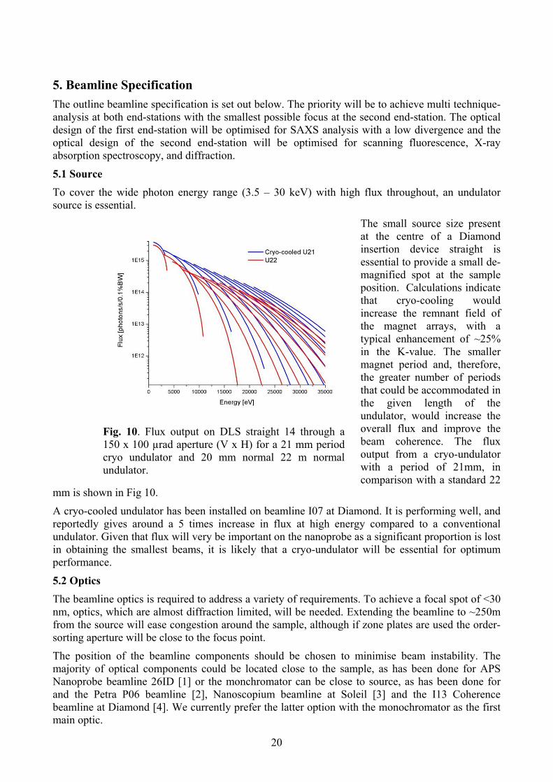
The small source size present at the centre of a Diamond insertion device straight is essential to provide a small de-magnified spot at the sample position. Calculations indicate that cryo-cooling would increase the remnant field of the magnet arrays, with a typical enhancement of ~25% in the K-value. The smaller magnet period and, therefore, the greater number of periods that could be accommodated in the given length of the undulator, would increase the overall flux and improve the beam coherence. The flux output from a cryo-undulator with a period of 21mm, in comparison with a standard 22
mm is shown in Fig 10.
Fig. 10. Flux output on DLS straight 14 through a 150 x 100 µrad aperture (V x H) for a 21 mm period cryo undulator and 20 mm normal 22 m normal undulator.
A cryo-cooled undulator has been installed on beamline I07 at Diamond. It is performing well, and reportedly gives around a 5 times increase in flux at high energy compared to a conventional undulator. Given that flux will very be important on the nanoprobe as a significant proportion is lost in obtaining the smallest beams, it is likely that a cryo-undulator will be essential for optimum performance.
5.2 Optics The beamline optics is required to address a variety of requirements. To achieve a focal spot of <30 nm, optics, which are almost diffraction limited, will be needed. Extending the beamline to ~250m from the source will ease congestion around the sample, although if zone plates are used the order-sorting aperture will be close to the focus point.
The position of the beamline components should be chosen to minimise beam instability. The majority of optical components could be located close to the sample, as has been done for APS Nanoprobe beamline 26ID [1] or the monchromator can be close to source, as has been done for and the Petra P06 beamline [2], Nanoscopium beamline at Soleil [3] and the I13 Coherence beamline at Diamond [4]. We currently prefer the latter option with the monochromator as the first main optic.
20

Fig. 11. Schematic of the beamline layout
A schematic plan view of the beamline is shown in Fig. 11. An interchangeable Si (111) or Si(311) double crystal monochromator would be employed for energy selection. For flux hungry experiments, a double multilayer monochromator would also be available. To reach a photon energy of 3.5 keV, the DCM will need to be inclined to 35°. As the beam is linearly polarised in the horizontal plane, the flux will be significantly reduced at the lowest energies (by a factor of up to 5) but we believe the benefits of improved stability from a horizontal monochromator outweighs this issue.
There is also a pair of horizontal deflecting mirrors. The first mirror would be mechanically bent to generate an intermediate focus. The second mirror would be planar, and coated with three stripes: Si, Cr and Pt, for harmonic suppression. The angle on the two mirrors would be changed in tandem so maintain a fixed exit beam. The horizontal deflection of the mirrors reduces the effect of slope errors and helps preserve beam coherence.
Nanofocusing can be achieved by three methods: diffraction, reflection and refraction. Diffractive focusing with Fresnel zone plates is most commonly employed in beamlines seeking sub-micron focusing. It is used at APS Nanoprobe and is our default option for the nanoprobe end-station. It is anticipated that 4-6 different FZP would be required to cover the entire photon energy range optimally. Rapid progress is, however, being made with other schemes. Osaka University has achieved <30 nm focusing using KB reflective optics and commercial KB optics are available that promise ~50 nm focal size. The current 26ID experience is that the zone plates degrade quite quickly unless used in a He or high vacuum environment. However Xradia are currently working on solutions to this so it may be possible to have the final end-station in an air enclosure. Be refractive lenses (planned for use in Petra P06 beamline) are a good choice for energies higher than 10 keV, but are not a viable choice for the nanoprobe end-station. For the mesoprobe end-station either KB mirrors or refractive lenses are possible options. We will monitor progress on the competing technologies and choose the best option during the final design phase of the beamline.
5.3 Sample environment and control Both temperature stability and vibrational stability are necessary to achieve small spot sizes. Thermal stability is essential. A thermal ante-chamber for both experimental hutches and a nanoprobe end-station a local enclosure will be required. Stability to 0.01 C may be required here; a recent test at Diamond with an active temperature controlled enclosure achieved a standard deviation of 0.9 mC over an 11.5 hour period. The heat loads from sample environments will need to be managed very carefully. Mechanics must have very low heat generation and be able to retain their position when powered off. These would be devices that have a crawling type motion. Such devices are available with integrated measuring systems for active feedback control. Stiffness is paramount and so all motions must be flexure based. The minimisation of acoustic noise is required, this can be done using baffles and absorbers.
There is a belief that designing a low vibration hutch and then using high stiffness mechanics is best approach. Diamond is looking at the possible use of a combination of both active and passive vibration isolation. This approach could lead to improvements of the floor vibrations by a factor of
21
10 at frequencies of about 5-25 Hz, and up to 5x at 25-50Hz. The I13 external building shows promising levels of vibration, which are lower than the main Diamond experimental floor. Diamond engineers have successfully constructed an enclosure, in the Diamond optics lab, that using piezo-based feedback has a vibrational amplitude of 2.5 nm, hence with further development we believe a vibrational amplitude of 1 nm is achievable in the nanoprobe end-station.
For both end-stations we will mount the sample and the final optics on the same platform with stiff positioning stages capable of a few nm movement steps. The technology to achieve interferometric feedback has been developed at the ALS and the APS, and is commercially available (5).
Environmental stages will enable measurements to be made in the presence of electric and magnetic fields, and at controlled temperatures, pressures and stresses, to monitor changes in real time. These will require very precise design on the nanoprobe, with careful heat management. Optical tweezers are another widely used technology for holding and manipulating nano objects; these have already been used successfully on I22 and will be available to this beamline. Alignment of the sample in the beam will be assisted by imaging samples with a visible light microscope or with the X-ray beam itself.
Detectors Efficient detection with large area and small multi-element detectors will be required to fully exploit the high flux and to minimise exposure of materials to the beam. To achieve cost savings it may be possible to use some of the detectors on both end-stations thus avoiding duplication.
The requirements for detectors for scattering experiments are challenging. High resolution across a wide dynamic range will be essential but access to dynamic processes will also be critical to the success of this beamline. State of the art technology will be required to fulfill the requirements potentially coming from the latest Hybrid high resolution, high dynamic range pixellated or fast CCD area detectors for scattering/diffraction.
For X-ray fluorescence and X-ray absorption spectroscopies, a solid state energy resolving detector is required. For an energy range of 3.5 - 30 keV both Si and Ge detectors will be required, as either will fail to be optimal over the entire energy range. Peter Cloetens has shown that for nano beamlines, a sample geometry with the sample normal to the beam and the detector at an angle of ca. 60o is efficient [6]. This set-up also avoids elongating the horizontal footprint of the beam on the sample. The incident beam is probably best measured with an ion chamber or scatter to a photodiode before the final optic as the space between the optic and the sample is very limited. For spectroscopy on conducting samples electron yield detection may provide a method of keeping the three dimensional sample volume to a minimum, for other detection methods ultra-thin samples or a tomographic approach will be required for 3D imaging. We acknowledge that ultra-thin dilute samples will present a challenge for spectroscopic measurements.
A hybrid pixellated detector such as a Pilatus detector is the best choice for the area detector, which will sit behind the sample for collecting diffraction in transmission mode. For monitoring strain in small grains in alloys, an analyser and a point detector would be used.
22

6. Costs Machine
Insertion Device (extra cost for cryo device) Front End XBPMS Optics Monochromator, double crystal (DCM) Monochromator, Multilayer (MLM) Mirrors, hard x-rays Slits Diagnostics (Diags, XBPMs, Hydrost, Autoca Miscellaneous Instrumentation Gas Bremsstrahlung collimator Gas Bremsstrahlung stop Experiment Shutter x 2 Tubes, Stands & Bellows Experimental Station - Nanoprobe X-ray optics (FZPs, KBs) Sample environments Sample Platform Sample Chamber Inline visualisation Experimental Station - Mesoprobe X-ray optics (FZPs, KBs, CRLs) Sample environments Laser Tweezers Sample Platform Inline visualisation Detectors Large Area Si Detector Si Fluorescence Ge Fluorescence X-ray Camera Area Detector Instrumentation Controls racks Vacuum Motion Systems PSS Radiation monitor (portable, installed) Oxygen Level Monitors COSHH System
23

24
Electrical Hardware incl. cabling Computing Network cabling Computer and Network Racks Network Equipment Workstations Peripherals Servers Storage Software
Infrastructure Hutches External Transfer line C&S External Building Beamline Alarm Panel Radiation Monitor Furniture Preparation Lab Basic Equipment Preparation Lab Equipment Small Workshop Equipment Combined SEM/FIB Total
Technical References
1 J. Maser et al, 8th
Conf.X-ray Microscopy IPAP Conf. Series 7, 26 (2006)
2 U. G. Falkenberg et al, Status of the Hard X-ray Micro/Nano-Probe beamline (P06) at Pera III, Petra Annual Report (2007)
3 A. Somogyi et al., AIP Conference Proceedings, 1234: 395-398. (2010)
4 C.Rau I13 – X-ray Imaging and Coherence beamline at Diamond, http://www.diamond.ac.uk/Beamlines/Beamlineplan/I13L/default.htm
5 http://www.xradia.com/products/ultraspx-s.php
6 http://www.esrf.eu/UsersAndScience/Experiments/TBS/ISG/events/EnergyResolvingBrainstorming/cloetens.pdf

Appendix I) Expressions of Support
Lead Affiliation Areas of Interest / Techniques
Materials Science
Asa H. Barber School of Engineering and Materials Science, Queen Mary University of London, London
Mechanical properties of synthetic and biological nanofibres, 3D nanoscale imaging using AFM, electrospinning of high strength nanofibres for ballistic and defence applications
Guillaume Beutier Laboratory SIMaP, Grenoble Coherent X-ray Diffractive Imaging of the atomic structure and strain fields within nanopillars, whose plastic behaviour is produced by a small number of dislocations. Important fundamental research for further miniaturization in the microelectronics industry. Nanoprobe and in situ AFM would be a powerful combination.
Nigel Brandon Dept of Earth Science and Engineering, Imperial College, London
Tomographic and XRF scanning at < 100nm of complex electrode microstructures to understand the fundamental chemical processes that take place in fuel cells, with the aim of developing more efficient technology for clean energy provision.
Richard Brydson Institute for Materials Research, University of Leeds
Imaging and Analysis of Individual Nano-objects under real conditions. Adventurous proposed experiments that would benefit greatly from an environmental SEM on the beamline.
Andy Bushby School of Engineering and Materials Science, Queen Mary University of London, London
nano-mechanics of synthetic and biological materials, especially using the technique of nanoindentation; nondestructive and high-throughput testing of polymer coatings; mechanics of hydrated and poroelastic tissues like cartilage; high resolution SEM imaging of hydrated biological tissues
DM-WG-PP-009 25

Lead Affiliation Areas of Interest / Techniques Markys Cain National Physical Laboratory, Teddington Structure and dynamics of ferroelectric switching; evolution of domain structure
under external stresses and electric fields; new polymeric ferroelectric materials; In-situ SAXS/WAXD imaging of ferroelectric switching in lead-free alkali niobate ceramics, intrinsic and composite ferroelectrics and polymer films.
Alison J Davenport School of Metallurgy and Materials, University of Birmingham
Use of XRD and XAS for the study of the corrosion of metals in wet environments’ specifically, the relationship between local corrosion sites and the chemistry and microstructure of alloys at sub micron resolution.
David Dunbar Department of Chemical & Biological Engineering, University of Sheffield
Polymer solar cells: linking structure variations at the nano- and microscale to performance in solar cell devices; self organisation in polymeric material e.g. phase separation in thin films of polymer blends for a range of applications, including organic electronics, nano-lithography for inorganic electronics, gaseous and liquid filtration membranes
David Dye Dept of Materials, Imperial College, London Study of strain within individual grains in alloys for high-performance applications, e.g. γ-TiAl to be used in future aero engines.
Karen Edler Department of Chemistry, University of Bath Properties of self- organized composite materials including surfactant templated Ti films used for dye sensitized solar cells; in-situ studies of nanocomposite and nanocrystal synthesis using microfluidics
Stephen Eichhorn Materials Science Centre, University of Manchester
Cellulose nanowhisker composites; All-cellulose nanocomposites; Bacterial cellulose nanocomposites; Tractography in electrospun brain mimics; use of Raman spectroscopy, synchrotron x-ray diffraction and molecular dynamics/mechanics modelling of polymeric materials
Patrick Fairclough Department of Chemistry, University of Sheffield
interactions and association within polymeric materials (block copolymers,weakly ordered systems such as polymer networks and gels) using microscopic, light scattering, SAXS/WAXD and spectroscopic techniques; organic electronics and solar cells; organisation of carbon nanotubes in polymer foams
DM-WG-PP-009 26

Lead Affiliation Areas of Interest / Techniques George Fern
Wolfson Centre for Materials Processing, Brunel University
X-ray diffraction, X-ray fluorescence and X-ray excited optical luminescence measurements of nano-sized (~30nm) particles of emissive materials, with the aim of optimising the efficiency for the development of improved lighting sources, scintillation detectors and biological markers.
Finn Giuliani Dept of Materials, Imperial College, London Deformation of ceramics, including the layered MAX phases
Guenter Goerigk Outstation JCNS-FRMII c/o Technische Universitaet Muenchen, Forschungszentrum Jülich, Germany
Time resolved in-situ ASAXS (Anomalous Small-Angle X-ray Scattering) studies on the decomposition kinetics of Copper-Cobalt alloys; Structural investigations of amorphous Silicon-Germanium alloys used in solar cell techniques; counterion condensation to polyelectrolytes by anomalous small-angle X-ray scattering; spinodal decomposition in metallic glasses
Chris Gourlay Dept of Materials, Imperial College, London Mechanical properties of Sn-based inter-metallics that are grown from solder-joint interfaces and are the origins of failure in solder joints
Himadri S. Gupta School of Engineering and Materials Science, Queen Mary University of London, London
Deformation and fracture mechanisms at the fibrillar level in bone using in-situ X-ray scattering and deformation methods; material origins of time-dependent mechanics of collagenous tissue; mechanics of hypermineralized nanocomposites; nanostructural origins of bone fragility in rickets and other metabolic bone diseases using transgenic mouse models
Ian Hamley Department of Chemistry, University of Reading
Mechanisms of self-healing in healable supra-molecular polymers based on π-π stacking interactions; mapping of shear-induced structure as a function of distance from shear plates in synthetic polymer and biopolymer solutions, texture/structure mapping in amyloid peptides and peptide conjugates, crystallization and grain boundary effects (epitaxial growth etc) in block copolymer melts and solids
Simon Hogg Dept of Materials, University of Loughborough
Structural studies of high-strength nanostructured Al-based alloys. Alloys consist of complex mix of phases at the nanoscale and require a nanoprobe to be able to study sufficiently small regions that contain a single phase.
Armin Hoell Institute für angewandte Materialforschung, Helmholtz Zentrum Berlin, Germany
metallic foams and interfaces between catalysts and proton-conducting membranes; anomalous scattering experiments which deliver chemically sensitive contrast on such systems
DM-WG-PP-009 27
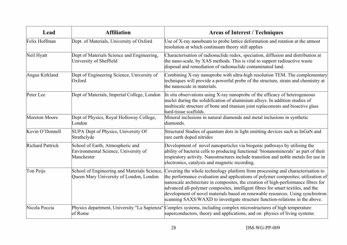
Lead Affiliation Areas of Interest / Techniques Felix Hoffman Dept. of Materials, University of Oxford Use of X-ray nanobeam to probe lattice deformation and rotation at the utmost
resolution at which continuum theory still applies
Neil Hyatt Dept of Materials Science and Engineering, University of Sheffield
Characterisation of radionuclide redox, speciation, diffusion and distribution at the nano-scale, by XAS methods. This is vital to support radioactive waste disposal and remediation of radionuclide contaminated land.
Angus Kirkland Dept of Engineering Science, University of Oxford
Combining X-ray nanoprobe with ultra-high resolution TEM. The complementary techniques will provide a powerful probe of the structure, strain and chemistry at the nanoscale in materials.
Peter Lee Dept of Materials, Imperial College, London In situ observations using X-ray nanoprobe of the efficacy of heterogeneous nuclei during the solidification of aluminium alloys. In addition studies of multiscale structure of bone and titanium joint replacements and bioactive glass hard-tissue scaffolds.
Moreton Moore Dept of Physics, Royal Holloway College, London
Mineral inclusions in natural diamonds and metal inclusions in synthetic diamonds.
Kevin O’Donnell SUPA Dept of Physics, University Of Strathclyde
Structural Studies of quantum dots in light emitting devices such as InGaN and rare earth doped nitrides
Richard Pattrick School of Earth, Atmospheric and Environmental Science, University of Manchester
Development of novel nanoparticles via biogenic pathways by utilising the ability of bacteria cells to producing functional ‘bionanominerals’ as part of their respiratory activity. Nanostructures include transition and noble metals for use in electronics, catalysis and magnetic recording.
Ton Peijs School of Engineering and Materials Science, Queen Mary University of London, London
Covering the whole technology platform from processing and characterisation to the performance evaluation and applications of polymer composites; utilization of nanoscale architecture in composites, the creation of high-performance fibres for advanced all-polymer composites, intelligent fibres for smart textiles, and the development of novel materials based on renewable resources. Using synchrotron scanning SAXS/WAXD to investigate structure function-relations in the above.
Nicola Poccia Physics department, University "La Sapienza" of Rome
Complex systems, including complex microstructures of high temperature superconductors, theory and applications, and on physics of living systems
DM-WG-PP-009 28

Lead Affiliation Areas of Interest / Techniques Stephen Skinner
Dept of Materials, Imperial College, London X-ray studies of the nanoscale element distribution in electrode materials of advanced fuel cells.
Jeremy Sloan Dept of Materials, University of Warwick. Nanometer to sub-micron composite materials, e.g. filled carbon nanotubes and metal (Nb, Mo, W) – chalcogenide mesostructures. MBE-fabricated materials containing nansocale precipitates and other inclusions.
Manoj Tiwari Science Division, Diamond Light Source Ltd Grazing incidence scanning X-ray fluorescence of contaminants in silicon device wafers and thin layered optics
Joachim Wagner Institut fuer Chemie, Universitaet Rostock, Rostock, Germany
Fluorescence correlation spectroscopy to measure self-diffusion coefficient of colloidal tracer particles using dominant resonant scattering near the absorption edges of selected atoms (example systems: polybrominated polymer particles and Fe- or Co- containing ferrofluids)
Phil Withers School of Materials, University of Manchester X-ray studies of the nanoscale element distribution in electrode materials of advanced fuel cells.
Rudolf Winter Materials Physics, Aberystwyth University, Aberystwyth
Using ASAXS, GISAXS, SAXS/WAXD and ellipsometry in the study of ceramic films with applications in the areas of photovoltaics (inorganic thin-film solar cells), fuel cells and sensors (oxygen vacancy conducting membranes) and catalysis (support structures).
Yanqui Zhu Dept of Engineering, University of Exeter Use of XAS, XRD and scanning XRF to better understand the chemistry , crystallography an d morphology, and their influence on the magnetic and optical properties of novel 1 dimensional nanostructures.
Life Sciences
Owen Addison Scoolf of Dentistry, University of Birmingham
Characterisation of the distribution of materials at the nanoscale in breakdown products derived from biomedical materials, devices and coatings on normal physiological functions.
Maisoon Al-Jawad Institute of Dentistry, Queen Mary University of London, London
Scanning small-angle and wide-angle X-ray scattering of the dentine-enamel junction in teeth, crystallography and microstructural studies of teeth affected by genetic abnormalities, amelogenin-enamel interactions in tooth formation, 3D mapping of texture in dental enamel and restorative materials
DM-WG-PP-009 29

Lead Affiliation Areas of Interest / Techniques Claire Baldock Wellcome Trust Centre for Matrix Research,
University of Manchester, Manchester Structural studies on microfibrillar components of the extracellular matrix (fibrillin-rich microfibrils and collagen-VI microfibrils), with the goal to understanding their alterations and mechanical consequences in disorders like Marfan syndrome, congenital muscular dystrophy and myopathy
Craig Boote School of Optometry and Vision Sciences, Cardiff University, Cardiff
To better understand the structural basis of corneal transparency and function; Elucidating the ultrastuctural basis of optic nerve head death in glaucoma; using synchrotron X-ray diffraction to determine the organisation of collagen fibrils in both normal and diseased corneas
David Bradley Faculty of Engineering and Physical Sciences, University of Surrey
XRF mapping of the distribution of minerals in bone and trace materials around malignant growth. The ability to do this at the nanoscale would provide important information regarding the metabolism of cancer.
Richard Brydson Inst for Materials Research, University of Leeds
Nanoparticles in biological and organic tissue for nanotoxicological studies in healthcare, aquatic environments, food matrices etc.; biomineralization of nanoparticles; nanoparticle and nanowire synthesis
Joanna Collingwood Inst of Science and Technology in Medicine, University of Keele
Sub cellular resolution XRF and XRD of distributions and aggregates of metals, including Fe, Cu and Zn, in tissues and isolated animal cells, with the aim at better understanding the basis of metal ion metabolism and the impact of transition metals in neurodegenerative disorders.
Fred Currell School of Maths and Physics, Queens University Belfast
Sub cellular XRF nanoscale mapping of elements following radiobiological damage caused by X-rays and electrons. The probe needs to map at scales smaller than the cell nucleus.
Michael J Farquharson
McMaster University X-ray fluorescence mapping of Cu, Fe and Zn within individual cells of hard and soft tissue, and the correlation with the physiology of normal tissue and tumour growth.
Michael A. Ferenczi Molecular Medicine Section National Heart and Lung Institute, Imperial College London
structure/function relationship in muscle contraction; using SAXS to study the three-dimensional sarcomeric structure; mutations affecting less-well understood proteins like troponin or tropomyosin & their role in cardiomyopathies
DM-WG-PP-009 30

Lead Affiliation Areas of Interest / Techniques Alister Hart Dept of Surgery and Cancer, Imperial
College, London XRF studies of chemical species of nano-sized metal (cobalt-chromium) wear debris derived from metal-on-metal hip replacements. it would enable the distinction between the many possible species of cobalt-chromium nanoparticles in the tissues from patients with failed hip replacements.
Matthew Johnson Stevenage Laboratory, GlaxoSmithKline Ltd. Nanoscale XRF studies of the surfaces of tablets or particles, which would lead to advancement in the understanding of physico/chemical properties measured throughout the manufacturing and biological processes in pharmaceuticals.
Carlo Knupp School of Optometry and Vision Sciences, Cardiff University, Cardiff
Molecular organisation of “fibrous proteins”, in particular that of collagens forming networks and that of the proteins in the muscle sarcomere; X-ray diffraction study of nature of collagen VI aggregates in the eye, as related to Sorsby’s Fundus Dystrophy and Age-Related Macular Degeneration; X-ray fibre diffraction of dynamic molecular changes in active muscle
Rob Mairs Dept of Radiation Oncology, Cancer ResearchUK Beatson Laboratories
Studies of the fundamental aspects of carcinogenesis including experimental therapy of xenografts in rodents as well as the irradiation of cultured cells.
Richard Martin Aston Research Centre for Healthy Aging, Aston University
Studies of biomedical implants where the new tissue growth is slow and typicallyonly a few µm thick. A nanoprobe of the order ~ 30 nm will enable the new growth and adhesion of the implant to existing tissue to be mapped and characterised in great detail.
Keith Meek School of Optometry and Vision Sciences, Cardiff University, Cardiff
Ultrastructure of connective tissues and in particular, the basis of transparency of the cornea, using X-ray diffraction and electron microscopy; the role of proteoglycans in maintaining corneal ultrastructure; quantifying the collagen orientation throughout the normal and myopic globe
Sandro Olivo Dept of Medical Physics and Bioengineering, University College, London
XRF determination of the elemental composition of biological samples at the sub-cellular level to correlate with the development of cancerous tumours.
Andrew Quantock School of Optometry and Vision Sciences, Cardiff University, Cardiff
X-ray diffraction and electron microscopic studies of organisation of collagen fibrils and proteoglycan macromolecules in corneal diseases and dystrophies; the molecular changes that accompany the development of corneal transparency; tissue engineering of the corneal epithelial, stromal and endothelial layers
DM-WG-PP-009 31

Lead Affiliation Areas of Interest / Techniques Pierre Rizkallah School of Medicine, Cardiff University,
Cardiff Structure-function relations in complexes of human immune system proteins and signal transduction proteins, including: mammalian mechanistic target of Rapomycin (mTOR), cardiac Ryanodine receptor; phospholipase Czeta
Giuseppe Schettino Centre for Cancer Research and Cell Biology, Queen’s University Belfast
Radio-biological damage caused by X-rays and electrons on the sub-cellular level, with the aim of improving cancer therapies.
Tony Stead School of Biological Sciences, Royal Holloway College, London
XRF mapping at spatial resolutions < 1 micron to understand the uptake of silver in pre-cut flowers with the aim of developing more efficient and less environmentally damaging ways of delaying plant senescence.
Pankaj Vadgama Interdisciplinary Research Centre in Biomedical Materials, Queen Mary Universityof London
Materials biocompatibility: aggregation of proteins at biomaterial interfaces to blood or tissues; mechanisms of protein membrane formation in microfluidic channels; formation of electropolymerized films for biosensing; biosensors; nanostructural mechanisms of antimicrobial actions of silver nanoparticles
Tim Wess School of Optometry and Vision Sciences, Cardiff University, Cardiff
Investigation of order and disorder in the structure of biological systems using synchrotron radiation and other complementary methods; Examples include study of important historical collagen based materials such as the Dead Sea Scrolls and the Domesday book; mineralisation, development and diagenesis of bone structures; the three dimensional structure of collagen
Environmental and Earth Science
Liane Benning School of Earth and Environment, University of Leeds
Quantification of the mechanisms and kinetic rates of in situ and real-time changes in the biogeochemical environments around microbial cells (bacteria, archae and eukaryotes – i.e., fungi) and their interaction with mineral phases.
Charles Cockell Geomicrobiology Research Group, Oepn University
Nanoscale XRF studies of the role of microbial populations on the degradation of shales with relevance to coastal erosion.
Adrian Finch School of Geography and Geosciences, University of St Andrews.
EXAFS and XRF mapping at the S, Sr and Ba edges of nanostructured (~10nm) bio-materials for use in the reconstruction of past (palaeo-)environments. Materials include coral skeletons and shells of bivalves and brachiopods.
DM-WG-PP-009 32

DM-WG-PP-009 33
Lead Affiliation Areas of Interest / Techniques Mark Hodson Dept of Geography and Environmental
Science, University of Reading Nanoscale XRF studies of metals in contaminated soils, which would enable the probing, at appropriate scales, of the partitioning of metals in earthworm tissues, in the calcium carbonate granules secreted by earthworms and in soil particles.
Craig Kennedy Conservation Group, Historic Scotland, Edinburgh
Understanding degradation and origins of materials found in archaeology and built heritage: for example, analysis of cement mineralogical degradation of sandstones, analysis of earth and clay minerals from earthen structures and fired bricks to determine origjns and degradation;
Paul F Schofield Department of Mineralogy Natural History Museum, London
XAS and scanning XRF to determine the elemental and valence state distributions (at the level <30mn) and the structure of amorphous and sub-micron precipitates and mineral surface coatings, in situ within heterogeneous materials such as soils and laterites. The work has application to mining, and remediation of contaminated soil
Sam Shaw School of Earth and Environment, University of Leeds
Using synchrotron radiation techniques to study molecular- and nano-scale mechanisms of mineral reactions which occur in the natural environment, including: natural mineral nanoparticle formation and their interaction with contaminants; study of marine carbonate minerals to determine ancient seawater chemistry and isotopic composition
Space Science
John Bridges Department of Physics and Astronomy, University of Leicester.
Nanoscae XRF and XANES studies of minerals in space dusts returned from the stardust mission to Comet Wild2, martian meteorites and from future return missions to Mars and the asteroids.
George Fraser
Department of Physics and Astronomy, University of Leicester.
Microanalysis using XRF and XAS to determine the elemental distribution and mineralogy of interstellar grains and samples returned from Mars. Advanced detector development.
Paul F Schofield Department of Mineralogy Natural History Museum, London
High resolution XAS of polyvalent transition metals in heterogeneous meteoric materials with the aim of understanding the processes involved in the evolution of solar nebula.
Alexandre Simionovici
Lab de Géodynamiques des Chaînes Alpines, Grenoble.
Mineralogy of cometary and interstellar grains using scanning XRF, XRD, XAS and tomography in the spectral range 100nm

34
Appendix II) Alternative Sources There are an increasing number of hard X-ray beamlines that will offer capability in the nanoscale.
APS: Hard X-ray Nanoprobe Beamline: 26 ID) 26ID is built by the Center for Nano Materials (CNM), which is adjacent to the APS ring building. The beamline is build for scanning microscopies with 30nm spatial resolution with additional full-field microscopy capacities. The instrument operates in the energy range of 3-30keV. The instrument aims for a wide field of applications based on its high sensitivity to elemental composition, crystallographic phase, and strain when applied at this length scale. Coherent diffraction techniques make possible the study of static and dynamic disorder and phase-sensitive imaging at a spatial resolution in principle beyond that of the probe itself. A significant engineering effort has been undertaken to achieve the high mechanical stability and position repeatability. The optomechanical platform includes sample stages controlled by an interferometric system. The device is now available from the X-Radia company.
Soleil: Microscopium Microscopium aims for diffraction and spectroscopy on the micro- and nano-length scale. A long straight section will host two canted undulators providing beam for a micro/nano scanning microscopy station and for tomography. For scanning microscopy it is planned to use an U20 undulator in a modified straight layout, similar to the mini-beta layout planned for I13 at Diamond. A prefocusing mirror will be used to generate a secondary source (at38m distance) and the beam will be further demagnified with a KB system located about 80m from the source.
ESRF: Microfocus Beamline ID13 ID13 may be counted to be one of the first upgraded beamlines at the ESRF. The beamline is operational since 2008. Originally designed to perform SAXS experiments recently a new extension has been built annex to the main ring building. This is the station where the coherent diffraction results have been reported claiming for achieving 10nm spatial resolution.
ESRF: ID22 NINA The upgraded beamline will have two branches: nano-scale imaging (NI) and nano-scale analysis (NA). The NI branch aims for a spatial resolution of <20nm, but at fixed wavelengths without spectroscopic capability. The priority for the NA branch is XAS spectra over 1000 eV with signal to noise sufficient for structural analysis. It will not achieve a spatial resolution better than 1µm. Thus neither branch will offer a capability of nanometre spatial resolution with XANES and XRD.
NSLS II: Submicron Resolution X-ray Spectroscopy/ Hard X-ray Nanoprobe
At NSLS II two hard X-ray nanobeamlines are being constructed one for spectroscopy, Submicron Resolution X-ray Spectroscopy, with a target beamsize from 1 micron to below 100 nm and a non-scanning beamline Hard X-ray Nanoprobe with an ultimate target beamsize of 1 nm. The latter will use multi-layer Laue lenses which are being developed at Brookhaven.
PETRA III: Hard X-Ray Micro Probe, Imaging: P06
The Hard X-ray Micro/Nano-Probe beamline P06 at PETRA III provides advanced visualisation by micro/nano analysis using different X-ray techniques: X-ray fluorescence (XRF), X-ray absorption spectroscopy (XAS), X-ray diffraction (XRD), imaging (absorption, phase contrast) and various tomographies. Use will also be made of the high degree of coherence of the beam. A canted 2m
DM-WG-PP-009
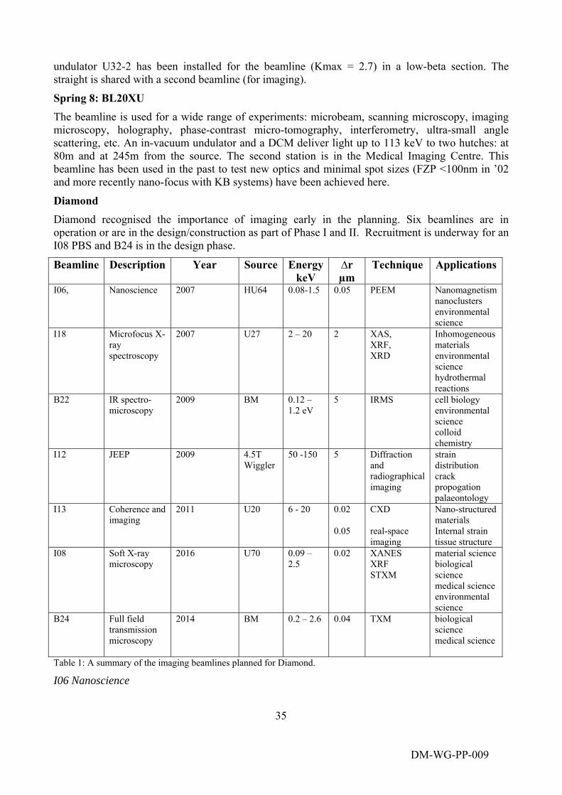
35
undulator U32-2 has been installed for the beamline (Kmax = 2.7) in a low-beta section. The straight is shared with a second beamline (for imaging).
Spring 8: BL20XU The beamline is used for a wide range of experiments: microbeam, scanning microscopy, imaging microscopy, holography, phase-contrast micro-tomography, interferometry, ultra-small angle scattering, etc. An in-vacuum undulator and a DCM deliver light up to 113 keV to two hutches: at 80m and at 245m from the source. The second station is in the Medical Imaging Centre. This beamline has been used in the past to test new optics and minimal spot sizes (FZP <100nm in ’02 and more recently nano-focus with KB systems) have been achieved here.
Diamond Diamond recognised the importance of imaging early in the planning. Six beamlines are in operation or are in the design/construction as part of Phase I and II. Recruitment is underway for an I08 PBS and B24 is in the design phase.
Beamline Description Year Source EnergykeV
∆r µm
Technique Applications
I06,
Nanoscience 2007 HU64 0.08-1.5 0.05 PEEM Nanomagnetism nanoclusters environmental science
I18
Microfocus X-ray spectroscopy
2007 U27 2 – 20 2 XAS, XRF, XRD
Inhomogeneous materials environmental science hydrothermal reactions
B22
IR spectro-microscopy
2009 BM 0.12 – 1.2 eV
5 IRMS cell biology environmental science colloid chemistry
I12
JEEP 2009 4.5T Wiggler
50 -150 5 Diffraction and radiographical imaging
strain distribution crack propogation palaeontology
I13
Coherence and imaging
2011 U20 6 - 20 0.02 0.05
CXD real-space imaging
Nano-structured materials Internal strain tissue structure
I08
Soft X-ray microscopy
2016 U70 0.09 – 2.5
0.02 XANES XRF STXM
material science biological science medical science environmental science
B24 Full field transmission microscopy
2014 BM 0.2 – 2.6 0.04 TXM biological science medical science
Table 1: A summary of the imaging beamlines planned for Diamond.
I06 Nanoscience
DM-WG-PP-009
DM-WG-PP-009
36
The beamline employs an electron microscope to image the photoemitted electrons ejected by the incident X-ray beam. Nanometre resolution has been achieved, but within a UHV environment that is ideally suited for studies of small structures such as magnetic nanoparticles supported on clean surfaces.
I18 Microfocus X-ray Spectroscopy
The beamline was designed as a multimodal beamline with good spatial resolution. Scanning X-ray fluorescence (XRF), absorption spectroscopy (XAS) and diffraction (XRD) can be made with a spatial resolution on the micron scale. The design priority was to achieve high quality absorption spectra, sufficient to analyse the local atomic structure in small grains and regions of inhomogeneous matter. Energy scans of 700 eV with good statistics can be recorded with a spatial resolution of 2 microns.
B22, Infra Red Microspectroscopy (IRMS)
This beamline operates at the extreme low energy end of the available photon spectrum from the bending magnet source. It is mainly intended for imaging with molecular sensitivity of tissue and cellular structures.
I12 JEEP
The beamline provides white and monochromatic beams at energies beyond 100 keV and with dimensions up to 200mm. It enables high intensity imaging and diffraction measurements in real time, and at resolutions of 5 microns or better, of large geological and engineering components under controlled environments.
I13 Coherence and Imaging is a long beamline that will exploit the high degree of coherence of the undulator beams at Diamond. The beamline is designed to perform high resolution imaging and tomography beyond today’s limits. The coherence branch is planned to perform coherent X-ray diffraction, initially at around 8KeV, later at higher energies up to 20 keV. It will generate 3d images of nanoscale structures by recombining the diffracted (speckle) patterns in the so called lensless imaging mode. The main scope of the real space imaging branch is related to full field imaging and tomography over a large field of view and high photon flux with spatial resolution down to < 50nm. It will not be used for spectroscopy.
I08 Low Energy Microscopy will be a high resolution scanning transmission X-ray microscope (STXM), operating in the low energy range from 250 eV to 3 keV.
The central theme of the beamline is the ability to obtain morphological and chemically-specific information on a full range of materials (inorganic/organic) under real (wet, ‘dirty’) conditions. The operating energy range encompasses a significant number of important K and L absorption edges for low-Z elements, and relatively thick (~10 – 20 μm) samples can be studied with both absorption and phase contrast techniques.
B24 Soft X-ray Full field Transmission Microscopy. A beamline will be built using a bending magnet source and operating in the soft X-ray energy region from 200 eV to 2.6 keV. the beamline will cover the ‘water window’ between the carbon edge at 284 eV and the oxygen edge at 543 eV and provide a new capability for imaging of cellular structures and tissue material