Diagnostic role of Doppler echocardiography in ... · 154 Diagnostic Role of Doppler...
7
1 5 4 DiagnosticRoleofDopplerEchocardiographyi Constrictive ericaritis JAEK.OH,MD, FACC,LIVK .HATLE,MD,JAMESB .SEWARD,MD,FACC, GORDONK .DANIELSON,MD, FACC,HARTZELLV .SCHAFF, MD, FACC, GUYS . REEDER,MD, FACC,A .JAMILTAJIK, MD, FACC Rochester,Minnesota Objectives . Thisstudywasconductedtoassessthediagnostic role r phyinconstrictivepericarditis . Background.Ithas beenobservedthatpatientswithconstric- tivepericarditishaveacharacteristicDopplerpatternofrespira- toryvariationinventricularfillingandcentralvenousflow velocities.However,theobservationwasbasedonasmallnumber ofpatientswithknowndiagnosis . Methods.We reviewedtheechphicfeaturesof28 patients(21menand7women ;meanage±SD55t15 years) withsapectedconstrictivepericarditiswhounderwentexplor- atory yorpericardiectomy . !results .Atoperation,constrictivepericarditiswasdiagnosed in25 patients, restrictionin1andnormalpericardium1n2 .Ofthe 25patientswithconstriction,correctpreoperativeDopplerdiag- nosiswasmadein22(88%)andDopplerechocardiography Constrictivepericarditisisanelusivediseaseentityinwhich theclinicalpresentationsmayresemblethoseofrestrictive cardiomyopathyorliverdisease .Evenafterexhaustive clinicalandhemodynamicinvestigations,itissometimes difficulttodifferentiateconstrictivepericarditisfromrestric- tivecardiomyopathy .Thediagnosticstudiesofteninclude echocardiography,computedtomography,cardiaccatheter- izationandoccasionallyendomyocardialbiopsy .Thediag- nosismayremainuncertainevenafterextensiveinvestiga- tions,andsurgicalexplorationisnecessaryasafinalstep . Hatleetal .(1)andAppletonetal .(2)describedcharac- teristicDopplerfindingsofconstrictivepericarditisthat differedfrompatternsofrestrictivecardiomyopathy (1,3) . However,theirobservationswerebasedonasmallnumber ofpatientswithknownconstrictivepericarditis .Since1986 FromtheDivisionofCardiovascularDiseasesandInternalMedicineand theDivisionofThoracicandCardiovascularSurgery .MayoClinicandMayo Foundation,Rochester .Minnesota .Dr .HatleisaVisitingScientistatthe DivisionofCardiovascularDiseasesandInternalMedicine,MayoClinicand MayoFoundation . ManuscriptreceivedAugust31,1992,revisedmanuscriptreceivedAugust 16,1993 .acceptedAugust27,1993 . Address for corres ndence; Dr.JaeK .Oh .DivisionofCardiovascular DiseasesandInternalMedicine .MayoClinic,200FirstStreetSW,Rochester, Minnesota55905 . ©1994bytheAmericanCollege of Cardiology JACCVol .23 .No.I January1994 :154-62 PERICARDITIS showedrestrictionin3.Intwopatientswithanormalpericar- dium,Dopplerfeatureswereconsistentwithconstrictioninone patientandwerenormalintheother.Intheonepatient with restriction,Dopplerechocardiographyshowedrestriction . In19 patientswithsurgicallyprovedconstriction,repeatDopplerstudy afterpericardiectornyshowednormalfindingsin14andrestric- tionin5 .Twelveofthe14patientswithnormalizedDoppler findingsbecameasymptomatic,whereasall5 withrestrictive Dopplerfeaturesremainedsymptomatic . Conclusions.Dopplerechocardiographyperformed simulta- neouslywithrespiratoryrecordingishighlysensitivefordiagnos- ingconstrictivepericarditis,anditappearstopredictfunctional responsetopericardiectomy . (j Am troll Cardiol1994,23 :154-62) inourclinicalechocardiographiclaboratory,acomprehen- siveDopplerexaminationhasbeenprospectivelyperformed tocharacterizetheintracardiacflowvelocitypatternsandto assesstheclinicalroleofDopplerechocardiographyin patientssuspectedofhavingconstrictivepericarditis . Methods Patients . Forinclusioninthisstudy,apatientmusthave hadsurgicalinspectionofthepericardium .From1986to 1990,28patientswithsuspectedconstrictivepericarditis underwentexploratorythoracotomyorpericardiectomy,or both,afteracomprehensivetwo-dimensionalandDoppler echocardiographicexamination(Table1) .Therewere21 menand7womenwithameanageof55±15years(range 23to76) .Ninepatientshadahistoryofviraloridiopathic pericarditis,eighthadundergonepreviouscardiacsurgery, threehadhadradiationtherapytothechest,onehadrheuma- toidarthritisandonehadtuberculosispericarditis.Underlying causecouldnotbeidentifiedintheremainingsixpatients . AllbuttwopatientswereinNewYorkHeartAssociation functionalclassIIIorIVatthetimeofpreoperativeechocar- diographicexaminations .Inthreepatients(11%),themain clinicalproblemwasrecurrentchestpain .Twopatients(7%) initiallypresentedwithcardiactamponade,buttheirsymp- 0735-10971941$$6 .00
Transcript of Diagnostic role of Doppler echocardiography in ... · 154 Diagnostic Role of Doppler...
1 54
Diagnostic Role of Doppler Echocardiography iConstrictive ericar itis
JAE K. OH, MD, FACC, LIV K. HATLE, MD, JAMES B. SEWARD, MD, FACC,
GORDON K. DANIELSON, MD, FACC, HARTZELL V. SCHAFF, MD, FACC,
GUY S . REEDER, MD, FACC, A. JAMIL TAJIK, MD, FACC
Rochester, Minnesota
Objectives . This study was conducted to assess the diagnostic
role r phy in constrictive pericarditis .
Background. It has been observed that patients with constric-
tive pericarditis have a characteristic Doppler pattern of respira-tory variation in ventricular filling and central venous flow
velocities. However, the observation was based on a small number
of patients with known diagnosis .
Methods. We reviewed the ech phic features of 28
patients (21 men and 7 women ; mean age ± SD 55 t 15 years)with sapected constrictive pericarditis who underwent explor-
atory
y or pericardiectomy .
!results . At operation, constrictive pericarditis was diagnosedin 25 patients, restriction in 1 and normal pericardium 1n 2 . Of the25 patients with constriction, correct preoperative Doppler diag-
nosis was made in 22 (88%) and Doppler echocardiography
Constrictive pericarditis is an elusive disease entity in whichthe clinical presentations may resemble those of restrictivecardiomyopathy or liver disease . Even after exhaustiveclinical and hemodynamic investigations, it is sometimesdifficult to differentiate constrictive pericarditis from restric-tive cardiomyopathy . The diagnostic studies often includeechocardiography, computed tomography, cardiac catheter-ization and occasionally endomyocardial biopsy. The diag-nosis may remain uncertain even after extensive investiga-tions, and surgical exploration is necessary as a final step .
Hatle et al . (1) and Appleton et al. (2) described charac-teristic Doppler findings of constrictive pericarditis thatdiffered from patterns of restrictive cardiomyopathy (1,3) .However, their observations were based on a small numberof patients with known constrictive pericarditis . Since 1986
From the Division of Cardiovascular Diseases and Internal Medicine andthe Division of Thoracic and Cardiovascular Surgery . Mayo Clinic and MayoFoundation, Rochester. Minnesota . Dr. Hatle is a Visiting Scientist at theDivision of Cardiovascular Diseases and Internal Medicine, Mayo Clinic andMayo Foundation .
Manuscript received August 31, 1992, revised manuscript received August16, 1993 . accepted August 27, 1993 .
Address for corres ndence; Dr. Jae K. Oh . Division of CardiovascularDiseases and Internal Medicine . Mayo Clinic, 200 First Street SW, Rochester,Minnesota 55905 .
©1994 by the American College of Cardiology
JACC Vol . 23 . No. IJanuary 1994 :154-62
PERICARDITIS
showed restriction in 3. In two patients with a normal pericar-dium, Doppler features were consistent with constriction in onepatient and were normal in the other. In the one patient withrestriction, Doppler echocardiography showed restriction . In 19patients with surgically proved constriction, repeat Doppler studyafter pericardiectorny showed normal findings in 14 and restric-tion in 5 . Twelve of the 14 patients with normalized Dopplerfindings became asymptomatic, whereas all 5 with restrictiveDoppler features remained symptomatic .
Conclusions. Doppler echocardiography performed simulta-neously with respiratory recording is highly sensitive for diagnos-ing constrictive pericarditis, and it appears to predict functionalresponse to pericardiectomy .
(j Am troll Cardiol 1994,23 :154-62)
in our clinical echocardiographic laboratory, a comprehen-sive Doppler examination has been prospectively performedto characterize the intracardiac flow velocity patterns and toassess the clinical role of Doppler echocardiography inpatients suspected of having constrictive pericarditis .
MethodsPatients . For inclusion in this study, a patient must have
had surgical inspection of the pericardium . From 1986 to1990, 28 patients with suspected constrictive pericarditisunderwent exploratory thoracotomy or pericardiectomy, orboth, after a comprehensive two-dimensional and Dopplerechocardiographic examination (Table 1) . There were 21men and 7 women with a mean age of 55 ± 15 years (range23 to 76). Nine patients had a history of viral or idiopathicpericarditis, eight had undergone previous cardiac surgery,three had had radiation therapy to the chest, one had rheuma-toid arthritis and one had tuberculosis pericarditis. Underlyingcause could not be identified in the remaining six patients .
All but two patients were in New York Heart Associationfunctional class III or IV at the time of preoperative echocar-diographic examinations . In three patients (11%), the mainclinical problem was recurrent chest pain . Two patients (7%)initially presented with cardiac tamponade, but their symp-
0735-10971941$$6 .00

JACC Vol. 23, No. IAmary 1494:154-62
Table 1 . Summary of Casts
toms and signs persisted after successful pericardiocentesis .Twenty-three patients (82 17c) had sinus rhythm and fivepatients had atrial fibrillation or flutter . On physical exami-nation, systolic blood pressure was 126 t 20 mm Hg (range80 to 190). Heart rate was --=-100 beatsimin in five patients(mean 85 ± 13; range 66 to 128) . Jugular venous pressurewas increased in all (mean 17 ± 4 cm H 20; range 9 to 25) .Pericardial knock was heard in 10 patients (36%) and pulsusparadoxus (inspinaory decline of systolic pressure 12 to25 mm Hg) was present in 4 patients (14%) . Pericardialcalcification on chest radiograph was detected in only onepatient (4%) . On the basis of history, physical examination,electrocardiogram and chest radiograph, constrictive peri-carditis was clinically suspected in 20 patients (71%) . Other
OH ET Id .
155CONSTRICTIVE PERICARDJTIS
*In some patients- long-term follow-up was obtained at a different time from the repeat Doppler study . The lange-term follow-up duration is indicated inparentheses . Roman numerals represent New York Heart Association functional class . C = constriction : Ca = cancer-. CABG = coronary artery bypass graft -,COPD = chronic obstructive pulmonary disease : CT = computed tomography . F = female : IP = idiopathic pericarditis ; LVEDP = left ventricular end-diastolic
pressure : M = male ; MR1 = magnetic resonance imaging ; MVR mitrul valve replacement : ND = not done : NL = normal ; PAP(s) = systolic pulmonary artery
pressures : PCWP = pulmonary capillary wedge pressure : PE pericardial effusion : R = restrictive : periop = perioperatively : RAP = right atria) pressure ;
RVEDP = right ventricular end-diastolic pressure : TP = thickened pericardium .
diagnoses considered were ventricular failure in three, liverdisease in two, mitral stenosis in one, fibrosing mediastinitisin one and pulmonary involvement from mixed connectivetissue disease in one .
Echocardiography examination . All examinations wereperformed with a commercially available cardiac ultrasound
instrument, using a 2 .5- or 3 .5-MHz transducer. Pulsed waveDoppler echocardiography was performed with simulta-neous respiratory recording by means of a nasal respirome-ter (1). Doppler spectra and respiratory tracings were simul-taneously recorded on a strip chart at varying paper speedsof 25, 50 and 100 mm/s . Mitral flow velocities were recordedfrom the apicall window, with the sample volume betweenthe leaflet tips . Tricuspid flow velocities were recorded from
Other Diagnostic Tests
CaseNo .
Age
Gender Probable CauseCT orMR[
_
Catheteriration (mm HOsurSurgicalDiagnosis
Follow-Up
RAP RVEDP PAP(s) PCWP--
LVEDP Interval Dapples 5ytn~tterms
Constrictive Pattern by Doppler Echocardiography
I 23/M IP TP - - - - C 13 mo NL I2 37!M IP TP 37 33 47
38 39 C 7 days NL II3 5opm !P TP - - - - C Imo NL I4 70/F IP ND Coronary arteries only C 7 no NIL I5 62/M IP ND 18 38 16 16 C 6 days NL I (I yr)6 54,1M IP ND - - - C 5 days NL 1 (2 mo)7 37/M IP TP C - - 1 (2 mo)8 66/M CAUG TP 25 26 44 27 29 C 23 days R III9 59!M CABG TP 12 9 29 12 '2 C 2 yr NL 110 57/F CABG.IOPD Normal 18 16 35 - 19 No C 3 yr - III11 65-INI CABG TP,?E - - - - - C 10 mo NL I12 61 ,'F CA13G TP 17 18 37 20 22 C 5 days R I11(2 .5 yr)13 57 .'M CABG TP 25 27 49 30 28 C 2 .5 rn ,.• R 111 (2 me)14 30+ NIVR TP C 2,5 mo NL I15 79/AI Radiation (Hodgkin's) NI) C 5 days NL Died in 6 mo16 64/F Radiation (breast Ca) TP. PE - C - Died in 6 mo17 621F Rheumatoid arthritis PE 1 5 18 37 16 It C 4 days NL (4 vr)18 46/M Tuberculosis TP. PE C 3 mo NL I19 64!M Unknown TP C 1 (4 me)20 63/M Unknown C 23 24 39 23 22 C 13 me NL I21 76/M Unknown C 16 20 33 19 20 C - - -22 A4Mf Hn1, no"n r - - - - C Imo IN L I2 3 73/M Unknown C 26 25 55 27 29 C 21 days R 11 (6 met
Restrictive I'Littern by Doppler Echocardiography
24 36/M IP C 21 24 33 24 25 C2 5 69/M CABG No C I I 16 26 16 16 R 4 days R26 60/M Unknown C C 6 days R Ili (2 mo)27 54/M Radiation (lymphoma) C 23 23 31 26 C - - Died periop
Normal Pattern by Doppler Echocardiography
28 23117 IP C No C - - (3 yr) chest pain

JACC Vol . 23, No . IJanuary 1994 :154-62
compared with the lower value . Doppler velocities of themitral valve, tricuspid valve, hepatic vein and pulmonaryvein during inspiratory and expiratory phase were comparedusing paired i tests, and a p value < 0 .05 was consideredstatistically significant .
ResultsTwo-dimensional echocardiography . Echocardiographic
examination was abnormal in all but one patient. Pericardialeffusion was detected in eight patients (297c), two of whomalso had evidence of cardiac tamponade and underwentpericardiocentesis (7). Doppler study after pericardiocente-sis demonstrated typical constrictive features . In the remain-ing six patients, there was a small to moderate pericardialeffusion without clinical features of tamponade . The pericar-dium was thickened in 10 patients (36%), including I patientwith heavy pericardial calcification. In one patient withprevious radiation to the chest, there was a large soft tissuemass compressing the right ventricular outflow tract . Abnor-mal septal motion or dilated inferior vena cava, oe both, waspresent in 20 patients (71%) . No significant valvuiar abnor-malities were seen except in one patient with moderatemitral regurgitation and one patient with moderate aorticregurgitation . Left ventricular cavity size and global systolicfunction were normal (ejection fraction L-5WE) in 23 patientsMW
Doppler echocardiegraphic features . Doppler featureswere characteristic of constriction in 23 patients, indicatedrestriction in 4 and were normal in 1 .
Constrictive pattern . Doppler velocity data in the pa-tients with typical constrictive profiles (Fig . 2 and 3) arelisted in Table 2. Usually, the magnitude of change in mitralE velocity between the inspiratory and expiratory phase wasgreater than that of A velocity ; E from 0 .55 to 0.85 ni/s (55%)versus A from 0.41 to 0 .53 m/s (29%) . Because both veloc-ities changed in the same direction with respiration, the EIAratio did not change significantly ; the mean E/A ratio was1 .46 with inspiration and 1 .62 with expiration. The E veloc-ity was higher than the A velocity in 14 patients (78%) withinspiration and in 17 patients (94%) with expiration . Meandeceleration of the E wave was slightly shorter with inspi-ration (146 ± 34 vs. 155 ± 41 ms, p = NS) .
Pulmonary venous flow in two patients showed that bothsystolic and diastolic flow velocities increased during theexpiratory phase (from 0.35 to 0.50 m/s and from 0.2 to0.4 m/s, respectively), but the degree of increment wasgreater for diastolic flow (Fig . 4) .
Hepatic venous Doppler velocities in 22 patients demon-strated expiratory diastolic flow reversals that ranged from0.20 to 0.50 m/s (mean 0.31 ± 0 .09). Inspiratory diastolicflow reversals were present in 17 patients, with velocitiesranging from 0.10 to 0 .40 m/s (mean 0 .14 ± 0.11) . In threepatients, inspiratory diastolic reversal flow velocities wereequal to or greater than expiratory diastolic reversals, sug-gestive of a superimposed restrictive hemodynamic process
OH ET AL,
157CONSTRICTIVE PERICARDITIS
Figure 2. Case 17. Typical mitral and tricuspid inflow Dopplerspectra of constrictive pericarditis . Upper panel shows mitral valve(MV) flow velocities. The first E velocity after the onset of inspira-tion (INSPI is 0 .5 m1s, which is much smaller than the expiratory(EXP) E velocity of 0 .9 m/s ; there is an 80% increase in E frominspiration to expiration. Deceleration time is shorter with inspira-tion (150 ms) than with expiration (190 ms) . Respiratory variation ofA velocity is smaller than that of E velocity (from 0 .6 to 0 .8 mis)with a 33c increase . Lower panel shows the opposite changes in thetricuspid (TV) flow velocities . The E velocity is 0 .8 with inspirationcompared with 0 .6 m/s after expiration . Reciprocal change in thedeceleration time of tricuspid valve flow E velocity is evident .
(Fig . 5BY During inspiration, systolic forward flow velocitywas higher than or equal to diastolic forward velocity in I Ipatients (507b); the opposite was noted in I I patients,including 4 with atrial fibrillation . Both hepatic vein andsuperior vena cava flow velocities were measured in 10patients with constrictive Doppler features . Although therewas no significant difference in forward flow velocities,hepatic vein diastolic reversal velocity was much higher thanthat from the superior venaa cava during expiration (Fig . 3)(0.28 t 0 .08 vs . 0.09 ± 0.05 m/s, p < 0 .002) .
Restrictive pattern. Doppler features were typical ofrestrictive physiology in four patients, with no significantrespiratory variation in either mitral E velocity (0 .93 ±0.22 m/s with inspiration and 0 .98 ± 0.17 m/s with expira-tion) or mitral A velocity (0 .40 m1ss with inspiration andexpiration) and with a shortened deceleration time (mean126 ± 28 Ins with inspiration and 130 ± 29 ins withexpiration) . Hepatic venous flow in these patients waspredominantly diastolic with increased inspiratory flow re-versals in all patients .
Normal pattern . The two-dimensional and Doppler ex-amination was completely normal in one patient .

158
OH ET AL.CONSTRICTIVE PERICARDITIS
Patients with atrial fibrillation . There were five patientswith atrial fibrillation at the time of preoperative Dopplerevaluation . Three patients had Doppler features of constric-tion : typical mitral inflow respiratory variations and expira-tory diastolic flow reversals in the hepatic vein (Fig . 6) . Two
Table 2. Doppler Velocity Data in Constrictive Pattern
Data presented are number of patients or mean value ± SD . A = peakvelocity of late filling with atria] contraction ; DT = deceleration time ; E _peak velocity of early rapid filling .
JACC Vol . 23, No . IJanuary 1994 :154-62
Figure 3. Case 7 . Typical hepatic venous and supe-rior vena cava Doppler flow pattern of constrictivepericarditis . A, Hepatic venous (HIV) flow shows amarked increase in diastolic reversal (DR) duringexpiration (EXP) compared with diastolic reversal(dr) during inspiration (INSP) (small arrowheads) .Note that the predominant hepatic forward flow isduring systole . H, Superior vena cava (SVC) Dopplerflow velocity from the same patient . Diastolic flowreversals during inspiration (dr) and expiration (DR)are less prominent than those recorded from thehepatic vein .
of the five patients had restrictive features ; mitral flowvelocity variation was related more to cardiac cycle lengththan to respiratory phase, and in the hepatic veins thediastolic flow reversals were prominent during inspiration .
Diagnostic accuracy of Doppler echocardiography com-pared with surgical observation . Operations were performedfrom I day to 4 months (mean 13 # 22 days) after theechocardiographic study . Constriction was diagnosed ifthere was bulging of the heart through the pericardial inci-sion as the dissection progressed or a decrease in right atrialpressure after excision of the thickened pericardium, orboth. Twenty-six patients underwent complete pericardiec-tomy, and two had a partial pericardiectomy . Three patientsalso had coronary artery bypass grafting . At operation, thediagnosis of constrictive pericarditis was confirmed in 25patients (89%) . Two patients (7%) had normal pericardium .The one remaining patient had a thickened pericardium, buthis predominant underlying pathologic condition wasthought to be a restrictive process because his hemodynamicvalues did not change after complete pericardiectomy . Pre-operative Doppler echocardiography in this patient showed arestrictive physiology .
Of the 25 patients with surgically proved constrictivepericarditis, preoperative Doppler findings were consistentwith constriction in 22 patients (sensitivity 88%) and purelyrestrictive hemodynamics in 3 patients (Table 3) . In twopatients with a normal pericardium at operation, Dopplerechocardiography showed features of constriction in one
No . Inspiration Expirationp
Value
MitralE (m/s) 21 0.55 ± 0.19 0 .85 ± 0.26 <0.01A (m/s) 18 0.41 ± 0.16 0 .53 t 0.18 < 0.01E/A 18 1 .46 t 0.71 1 .62 t 0.52 NSDT (ms) 18 146 ± 34 155 t 41 NS
TricuspidE (m/s) 13 0.65 ± 0.15 0.42[0.13 <0.001A ,m/s) 8 0.40 ± 0.15 0.31 t 0.11 < 0.03E/A 8 2 .03 ± 0.97 1 .6 t 0.8 < 0.001DT (ms) 6 142 ± 31 142 t 34 NS
Hepatic vein 22Systolic (m/s)Forward 0 .29 ± 0.14 0.21 t 0 .14 < 0.02Reversal -0.05 ± 0.09 -0.14 ± 0 .13 <0.001
Diastolic (m/s)Forward 0 .3. •̀ ± 0.18 0.20 ± 0 .18 < 0.01Reversal -0.14 ± 0.11 -0.31 ± 0 .09 < 0.0001
JACC Vol. 23, No . IJanuary 1994 :154-62
11I I . . . . II ~
. . . . ~ . . I
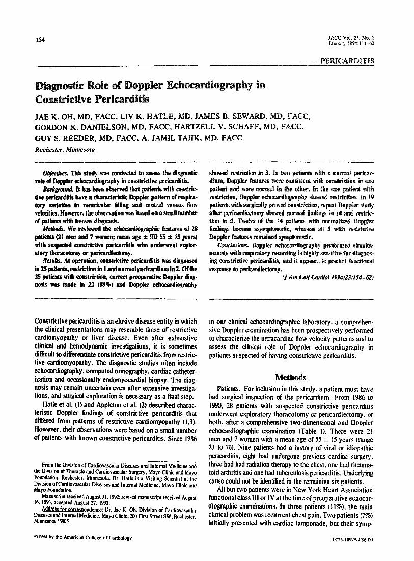
Figure 4 . Pulmonary venous Doppler flow velocity recording bytransesophageal echocardiography in a patient with constriction,The recording was performed simultaneously with respirometer(RESP) and pressure recordings from the pulmonary capillarywedge (PWP) and left ventricle (LV) . Pulmonary venous diastolicforward flow velocity after the onset of expiration (double arrow-heads) is much higher than that after inspiration (single arrowhead) .The difference in pulmonary venous forward flow velocities corre-sponds to the difference in filling pressure gradient (pulmonarycapillary wedge to left ventricle) during inspiration (insp) andexpiration (exp) . This figure was obtained from a patient withconstriction after completion of the current study . The number inparentheses indicates systolic left ventricular pressure in mm Hg .
(the patient had severe chronic obstructive lung disease) andwas normal in the other patient with recurrent (pericardial)chest pain and computed tomographic findings of a thick-ened pericardium . Therefore, of 23 patients with a preoper-ative diagnosis of constriction by Doppler echocardiogra-phy . 22 patients (96%) were found to have constriction atoperation and I patient with chronic obstructive pulmonarydisease had a normal pericardium . Doppler features wereconsistent with restrictive physiology in four patients, threewere diagnosed as having constriction and one as restrictionat operation. The patient with a normal Doppler study had anormal pericardium at operation .
Additional diagnostic tests . Computed tomography .Computed tomography or magnetic resonance imaging wasperformed in 23 and 1 patient(s), respectively (Table 1) .Pericardial thickening or calcification, or both, were noted in22 patients ; I patient (5%) did not have constriction atoperation. There was no constriction in two patients withnormal computed tomograms .
Cardiac catheterization . Fourteen patients underwentpreoperative left and right heart catheterization I to 31 days(mean 8) before (1 patient) or after (13 patients) echocardio-graphic examination . In all, equalization of end-diastolicpressures or "square root sign," or both, was demonstrated .No constriction was found at operation in 2 (14%) of these 14patients . Twelve patients underwent coronary angiographyand seven of them were found to have a significant coronaryartery disease (>70% lumen narrowing of at least onecoronary artery) .
OH ET AL .
159CONSTRICTIVE PERICARDITtS
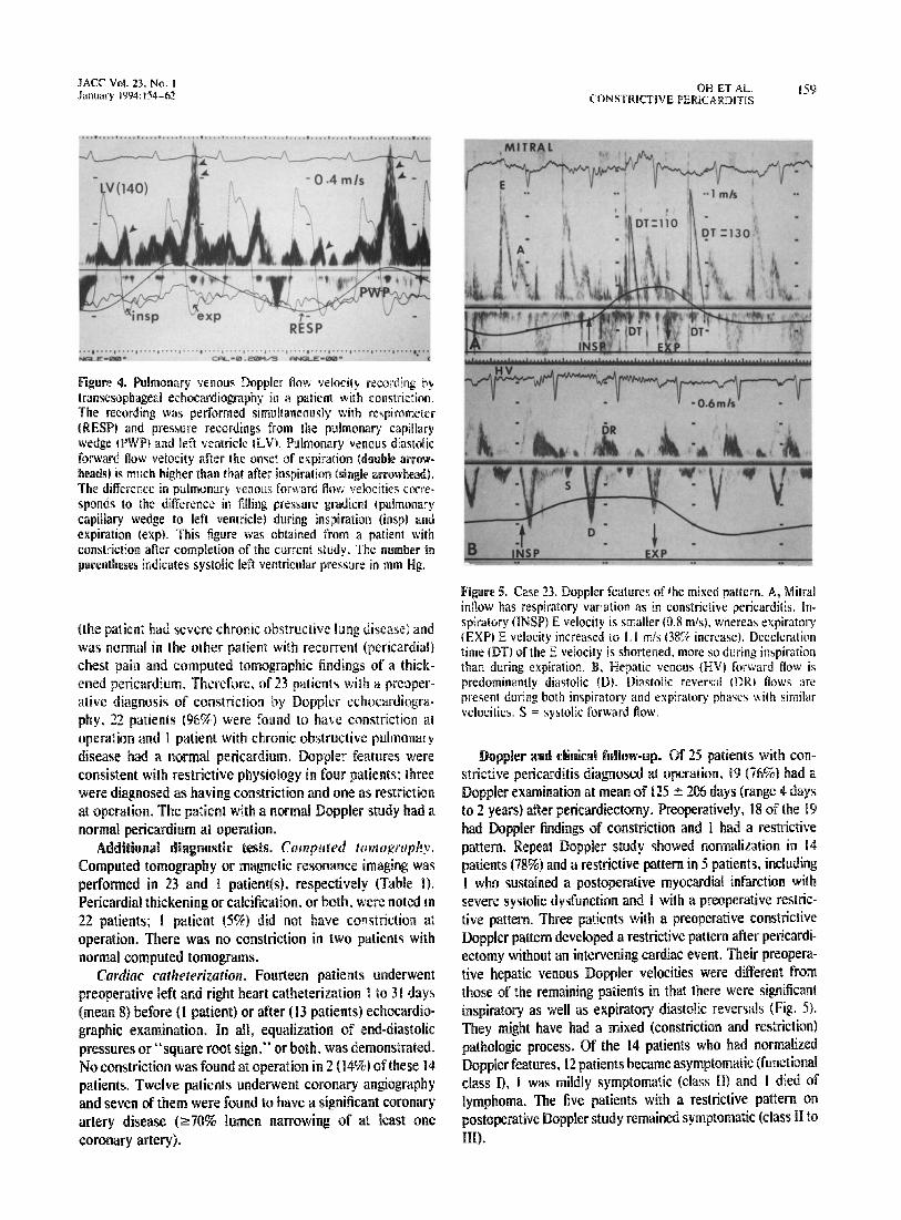
Figure 5 . Case 23 . Doppler features of the mixed pattern . A, Mitralinflow has respiratory variation as in constrictive pericarditis . In-spiratory (INSP) E velocity is smaller (0.8 m/s), whereas expiratoryIEXP) E velocity increased to 1 .1 m/s (38% increase) . Decelerationtinge (DT) of the E velocity is shortened, more so during inspirationthan during expiration . B, Hepatic venous (HV) forward flow ispredominantly diastolic (D) . Diastolic reversal (DR) flows arepresent during both inspiratory and expiratory phases with similarvelocities . S = systolic forward Pow .
Doppler and clinical follow-up . Of 25 patients with con-strictive pericarditis diagnosed at operation, 19 (76%) had aDoppler examination at mean of 125 ± 206 days (range 4 daysto 2 years) after pericardiectomy . Preoperatively, 18 of the 19had Doppler findings of constriction and I had a restrictivepattern . Repeat Doppler study showed normalization in 14patients (789o) and a restrictive pattern in 5 patients, includingI who sustained a postoperative myocardial infarction withsevere systolic dysfunction and I with a preoperative restric-tive pattern . Three patients with a preoperative constrictiveDoppler pattern developed a restrictive pattern after pericardi-ectomy without an intervening cardiac event . Their preopera-tive hepatic venous Doppler velocities were different fromthose of the remaining patients in that there were significantinspiratory as well as expiratory diastolic reversals (Fig . 5) .They might have had a mixed (constriction and restriction)pathologic process. Of the 14 patients who had normalizedDoppler features, 12 patients became asymptomatic (functionalclass I), 1 was mildly symptomatic (class 11) and I died oflymphoma . The five patients with a restrictive pattern onpostoperative Doppler study remained symptomatic (class 11 toIII) .

160 OH ET AL .CONSTRICTIVE PERICARDITIS
DiscussionHemodynamles and Doppler echocardiographic features of
constrictive pericarditis. Hatle et al . (1) and Appleton et al .(2) described characteristic Doppler patterns of constrictivepericarditis based on respiratory variation in transvalvularand central venous flow velocity profiles . The proposedunderlying mechanisms of the Doppllek features in constric-tion are the dissociation between intrathoracic and intracar-diac pressures and an interventricular "coupling" phenom-enon, resulting in a septal shift (Fig . 7) similar to that seen intamponade (8,9) . Normally, a decrease in intrathoracic pres-sure during inspiration is directly transmitted to the intra-cardiac cavity . Pulmonary veins are intrathoracic, and innormal subjects, pressure changes with respiration in pul-
Table 3. Sensitivity of Doppler Echocardiography
I
C = constriction; R = restriction .
Constriction25
RestrictionI
IDoppler
normal
C
R_
R1
22
3
l
3ACC Vol . 23, No. IJanuary 1994 :154--62
Figure 6. Case 5. Mitral inflow and hepatic ve-nous pulsed wave Doppler velocity recordingsfrom a patient with constriction and atrial fibrilla-tion . A, Despite the varying cardiac cycles, mitralinflow during the first beat after the onset ofinspiration is significantly smaller than the firstbeat after the onset of a°-oration . There is dia-stolic mitral regurgitation (arrows) due to high leftventricular diastolic pressure . DT = decelerationtime. B, Hepatic vein flow shows predominantdiastolic forward flow as expected in atrial fibril-lation . There is a marked increase in diastolicreversals during expiration (expir), which is char-acteristic of constriction . insp = inspiration .
monary veins parallel the pressure changes in the intracar-diac chambers. Therefore, the pressure gradient from thepulmonary vein to the left ventricle shows only a minimalchange with respiration .
Figure 7 . Schematic of respiratory variation in iransvalvular andcentral venous flow velocities in constrictive pericarditis . Withinspiration, the driving pressure gradient from the pulmonary cap-illaries to the left cardiac chambers decreases, resulting in a de-crease in mitral inflow and diastolic pulmonary venous (PV) flowvelocity . The decreased left ventricular filling results in ventricularseptal shift to the left (small arrow), allowing augmented flow to theright-sided chambers shown as increased tricuspid inflow and dia-stolic hepatic venous (HV) flow velocity because the cardiac volumeis relatively fixed as a result of the thickened shell of pericardium .The opposite changes occur during expiration (see text). D =diastole ; LA = left atrium ; LV = left ventricle ; RA = right atrium :RV = right ventricle ; S = systole.

162
OH ET AL.CONSTRICTIVE PERICARDITIS
of inspiration or expiration . When diastole begins at end-inspiration, diastolic flow reversals in the hepatic vein maybe prominent even before the onset of expiration, especiallywith a slight time lag between actual timing of respirationand its recording by respirometer on the monitor or stripchart (Fig . 3A).
Although this study represents the largest comprehensiveDoppler evaluation of constrictive pericarditis, the numberof patients is still small. Hence, the prognostic implication ofrestrictive or mixed Doppler findings in the setting of thick-ened pericardium needs to be explored further .
Clinical implications . Echocardiography is usually one ofthe initial diagnostic modalities used in patients with heartfailure, dyspnea, peripheral edema, jugular venous disten-sion or abnormal cardiac auscultation, and it may providethe first diagnostic-clue for constrictive pericarditis, as it didin 29% of our pati! . nts . Because it has been shown that thereis no respiratory -ariation in mitral and pulmonary venousDoppler velocities in patients with restrictive cardiomyopa-thy (20,21), pericardial exploration and pericardiectomy maybe recommended if a patient with characteristic symptomsand signs of constriction hak typical two-dimensional andDoppler features of constriction as described . If a patient hastypical echocardiographic Doppler features of constrictionbut has atypical or comorbid clinical features (chronic ob-structive lung disease, arrhythmia, mechanical ventilation ora different clinically suspected diagnosis for heart failure)that can mimic Doppler findings of constriction, computedtomography or magnetic resonance imaging will be neces-sary to document thickened pericardium before surgicalconsideration. The restrictive Doppler pattern poses a moredifficult problem in a patient with clinical features of con-striction because it does not exclude constriction . Thispatient must undergo further diagnostic evaluation withcomputed tomography, cardiac catheterization or even en-domyocardial biopsy because some patients may presentwith combined constriction and restriction . Moreover, itappears that a restrictive Doppler pattern, either alone or incombination with the constrictive pattern, predicts a lesssatisfactory functional response to pericardiectomy . Herno-dynamic assessment by cardiac catheterization does notappear to provide further diagnostic information or alter thetherapeutic approach once the diagnosis of constriction ismade by two-dimensional Doppler echocardiography orcomputed tomography. In selected patients, however, pre-operative coronary angiography will be necessary .
We thank Gail M . Ludens for excellent help in preparation of the manuscriptand appreciate Dr. Kent R. Bailey and Ruth Cha for statistical review of thedata.
References1 . Hatle LK . Appleton CP, Popp RL. Differentiation of constrictive peri-
carditis and restrictive cardiomyopathy by Doppler echocardiography .Circulation 1989 :79:357-70 .
2 . Appleton CP, Halle LK. Popp RL . Central venous flow velocity patternscan differentiate constrictive pericarditis from restrictive cardiomyopathy[abstract] . J Am Coll Cardiol 1987 :9:119A .
3 . Appleton CP, Halle LK, Popp RL . Demonstration of restrictive ventric-ular physiology by Doppler echocardiography . J Am Coll Cardiol 1988 ;11 :757-68.
4 . Helmcke F, Nanda NC, Hsiung MC . et al . Color Doppler assessmentof mitral regurgitation with orthogonal planes . Circulation 1987 :75 :175-83 .
5 . Perry GJ . Helmeke F, Nanda NC . Byard C . Soto B . Evaluation of aorticinsufficiency by Doppler color flow mapping . J Am Coll Cardiol 1987 ;9 :952-9 .
6 . Appleton CP, Hatle LK, Popp RL . Superior vena cava and hepatic veinDoppler echocardiography in healthy adults . J Am Coll Cardiol 1987 ;10 :1032-9.
7 . Callahan JA, Seward JB, Tajik AJ, et al . Pericardiocentesis assisted bytwo-dimensional echocardiography . J Thorac Cardiovasc Surg 1983 :85 :877-9 .
8 . Appleton CP, Hatle LK, Popp RL . Cardiac tamponade and pericardialeffusion: respiratory variation in transvalvular flow velocities studied byDoppler echocardiography . J Am Coll Cardiol 1988:11 :1020-30.
9 . Santamore WP, Bartlett R . Van Buren SJ . Dowd MK. Katcher MA .Ventricular coupling in constrictive pericarditis . Circulation 1986:74 :597-602.
10 . Hansen AT, Eskildsen P . Gotzsche H . Pressure curves from the rightauricle and the right ventricle in chronic constrictive pericarditis . Circu-lation 1951 :3 :881-8 .
i i . Shabetai R . Fowler NO. Guntheroth WG . The hemodynamics of cardiactamponade and constrictive pericarditis . Am J Cardiol 1970 :26 :00-9 .
12 . Meaney E . Shabetai R . Bhargava V, et al . Cardiac amyloidosis, constric-tive pericarditis and restrictive cardiomyopathy . Am J Cardiol 1976 :38 :547-56 .
13 . Fowler NO . Constrictive pericarditis: new aspects . Am J Cardiol 1982 ;50 :1014-7 .
14 . Bloomfield RA, Lauson HD . Cournand A . Breed ES . Richards DW Jr .Recording of right heart pressures in normal subjects and in patients withchronic pulmonary disease and various types of cardio-circulatory dis-ease . J Clin Invest 1946 ;25 :639-64 .
15. Benotti JR, Grossman W, Cohn PF . Clinical profile of restrictive cardio-myopathy . Circulation 1980;61 :1206-12 .
16. Siegel RJ . Shah PK, Fishbein MC . Idiopathic restrictive cardiomyopathy.Circulation 1984.70 :165-9 .
17. Appleton CP, Hatle LK . Popp RL . Relation of transmitral flow velocitypatterns to left ventricular diastolic function : new insights from a com-bined hemodynamic and Doppler echocardiographic study. J Am CollCardiol 19 ;12:426-40 .
18. Agatston AS, Rao A, Price RJ . Kinney EL. Diagnosis of constrictivepericarditis by pulsed Doppler echocardiography . Am J Cardiol 1984 ;54:929-30.
19. von Bibra H. Schober K . Jenni R, Busch R, Sebening H, Blomer H .Diagnosis of constrictive pericarditis by pulsed Doppler echocardiogra-phy of the hepatic vein . Am J Cardiol 1989 :63 :483-8 .
20. Schiavone WA, Calatiore PA, Salcedo EE. Transesophageal Dopplerechocardiographic demonstration of pulmonary venous flow velocity inrestrictive cardiomyopathy and constrictive pericarditis. Am J Cardiol1989;63:1286-8 .
21 . Klein AL, Tajik AJ. Doppler assessment of diastolic function in cardiacamyloidosis . Echocardiography 1991 ;8 :233-5I .
JACC Vol . 23 . No . IJanuary 1994 :154-62