Diagnosis of bile duct hepatocellular carcinoma thrombus ...
7
BRIEF ARTICLE Diagnosis of bile duct hepatocellular carcinoma thrombus without obvious intrahepatic mass Xue-Ying Long, Yi-Xiong Li, Wei Wu, Lang Li, Jue Cao Xue-Ying Long, Lang Li, Jue Cao, Department of Radiology, Xiangya Hospital, Central South University, Changsha 410008, Hunan Province, China Yi-Xiong Li, Wei Wu, Department of General Surgery, Xiangya Hospital, Central South University, Changsha 410008, Hunan Province, China Author contributions: Long XY, Li YX, Wu W, Li L and Cao J performed the research; Long XY, Li YX and Wu W analyzed the data; Long XY and Li L wrote the paper. Correspondence to: Yi-Xiong Li, Professor, Department of General Surgery, Xiangya Hospital, Central South University, Changsha 410008, Hunan Province, China. [email protected] Telephone: +86-731-84327021 Fax: +86-731-84327332 Received: February 24, 2010 Revised: April 15, 2010 Accepted: April 22, 2010 Published online: October 21, 2010 Abstract AIM: To study the diagnosis of hepatocellular carcino- ma (HCC) presenting as bile duct tumor thrombus with no detectable intrahepatic mass. METHODS: Six patients with pathologically proven bile duct HCC thrombi but no intrahepatic mass dem- onstrated on the preoperative imaging or palpated intrahepatic mass during operative exploration, were collected. Their clinical and imaging data were retro- spectively analyzed. The major findings or signs on comprehensive imaging were correlated with the surgi- cal and pathologic findings. RESULTS: Jaundice was the major clinical symptom of the patients. The elevated serum total bilirubin, direct bilirubin and alanine aminotransferase levels were in concordance with obstructive jaundice and the underly- ing liver disease. Of the 6 patients showing evidence of viral hepatitis, 5 were positive for serum alpha fetopro- tein and carbohydrate antigen 19-9, and 1 was positive for serum carcinoembryonic antigen. No patient was correctly diagnosed by ultrasound. The main features of patients on comprehensive imaging were filling defects with cup-shaped ends of the bile duct, with large filling defects presenting as casting moulds in the expanded bile duct, hypervascular intraluminal nodules, debris or blood clots in the bile duct. No obvious circular thicken- ing of the bile duct walls was observed. CONCLUSION: Even with no detectable intrahepatic tumor, bile duct HCC thrombus should be considered in patients predisposed to HCC, and some imaging signs are indicative of its diagnosis. © 2010 Baishideng. All rights reserved. Key words: Hepatocellular carcinoma; Obstructive jaun- dice; Bile duct tumor thrombus; Diagnosis; Diagnostic imaging Peer reviewers: Takashi Kobayashi, MD, PhD, Department of Surgery, Showa General Hospital, 2-450 Tenjincho, Kodaira, Tokyo 187-8510, Japan; Michael L Schilsky, MD, Associate Professor of Medicine and Surgery, Yale University School of Medicine, 333 Cedar Street, LMP 1080, New Haven, CT 06520, United States Long XY, Li YX, Wu W, Li L, Cao J. Diagnosis of bile duct hepatocellular carcinoma thrombus without obvious intrahe- patic mass. World J Gastroenterol 2010; 16(39): 4998-5004 Available from: URL: http://www.wjgnet.com/1007-9327/full/ v16/i39/4998.htm DOI: http://dx.doi.org/10.3748/wjg.v16.i39. 4998 INTRODUCTION Obstructive jaundice associated with hepatocellular car- cinoma (HCC) is not common, with an incidence of 0.5%-13% in patients with HCC [1-11] . This type of HCC is also known as icteric type of HCC (IHCC) [5-7] . Bile duct tumor thrombus (BDTT) is the leading cause of obstruc- 4998 World J Gastroenterol 2010 October 21; 16(39): 4998-5004 ISSN 1007-9327 (print) ISSN 2219-2840 (online) © 2010 Baishideng. All rights reserved. Online Submissions: http://www.wjgnet.com/1007-9327office [email protected] doi:10.3748/wjg.v16.i39.4998 October 21, 2010|Volume 16|Issue 39| WJG|www.wjgnet.com
Transcript of Diagnosis of bile duct hepatocellular carcinoma thrombus ...

BRIEF ARTICLE
Diagnosis of bile duct hepatocellular carcinoma thrombus without obvious intrahepatic mass
Xue-Ying Long, Yi-Xiong Li, Wei Wu, Lang Li, Jue Cao
Xue-Ying Long, Lang Li, Jue Cao, Department of Radiology, Xiangya Hospital, Central South University, Changsha 410008, Hunan Province, China Yi-Xiong Li, Wei Wu, Department of General Surgery, Xiangya Hospital, Central South University, Changsha 410008, Hunan Province, ChinaAuthor contributions: Long XY, Li YX, Wu W, Li L and Cao J performed the research; Long XY, Li YX and Wu W analyzed the data; Long XY and Li L wrote the paper.Correspondence to: Yi-Xiong Li, Professor, Department of General Surgery, Xiangya Hospital, Central South University, Changsha 410008, Hunan Province, China. [email protected]: +86-731-84327021 Fax: +86-731-84327332Received: February 24, 2010 Revised: April 15, 2010Accepted: April 22, 2010Published online: October 21, 2010
AbstractAIM: To study the diagnosis of hepatocellular carcino-ma (HCC) presenting as bile duct tumor thrombus with no detectable intrahepatic mass.
METHODS: Six patients with pathologically proven bile duct HCC thrombi but no intrahepatic mass dem-onstrated on the preoperative imaging or palpated intrahepatic mass during operative exploration, were collected. Their clinical and imaging data were retro-spectively analyzed. The major findings or signs on comprehensive imaging were correlated with the surgi-cal and pathologic findings.
RESULTS: Jaundice was the major clinical symptom of the patients. The elevated serum total bilirubin, direct bilirubin and alanine aminotransferase levels were in concordance with obstructive jaundice and the underly-ing liver disease. Of the 6 patients showing evidence of viral hepatitis, 5 were positive for serum alpha fetopro-tein and carbohydrate antigen 19-9, and 1 was positive for serum carcinoembryonic antigen. No patient was
correctly diagnosed by ultrasound. The main features of patients on comprehensive imaging were filling defects with cup-shaped ends of the bile duct, with large filling defects presenting as casting moulds in the expanded bile duct, hypervascular intraluminal nodules, debris or blood clots in the bile duct. No obvious circular thicken-ing of the bile duct walls was observed.
CONCLUSION: Even with no detectable intrahepatic tumor, bile duct HCC thrombus should be considered in patients predisposed to HCC, and some imaging signs are indicative of its diagnosis.
© 2010 Baishideng. All rights reserved.
Key words: Hepatocellular carcinoma; Obstructive jaun-dice; Bile duct tumor thrombus; Diagnosis; Diagnostic imaging
Peer reviewers: Takashi Kobayashi, MD, PhD, Department of Surgery, Showa General Hospital, 2-450 Tenjincho, Kodaira, Tokyo 187-8510, Japan; Michael L Schilsky, MD, Associate Professor of Medicine and Surgery, Yale University School of Medicine, 333 Cedar Street, LMP 1080, New Haven, CT 06520, United States
Long XY, Li YX, Wu W, Li L, Cao J. Diagnosis of bile duct hepatocellular carcinoma thrombus without obvious intrahe-patic mass. World J Gastroenterol 2010; 16(39): 4998-5004 Available from: URL: http://www.wjgnet.com/1007-9327/full/v16/i39/4998.htm DOI: http://dx.doi.org/10.3748/wjg.v16.i39.4998
INTRODUCTIONObstructive jaundice associated with hepatocellular car-cinoma (HCC) is not common, with an incidence of 0.5%-13% in patients with HCC[1-11]. This type of HCC is also known as icteric type of HCC (IHCC)[5-7]. Bile duct tumor thrombus (BDTT) is the leading cause of obstruc-
4998
World J Gastroenterol 2010 October 21; 16(39): 4998-5004 ISSN 1007-9327 (print) ISSN 2219-2840 (online)
© 2010 Baishideng. All rights reserved.
Online Submissions: http://www.wjgnet.com/[email protected]:10.3748/wjg.v16.i39.4998
October 21, 2010|Volume 16|Issue 39|WJG|www.wjgnet.com

Long XY et al . Intrabile duct HCC
tion in IHCC. It has been shown that HCC is smaller in patients with biliary tumor thrombi than in those without biliary tumor thrombi, with a mean tumor size of 3.8 ± 2.1 cm vs 6.7 ± 4.6 cm[8]. Several studies[9-11] concerning IHCC reported that primary hepatic parenchymal tumor is detectable in most patients with IHCC while no obvi-ous intrahepatic tumor is detectable in only 2.9%-25.0% of patients with biliary HCC thrombi. We encountered 6 patients with this special type of IHCC. Postoperative pathologic examinations “surprisingly” proved that the bile duct nodules leading to obstructive jaundice were HCCs. However, neither intrahepatic mass nor portal vein thrombus was identified on the preoperative imaging or even during explorative surgery. This type of IHCC is very rare and difficult to diagnose, and only few cases have been occasionally reported[12-17], without their fea-tures summarized. We retrospectively analyzed the clinical and imaging data about 6 patients with emphasis laid on the diagnostic imaging correlated with pathologic and surgical data, and clinical features and imaging signs that might lead to the diagnosis.
MATERIALS AND METHODSEthicsThis study was approved by our institutional review board. Informed consent was not obtained from the patients as this was a limited, anonymous retrospective review of pa-tient data.
PatientsSix patients including 5 men and 1 woman at the age of 47-64 years were confirmed with bile duct HCC by sur-gery and histology between January 2000 and November 2008 at our hospital. Their medical records were thor-oughly reviewed and cross-checked.
Jaundice was the predominant symptom of the pa-tients, and presented as an initial symptom of 4 patients. The time from the onset of jaundice to admission was 7 d-3 mo (median 1 mo). The other main clinical symp-toms were fatigue, right upper quadrant abdominal pain or upper abdominal pain, abdominal distension, loss of appetite and loss of weight. Liver function reserve was Child grade A and B in 4 and 2 patients, respectively.
MethodsLaboratory data about the patients before surgery were recorded and analyzed.
Each patient underwent two or more preoperative diagnostic imaging procedures, including transabdomi-nal ultrasonography (US), computed tomography (CT) with plain scan and arterial and portal phase contrast enhanced scans, magnetic resonance imaging (MRI) with magnetic resonance cholangiopancreatography (MRCP), endoscopic retrograde cholangiopancreatography (ERCP) and percutaneous transhepatic cholangiography (PTC). All available imaging data including diagnosis reports and images were retrospectively reviewed by two radiologists
with more than 5 years of experience in abdominal imag-ing. A consensus was reached with the main findings or signs recorded.
Surgical records and pathologic reports were also re-viewed and correlated with the major findings or signs on comprehensive imaging.
RESULTSBlood testThe levels of total serum bilirubin (TBIL), direct biliru-bin (DBIL), alanine aminotransferase (ALT), hepatitis B surface antigen (HBsAg), α-fetoprotein (AFP), carbo-hydrate antigen 19-9 (CA19-9), and carcinoembryonic antigen (CEA) of each patient are listed in Table 1.
US Transabdominal US was performed, showing dilatated intrahepatic ducts with nodules in hilar bile ducts but no intrahepatic mass. The intraluminal nodules were hypoechoic, slightly hyperechoic, and mixed echoic in 4, 1, and 1 patients, respectively. Three and 1 patients were diagnosed as hilar cholangiocarcinoma and choledocho-lithiasis, respectively. Further evaluation was needed in 2 patients.
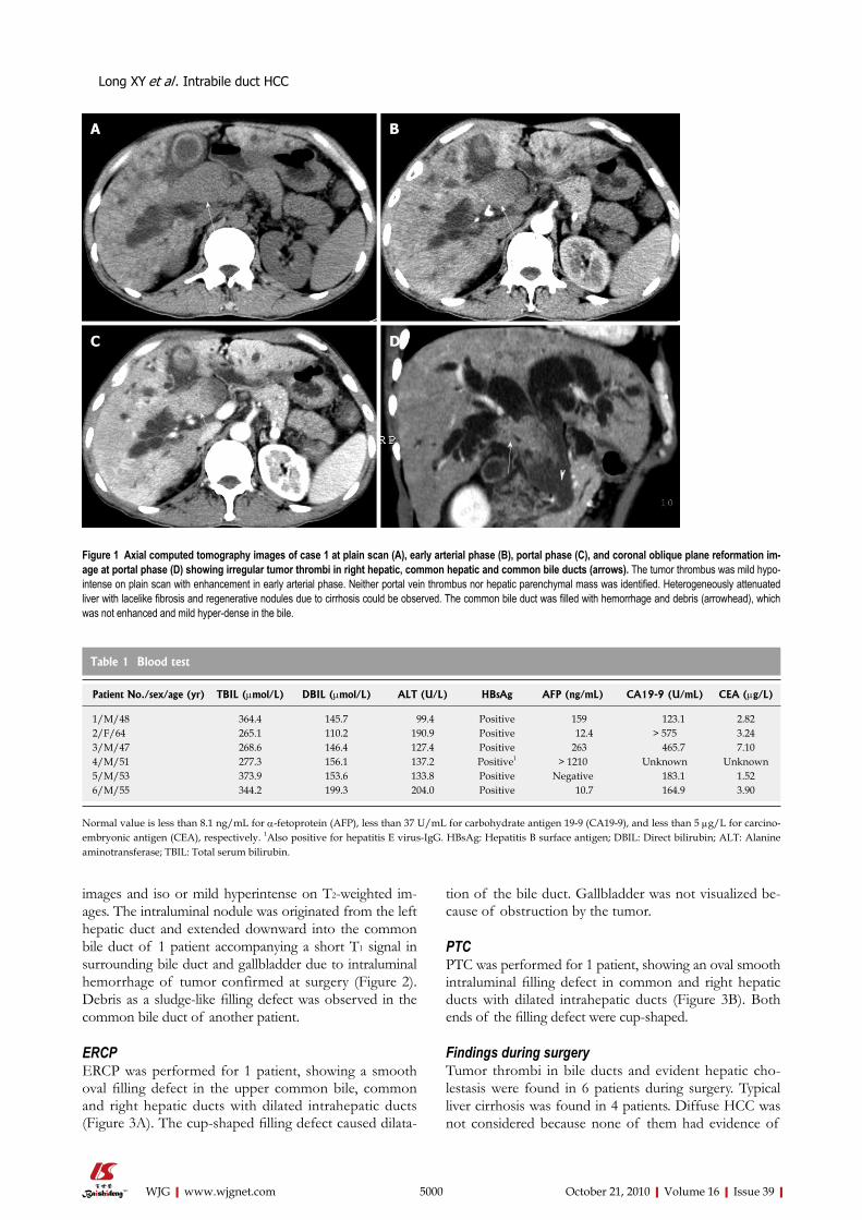
CT CT with pre-contrast scan and dual-phase contrast-enh-anced scan was performed for 5 patients, showing dilated bilateral intrahepatic ducts with an intraductal nodule ob-structing the hilar bile duct and/or common bile duct, but no tumor thrombus in the portal vein or systemic vein and no obvious mass in the hepatic parenchyma. These intra-ductal nodules were relatively mild hypodense to the hepatic parenchyma on pre-contrast images. During the arterial phase, they showed different degrees of enhancement and were relatively isodense or mildly hyperdense to the hepatic parenchyma. The enhancement of intraductal nodules was relatively lower in portal phase than that of hepatic paren-chyma. In three lesions with the longest diameter greater than 3.0 cm, the intraluminal nodules appeared as cast moulds in the dilated ducts without obvious thickening of the walls. Non-enhanced sludge, which was mildly hyper-dense in the bile, was observed in the common bile duct of 2 patients (Figure 1). The sludge was found to be tumor de-bris or hemorrhage of tumor at surgery. CT showed signs of liver cirrhosis in 4 patients, such as splenomegaly, varices, heterogeneously attenuated liver with lacelike fibrosis and regenerative nodules, and irregular or nodular liver surface. A small amount of ascites was present in 1 patient.
MRI combined with MRCPConventional non-enhanced MRI combined with MRCP was performed for 3 patients, showing no mass in the hepatic parenchyma or portal vein. MRCP images showed moderate-severe dilatation of bilateral hepatic ducts with columnar or plugged filling defect of bile ducts in the hilar area. The filling defects were hypointense on T1-weighted
4999 October 21, 2010|Volume 16|Issue 39|WJG|www.wjgnet.com
images and iso or mild hyperintense on T2-weighted im-ages. The intraluminal nodule was originated from the left hepatic duct and extended downward into the common bile duct of 1 patient accompanying a short T1 signal in surrounding bile duct and gallbladder due to intraluminal hemorrhage of tumor confirmed at surgery (Figure 2). Debris as a sludge-like filling defect was observed in the common bile duct of another patient.
ERCPERCP was performed for 1 patient, showing a smooth oval filling defect in the upper common bile, common and right hepatic ducts with dilated intrahepatic ducts (Figure 3A). The cup-shaped filling defect caused dilata-
tion of the bile duct. Gallbladder was not visualized be-cause of obstruction by the tumor.
PTCPTC was performed for 1 patient, showing an oval smooth intraluminal filling defect in common and right hepatic ducts with dilated intrahepatic ducts (Figure 3B). Both ends of the filling defect were cup-shaped.
Findings during surgeryTumor thrombi in bile ducts and evident hepatic cho-lestasis were found in 6 patients during surgery. Typical liver cirrhosis was found in 4 patients. Diffuse HCC was not considered because none of them had evidence of
5000 October 21, 2010|Volume 16|Issue 39|WJG|www.wjgnet.com
Figure 1 Axial computed tomography images of case 1 at plain scan (A), early arterial phase (B), portal phase (C), and coronal oblique plane reformation im-age at portal phase (D) showing irregular tumor thrombi in right hepatic, common hepatic and common bile ducts (arrows). The tumor thrombus was mild hypo-intense on plain scan with enhancement in early arterial phase. Neither portal vein thrombus nor hepatic parenchymal mass was identified. Heterogeneously attenuated liver with lacelike fibrosis and regenerative nodules due to cirrhosis could be observed. The common bile duct was filled with hemorrhage and debris (arrowhead), which was not enhanced and mild hyper-dense in the bile.
A B
C D
Table 1 Blood test
Patient No./sex/age (yr) TBIL (µmol/L) DBIL (µmol/L) ALT (U/L) HBsAg AFP (ng/mL) CA19-9 (U/mL) CEA (µg/L)
1/M/48 364.4 145.7 99.4 Positive 159 123.1 2.822/F/64 265.1 110.2 190.9 Positive 12.4 > 575 3.243/M/47 268.6 146.4 127.4 Positive 263 465.7 7.104/M/51 277.3 156.1 137.2 Positive1 > 1210 Unknown Unknown5/M/53 373.9 153.6 133.8 Positive Negative 183.1 1.526/M/55 344.2 199.3 204.0 Positive 10.7 164.9 3.90
Normal value is less than 8.1 ng/mL for α-fetoprotein (AFP), less than 37 U/mL for carbohydrate antigen 19-9 (CA19-9), and less than 5 µg/L for carcino-embryonic antigen (CEA), respectively. 1Also positive for hepatitis E virus-IgG. HBsAg: Hepatitis B surface antigen; DBIL: Direct bilirubin; ALT: Alanine aminotransferase; TBIL: Total serum bilirubin.
Long XY et al . Intrabile duct HCC

tumor invasion of the portal vein or system vein. No obvious intrahepatic mass was palpated in all patients. Diffuse miliary peritoneum metastasis was observed in 1 patient and thought to be infiltration of the tumor in hilar duct. The tumor thrombi were dark brown, dark red or yellowish brown in color, and soft or slightly elastic, relatively friable, and extremely vascular tending to bleed even on light touch. Most of them were easily separated from the bile duct walls. Sludge-like debris or small blood
clots were found in the common bile ducts of 4 patients and blood-stained bile was found in the intrahepatic ducts of 2 patients. Removal of tumor thrombi was attempted in 6 patients and was successful without active hemor-rhage in 4 patients. Partial hepatectomy was performed for 2 patients, and aborted in 4 patients due to the poor liver function reserve or peritoneal metastasis. Further exploration after clearance of thrombi revealed relatively smooth internal walls of common bile duct (CBD) and
5001 October 21, 2010|Volume 16|Issue 39|WJG|www.wjgnet.com
Figure 2 Magnetic resonance and magnetic resonance cholangiopancreatography images of Case 2. A: Coronal T1 weighted image showing hypo-intense tumor thrombus (large arrows) in left hepatic, common hepatic and common bile ducts; B: Coronal T2 weighted image showing the tumor thrombus as iso- to slight hyper-intense; C: Thick slice magnetic resonance cholangiopancreatography image showing bilateral dilated intrahepatic ducts with “vanished” common hepatic and common bile ducts; D: Thin slice magnetic resonance cholangiopancreatography image showing filling defect in the hilar. Neither portal vein thrombus nor hepatic parenchymal mass is identified. The signal intensity of liver is heterogeneous due to cirrhosis. Blood clots or hemorrhage can be observed in gallbladder (small arrows), common hepatic and common bile ducts (arrowheads), which are hyper-dense on T1 weighted image, and slight hyper-intense on T2 weighted image.
A B
C D
Figure 3 Endoscopic retrograde cholangiopancreatography image of case 3 (A) and percutaneous transhepatic cholangiography image of case 4 (B) showing tumor thrombus (arrows) in right hepatic duct and common hepatic duct as a smooth oval filling defect on endoscopic retrograde cholangiopan-creatography image and a smooth oval filling defect on percutaneous transhepatic cholangiography image, respectively.
A B
Long XY et al . Intrabile duct HCC

common hepatic duct (CHD). The resected liver tis-sue revealed a small hepatic parenchymal tumor in each, 0.5 cm × 1.0 cm and 0.8 cm × 1.5 cm in size.
PathologyThe pathologic reports of intrabiliary thrombi revealed HCC but no variants of cholangiocarcinoma, mixed type of HCC or cholangiocarcinoma in 6 patients. Of the 6 patients, 2 had poorly-differentiated HCC, 3 had poorly-moderately differentiated HCC, and 1 had moderately-differentiated HCC, which accompanied hemorrhage or necrotic tissue in most of the 6 patients.
DISCUSSIONPortal vein tumor thrombus (PVTT) is frequently seen in HCC patients. However, HCC presented as biliary duct tumor thrombus (BDTT) is a relatively rare entity. Intra-hepatic tumor or PVTT is evident in most of patients with HCC presented as BDTT, yet few patients with HCC thrombi in the bile duct but without any detectable intra-hepatic mass or PVTT have been reported[9-17]. In this cir-cumstance, it is difficult but still important to establish the correct diagnosis, especially to differentiate it from cholan-giocarcinoma or diffuse-type HCC. Because the therapeu-tic plan for HCC presented as BDTT may be substantially different from that for cholangiocarcinoma and diffuse-type HCC, surgery can often be offered when the disease is still localized[1,2,10-20], while percutaneous transhepatic cholangial drainage (PTCD) combined with transcatheter arterial chemoembolization (TACE) serves as an effective alternative therapy especially when the tumor is unresect-able[1,6,7]. We retrospectively analyzed the clinical and imag-ing data about these patients, and found that some features might be helpful for the diagnosis.
Jaundice is the predominant clinical presentation of this disease[1-21]. Causes of obstructive jaundice in this type of HCC include intraluminal growth of tumor leading to obstruction of intra or extrahepatic ducts, tumor tis-sue fragments and/or hemorrhages or blood clots due to necrosis, bleeding, and detachment of intraductal tumors, giving rise to the obstruction, which are similar to the re-ported findings in IHCC[3].
Apart from jaundice, there are also other non-specif-ic symptoms such as fatigue, abdominal pain, abdominal distension and loss of appetite. A differential diagnosis between this and other common diseases causing ob-structive jaundice such as cholangiocarcinoma and cho-ledocholithiasis is essential.
In this study, all the 6 patients were positive for the markers of chronic viral hepatitis. The proportion of liver cirrhosis was relatively high with typical liver cirrhosis found in 4 patients. The elevated serum ALT level in our patients might be associated with the underlying disease. The majority of patients were middle-aged or old males. These features were also found in common types of HCC.
Serum tumor markers may be helpful in the diagnosis of this disease[22]. Positive serum AFP supports the di-agnosis of HCC, while CEA level is frequently elevated
in patients with cholangiocarcinoma. In this study, the positive ratios for AFP and CA19-9 were high, suggest-ing that positive AFP and CA19-9 support the diagnosis. However, positive CA19-9 may also frequently be seen in cholangitis, bile duct stones and biliary or pancreatic tumors leading to obstructive jaundice[23]. Thus, its value for differential diagnosis has to be further evaluated in studies with more cases.
Several diagnostic imaging methods play an important role in the diagnosis of obstructive jaundice[24-31]. The location and extent of obstruction can be reliably demon-strated using proper techniques, and the cause of obstruc-tion can often be inferred by analyzing the morphology of obstruction site and other relevant signs. Transabdominal US is the most widely used and usually the initial tool to evaluate obstructive jaundice. However, none of our pa-tients was correctly diagnosed by transabdominal US, and most of them were misdiagnosed as cholangiocarcinoma. Tamada et al[29] reported that intraductal US (IDUS) can distinguish between tumor thrombi caused by HCC and polypoid type cholangiocarcinoma. However, IDUS has not been widely accepted because it is invasive and requires special equipments and specific expertise. CT, MRI com-bined with MRCP, ERCP and PTC are also widely used to evaluate obstructive jaundice. Jung et al[30] compared the CT features of HCC invading bile ducts with those of in-traductal papillary cholangiocarcinoma, and found that the presence of parenchymal mass is the distinct difference between them. Since no parenchymal mass was detectable on cross-sectional images in our patients, we searched for other signs leading to the diagnosis. After analyzing the intrabiliary lesions on comprehensive images with surgical correlation, we found that the following features may be helpful in the diagnosis of this special type of IHCC.
First, the tumor thrombi are typically intraluminal pol-ypoid lesions with irregular or smooth surface depending on the presence of necrosis. Lesions can be either small or larger. Small lesions are localized in 1 or 2 ducts while large lesions can form “biliary duct casts” and extend inferiorly. Because of rapid growth of the tumor with no apparent fibrosis of the duct wall, the ducts can often be expanded into a fusiform shape at the site of lesion. Cup-shaped filling defects can also be observed in dilated ducts around the lesion on cholangiographic images due to the expanded growth pattern of the lesion.
Second, the tumor thrombi are hypervascular. It is well known that HCC is a hypervascular tumor. Detection of vascularity in bile duct thrombi can reliably rule out the di-agnosis of bile duct stones, and differentially diagnose be-tween HCC and cholangiocarcinoma. The bile duct throm-bi with the characteristic early enhancement pattern on dual-phase contrast-enhanced CT[30] or dynamic contrast-enhanced MRI[28] are important to differentiate HCC from cholangiocarcinoma. The results of this study support this finding. The hypervascularity of bile duct tumor thrombi was confirmed during surgery in this study. It was reported that color Doppler sonography can also effectively detect tumor vascularity of bile duct HCC thrombi[31].
Third, intraluminal fragments or blood clots may
5002 October 21, 2010|Volume 16|Issue 39|WJG|www.wjgnet.com
Long XY et al . Intrabile duct HCC

present. Because the tumor thrombi are loose, fragile and prone to necrosis, detachment of parts of the lesions and hemorrhage may frequently occur, and those that are relatively large can lead to free thrombi in the ducts. CT can demonstrate irregular or sludge-like lesions in the ducts with no enhancement. MRI and MRCP may be even superior over CT in demonstrating such lesions[28]. Hemorrhagic lesions or blood clots have a high signal on T1WI, and a low signal on T2WI. Debris in bile appears as sludge in bile ducts and gallbladder.
Fourth, no apparent circular thickening of the duct walls or constriction of the ducts is present. Cholangio-carcinoma is often associated with the thickening of bile duct walls, often leading to constriction of nearby ducts. No apparent thickening of the duct walls, especially no circular thickening of the walls, was observed in our pa-tients, and ducts were compacted rather than constricted or narrowed due to the tumor.
Fifth, no portal vein thrombus is present. It might be due to the relatively early stage of the disease in our pa-tients, and it is critical for differentiating it from diffuse-type HCC.
Sixth, cirrhosis of the background liver may support the diagnosis. A relatively high percentage of cirrhosis was observed on preoperative images and during surgery in our patients. “Downstream duct dilatation”, a sign stand-ing for the dilatated bile duct below the level of intralumi-nal nodule, has been described by Jung et al[30] in patients with intraductal cholangiocarcinoma, which is thought to be related to mucin produced by the tumor. However, “downstream duct dilatation” was also present in 2 out of 6 patients in our case study, which was contributed to the obstruction by blood clots or fragments in the common bile ducts.
The reasons why HCC is present as intrabile duct tu-mor thrombi without detectable primary hepatic tumor are as follows. The tumor may originate from canceriza-tion of ectopic hepatocytes in the bile duct wall[17], or the primary tumor is just too small to be identified, or the tumor located at the origin of or close to the intrahepatic duct grows intraluminally and stretchs inferiorly. Although no primary hepatic tumor was demonstrated on preopera-tive imaging or palpated during operation in our patients, the resected tissues revealed small hepatic tumors in 2 pa-tients. Moreover, since deeply seated small hepatic tumor is hard to palpate during intraoperative exploration, espe-cially in patients with marked cirrhosis, it is hard to rule out the potentiality of small primary intrahepatic HCC in the other 4 patients who did not undergo partial hepatic resection. So, it is recommended that intraoperative ul-trasonography (IOUS) should be performed to find the potential intrahepatic tumor or to determine the resection level before the operator decides to perform the resection.
If this disease is suspected, it is still important to look for more sensitive techniques such as CT during arterial portography (CTAP) and superparamagnetic iron oxide (SPIO)-enhanced MRI to find possible primary tumors. CTAP is generally accepted as the most sensitive technique
to detect small HCC, but it is only performed for selected patients due to its invasiveness. SPIO-enhanced MRI has emerged as another effective technique to detect small HCC, but its value in evaluating bile duct tumor has not yet fully investigated. Unless the clinicians or the radiolo-gists take HCC into consideration, these techniques can be first adopted in the diagnosis of obstructive jaundice. Thus, our study may help the clinicians and radiologists to consider this disease before such techniques are applied.
There are some limitations in our study. First, it is a retrospective analysis of a limited number of cases. Sec-ond, although dynamic contrast-enhanced MRI is used as a conventional technique for the diagnosis of HCC in our hospital, it has not been routinely performed for the evaluation of obstructive jaundice. Third, contrast studies with other types of HCC or other tumors with intraluminal growth are not available due to the limited number of cases. Further study is needed to verify the diagnostic value of the features listed.
COMMENTSBackgroundHepatocellular carcinoma (HCC) thrombus in the bile duct is a rare cause of obstructive jaundice. Although it is rarely encountered, its correct diagnosis, especially differentiating it from other causes of biliary obstruction such as chol-angiocarcinoma, is very important. Usually, the presence of primary intrahepatic tumors is the key to its diagnosis. However, since few cases of HCC thrombi in the bile duct with no detectable intrahepatic mass have been reported, its diag-nosis is even difficult.Research frontiersSix patients with this rare disease were reported. Their clinical and imaging data were retrospectively analyzed with a review of the literature. Some clinical features and imaging signs that may favor the diagnosis were summarized. The study may be helpful for a better understanding of the disease, especially for its diagnosis.Innovations and breakthroughsLittle is known about the diagnosis of this rare disease. More accurate diagno-sis were introduced in this study by describing their clinical features and imag-ing signs.ApplicationsThis research may evoke the attention of clinicians to the diagnosis of bile duct hepatocellular carcinoma thrombi without an intrahepatic tumor demonstrated on the diagnostic imaging. If certain clinical features and imaging signs are pre-sented, the diagnosis of the disease can be considered.Peer reviewThe manuscript presents an interesting series of patients with HCC presented as biliary tract obstruction leading to jaundice. The clinical and imaging data help find the features that differentiate this tumor from cholangiocarcinoma.
REFERENCES1 Lau W, Leung K, Leung TW, Liew CT, Chan MS, Yu SC, Li
AK. A logical approach to hepatocellular carcinoma pre-senting with jaundice. Ann Surg 1997; 225: 281-285
2 Qin LX, Tang ZY. Hepatocellular carcinoma with obstruc-tive jaundice: diagnosis, treatment and prognosis. World J Gastroenterol 2003; 9: 385-391
3 Lai EC, Lau WY. Hepatocellular carcinoma presenting with obstructive jaundice. ANZ J Surg 2006; 76: 631-636
4 Wang HJ, Kim JH, Kim JH, Kim WH, Kim MW. Hepatocel-lular carcinoma with tumor thrombi in the bile duct. Hepato-gastroenterology 1999; 46: 2495-2499
5 Lin TY, Chen KM, Chen YR, Lin WS, Wang TH, Sung JL.
5003 October 21, 2010|Volume 16|Issue 39|WJG|www.wjgnet.com
COMMENTS
Long XY et al . Intrabile duct HCC

Icteric type hepatoma. Med Chir Dig 1975; 4: 267-2706 Huang JF, Wang LY, Lin ZY, Chen SC, Hsieh MY, Chuang
WL, Yu MY, Lu SN, Wang JH, Yeung KW, Chang WY. In-cidence and clinical outcome of icteric type hepatocellular carcinoma. J Gastroenterol Hepatol 2002; 17: 190-195
7 Huang GT, Sheu JC, Lee HS, Lai MY, Wang TH, Chen DS. Icteric type hepatocellular carcinoma: revisited 20 years later. J Gastroenterol 1998; 33: 53-56
8 Yeh CN, Jan YY, Lee WC, Chen MF. Hepatic resection for hepatocellular carcinoma with obstructive jaundice due to biliary tumor thrombi. World J Surg 2004; 28: 471-475
9 Peng BG, Liang LJ, Li SQ, Zhou F, Hua YP, Luo SM. Surgi-cal treatment of hepatocellular carcinoma with bile duct tumor thrombi. World J Gastroenterol 2005; 11: 3966-3969
10 Peng SY, Wang JW, Liu YB, Cai XJ, Xu B, Deng GL, Li HJ. Hepatocellular carcinoma with bile duct thrombi: analy-sis of surgical treatment. Hepatogastroenterology 2004; 51: 801-804
11 Qin LX, Ma ZC, Wu ZQ, Fan J, Zhou XD, Sun HC, Ye QH, Wang L, Tang ZY. Diagnosis and surgical treatments of he-patocellular carcinoma with tumor thrombosis in bile duct: experience of 34 patients. World J Gastroenterol 2004; 10: 1397-1401
12 Badve SS, Saxena R, Wagholikar UL. Intraductal hepatocel-lular carcinoma with normal liver--case report. Indian J Can-cer 1991; 28: 165-167
13 Buckmaster MJ, Schwartz RW, Carnahan GE, Strodel WE. Hepatocellular carcinoma embolus to the common hepatic duct with no detectable primary hepatic tumor. Am Surg 1994; 60: 699-702
14 Cho HG, Chung JP, Lee KS, Chon CY, Kang JK, Park IS, Kim KW, Chi HS, Kim H. Extrahepatic bile duct hepato-cellular carcinoma without primary hepatic parenchymal lesions--a case report. Korean J Intern Med 1996; 11: 169-174
15 Kashiwazaki M, Nakamori S, Makino T, Omura Y, Yasui M, Ikenaga M, Miyazaki M, Hirao M, Takami K, Fujitani K, Mishima H, Sugiura T, Tsujinaka T. [An icteric type hepato-cellular carcinoma with no detectable tumor in the liver but with an intrabile duct recurrent tumor] Gan To Kagaku Ryoho 2007; 34: 2099-2101
16 Makino T, Nakamori S, Kashiwazaki M, Masuda N, Ike-naga M, Hirao M, Fujitani K, Mishima H, Sawamura T, Takeda M, Mano M, Tsujinaka T. An icteric type hepatocel-lular carcinoma with no detectable tumor in the liver: report of a case. Surg Today 2006; 36: 633-637
17 Tsushimi T, Enoki T, Harada E, Orita M, Noshima S, Ma-suda M, Hamano K. Ectopic hepatocellular carcinoma aris-ing in the bile duct. J Hepatobiliary Pancreat Surg 2005; 12: 266-268
18 Jan YY, Chen MF, Chen TJ. Long term survival after ob-struction of the common bile duct by ductal hepatocellular carcinoma. Eur J Surg 1995; 161: 771-774
19 Peng SY, Wang JW, Liu YB, Cai XJ, Deng GL, Xu B, Li HJ.
Surgical intervention for obstructive jaundice due to biliary tumor thrombus in hepatocellular carcinoma. World J Surg 2004; 28: 43-46
20 Shiomi M, Kamiya J, Nagino M, Uesaka K, Sano T, Hay-akawa N, Kanai M, Yamamoto H, Nimura Y. Hepatocellular carcinoma with biliary tumor thrombi: aggressive operative approach after appropriate preoperative management. Sur-gery 2001; 129: 692-698
21 Kojiro M, Kawabata K, Kawano Y, Shirai F, Takemoto N, Nakashima T. Hepatocellular carcinoma presenting as in-trabile duct tumor growth: a clinicopathologic study of 24 cases. Cancer 1982; 49: 2144-2147
22 Snarska J, Szajda SD, Puchalski Z, Szmitkowski M, Cha-bielska E, Kaminski F, Zwierz P, Zwierz K. Usefulness of examination of some tumor markers in diagnostics of liver cancer. Hepatogastroenterology 2006; 53: 271-274
23 Mann DV, Edwards R, Ho S, Lau WY, Glazer G. Elevated tumour marker CA19-9: clinical interpretation and influence of obstructive jaundice. Eur J Surg Oncol 2000; 26: 474-479
24 Park SJ, Han JK, Kim TK, Choi BI. Three-dimensional spiral CT cholangiography with minimum intensity projection in patients with suspected obstructive biliary disease: com-parison with percutaneous transhepatic cholangiography. Abdom Imaging 2001; 26: 281-286
25 Yeh TS, Jan YY, Tseng JH, Chiu CT, Chen TC, Hwang TL, Chen MF. Malignant perihilar biliary obstruction: magnetic resonance cholangiopancreatographic findings. Am J Gastro-enterol 2000; 95: 432-440
26 Lau WY, Leow CK, Leung KL, Leung TW, Chan M, Yu SC. Cholangiographic features in the diagnosis and manage-ment of obstructive icteric type hepatocellular carcinoma. HPB Surg 2000; 11: 299-306
27 Kirk JM, Skipper D, Joseph AE, Knee G, Grundy A. Intralu-minal bile duct hepatocellular carcinoma. Clin Radiol 1994; 49: 886-888
28 Tseng JH, Hung CF, Ng KK, Wan YL, Yeh TS, Chiu CT. Icteric-type hepatoma: magnetic resonance imaging and magnetic resonance cholangiographic features. Abdom Imag-ing 2001; 26: 171-177
29 Tamada K, Isoda N, Wada S, Tomiyama T, Ohashi A, Satoh Y, Ido K, Sugano K. Intraductal ultrasonography for hepa-tocellular carcinoma with tumor thrombi in the bile duct: comparison with polypoid cholangiocarcinoma. J Gastroen-terol Hepatol 2001; 16: 801-805
30 Jung AY, Lee JM, Choi SH, Kim SH, Lee JY, Kim SW, Han JK, Choi BI. CT features of an intraductal polypoid mass: Differentiation between hepatocellular carcinoma with bile duct tumor invasion and intraductal papillary cholangiocar-cinoma. J Comput Assist Tomogr 2006; 30: 173-181
31 Wang JH, Chen TM, Tung HD, Lee CM, Changchien CS, Lu SN. Color Doppler sonography of bile duct tumor thrombi in hepatocellular carcinoma. J Ultrasound Med 2002; 21: 767-772; quiz 773-774
S- Editor Wang JL L- Editor Wang XL E- Editor Ma WH
5004 October 21, 2010|Volume 16|Issue 39|WJG|www.wjgnet.com
Long XY et al . Intrabile duct HCC