Diagnosis and Treatment of Pleural Effusion
24
Arch Bronconeumol. 2006;42(7):349-72 349 Introduction The pleural space, between the parietal pleura covering the chest wall and the visceral pleura covering the lung, contains—in a healthy person—a few milliliters of fluid that acts as a lubricant between the 2 surfaces. Pathological accumulation of fluid in this space is called pleural effusion. 1 Etiology, Pathogenesis, and Epidemiology Pleural fluid originates in the pleural capillaries (mainly those of the parietal pleura), lymphatics, intrathoracic blood vessels, the interstitial pulmonary space, and the peritoneal cavity. It is reabsorbed mainly through the lymphatics of the parietal pleura. The mechanisms that cause pleural effusion all result in an increase in the production or a decrease in the removal of pleural fluid and may be related to changes in hydrostatic capillary, intravascular or extravascular colloid osmotic, and negative intrathoracic pressures (Table 1). The prevalence of pleural effusion is slightly in excess of 400/100 000 population. Congestive heart failure is the most common cause of pleural effusions overall. However, the predominant etiologies among the exudates are pneumonia, malignancy, and pulmonary embolism. Table 2 shows the most common causes of pleural effusion. Methods Used to Investigate Pleural Disease Medical History Patients with pleural effusions should be studied systematically. As a first step a complete medical history should be taken with special emphasis on the patient’s history of exposure to asbestos, current and recent medications, and the prior or current presence of entities such as heart disease, tuberculosis, neoplastic disease, and connective tissue disease. Secondly, a complete physical examination should be performed. Based on the overall picture provided by the clinical variables, medical history, physical examination, results of basic laboratory tests, and of any additional tests ordered because of a suspected diagnosis, it is possible to establish a diagnosis before thoracentesis and order the pertinent tests. Radiographic Techniques An effusion of more than 75 mL is often visible on chest radiographs. Pleural effusions can be either free flowing or loculated and either typically or atypically sited (subpulmonic, fissural, or mediastinal) sited. The amount of fluid varies. When there is a doubt in the case of small effusions the existence of pleural fluid should be confirmed by chest ultrasound or radiographically using a lateral decubitus projection on the affected side. Anomalies in the lung parenchyma can help to confirm the suspected diagnosis, and computed tomography can contribute useful additional information. Correspondence: Dr. V. Villena Garrido. Servicio de Neumología. Hospital 12 de Octubre. Avda. de Córdoba, s/n. 28041 Madrid. España. E-mail: [email protected] Manuscript received November 10, 2005. Accepted for publication November 22, 2005. RECOMMENDATIONS OF THE SPANISH SOCIETY OF PULMONOLOGY AND THORACIC SURGERY (SEPAR) Diagnosis and Treatment of Pleural Effusion Victoria Villena Garrido (coordinator), a Jaime Ferrer Sancho, b Hernández Blasco, c Alicia de Pablo Gafas, d Esteban Pérez Rodríguez, e Francisco Rodríguez Panadero, f Santiago Romero Candeira, c Ángel Salvatierra Velázquez, g and Luis Valdés Cuadrado. h Assembly on Transplantation and Transplant Techiques SEPAR. a Hospital Universitario 12 de Octubre, Madrid, Spain. b Hospital Vall d’Hebron, Barcelona, Spain. c Hospital General Universitario, Alicante, Spain. d Clínica Puerta de Hierro, Madrid, Spain. e Hospital Ramón y Cajal, Madrid, Spain. f Hospital Virgen del Rocío, Seville, Spain. c Hospital General Universitario, Alicante, Spain. g Hospital Reina Sofía, Cordoba, Spain. h Hospital de Conxo, Santiago de Compostela, La Coruña, Spain. 143.719 TABLE 1 Mechanisms of Production of Pleural Effusion Systemic increase in hydrostatic pressure Decreased oncotic pressure in the microvascular circulation Increased permeability of the pleural microvascular circulation Increase in pulmonary interstitial fluid Impaired lymphatic drainage Movement of fluid from other cavities or sites such as the peritoneal, retroperitoneal, or subarachnoid spaces, or catheters Decrease in negative pressure within the pleural space Vascular rupture in the chest Rupture of the thoracic duct
Transcript of Diagnosis and Treatment of Pleural Effusion

Arch Bronconeumol. 2006;42(7):349-72 349
Introduction
The pleural space, between the parietal pleuracovering the chest wall and the visceral pleura coveringthe lung, contains—in a healthy person—a fewmilliliters of fluid that acts as a lubricant between the 2surfaces. Pathological accumulation of fluid in thisspace is called pleural effusion.1
Etiology, Pathogenesis, and Epidemiology
Pleural fluid originates in the pleural capillaries(mainly those of the parietal pleura), lymphatics,intrathoracic blood vessels, the interstitial pulmonaryspace, and the peritoneal cavity. It is reabsorbed mainlythrough the lymphatics of the parietal pleura. Themechanisms that cause pleural effusion all result in anincrease in the production or a decrease in the removalof pleural fluid and may be related to changes inhydrostatic capillary, intravascular or extravascularcolloid osmotic, and negative intrathoracic pressures(Table 1).
The prevalence of pleural effusion is slightly inexcess of 400/100 000 population. Congestive heartfailure is the most common cause of pleural effusionsoverall. However, the predominant etiologies among theexudates are pneumonia, malignancy, and pulmonaryembolism. Table 2 shows the most common causes ofpleural effusion.
Methods Used to Investigate Pleural Disease
Medical History
Patients with pleural effusions should be studiedsystematically. As a first step a complete medicalhistory should be taken with special emphasis on thepatient’s history of exposure to asbestos, current andrecent medications, and the prior or current presence ofentities such as heart disease, tuberculosis, neoplasticdisease, and connective tissue disease. Secondly, acomplete physical examination should be performed.Based on the overall picture provided by the clinicalvariables, medical history, physical examination, resultsof basic laboratory tests, and of any additional testsordered because of a suspected diagnosis, it is possibleto establish a diagnosis before thoracentesis and orderthe pertinent tests.
Radiographic Techniques
An effusion of more than 75 mL is often visible onchest radiographs. Pleural effusions can be either freeflowing or loculated and either typically or atypicallysited (subpulmonic, fissural, or mediastinal) sited. Theamount of fluid varies. When there is a doubt in thecase of small effusions the existence of pleural fluidshould be confirmed by chest ultrasound orradiographically using a lateral decubitus projection onthe affected side. Anomalies in the lung parenchymacan help to confirm the suspected diagnosis, andcomputed tomography can contribute useful additionalinformation.
Correspondence: Dr. V. Villena Garrido.Servicio de Neumología. Hospital 12 de Octubre. Avda. de Córdoba, s/n. 28041 Madrid. España.E-mail: [email protected]
Manuscript received November 10, 2005. Accepted for publication November 22, 2005.
RECOMMENDATIONS OF THE SPANISH SOCIETY OF PULMONOLOGY AND THORACIC SURGERY (SEPAR)
Diagnosis and Treatment of Pleural Effusion
Victoria Villena Garrido (coordinator),a Jaime Ferrer Sancho,b Hernández Blasco,c Alicia de Pablo Gafas,d
Esteban Pérez Rodríguez,e Francisco Rodríguez Panadero,f Santiago Romero Candeira,c Ángel Salvatierra Velázquez,g
and Luis Valdés Cuadrado.h Assembly on Transplantation and Transplant Techiques SEPAR.
aHospital Universitario 12 de Octubre, Madrid, Spain.bHospital Vall d’Hebron, Barcelona, Spain.cHospital General Universitario, Alicante, Spain.dClínica Puerta de Hierro, Madrid, Spain.eHospital Ramón y Cajal, Madrid, Spain.fHospital Virgen del Rocío, Seville, Spain.cHospital General Universitario, Alicante, Spain.gHospital Reina Sofía, Cordoba, Spain.hHospital de Conxo, Santiago de Compostela, La Coruña, Spain.
143.719
TABLE 1Mechanisms of Production of Pleural Effusion
Systemic increase in hydrostatic pressureDecreased oncotic pressure in the microvascular circulationIncreased permeability of the pleural microvascular circulationIncrease in pulmonary interstitial fluidImpaired lymphatic drainageMovement of fluid from other cavities or sites such as the
peritoneal, retroperitoneal, or subarachnoid spaces, or cathetersDecrease in negative pressure within the pleural spaceVascular rupture in the chestRupture of the thoracic duct

VILLENA GARRIDO V ET AL. DIAGNOSIS AND TREATMENT OF PLEURAL EFFUSION
350 Arch Bronconeumol. 2006;42(7):349-72
Thoracentesis
Pleural fluid should always be investigated usingthoracentesis except when the suspected effusion isclearly secondary to a specific underlying disease (forexample heart failure) (level C recommendation; seeexplanation at the end of this article). The morbidityassociated with thoracentesis carried out by anexperienced operator is low. In the case of small effusions,thoracentesis can be undertaken if the distance betweenthe horizontal line of the pleural effusion and the chestwall is more than 1 cm on an ipsilateral decubitus view.Otherwise, ultrasound guidance is necessary. Since
thoracentesis can cause bleeding in patients with a plateletcount under 50 000/µL, these patients must receive priorcoagulation therapy. The most common complications arevagal reaction (10%-14%) and pneumothorax (3%-8%).A chest radiograph is not essential after thoracentesisexcept when complications such as pneumothorax aresuspected (level D recommendation).
The following properties of the fluid sample areanalyzed: color, appearance (pus in the case ofempyema, milky with lipid effusion, and bloody inhemothorax), and smell (putrid in infections caused byanaerobic microorganisms, and ammoniac in the case ofurinothorax). Hemorrhagic fluid is more likely in
TABLE 2The Most Common Causes of Pleural Effusion
Physical agentsChest injuryElectrical burnRadiation therapy Iatrogenic causes
PharmacotherapyNitrofurantoinBromocriptineProcarbazineDantroleneMitomycinMetronidazolePropylthiouracilPractololMethysergideMethotrexateAmiodaroneErgotamineBleomycinMinoxidil
Decrease in oncotic pressureChronic hepatic diseaseNephrotic syndrome Hypoalbuminemia from various processes
Cardiovascular diseasesHeart failurePulmonary embolism Constrictive pericarditisObstruction of the superior vena cava Fontan proceduresSplenic vein thrombosis Rupture of a dissecting aortic aneurysmCholesterol embolismHeart bypass surgeryPostinfarct-postpericardiotomy
InfectionsBacterial: pneumonia or systemic infectionTuberculosisParasitosisMycosisViral: respiratory, hepatic, cardiotropicOther pathogens
Neoplastic disease MesotheliomaCancersLymphoproliferative syndromesSarcomasMyelomaOthers
Diseases of the immune systemRheumatoid arthritisDisseminated lupus erythematosusDrug-induced lupusMixed connective tissue diseaseAnkylosing spondylitisSjögren’s syndromeAngioimmunoblastic lymphadenopathyChurg-Strauss vasculitisWegener’s granulomatosisFamilial Mediterranean feverSarcoidosisExtrinsic allergic alveolitis Allergic bronchopulmonary aspergillosisPost lung transplant rejection
Infradiaphragmatic and digestive diseaseEsophageal rupture Sclerotherapy of esophageal varicose veinsStrangulated transdiaphragmatic herniaAbdominal surgeryPeritonitisInflammatory intestinal diseaseSpleen disease: rupture, infarction, angiomaSubphrenic, hepatic, or splenic abscessBile duct obstructionPancreatitis and pancreatic pseudocystsOvarian hyperstimulation syndromeMeig’s syndromePostpartumLiver transplantAscites from other causes
OthersBenign asbestos-related effusionUremiaYellow nail syndromeLymphangioleiomyomatosisHistiocytosis XTrapped lungMyxedemaFetal pleural effusionAmyloidosis

VILLENA GARRIDO V ET AL. DIAGNOSIS AND TREATMENT OF PLEURAL EFFUSION
Arch Bronconeumol. 2006;42(7):349-72 351
effusions caused by malignancy, trauma, or pulmonaryembolism.2 Table 3 lists the tests usually carried out onpleural fluid.
Biochemical parameters. Proteins, lactatedehydrogenase (LDH), and albumin are measured inpleural fluid to differentiate between transudate andexudate. This method is discussed in greater detailbelow. Glucose levels in pleural fluid and blood arecompared. The pH value, which should be measuredwith a blood gas analyzer,3 is generally between 7.45and 7.55 for transudates and between 7.30 and 7.45 forexudates. In small effusions, the use of local anesthesiamay give rise to an artificially low pH value.4 Thecombination of a low pH (under 7.30) and low glucosevalues (under 60 mg/dL) occurs in complicatedparapneumonic effusions, and effusions secondary tomalignancy, tuberculosis, rheumatoid arthritis,esophageal rupture, and, less often, systemic lupuserythematosus. Low pH values are also occasionallyassociated with hemothorax, pulmonary embolism, andpancreatic pleural effusion. In parapneumoniceffusions, low pH and glucose levels are associated witha higher probability that chest drainage will benecessary.5 In malignant effusions this combinationindicates more extensive involvement of the pleura, asituation in which the sensitivity of cytology increasesand the likelihood of successful pleurodesis decreases;this combination is associated with shorter survival.6
Cholesterol, as well as being a useful marker fordifferentiating between transudative and exudativeeffusions, also helps to distinguish between chylothoraxand pseudochylothorax when analyzed in conjunctionwith the triglycerides. This topic is discussed in therelevant section below. Pleural amylase levels can bemuch higher than the upper limit of normal in serum,particularly in effusions caused by pancreatitis,malignancy, or esophageal rupture. Similar high levels
occur, although less often, in effusions secondary toruptured ectopic pregnancy, tuberculosis,hydronephrosis, parapneumonic effusion, hepaticcirrhosis, and heart failure. The source of the amylase issalivary in esophageal rupture and tumors.
Optionally, other biochemical parameters can bemeasured, including adenosine deaminase (ADA),interferon γ, antinuclear antibodies, rheumatoid factor,and tumor markers whose diagnostic value is analyzedwith respect to the diseases for which they may haveclinical application.
Total white blood cell and differential counts. Redblood cell count. The white blood cell count has nodiagnostic value, and can rise to over 10 000 µL ineffusions caused by pneumonia, pancreatic disease,pulmonary embolism, pericardiotomy, and systemiclupus erythematosus. Polymorphonuclear cells tend topredominate in recent effusions and lymphocytes inlong-standing ones. The neutrophilic pleural effusionsinclude the following: effusions caused by pneumonia,pancreatitis, subphrenic abscess, pulmonary embolism,and the early stages of pleural tuberculosis. Theprincipal cause of pleural eosinophilia (over 10%eosinophils) is the presence of air or blood in thepleural space. Eosinophilia is also found, albeit morerarely, in benign asbestos-related effusions, and in thosecaused by drug treatment, pulmonary embolism, fungi,parasites, eosinophilic pulmonary infiltrationsyndromes (such as Churg-Strauss syndrome), andacute or chronic eosinophilic pneumonia. If basophilsexceed 10% of the differential count, the spread ofleukemia to the pleura should be suspected.
Although only a few milliliters of blood will colorlarge quantities of pleural fluid, the red blood cell countin hemorrhagic effusions usually exceeds 100 000 µL,and pleural hematocrit levels should be determined insuch cases. Hemothorax is defined as a hematocrit level
TABLE 3Study of Pleural Fluid and Biopsy Specimens*
Specimens Diagnostic Thoracentesis Pleural Biopsy
Biochemical analysisBiochemistry: glucose, proteins, LDH, cholesterol,† Dry tube or with heparinTriglycerides,† amylase†pH Syringe with heparin in anaerobiosisADA,† IFN-γ,† ANA,† RF,† others† Dry tubeCells: white blood cell count and differential, Tube with EDTA (pink or mauve top)hematocrit†
MicrobiologyGram stain† Tube without heparinCulture of aerobic and anaerobic bacteria† Blood culture bottlesFungal cultures† Tube without heparin Tissue in saline solutionMycobacterium tuberculosis culture and smear test† 100 mL flask without heparin Tissue in saline solution
Pathology††PF for cytology and other cytologic studies Heparinized or citrated tubeTissue for histology Tissue in formol or fresh tissue
*LDH indicates lactate dehydrogenase; ADA, adenosine deaminase; IFN-γ, interferon-γ; ANA, antinuclear antibodies; RF, rheumatoid factor; EDTA,ethylenediaminetetraacetic acid; and PF, pleural fluid.†Optional.††Samples not processed for several hours should be stored at room temperature.

in pleural fluid 50% higher than that of peripheralblood. When the red blood cell count is low, errors arecommon when automated cell counters are used.
Cultures. Pleural fluid cultures for fungi and forbacteria in aerobic and anaerobic media should beordered whenever such infections are suspected. Thevalue of polymerase chain reaction in the diagnosis oftuberculosis is discussed in the relevant section below.
Cytology. Pleural fluid cytology is among the toolsoffering the highest yield for diagnosing malignancy.The sensitivity of this test ranges from 40% to 87%depending mainly on the cytologist’s training, theextent of pleural involvement, and tumor type (yield ishigher in adenocarcinoma). Cytology of sequentialspecimens increased yield up to 30% in some studies(C). Immunocytochemical techniques use variousantibodies to differentiate between epithelial andmesothelial cells. Since no single technique is totallyspecific, a panel comprising at least 4 tests isrecommended. The use of cytology to diagnoserheumatoid arthritis is discussed below.
Pleural Biopsy
Pleural tissue specimens for use in diagnostic studiesshould be obtained in patients with exudative effusionsof unknown cause. Various methods are used to obtainsuch specimens. These are described below from leastto greatest complexity.
Pleural needle biopsy. This is the simplest way ofobtaining pleural biopsies. Abrams and Cope needlesare the tools most often used, and the diagnostic yield issimilar in both cases. At least 4 samples of the parietalpleura must be obtained for pathology plus 1 forMycobacterium tuberculosis culture (D).7 Theprocedure requires only local anesthesia, andhospitalization is not necessary in most cases. Needlebiopsy can establish a firm diagnosis of tuberculouspleuritis (with a sensitivity of more than 85%),malignancy (a sensitivity of 45% to 60% that can becomplemented by pleural cytology), and pleuralamyloidosis. Diagnostic yield is increased in patientswith cancer by the use of ultrasound or computedtomography guidance during the procedure. Needlebiopsy is contraindicated in patients with a plateletcount under 50 000 µL, skin infection in the incisionarea, respiratory insufficiency (because of the danger ofpneumothorax), and when the effusion is very small(because of the risk of injury to the abdominal viscera).When performed by an operator with skill andexperience, this procedure is associated with fewcomplications. Possible complications includepneumothorax, which occurs in under 10% of cases inmost series, infection of the pleural cavity, hemothorax,and laceration of the liver or spleen. A chest radiographshould be obtained after pleural biopsy to rule outpneumothorax.
Thoracoscopy. A thoracoscope facilitatesexamination of the pleural cavity and biopsy of theparietal and visceral pleura under visual guidance.Thoracoscopy can be performed with local anesthesiaand sedation. The diagnostic yield for cancer is over90%, and this procedure is particularly recommended inpatients with a history of asbestos exposure (because ofthe possibility of mesothelioma). If clearly malignantlesions are observed, pleurodesis can be carried outimmediately during the procedure.
Thoracotomy. Thoracotomy is only indicated in veryspecific situations and only when other diagnosticmethods have failed.
Other Diagnostic Methods
Occasionally, the diagnosis of patients with pleuraleffusion requires extrapleural study.
Fiberoptic bronchoscopy. Fiberoptic bronchoscopy isindicated if there are pulmonary symptoms (hemoptysis,stridor, or asymmetric chest sounds) or lesions in thelung parenchyma such as nodules or atelectasis.
Chest ultrasound. Ultrasonography is most useful forlocating small or encapsulated effusions, identifying theexistence of pockets, detecting pleural masses, and as aguide for pleural biopsies, and punctures.
Although some authors have proposed the use ofultrasound imaging for differentiating betweentransudates and exudates, the specificity of thetechnique for this purpose is low.
Computed tomography. Computed tomography is usedto investigate the mediastinum and the lung parenchyma,to detect pleural masses, and as a guide for biopsies.When used appropriately, this technique can also help toestablish a diagnosis of pleural effusion secondary topulmonary embolism. If the clinical findings or results oflaboratory tests point to an abdominal disease as thecause of the patient’s condition, abdominal imaging withcomputed tomography or ultrasound can be used to ruleout such disease.
Positron emission tomography. This imagingtechnique can be useful in the identification ofmalignant effusions, although experience in the study ofpleural disease is still scant.
Other studies. Depending on the suspected diagnosis,other studies can be ordered, including serumautoantibodies, Doppler ultrasound of the lower limbs, etc.
Key Points
– Thoracentesis is indicated in all patients with pleuraleffusion of unknown cause when the effusion is largeenough, that is, when the distance between the line of theeffusion and the chest wall on an ipsilateral decubitusradiographic view exceeds 1 cm (D).
VILLENA GARRIDO V ET AL. DIAGNOSIS AND TREATMENT OF PLEURAL EFFUSION
352 Arch Bronconeumol. 2006;42(7):349-72

– Ultrasound guidance of thoracentesis is useful insmall or loculated effusions (C).
– A chest radiograph is not requisite after thoracentesisunless pneumothorax is suspected (D).
– The appearance and smell of pleural fluid should beevaluated (D).
– The following tests are indicated in nonpurulenteffusions: white blood cell and differential counts, pH,proteins, and LDH.
– Other biochemical parameters in pleural fluid thatare useful in the assessment of these patients are glucose,cholesterol, triglycerides, albumin, ADA and interferon γ(D). Pleural fluid/serum ratios are also relevant.
– Pleural fluid cytology is indicated in all cases ofpleural effusion, and repetition of such studies increasessensitivity (C).
– At least 4 pleural biopsy specimens should be sentfor histology and 1 for M tuberculosis culture (D).
– If diagnosis is not confirmed by the less invasivetests, thoracoscopy should be considered (C).
– Fiberoptic bronchoscopy is indicated when there arepulmonary symptoms or radiographic abnormalities inthe lung parenchyma (C).
Diagnostic Algorithm
Figure 1 shows the general diagnostic algorithmrecommended for the evaluation of these patients.Progress from one step to the next is determined by thelack of an etiologic diagnosis and the absence of anycontraindications to each succeeding diagnostic test.Firstly, a full medical history should be obtained and acomplete physical examination undertaken. Radiographic
findings may help to guide the initial suspected diagnosis.A malignant etiology is more likely when a mass oratelectasis is found, or when the effusion is massive.8Thoracentesis is indicated when the cause is not obviousand there is sufficient volume. This technique providesan etiologic diagnosis in 25% of patients, and it has beenshown to be useful in guiding the diagnosis of patients inup to 90% of cases.1
Pleural biopsy is indicated in the case of exudates ofunknown cause. Two procedures are used to obtain suchspecimens: percutaneous needle biopsy of the parietalpleura with or without guidance (ultrasound orcomputed tomography) and thoracoscopy. The choiceof technique will depend on the initial suspectedetiology (the sensitivity of each method for thesuspected etiology should be considered), the patient’sclinical condition, the availability of means, and theoperator’s experience in each technique.
Tuberculosis and malignancy are unlikely inclinically stable patients with no history of exposure toasbestos who do not present with weight loss or feverand have under 95% lymphocytes in pleural fluid andan effusion that occupies less than one third of thehemithorax. Conversely, the likelihood of a malignantetiology is very high in afebrile patients with bloodyeffusions who have had symptoms for more than 1month and present with pleural masses, atelectasis, orenlarged nodes on chest radiography or computedtomography.9 The justification for using thoracoscopyor occasionally thoracotomy should be evaluated on acase-by-case basis taking into account the likely pretestdiagnosis, the benefits of confirming the diagnosis, andthe risks inherent in the procedure.
Despite the diagnostic tests available, in most caseseries the etiology of pleural effusions remains
VILLENA GARRIDO V ET AL. DIAGNOSIS AND TREATMENT OF PLEURAL EFFUSION
Arch Bronconeumol. 2006;42(7):349-72 353
TABLE 4Pathophysiologic Classification of Transudates
by Mechanism*
Increased hydrostatic pressurePulmonary venous hypertension: heart failure, volume
overload, nephrotic syndrome, glomerulonephritisSystemic venous hypertension: pulmonary embolism,* atrial
or cavopulmonary anastomosis (Fontan procedure)
Decreased oncotic pressureHypoalbuminemiaLymphatic obstruction Obstruction of the superior vena cava Thrombosis of the brachiocephalic trunkMetastatic cancer. Malignancy
Decreased pleural pressurePulmonary atelectasis
Connection with other cavities containing transudatesPeritoneal cavity. Ascites: cirrhosis (portal hypertension),
peritoneal dialysis, Meig’s syndromeRetroperitoneal cavity. Urinoma: urinothoraxSubarachnoid space. Spinal fluid: thecal-pleural or
ventricular fistulasInfusion recipientsPerforation or erosion caused by central venous catheters
Excessive productionFibrous tumorsMeig’s syndrome
*Mechanisms not yet fully demonstrated
Figure 1. General diagnostic algorithm for patients with pleural effusion.CT indicates computed tomography.
Diagnosis
Pleural Effusion
Medical History and Physical Examination
Thoracentesis
Pleural BiopsyUsing
PercutaneousNeedle or
Thoracoscopy
Thoracotomy
Observation
Diagnosis
DiagnosisTransudateChylothoraxEmpyema
Chest UltrasoundChest CTFiberopticBronchoscopyOther TestsDepending onSuspected Diagnosis

VILLENA GARRIDO V ET AL. DIAGNOSIS AND TREATMENT OF PLEURAL EFFUSION
354 Arch Bronconeumol. 2006;42(7):349-72
unknown after study in 5% to 10% of patients diagnosedwith this condition.10
Characteristics According to Etiology
Transudates. Distinguishing Transudates From Exudates
Transudate is the term used to denote an accumulationof fluid in the pleural space when the surface of themembranes enclosing this space is not directly affectedby the pathological process. Transudates are the result ofalterations in the pressures that regulate the passage ofliquid though the pleural space. Increased pressure in theleft cardiac chambers (especially the left atrium) is themost common etiology for the production of pleuraltransudate. Table 4 shows other, less common, etiologicand pathological mechanisms, although the influence ofsome of these has not been definitively demonstrated.
It is generally accepted that differentiating betweentransudates and exudates is a useful first step in thestudy of any pleural effusion of unknown cause.Excluding rare exceptions, once an effusion has beenclassified as a transudate, further diagnostic proceduresor studies of the pleural zone are unnecessary.
The clinical picture obtained from an interpretationof the medical history and the findings of physicalexamination and noninvasive tests appears to be the bestinitial approach to differentiating between transudatesand exudates.
The etiology of a pleural effusion is, however, oftendifficult to establish, and thoracentesis is a useful tool
for confirming diagnosis and/or ruling out otherassociated diseases.
The gross appearance of pleural fluid can helpdifferentiate between transudates and exudates.
However, effusions caused by heart failure or hepatichydrothorax can be bloody, and chylothorax (milkyeffusions) secondary to hepatic cirrhosis are oftentransudates. In fact, assessment of the appearance ofpleural fluid does not appear to facilitate a more precisedifferentiation than that obtained solely on the basis ofthe clinical picture prior to thoracentesis.
However, the biochemical criteria have been shownto have a higher specificity and sensitivity than theclinical picture for distinguishing transudative fromexudative pleural fluid. Several biochemical parametersare used to differentiate between the 2 types of fluid,and various cutoff points have been proposed (Table5).11 The most commonly used and most precise criteriaare those defined by Light and colleagues.1 Accordingto Light’s method, an effusion is considered to be anexudate if it fulfills any of the following criteria:
– A pleural fluid/serum protein ratio greater than 0.5.– A pleural fluid/serum LDH ratio greater than 0.6.– A pleural fluid LDH level more than two thirds of
the upper limit of normal for serum LDH levels.
Light’s criteria have a sensitivity for exudates ofnearly 100%, and the main drawback is their lowerspecificity, which results in between 15% and 30% oftransudates being classified as exudates.
This error may lead to patients with transudativeeffusions undergoing inappropriate invasive proceduresassociated with morbidity and not receiving appropriatetreatment for the underlying causative disease. The useof other distinguishing criteria, such as theserum/effusion gradient of albumin or total protein (thelatter being equally precise but more economical thanthe former, with a cutoff point of 3.1) reduces thenumber of false positives for exudates in patientsreceiving effective diuretic treatment.12
Empyema and Parapneumonic Effusion
Parapneumonic pleural effusion is defined as aneffusion associated with bacterial pneumonia, abscess,or bronchiectasis.
Pathogenesis. Parapneumonic effusions pass through 3phases: the exudative stage, the fibropurulent stage, andthe organizing stage. The exudative stage is characterizedby the accumulation of sterile pleural fluid secondary toan increase in capillary permeability caused by therelease of various cytokines: interleukin (IL) 6, IL-8,tumor necrosis factor-α, and vascular endothelial growthfactor. In these patients, pleural fluid has a glucose levelabove 60 mg/dL, a pH of more than 7.20, and theeffusion can be resolved with antibiotics. In thefibropurulent stage, bacterial invasion of the pleuralspace leads to endothelial injury, which gives rise to adecrease in fibrinolytic response, consequent depositionof fibrin on both pleural surfaces, and the possible
TABLE 5Biochemical Parameters Proposed in the Literature
for the Identification of Pleural Transudates1,11*
Parameter in PF Cutoff Points
Proteins <3 g/dLLDH <2/3 the upper limit for serum LDHCholesterol <45 mg/dL
<50 mg/dL<55 mg/dL<60 mg/dL
Cholinesterase <1390 U/L<1/10 the upper limit for serum cholinesterase<1600 U/L<1700 U/L
sL-selectin <240 ng/mL
PF/serum ratios Cutoff points
Proteins <0.5<0.6
LDH <0.6<0.9
Cholesterol <0.3Cholinesterase <0.23
<0.27<0.29
Bilirubin <0.6
Serum-PF gradient Cutoff points
Albumin >1.2Proteins >3.1
*LDH indicates lactate dehydrogenase; PF, pleural fluid; and sL-Selectin, solubleselectin.

formulation of loculations. At this stage, pleural fluidcontains a large number of polymorphonuclear cells,bacteria, and cell detritus. The increase in local metabolicactivity can justify the fall in pH and glucose and theincrease in LDH levels. During the organizing stage,various growth factors appear, including basic fibroblastgrowth factor, platelet-derived growth factor,transforming growth factor-β, establishing the final phasecharacterized by the deposition of fibrin and eventuallyfibrous collagen tissue. These 3 stages are usuallysequential and progressive, as shown in the classificationdefined by Light and Lee (Table 6).1 Although thesepatients must be treated promptly, in 50% disease doesnot progress to the fibroproliferative phase even 3 weeksafter the start of the process, so that a chest drainagetube, fibrinolytic agents, and video-assisted thoracoscopy(VAT) can sometimes be effective in the later stages.
Microbiology. Parapneumonic effusions occur duringthe clinical course of more than 57% of bacterialpneumonias, and 5% to 10% of these patients developempyema.13 The presence of parapneumonic pleuraleffusion should be considered in all patients with bacterialpneumonia. It can affect patients at any age, but is morecommon in aging adults and children and especially inpatients with chronic conditions such as diabetes,alcoholism, and aspiration risk factors.14,15 When pleuraleffusion is associated with nosocomial pneumonia theprognosis is poor, patients recover more slowly, length ofhospital stay is longer, and the microbiology is different.
The microorganisms most often isolated incommunity-acquired pneumonias are gram positiveaerobic and anaerobic bacteria, while those associatedwith nosocomial pneumonia are staphylococci andgram-negative aerobes (A). Empyemas caused by gram-negative bacteria are more common in patients withcomorbidity, especially diabetes or alcoholism.14
VILLENA GARRIDO V ET AL. DIAGNOSIS AND TREATMENT OF PLEURAL EFFUSION
Arch Bronconeumol. 2006;42(7):349-72 355
TABLE 6Parapneumonic Pleural Effusion and Empyema: Light’s Classification and Corresponding Treatment1*
Category Type Characteristics Treatment
1 Nonsignificant <1 cm thick on an ipsilateral Antibioticdecubitus view.
Thoracocentesis not required
2 Typical parapneumonic >1 cm thick, glucose >40 mg/dL, Antibiotic + consider therapeutic thoracentesispH >7.20, negative Gram stain and culture
3 Borderline complicated pH, 7-7.20 or LDH >1000 Antibiotic + pleural drainage tube Negative Gram stain and culture + consider fibrinolytics
4 Simple complicated pH <7.0. Positive Gram stain or culture. Antibiotic + pleural drainage tube + fibrinolyticsNot loculated, no pus
5 Complex complicated pH <7.0. Positive Gram stain or culture. Antibiotics + pleural drainage tube + fibrinolytics Multiloculated + consider VAT
6 Simple empyema Frank pus. Antibiotics + pleural drainage tube + fibrinolytics Single loculation or free flowing fluid + consider VAT
7 Complex empyema Frank pus. Multiple loculations. Antibiotics + pleural drainage tube + fibrinolytics Often requires decortication + VAT, + other surgical procedures if VAT fails
*LDH indicates lactate dehydrogenase; and VAT, video-assisted thoracoscopy.
Figure 2. Therapeutic algorithm for parapneumonic pleural effusion.LDH indicates lactate dehydrogenase.*Evaluate the presence of pleural pockets.†Consider placement of a small-bore catheter and treatment with fibrinolytics. ††Streptokinase 250 000 U/d for 3 days or urokinase 100 000 U/d for 3days are equally safe and effective.§Rescue thoracotomy, after failure of video-assisted thoracoscopy inorganizing empyemas.
Medical History and Physical ExaminationPresumptive Diagnosis: Parapneumonic
Pleural EffusionLaboratory Tests and Chest Radiography
Start Antibiotic Therapy
Pleural Effusion >1 cm
Thoracentesis
Purulent Fluid No
Yes pH, Gram Stain, and Culture
Gram Stain or Culture (+)pH <7.20 or Glucose
<40 mg/dL or LDH >1000 U/L
Antibiotic Therapy+
Pleural Drainage†
Fibrinolytic Agents††
Video-AssistedThoracoscopyDecortication§
AntibioticTherapy
NoYes
Ultrasound*

The proportion of cases in which the causativemicroorganism is isolated varies greatly, and increasesdepending on whether the parapneumonic effusion issimple, complicated, or empyema.
Diagnosis. The presence of microorganisms orpurulent content in pleural fluid confirms the diagnosisof parapneumonic effusion, while pus indicatesempyema. In the absence of these signs, the diagnosisof parapneumonic effusion is presumptive.
Parapneumonic pleural fluid is a predominantlypolymorphonuclear exudate, and the effusion evolves inparallel with the resolution of the pneumonia inresponse to antibiotic treatment.13
Pleural fluid must be tested for infection in allpatients.13 However, cultures are negative in mostpatients and in such cases pH and biochemical markersare a valuable diagnostic and prognostic aid. The pHvalue is the parameter that best identifies infectedparapneumonic effusions (level A recommendation).5However, a pH under 7.20 does not have 100%sensitivity.16 In such cases, a glucose level below 40mg/dL and an LDH level in excess of 1000 U/L can beuseful alternatives for identifying infectedparapneumonic effusions. In loculated pleural effusions,the pH can vary between one pocket and another.17
Treatment. Antibiotic therapy forms the basis of thetreatment of all parapneumonic effusions, but there isstill debate about the indication and timing of otherpleural treatments.18
Figure 2 shows the treatment algorithm for thesepatients. The American College of Chest Physiciansdeveloped a consensus statement on the medical andsurgical treatment of parapneumonic effusions usingevidence-based methods.19 This document defines 4 riskcategories: a) category 1 (very low risk): effusion of lessthan 1 cm on ipsilateral decubitus film, with negativeGram stain and culture and unknown pH; b) category 2(low risk): effusion greater than 1 cm, with negativeGram stain and culture and a pH value above 7.20; c)category 3 (moderate risk): free-flowing effusionoccupying more than half the hemithorax, loculated, orwith thickened parietal pleura, positive Gram stain orculture, or pH less than 7.20; and d) category 4 (highrisk): purulent pleural fluid. The consensus statementmakes the following recommendations, which should beinterpreted with caution because of the methodologicalproblems affecting the articles analyzed:
1. Patients with category 1 and 2 parapneumoniceffusion may not require pleural drainage (D).
2. Pleural drainage is recommended in category 3and 4 effusions (C).
3. Therapeutic thoracentesis alone or drainage tubealone appear to be inadequate for the treatment of manypatients with category 3 and 4 effusions (C). Nevertheless,in some cases these measures may be effective and resultin complete resolution. Careful monitoring isrecommended during the initial stage of the disease, andfurther measures are unnecessary when the effusionresolves completely (D).
4. Fibrinolytics, VAT, and surgery are acceptableadditional treatments for patients with category 3 and 4parapneumonic infusions (C).
ANTIBIOTICS. In all cases, empiric antibiotic treatmentmust be started as early as possible and subsequentlyadjusted in light of the results of cultures (D). Theantibiotic regimen should be chosen taking into accountwhether the pneumonia is community-acquired ornosocomial, the characteristics of the patient, themicrobiological peculiarities of the local geographicalarea, and the activity of the chosen antibiotic in pleuralfluid (taking into consideration, for example, that thepH of pleural fluid is acid and the penetrative capacityof the antibiotic may be decreased in the presence of athickened pleura, particularly in empyema).
The penetration of cephalosporins into the pleuralspace is slow, but concentrations are stable andpersistent. The penetration of quinolones is better thanthat of the penicillins, and the pleural penetration ofaminoglycosides is reduced in empyema.
Guidelines on the diagnosis and treatment ofpneumonia have recently been published in Spain.20,21
The treatment regimen for cases of complicatedparapneumonic effusions or empyema must includecoverage for anaerobic bacteria. Duration of treatmentdepends on the bacteriology, the effectiveness ofdrainage, and the resolution of symptoms.22 Resolutionusually takes over 2 weeks, and monitoring with seruminflammatory markers, such as C-reactive protein, canbe useful, particularly in the case of indolent disease.
PLEURAL DRAINAGE. The optimum size of the catheteris still a cause of debate. In a review of hundreds ofcases, it was concluded that excellent results can beobtained with a small-bore catheter used in conjunctionwith fibrinolytic agents. However, no randomized trialshave been carried out (C).
INTRAPLEURAL FIBRINOLYTICS. The conclusion of arecent Cochrane review23 was that intrapleuralfibrinolytic treatment provided significant benefits,reducing the length of hospital stay and the number ofcases requiring surgery as well as the duration of feverand/or pleural drainage. However, owing to the paucityof randomized controlled trials undertaken to date andthe fact that only a small number of patients have beenstudied, the available evidence is insufficient to supportroutine use of this treatment. In a recent double-blindrandomized trial enrolling a considerable number ofpatients undertaken by the MIST1 group,15 streptokinasefailed to produce better results than placebo withrespect to mortality, need for surgery, radiographicevolution, and length of hospital stay. The routine use ofthis drug is not, therefore, recommended (B). It is,however, likely that this treatment will prove beneficialin certain groups of patients and under certaincircumstances, but additional studies are required toidentify these groups and conditions. Streptokinase andurokinase (250 000 U/d and 100 000 U/d, respectively,for 3 days) are equally effective, but the former is
VILLENA GARRIDO V ET AL. DIAGNOSIS AND TREATMENT OF PLEURAL EFFUSION
356 Arch Bronconeumol. 2006;42(7):349-72

VILLENA GARRIDO V ET AL. DIAGNOSIS AND TREATMENT OF PLEURAL EFFUSION
Arch Bronconeumol. 2006;42(7):349-72 357
associated with a higher incidence of nonlethalcomplications.19
SURGICAL PROCEDURES. The options for surgicaltreatment are thoracotomy with decortication,minithoracotomy, VAT, and rib resection with opendrainage. Of these, VAT is the option most widely used inthe last decade. This procedure is, in general, associatedwith more favorable outcomes, reducing the length ofhospital stay, the number of postoperative complications,and the duration of surgery, although randomized trialsare needed in this area (C).19 In organizing empyema,however, rescue thoracotomy is required in between 10%and 29% of cases due to failure of VAT.
Prognosis. It is difficult to clearly define prognosticfactors because such a wide range of treatmentapproaches are used. In the opinion of many authors,purulent pleural fluid, delay in starting pleural drainage,diabetic comorbidity, and loculation are all associatedwith a poor prognosis. Yet in a prospective study of 85patients managed on a similar protocol (antibiotics,pleural drainage, and fibrinolytics), purulent pleuralfluid was the only variable found to have predictivevalue, and even in this variable the predictive value wasnot high enough to be clinically useful.16
Key Points
– The presence of parapneumonic pleural effusionshould be considered in all patients with bacterialpneumonia (C).
– Whereas the causative pathogen is isolated in only alow percentage of cases in parapneumonic effusionsoverall, in complicated parapneumonic effusion andempyemas the etiologic agent is established in over 50%of cases (A).
– While, gram-positive aerobes are themicroorganisms most commonly isolated in community-acquired pneumonia, staphylococci and gram-negativeaerobes are associated with nosocomial pneumonia.
– The pH value is the parameter that best distinguishesand identifies infected parapneumonic effusions (A).
– It is often necessary to establish a differentialdiagnosis because of overlapping clinical signs,biochemistry, and/or appearance of pleural fluid (B).
– Patients with category 1 and 2 parapneumonicpleural effusion with a pH greater than 7.20 and negativebacteriology may not require pleural drainage (D).
– Pleural drainage is recommended for the managementof patients with class 3 and 4 effusions in the presence of∫< pH lower than 7.20 and/or positive Gram stain or culturewith or without purulent fluid (C).
– Therapeutic thoracentesis alone or drainage tubealone appear to be insufficient in the treatment of manypatients with category 3 and 4 parapneumonic effusions(C), although in individual cases these measures may beeffective and lead to complete resolution. Patients mustbe monitored carefully in the first stage of the disease,and no further measures are necessary if resolution iscomplete (D).
– Fibrinolytics, video thoracoscopic interventions andsurgery are acceptable additional treatments for patientswith category 3 and 4 parapneumonic effusions (C).
– All these patients must receive antibiotics. Earlyempiric treatment is recommended in all these patients,with adjustment of the treatment regimens, if necessary,when the results of cultures are available.
– While there is no consensus on the ideal tube size forpleural drainage, a small-bore catheter used inconjunction with fibrinolytics is probably useful (C).
– Although intrapleural fibrinolytic therapy canprovide benefits and shorten the length of in-hospital stay(B), the routine use of these agents is not recommended(B).
– Streptokinase 250 000 U/d or 3 days and urokinase100 000 U/d for 3 days, are equally effective and saferegimens (C).
– Surgical treatment should be considered in cases inwhich chest drainage has failed (B).
– VAT is the most widely used surgical treatment, andit produces favorable outcomes. Randomized trials are,however, needed (C).
– No definitive prognostic factors have been identified,but purulent fluid is the most consistent indicator. Earlydiagnosis and treatment is essential in all cases (C).
Pleural Tuberculosis
The frequency of tuberculous pleural effusion ishighly variable and depends on the incidence oftuberculosis in each country. In Spain, this entityrepresents a considerable problem because it isestimated that the pleura is affected in 23.3% of allpatients with tuberculosis.24
Pathogenesis. In tuberculosis, pleural effusion iscaused by the rupture of a subpleural caseous focus intothe pleural space, generally 6 to 12 weeks after primaryinfection. Several studies have indicated that tuberculouspleurisy appears to be due to a delayed hypersensitivityreaction rather than to the direct action of the bacillus,and that infection with M tuberculosis triggers a seriesof poorly understood immunological reactions.25
In a minority of cases, tuberculous pleural effusioncan take the form of pseudochylothorax or empyema.Pseudochylothorax develops in long-standing effusions.Tuberculous empyema is a pleural infection by Mtuberculosis that produces an accumulation of purulentpleural fluid. It generally occurs in patients who havehad pulmonary or pleural tuberculosis, and often some10 years may elapse before the empyema is detected.
TABLE 7Sensitivity of Each of the Criteria Used to Diagnose
Tuberculous Pleural Effusion25
Criteria n Percentage
Ziehl-Neelsen staining of pleural fluid 14/254 (5.5)Culture of Mycobacterium tuberculosis 93/254 (36.6)
in pleural fluidZiehl-Neelsen staining of pleural tissue 64/248 (25.8)M tuberculosis culture in pleural tissue 140/248 (56.4)Caseating granulomas 198/248 (79.8)

VILLENA GARRIDO V ET AL. DIAGNOSIS AND TREATMENT OF PLEURAL EFFUSION
358 Arch Bronconeumol. 2006;42(7):349-72
Diagnosis. The diagnosis of pleural tuberculosis isconfirmed by the identification of the bacillus in pleuralfluid or pleural biopsy, or visualization of granulomasin the pleura. Most cases of tuberculous effusion are notcharacterized by any specific clinical signs orsymptoms that distinguish them from other types of
effusions. Furthermore, neither chest radiographs northe tuberculin skin test provide sufficient information toestablish a diagnosis.
Pleural fluid analysis is very useful for investigating thepossibility of tuberculosis, since the effusion is almostalways an exudate and is predominantly lymphocytic in93% of cases25; although polymorphonuclear cells maypredominate in patients whose symptoms have onlystarted within the preceding 2 weeks.1 If the effusion iseosinophilic, it is unlikely to be tuberculous.
A definitive diagnosis of tuberculous pleural effusionis established by the isolation of M tuberculosis inpleural fluid or biopsy (A). The yield of Ziehl-Neelsenstain and pleural fluid culture is low, but can beincreased by concurrent culture of a pleural tissuespecimen (Table 7).25 Sputum culture is useful if thelungs are involved, but the yield of this technique isscant when no parenchymal lesions are visible on achest radiograph except in patients with humanimmunodeficiency virus infection. Demonstration ofgranulomas in a biopsy specimen is diagnostic oftuberculous pleural effusion if the following entities areruled out: sarcoidosis, rheumatoid arthritis, tularemia,and fungal disease (A).1 The yield is approximately80%,25 but can rise to 86% if a specimen is sent forculture, and to 98% if thoracoscopy is performed.
In recent years, the usefulness of a series ofbiochemical markers in the diagnosis of tuberculouspleural effusion has been investigated. These include,among others, ADA, IFN-γ, lysozyme, IL-2, soluble IL-2 receptors, IL-1, tuberculostearic acid, and activated Tlymphocytes. In our opinion, the clinical utility of only2 of these markers—ADA and IFN-γ—has beendemonstrated. Since there is no universally acceptedcutoff point for either of these parameters, each hospitalmust establish its own level depending on casesmanaged by the facility and the test methods used.
In a meta-analysis, a maximum combined sensitivityand specificity of 93% was found for ADA.26 Thespecificity of this parameter may be higher when it isused in conjunction with a lymphocyte/neutrophil ratioin pleural fluid of 0.75 or greater.27
While a single low ADA does not rule out the diagnosisof tuberculous effusion, persistently low ADA levels doappear to exclude it. High ADA values can also be foundin some nontuberculous exudates,26,28 including certainmalignant effusions (mainly those caused by lymphomas,adenocarcinomas, and mesothelioma), rheumatoidarthritis, intracellular infections, parapneumonic effusions,and most empyemas.
In tuberculous pleural effusions, the ADA isoenzymethat rises is ADA-2,29 and in nontuberculous effusionsaccompanied by a raised ADA level, the isoenzyme withhigh activity is ADA-1. However, even the combined use of ADA, ADA-2, and the 2’-deoxyadenosinedeaminase/ADA ratio does not fully distinguish betweentuberculous and nontuberculous effusions. ADA analysisis more important in the study of pleural effusions inpatients aged under 35 years, a cohort that has aconsiderably higher prevalence of tuberculosis in Spain.Consequently the positive predictive value of ADA is
Figure 3. Impact of the prevalence of tuberculous pleural effusions on thepositive predictive value of a high concentration of adenosine deaminasefor the diagnosis of such effusions (sensitivity 100%, specificity 95%).
100
80
60
40
20
0
Postitive P
red
ictive V
alu
e
5 10 15 20 25 30 35 40 45 50 55 60 65 70 75
Prevalence of Tuberculous Pleural Effusion, %
Figure 4. Algorithm for the diagnosis of pleural tuberculosis. TBPEindicates tuberculous pleural effusion; TB, tuberculosis; and ADA,adenosine deaminase.*Patients aged under 35 years should also be included in this group ifADA analysis is not available or if there is no experience locally with thistechnique.
Suspected TBPE
Age <35 Years Age >35 Years*
Pleural Biopsy
GranulomasYes
Exclude Causes ofADA False Positives
Exclude OtherPleural
Granulomatoses
Reassess
Pleural TB
No
TB CulturePositive
No Granulomasor
Mycobacteria
Elevated ADA andLymphocytes/Neutrophils
>0.75
ThoracentesisThoracentesis

VILLENA GARRIDO V ET AL. DIAGNOSIS AND TREATMENT OF PLEURAL EFFUSION
Arch Bronconeumol. 2006;42(7):349-72 359
higher in these patients (Figure 3). Given that theprevalence of malignant pleural effusion in thesepatients is low, in hospitals with experience in ADAanalysis the presence of high ADA activity and alymphocyte/neutrophil ratio in pleural fluid above 0.75,a high probability diagnosis of tuberculous effusioncould be established and treatment started after rulingout the other etiologies mentioned above that give rise tofalse positives in ADA (Figure 4).
IFN-γ is a lymphokine released by CD4+ sensitizedlymphocytes that increases the mycobactericidalactivity of macrophages. Several study havedemonstrated its usefulness in the diagnosis of pleuraleffusions caused by tuberculosis.26,28,30 In a recent meta-analysis, the maximum joint sensitivity and specificityof this marker was 96%.26 In clinical practice, thedecision concerning the best parameters to use shouldtake into account the high yield of ADA, the higher costof IFN-γ, and the experience of the laboratory carryingout the analysis.31
Polymerase chain reaction is based on theamplification of the deoxyribonucleic acid of themycobacteria. The sensitivity of this technique in thediagnosis of tuberculous pleural disease varies between20% and 80%. Results, which depend on the techniqueused32 on the number of bacilli in the fluid sampleanalyzed, are positive in 100% of tuberculous effusionswith positive culture and 30% to 60% of fluids withnegative culture. As the specificity of this techniqueranges between 78% and 100%, its use in clinicalpractice is not recommended.
Treatment. The currently recommended treatment fortuberculous pleural effusion is an initial regimen ofrifampicin, isoniazid, and pyrazinamide for 2 monthsfollowed by rifampicin and isoniazid for 4 months(A).33 Ethambutol should be added to this regimen inareas with a high incidence of primary resistance toantituberculous drugs (over 4%) or when the patient hasreceived prior treatment with such drugs.
A paradoxical increase in the effusion occurs in up to16% of patients after start of treatment. Approximately50% of patients present pleural thickening a year afterstarting treatment, but there is no agreement about thecharacteristics of the associated pleural fluid. Neitherrepeated thoracentesis34 (A) nor corticosteroid therapy35
(B) help to prevent this development. The functionalrepercussions of this thickening are slight in most cases.
Patients with tuberculous empyema should receivetreatment with the 4 drugs mentioned above atmaximum doses.
An antibiogram should be obtained to determine thesensitivity of the causative pathogen. It is advisable tomeasure concentrations of each drug in pleural fluidsince penetration can be reduced as a result of pleuralthickening and this may result in subtherapeutic levelsand acquired resistance.
Placement of a chest tube is necessary in thesepatients. Fibrinolytics are sometimes useful, andthoracoscopy, decortication, and even thoracostomy maybe necessary.
Key Points
– When tuberculosis is the suspected etiology, bothpleural fluid and a biopsy specimen should be culturedbecause the identification of M tuberculosis will confirmthe diagnosis (A).
– Visualization of granulomas in the biopsy specimenconfirms the diagnosis provided the following etiologiesare ruled out: sarcoidosis, rheumatoid arthritis, tularemia,and fungal diseases (A).
– In patients aged under 35 years, the presence ofelevated ADA levels and a lymphocyte/neutrophil ratioabove 0.75 indicates a high probability that the effusionis tuberculous if the causes of possible false positives areruled out. When only one of these 2 criteria is fulfilled, abiopsy is essential (B).
– Pleural tuberculosis should be treated withrifampicin, isoniazid, and pyrazinamide for 2 months atthe usual doses. This should be followed by a regimen ofrifampicin and isoniazid for a further 4 months (A).Corticosteroid therapy does not prevent pleuralthickening in tuberculous effusions (B).
Malignant Pleural Effusion
The tumors that most often produce malignant pleuraleffusion are lung and breast carcinomas and lymphomas,but almost any tumor can cause this condition.
Diagnosis. A diagnosis of malignancy can only beconfirmed by the detection of neoplastic cells in pleuralfluid or tissue specimens. The yield of cytologyreported in the literature varies considerably betweenstudies depending on the extent of pleural involvementand the type of primary tumor involved (for example,the yield in squamous cell carcinoma—a growthcharacterized by cells closely linked by intercellularbridges—is lower than that obtained in other, lessdense, neoplasms such as small cell tumors). The yieldof cytology is better in malignant effusions associatedwith a low pH because of the close relationship betweenlow pH and extensive neoplastic involvement of thepleura.1,36
Although clearly positive tumor markers in pleuralfluid are not definitively diagnostic, they may help toidentify appropriate candidates for more invasivetechniques such as thoracoscopy. The principaldrawback of such markers is their low sensitivity andspecificity, and for this reason the use of a panel ofmarkers is recommended because it can increase theyield of cytology in approximately one third of cases.
Flow cytometry can complement cytology in somecases,37 especially in lymphocyte-predominant effusionswhen lymphoma is suspected.1
PLEURAL NEEDLE BIOPSY. Most guidelinesrecommend the addition of a biopsy procedure wheninitial cytology is negative and etiology is still unknownafter 2 weeks of evolution.38,39 Percutaneous pleuralbiopsy is recommended in such cases,40,41 but in light ofthe advances in imaging techniques some authors prefercomputed tomography or ultrasound guided needle

biopsy. This technique could replace blind biopsy inover two thirds of cases.42
Pleural needle biopsy has a lower yield than cytologyin malignant pleural effusions, even when bothprocedures are repeated, but sensitivity increases whenthe results of both tests are combined.
When pleural needle biopsy is compared tothoracoscopy, the superiority of the latter is clear,43,44
but the choice between blind needle biopsy andthoracoscopy should be made taking into account theexperience of the operator in both techniques, theavailability of means, and the clinical aggressivity ofthe effusion. While a needle biopsy can be performedwithout hospitalization, thoracoscopy is more complexand the patient must always be admitted.
Thoracoscopy does, however, facilitate bothdiagnosis and therapeutic application of talc to control arecurrent effusion).
Treatment. When a diagnosis of malignant effusionhas been confirmed in most cases it will be necessary toconsider palliative treatment aimed mainly at alleviatingthe dyspnea caused by the tendency of the effusion torecur.
THERAPEUTIC THORACENTESIS. This procedure must beconsidered in almost all dyspneic patients with malignantpleural effusions to ascertain its effect on the dyspnea andto measure the rate of recurrence of the effusion. In thecase of a massive effusion occupying a hemithorax andcontralateral mediastinal shift, this therapeutic procedureis urgent and it may also be necessary to proceed directlyto chest tube drainage followed by pleurodesis.
If pleural fluid pressure is not being monitored,aspiration of more than 1500 mL is not advised.45
If dyspnea is not noticeably alleviated by thoracentesis,the possibility that the lung parenchyma has beensignificantly impaired by lymphangitic carcinomatosis,atelectasis, thromboembolism, or tumor embolism shouldbe considered.
A centrally located mediastinum, particularly when it isretracted ipsilaterally to the effusion, suggests the presenceof a proximal bronchial obstruction or a lung trapped bytumor or fibrin. In such cases particular caution should beexercised when therapeutic thoracentesis is consideredbecause the lung will not reexpand Monitoring pleuralpressure during removal of fluid is highly recommended insuch cases, and fluid removal should be stopped if apleural pressure of -20 cm H2O is reached.45
PLEURODESIS. Pleurodesis is recommended in patientswho have a malignant pleural effusion and are expectedto survive more than a few weeks, especially in the caseof tumors that are refractory to chemotherapy.Chemotherapy should be tried prior to pleurodesis insmall cell lung cancer, lymphoma, metastatic breastcarcinoma, and other neoplasms that are clearlysensitive to such therapy. However, the decision to usepleurodesis should not be delayed if the response of theeffusion to chemotherapy is not satisfactory. Before asclerosing agent is injected, the ability of the lung to
reexpand should be confirmed. A trapped lung shouldbe suspected if fluid removal generates highly negativepleural pressures, the pH of pleural fluid is less than7.20, or computed tomography reveals markedthickening of the visceral pleural, a condition that willmake pleurodesis impossible or very complicated.
CHOICE OF SCLEROSING AGENT. Over 30 sclerosingagents are discussed in the literature. Uneven resultshave been reported, but the most important agents aretalc and tetracyclines and their derivatives.46
– Talc can be administered in a saline solution (slurry)or instilled in powder form using thoracoscopy (poudrage).
Although in one recent multicenter trial no cleardifferences were found between these 2 methods ofapplication except in pleural metastases secondary tolung and breast cancer,47 a Cochrane meta-analysisshows that better results were obtained withthoracoscopic talc poudrage than with slurry (B).48 Thisis probably because the talc, which is not water-soluble,tends to accumulate in the lower region of the pleuralcavity in slurry applications, thereby producingirregular adhesions and multiloculations. Talc with alarge particle size is recommended because it causesless extrapleural dissemination.49 The doserecommended is approximately 5 g.38
– Tetracycline derivatives. Repeated applications ofdoxycycline are required to achieve a success rate ofaround 70%. This agent usually causes very intense painand is also potentially hepatotoxic. Minocycline cancause serious, although rare, complications, includinghypersensitivity reactions, vestibular symptoms, andeven hemothorax.
– Other sclerosing agents. Bleomycin, which isexpensive and potentially toxic, is no more effectivethan other candidate agents. Silver nitrate was first usedby Spengler in 1906, and is now once again in the newsbecause of some experimental studies. However thissubstance appears to cause more alveolar inflammationthan talc and such complication could, in turn, lead to ahigher risk of further deterioration of respiratoryfunction in elderly patients or patients whose clinicalsituation is delicate.
ALTERNATIVES TO PLEURODESIS IN MALIGNANT
EFFUSIONS. INSERTION OF A PLEUROPERITONEAL SHUNT. Theinsertion of a pleuroperitoneal shunt may be indicated inpatients with recurrent effusion when re-expansion isimpossible because the lung is trapped by tumor or fibrin.
PARIETAL PLEURECTOMY. This procedure should berestricted to patients in good general health, and thereare currently very few indications for this intervention(certain cases of mesothelioma).
INTRAPLEURAL CATHETER CONNECTED TO A DRAINAGE BAG
OR VACUUM FLASK. Continuous drainage can be a goodprocedure for use in patients with a short lifeexpectancy,50 as an alternative to performing repeateddrainage procedures.
VILLENA GARRIDO V ET AL. DIAGNOSIS AND TREATMENT OF PLEURAL EFFUSION
360 Arch Bronconeumol. 2006;42(7):349-72

Pleural mesothelioma. Pleural mesothelioma is amalignant neoplasm originating in the pleura. Itdevelops mainly as a result of asbestos exposure at sometime during the previous 20 to 40 years, and in Spain itis usually associated with occupations related to theconstruction, shipbuilding, and transport industries.51
The 3 principal histologic subtypes are epithelial,sarcomatous, and mixed. Pleural biopsy specimens arerequired for diagnosis, and using thoracoscopy orthoracotomy will improve the yield. Pleuralmesothelioma must be distinguished from benignmesothelial hyperplasia and metastatic pleuraladenocarcinoma. Today, the use of a panel ofimmunohistochemical markers is almost indispensable(D). A positive reaction to calretinin, HBME-1, andcytokeratin 5/6 suggests mesothelioma, while a positiveresult for the carcinoembryonic antigen, B72.3, thehuman epithelia antigen (Ber-EP4), MOC 31, or BG8 isindicative of adenocarcinoma metastatic to the pleura.
The most commonly used of the many stagingclassifications that exist is the system proposed by theInternational Mesothelioma Interest Group.52 Survival isvery variable, with a mean of 9 to 12 months, and severalprognostic factors have been described, mainly generalpatient characteristics or cytohistologic parameters. Inrecent years, a trimodal treatment has been proposed—extrapleural pneumonectomy, radiation therapy, andchemotherapy—and it is possible that this approach mayimprove survival in patients with completely resectableepithelial tumors (resection margins free of tumor) andno extrapleural lymph node involvement (D).53 Beforethis treatment is undertaken, the patient’s ability toundergo a surgical intervention should be evaluated, andtumor extension should be studied with computedtomography, echocardiogram, magnetic resonanceimaging, positron emission tomography, andmediastinoscopy. In patients who are not eligible forsurgery the only treatment that has been shown toincrease survival is a combination of cisplatin andpemetrexed (A).54 When necessary, palliative treatments
for pain and dyspnea, such as pleurodesis, should beundertaken along with prophylactic radiotherapy of thechest wall to prevent tumor spread to puncture sites.
Key Points
– In most cases of malignant pleural effusions therapeuticthoracentesis should be considered to relieve dyspnea (D).
– Pleurodesis is recommended if the effusion isrecurrent and the patient is expected to live more than afew weeks (D).
– Trapped lung should be suspected if fluid removalgenerates highly negative pleural pressures, the pH ofpleural fluid is under 7.20, or computed tomography revealsmarked thickening of the visceral pleura; this condition willmake pleurodesis impossible or very difficult (D).
– Talc is the most effective sclerosing agent forpleurodesis (B).
– An intrapleural catheter connected to a bag can be agood solution for patients with a short life expectancy (D).
– Pleuroperitoneal shunt may be indicated in patientswith recurrent effusion and trapped lung (D).
– A panel of immunohistochemical markers isessential for the diagnosis of pleural mesothelioma (D).
– In the management of patients with pleuralmesothelioma, improved survival is achieved with acombination of cisplatin and pemetrexed in patients whoare not candidates for surgery (A) and possibly with atrimodal treatment approach (extrapleural pneumo-nectomy, radiotherapy, and chemotherapy) in patientswith completely resectable epithelial tumors and noextrapleural nodal involvement (D).
Pleural Effusion in Connective Tissue Disease
Connective tissue diseases are a heterogeneous groupof immunologically–mediated inflammatory diseasesthat share clinical characteristics, such as articularinvolvement of the serous membranes and bloodvessels. They are characterized pathologically byconnective tissue lesions, fibrinoid degeneration, andthe formation of granulomas and fibrosis.55
VILLENA GARRIDO V ET AL. DIAGNOSIS AND TREATMENT OF PLEURAL EFFUSION
Arch Bronconeumol. 2006;42(7):349-72 361
TABLE 8Pleural Effusion in Less Common Systemic Diseases*
Connective tissue disease Clinical Frequency of PE Characteristics of PF Type of PE
Polymyositis/dermatomyositis Muscle weakness, facial exanthema Very rare Not described SmallSjögren’s syndrome Mucosal dryness 1% Lymphocytic exudate Small, unilateralMixed connective tissue Mixture of SLE, scleroderma <10% Exudate Small
disease and PM/DM. snRNP Titers (+)Churg-Strauss syndrome Asthma, eosinophilia, sinusitis, Rare Eosinophilic exudate Bilateral
mononeuritis multiplexWegener’s granulomatosis Necrotizing granulomatous vasculitis, 5%-55% PMN exudate Small, unilateral
glomerulonephritis, involvement of the upper and lower airways.
Angioimmunoblastic Generalized lymphadenopathy, 12% Lymphocytic exudate Unilateral or bilaterallymphadenopathy hepatosplenomegaly, anemia
Ankylosing spondylitis Back pain and spinal rigidity <1% PMN exudate Small, unilateralFamilial Mediterranean fever Acute, recurrent polyserositis 40% PMN exudate SmallEosinophilia-myalgia syndrome Ingestion of contaminated L-tryptophan 12% Eosinophilic exudate BilateralTemporal arteritis Headache, mandibular claudication, Rare PMN exudate Small, unilateral
increased GSR*PE indicates pleural effusion; PF, pleural fluid; SLE, systemic lupus erythematosus; PMN, polymorphonuclear; PM/DM, polymyositis/dermatomyositis; snRNP, smallnuclear ribonucleoprotein; and GSR, global sedimentation rate.

The lungs and the pleura are target organs because theyare highly vascularized and their connective tissue contentis high.56 However, there are discrepancies between thehigh degree of pleural involvement described inpostmortem studies and the few published worksdescribing the characteristics of pleural effusion in thissetting. The grade of current scientific evidence is basedon publications describing isolated cases or case series.
We will deal in greater detail with the 2 mostcommon and best known connective tissue disorders.The others are summarized in Table 8.
Systemic Lupus Erythematosus. The lungs and pleuraare affected by the autoantibodies and immune complexesthat characterize this disease, which mainly affectswomen of childbearing age. Up to 50% of patients willhave pleural effusion (75%-93% in post-mortem series).
Patients usually present with symptoms such as fever,cough, and pleuritic chest pain.57 The effusion is usuallysmall and bilateral (50%) and associated withcardiomegaly and alveolar infiltrates or basal atelectasis.Patients with systemic lupus erythematosus can alsodevelop pleural effusion secondary to other processes(pulmonary embolism, pneumonia, nephrotic syndrome,etc). The first step should be to rule out the possibility of alupus-like syndrome caused by pharmacotherapy(chlorpromazine, hydralazine, isoniazid, methyldopa,minocycline, procainamide, or quinidine) because in thesecases the effusion will resolve spontaneously when drugtreatment is stopped.1
The pleural fluid is a serous or serohemorrhagicexudate, which usually has normal glucose and pHvalues and an LDH under 500 U/L. An antinuclearantibody titer in pleural fluid of greater than 1/320, apleural fluid/serum antinuclear antibody ratio of greaterthan 1, a homogeneous immunofluorescence pattern, orthe presence of lupus erythematosus cells are consistentwith a diagnosis of pleural effusion caused by systemiclupus erythematosus. However, since the results aresimilar to those obtained from serum, these tests are notrecommended in the initial pleural fluid specimen.Pleural biopsy can be useful if immunofluorescencestudies are performed as these reveal the mottled anddiffuse stain pattern of the nuclei.58 Response totreatment with corticosteroids is usually good.
Rheumatoid Arthritis. The most common pulmonarymanifestation of rheumatoid arthritis is pleuritis, with orwithout pleural effusion (50% of all patients presentpleural adhesions or effusion on autopsy). However, inthe largest clinical series, the frequency of associatedpleural effusion is estimated at less than 4% of patientswith rheumatoid arthitis.59 Although rheumatoidarthritis is 3 times more common among women, thefrequency of associated pleural effusion is 4 times morecommon among men. Although often asymptomatic;these effusions can be associated with chest pain andfever. In most cases they are unilateral and occupy lessthan half the hemithorax.
Comorbid cardiomegaly caused by pericarditis andpulmonary nodules can occur; up to 80% of patients
present subcutaneous nodules. The level of clinicalactivity in the pleural and articular regions may vary.1
The gross appearance of pleural fluid in rheumatoidarthritis varies from clear to purulent (pseudo-chylothorax) and the biochemical characteristics aresimilar to those of synovial fluid: exudates with lowglucose (under 40 g/L) and pH levels (under 7.20),elevated LDH (over 700 U/L) and low complementaryvalues.60 High cholesterol levels and cholesterol crystalsare sometimes found in chronic pleural effusions.
Although low specificity has been reported for bothrheumatoid factor titers in pleural fluid (above 1/320) andthe pleural fluid/serum ratio greater than 1 using latexagglutination, a large recent study using nephelometryobtained better results in pleural fluid with cutoff pointsof 20 U/mL (sensitivity 87%, specificity 95%,) and 60U/mL (sensitivity 52%, specificity 99%).61
The cytologic characteristics of pleural fluid maysuggest a diagnosis of rheumatoid arthritis: 2 types ofmultinucleated macrophages (the first thin and elongatedand, the second large and round) together with necroticbackground material and a noticeable paucity ofmesothelial cells.62 The existence of rheumatoid arthritiscells or ragocytes is nonspecific. Although blind pleuralbiopsy can sometimes demonstrate these rheumatoidnodules, in most cases it will reveal only nonspecificinflammatory changes. The characteristic thoracoscopicpicture that has been described is a granular, inflamedand thickened parietal pleura with numerous vesiclessome 0.5 mm in diameter. This is similar to thehistologic picture associated with rheumatoid nodulesand synovitis secondary to rheumatoid arthritis.63
Effusions caused by rheumatoid arthritis can betransient, persistent, or recurrent.
They can improve after drainage, which isrecommended when clinically indicated. No controlledtrials have been carried out to evaluate the effectivenessof nonsteroidal antiinflammatory drugs or corticosteroids(either systemic or intrapleural) in the management ofpersistent or recurrent pleural effusion. These effusionscan spontaneously develop into empyemas.
Pleural Effusion and Cardiac or Vascular Disease
Pleural effusion secondary to congestive heart failure.Congestive heart failure is the most common cause oftransudate and probably of all pleural effusions in adults.The effusion is the result of an increase in hydrostaticpressure (pulmonary venous hypertension) that results inthe movement of fluid into the interstitial pulmonaryspace and from there to the pleural space. Most pleuraleffusions caused by congestive heart failure are bilateral(75%) and usually larger on the right. Unilateralconfusions occur predominantly on the right side (2 to 1ratio) and are occasionally intrafissural (“pseudotumor”or “evanescent tumor”). Thoracentesis is only indicatedin the presence of fever, pleuritic chest pain, or othersigns that might indicate another intercurrent disease.1The pleural fluid is predominantly lymphocytic and clearyellow in color. It fulfills the criteria of a transudate,
VILLENA GARRIDO V ET AL. DIAGNOSIS AND TREATMENT OF PLEURAL EFFUSION
362 Arch Bronconeumol. 2006;42(7):349-72
although diuretics may increase the concentration ofcertain components to the exudative range.64
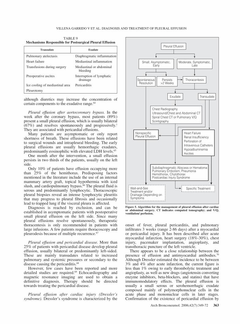
Pleural effusion after aortocoronary bypass. In theweek after the coronary bypass, most patients (89%)present a small pleural effusion, which is usually bilateral(67%) and resolves spontaneously and progressively.1They are associated with pericardial effusions.
Many patients are asymptomatic or only reportshortness of breath. These effusions have been relatedto surgical wounds and intrapleural bleeding. The earlypleural effusions are usually hemorrhagic exudates,predominantly eosinophilic with elevated LDH levels.65
One month after the intervention, a small effusionpersists in two thirds of the patients, usually on the leftside.
Only 10% of patients have effusion occupying morethan 25% of the hemithorax. Predisposing factorsmentioned in the literature include the use of an internalmammary artery graft, topical hypothermia with icedslush, and cardiopulmonary bypass.66 The pleural fluid isserous and predominantly lymphocytic. Thoracoscopicpleural biopsies reveal an intense lymphocytic pleuritisthat may progress to pleural fibrosis and occasionallylead to trapped lung if the visceral pleura is affected.
Diagnosis is reached by exclusion, and can beestablished in asymptomatic patients with postoperativesmall pleural effusion on the left side. Since manypleural effusions resolve spontaneously, therapeuticthoracentesis is only recommended in patients withlarge infusions. A few patients require thoracoscopy andpleurodesis because of multiple recurrence.67
Pleural effusion and pericardial disease. More than25% of patients with pericardial disease develop pleuraleffusion, usually bilateral or predominantly left-sided.These are mainly transudates related to increasedpulmonary and systemic pressures or secondary to thedisease causing the pericarditis.68
However, few cases have been reported and moredetailed studies are required.69 Echocardiography andmagnetic resonance imaging are used to obtain adefinitive diagnosis. Therapy should be directedtowards treating the pericardial disease.
Pleural effusion after cardiac injury (Dressler’ssyndrome). Dressler’s syndrome is characterized by the
onset of fever, pleural pericarditis, and pulmonaryinfiltrates 3 weeks (range 2-86 days) after a myocardialor pericardial injury. It has been described after acutemyocardial infarction, heart surgery (18%-30%), chestinjury, pacemaker implantation, angioplasty, andtransthoracic puncture of the left ventricle.
There appears to be a close relationship between thepresence of effusion and antimyocardial antibodies.70
Although Dressler estimated the incidence to be between3% and 4% after acute infarction, the current figure isless than 1% owing to early thrombolytic treatment andangioplasty, as well as new drugs (angiotensin convertingenzyme inhibitors, beta-blockers, and statins) that haveimmunomodulatory effects. The pleural effusion isusually a small serous or serohemorrhagic exudatecomposed mainly of polymorphonuclear cells in theacute phase and mononuclear cells in later stages.Confirmation of the existence of pericardial effusion by
VILLENA GARRIDO V ET AL. DIAGNOSIS AND TREATMENT OF PLEURAL EFFUSION
Arch Bronconeumol. 2006;42(7):349-72 363
Figure 5. Algorithm for the management of pleural effusion after cardiacor abdominal surgery. CT indicates computed tomography; and V/Q,ventilation/ perfusion.
Pleural Effusion
ThoracentesisSpontaneousResolution
Exudate Transudate
Chest Radiography
Ultrasound/Chest and Abdominal CT
Spiral Chest CT or Pulmonary V/Q
Scintigraphy
Subdiaphragmatic Abscess or HematomaPulmonary Embolism, PneumoniaHemothorax, ChylothoraxPostcardiac Injury Syndrome
Specific TreatmentWait-and-SeeTreatment and/orDrainage Depending onSymptoms
NonspecificPleural Effusion
Heart Failure
Renal Insufficiency
Perforation of
Intravenous Catheters
Hypoalbuminemia
Ascites
Persists>2 Weeks
Small, Asymptomatic,Early
Moderate, Symptomatic,Late
TABLE 9Mechanisms Responsible for Postsurgical Pleural Effusion
Transudate Exudate
Pulmonary atelectasis Diaphragmatic inflammation
Heart failure Mediastinal inflammation
Transfusions during surgery Mediastinal or abdominal bleeding
Preoperative ascites Interruption of lymphatic drainage
Ice cooling of mediastinal area Pericarditis
Pleurotomy
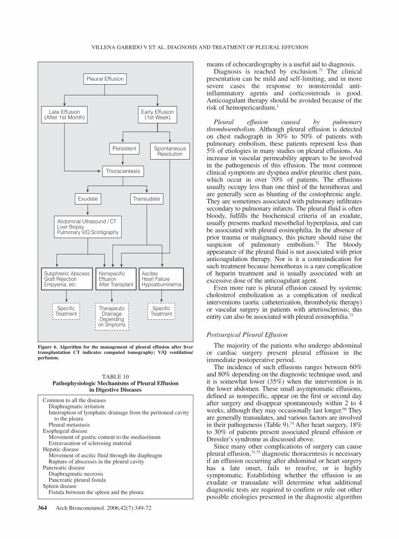
VILLENA GARRIDO V ET AL. DIAGNOSIS AND TREATMENT OF PLEURAL EFFUSION
364 Arch Bronconeumol. 2006;42(7):349-72
means of echocardiography is a useful aid to diagnosis.Diagnosis is reached by exclusion.71 The clinical
presentation can be mild and self-limiting, and in moresevere cases the response to nonsteroidal anti-inflammatory agents and corticosteroids is good.Anticoagulant therapy should be avoided because of therisk of hemopericardium.1
Pleural effusion caused by pulmonarythromboembolism. Although pleural effusion is detectedon chest radiograph in 30% to 50% of patients withpulmonary embolism, these patients represent less than5% of etiologies in many studies on pleural effusions. Anincrease in vascular permeability appears to be involvedin the pathogenesis of this effusion. The most commonclinical symptoms are dyspnea and/or pleuritic chest pain,which occur in over 70% of patients. The effusionsusually occupy less than one third of the hemithorax andare generally seen as blunting of the costophrenic angle.They are sometimes associated with pulmonary infiltratessecondary to pulmonary infarcts. The pleural fluid is oftenbloody, fulfills the biochemical criteria of an exudate,usually presents marked mesothelial hyperplasia, and canbe associated with pleural eosinophilia. In the absence ofprior trauma or malignancy, this picture should raise thesuspicion of pulmonary embolism.72 The bloodyappearance of the pleural fluid is not associated with prioranticoagulation therapy. Nor is it a contraindication forsuch treatment because hemothorax is a rare complicationof heparin treatment and is usually associated with anexcessive dose of the anticoagulant agent.
Even more rare is pleural effusion caused by systemiccholesterol embolization as a complication of medicalinterventions (aortic catheterization, thrombolytic therapy)or vascular surgery in patients with arteriosclerosis; thisentity can also be associated with pleural eosinophilia.73
Postsurgical Pleural Effusion
The majority of the patients who undergo abdominalor cardiac surgery present pleural effusion in theimmediate postoperative period.
The incidence of such effusions ranges between 60%and 80% depending on the diagnostic technique used, andit is somewhat lower (35%) when the intervention is inthe lower abdomen. These small asymptomatic effusions,defined as nonspecific, appear on the first or second dayafter surgery and disappear spontaneously within 2 to 4weeks, although they may occasionally last longer.66 Theyare generally transudates, and various factors are involvedin their pathogenesis (Table 9).74 After heart surgery, 18%to 30% of patients present associated pleural effusion orDressler’s syndrome as discussed above.
Since many other complications of surgery can causepleural effusion,71,75 diagnostic thoracentesis is necessaryif an effusion occurring after abdominal or heart surgeryhas a late onset, fails to resolve, or is highlysymptomatic. Establishing whether the effusion is anexudate or transudate will determine what additionaldiagnostic tests are required to confirm or rule out otherpossible etiologies presented in the diagnostic algorithm
Figure 6. Algorithm for the management of pleural effusion after livertransplantation CT indicates computed tomography; V/Q ventilation/perfusion.
Pleural Effusion
Persistent SpontaneousResolution
Exudate Transudate
Abdominal Ultrasound / CTLiver BiopsyPulmonary V/Q Scintigraphy
Subphrenic AbscessGraft RejectionEmpyema, etc
SpecificTreatment
NonspecificEffusionAfter Transplant
TherapeuticDrainage
Dependingon Smptoms
AscitesHeart FailureHypoalbuminemia
SpecificTreatment
Thoracentesis
Early Effusion(1st Week)
Late Effusion(After 1st Month)
TABLE 10Pathophysiologic Mechanisms of Pleural Effusion
in Digestive Diseases
Common to all the diseasesDiaphragmatic irritationInterruption of lymphatic drainage from the peritoneal cavity
to the pleuraPleural metastasis
Esophageal diseaseMovement of gastric content to the mediastinumExtravasation of sclerosing material
Hepatic diseaseMovement of ascitic fluid through the diaphragmRupture of abscesses in the pleural cavity
Pancreatic diseaseDiaphragmatic necrosisPancreatic pleural fistula
Spleen diseaseFistula between the spleen and the pleura

VILLENA GARRIDO V ET AL. DIAGNOSIS AND TREATMENT OF PLEURAL EFFUSION
Arch Bronconeumol. 2006;42(7):349-72 365
(Figure 5). Nonspecific postsurgical effusion is,therefore, a diagnosis of exclusion.
Effusions caused by abdominal or cardiac surgery aremanaged by treating the etiologic cause, and drainageusing a range of techniques is indicated in nonspecificpostoperative effusions when required by the patient’sclinical status.
Pleural effusion after liver transplantation. Over 50%of patients who undergo a liver transplant subsequentlydevelop a nonspecific unilateral or bilateral pleuraleffusion.76 The effusion starts in the first 7 days after theintervention, is asymptomatic, and usually resolvesspontaneously within a month, although it cansometimes persist for more than 6 months. The pleuralfluid is usually a transudate of multifactorial origin(pretransplant ascites, hypoalbuminemia, administrationof intravenous fluids and transfusions) but may be anexudate, when the cause is diaphragmatic irritationrelated to the surgery. In persistent transudativeeffusions, all other possible etiologies must be excluded.If the nonresolving effusion is an exudate, the followingmust be ruled out: subphrenic abscess or hematoma,hemothorax secondary to anticoagulation therapy, graftrejection, pulmonary embolism, pneumonia, andanastomosis obstruction in suprahepatic veins. A
diagnosis of nonspecific postsurgical effusion can onlybe established when all other possible etiologies havebeen excluded.
The diagnostic algorithm is shown in Figure 6.
Pleural effusion after lung transplantation. After lungtransplantation, the 2 pleural cavities are connected untilthe development of new adhesions separates them.Consequently, any pleural effusion, whatever the cause,may be bilateral, and any intervention undertaken on onecavity will also affect the other. Pleural effusion occursduring the early postoperative period after lungtransplantation in all patients and is multifactorial inorigin (increased permeability caused by ischemia-reperfusion injury, interruption of lymphatic drainage,acute early temporary rejection, mediastinal bleeding).77
The effusion is a predominantly neutrophilic bloodyexudate that resolves within a few days. No tests arerequired unless it persists for more than 3 weeks, thepatient presents symptoms such as fever or pleuriticchest pain, or new fluid accumulates.
The first step in such cases is to performtransbronchial biopsies in order to rule out acute graftrejection because this is the most common etiology forpleural effusion at any time after lung transplantation.78
Once acute rejection has been ruled out, pleural fluid
Figure. 7. Algorithm for themanagement of pleural effusion afterlung transplantation. TBB indicatestransbronchial biopsy.
Pleural Effusion
Late Onset EffusionImmediately After Transplantation
BloodyHemothorax
Reoperation Becauseof Mediastinal
BleedingPersistence
Bronchoscopy With BTB
Acute RejectionNo RejectionSpontaneous Resolution
Thoracentesis
Chylothorax TransudateExudate
Heart FailureHypoalbuminemia
Thoracic Duct InjuryCaused by Surgical Procedures
Empyema, Pneumonia,Pulmonary Embolism, Neoplasm
SpecificTreatment
SurgicalTreatment
SpecificTreatment

obtained by thoracentesis should be tested (Figure 7).Hemothorax is associated with increased postoperativemortality.77
Pleural Effusion and Benign Digestive Disease
Many digestive diseases cause pleural effusion, andthe mechanisms are diverse (Table 10).79,80 Figure 8shows the benign digestive diseases associated withpleural effusions. These may evolve with or withoutabdominal symptoms.81,82
These effusions are managed by treating the
causative disease. In patients who are experiencingsevere symptoms (mainly dyspnea) pleural fluid can beevacuated. It should not be forgotten, however, that inthe case of effusions accompanied by severe ascites, theremoval of ascitic fluid will reduce the volume of thepleural effusion.
Since there is no evidence that any one drainagetechnique is better than another, the decision concerningwhich technique should be used must be taken on acase-by-case basis taking into account symptoms, riskfactors, and comorbidity.
Drug-Induced Pleural Effusion
Drugs have been shown to cause pleural effusion,albeit very rarely. The mechanisms involved are poorlyunderstood, although several hypothesis have beenpostulated including hypersensitivity reaction and directtoxicity through inflammatory or oxidative processes.The list of drugs that cause pleural effusion grows dailyand currently includes a broad range includingcardiovascular, antiinflammatory, chemotherapeutic, andantibiotic agents. The best known are amiodarone,nitrofurantoin, methysergide, bromocriptine, and theergoline derivatives. If a drug-related etiology issuspected, the treating physician should consult acomprehensive list of causative agents such as can befound in the literature.1,83 Another source of informationis the web site www.pneumotox.com. Any cliniciancarrying out a differential diagnosis of a pleural effusionmust consider the possibility that it may be druginduced, and this makes a medical history indispensable.In order to establish a causal relationship, the physicianmust know what medications the patient has beentaking, whether the timing of presentation is consistentwith a diagnosis of drug-related effusion and, if possible,must demonstrate resolution of the effusion onwithdrawal of the medication. This is necessary becausethere are no specific findings that confirm this diagnosis.The pleural effusion can be unilateral or bilateral and isoften associated with pneumonitis. The pleural fluid isoccasionally eosinophilic, although this finding has nodiagnostic value. Pleural biopsy in such cases usuallyonly reveals nonspecific inflammation. As a generalrule, other plausible causes for pleural effusion shouldbe ruled out before attributing the effusion to a drug.Treatment obviously consists in withdrawal of thecausative medication, and in most cases the effusion willthen resolve.
Asbestos-Related Benign Pleural Disease
Asbestos is associated with a number of pleuralmanifestations.84,85
Benign pleural disease is the most commonrespiratory condition caused by asbestos exposure.
It occurs in around 50% of people who have beencontinuously exposed to asbestos in the workplace.
The latency period tends to be more than 20 years,and the incidence is directly proportional to theintensity of exposure and the time lapsed since initial
VILLENA GARRIDO V ET AL. DIAGNOSIS AND TREATMENT OF PLEURAL EFFUSION
366 Arch Bronconeumol. 2006;42(7):349-72
Figure 8. Digestive diseases that cause pleural effusion. PF indicatespleural fluid; LDH, lactate dehydrogenase; and CT, computedtomography.
Cirrhosis With or Without Ascites
Nonspecific Exudate
Polymorphonuclear ExudateEmpyema if Fistula PresentPF/Serum Bilirubin Ratio>1 if Fistula Present
Hydatids in PF
Nonspecific ExudateChocolate Brown Fluid
Chronic
Small/Moderate, Exudate, High Amylase>50 000 Leukocytes/mL,pH<7.35, High LDH
Moderate/Large, Exudate,Very High Amylase
Diagnosis WithAbdominalUltrasound/CT
Acute
Pa
nc
rea
ticD
ise
ase
Infarct
Abscess
Sp
lee
nD
ise
ase
NonspecificExudate
He
pa
tic D
ise
ase
Exudate
ViralHepatitis
Abscess
Amebiasis
Hydatidosis
Transudate
Acute Chest PainPneumothoraxExudate, pH <6,Elevated AmylaseEmpyema
Mild Pain or AsymptomaticExudate, Normal pH andAmylase
Nonspecific ExudateSuspicion Becauseof Medical History
Eso
ph
ag
ea
l Dis
ea
se
Severe
Rupture
Sclerosis of VaricoseVeins
Mild

exposure. A history of asbestos exposure should takethe form of a complete occupational history including,in date order, all the patients’ jobs, the specific activitiesinvolved, and the materials handled throughout theirwhole working lives. The recommended method forscreening patients for asbestos-related benign pleuraldisease is plain chest radiograph using obliqueprojections if possible. However, if better definition ofthe lesions is needed, high resolution computedtomography using 2 cm collimation is the best tool.
Pleural plaques are the most common complicationassociated with asbestos exposure, although thiscondition has also been reported after tuberculosis,hemothorax, and chest injuries. These plaques arehyalinized collagen formations that are nearly alwaysfound on the parietal pleura and are usuallycircumscribed. The prevalence of plaques appears to
correlate directly with the intensity of exposure and thelatency period. They are usually bilateral, and the besttool for their detection is chest computed tomography,although oblique chest radiography may suffice in somecases. In 30% of cases plaques are found in conjunctionwith pulmonary asbestosis. The possible effect ofpleural plaques on respiratory function has been theobject of many studies. To date, no conclusive evidencehas been found of any effect. This means that any lungfunction abnormalities observed may be due toconcomitant smoking or undetected pulmonary fibrosis.Although the pleural plaque formations are associatedwith a higher risk of developing asbestosis or neoplasticdisease because they indicate a greater level of exposureto asbestos, it is not thought that they themselvesbecome malignant.
Diffuse pleural fibrosis leads to a thickened visceralpleura that tends to limit respiratory movements. Itsfrequency correlates directly with the duration andintensity of exposure.
Consequently, the patients present a restrictiveventilatory defect, and this may progress to respiratoryfailure in the advanced stages of the disease. In suchcases an improvement has been reported withnoninvasive mechanical ventilation.
Asbestos-related pleural effusion occurs in patientswith a history of exposure to asbestos who present withan exudative effusion but no signs of malignancy whenmonitored over a period of at least 3 years. The effusionis usually unilateral, predominantly left-sided, andserosanguineous.
Except for suggestive pleural calcification, there areno specific diagnostic findings in the examination ofeither pleural fluid or tissue. Diagnosis must, therefore,be based on the criteria discussed below and on ongoingmonitoring.
Thoracoscopy is recommended in patients withpersistent pleural effusion in order to rule outmesothelioma before establishing a diagnosis of benignpleural disease. In the long term, the effusion recurs in30% of patients, and diffuse pleural fibrosis, roundedatelectasis, and mesothelioma develop in 20%, 20%,and 5% of cases, respectively.
Rounded atelectasis is a benign lesion usually causedby exposure to asbestos. The peripheral lung is trappedby the underlying pleural layer when the thickeningpleura compresses the lung and gives rise to atelectasis.In plain chest radiography an increase in basal densitycan be observed together with thickening of the adjacentpleura. These characteristics are more clearly defined inchest computed tomography, which may reveal thefollowing pathognomonic findings: a pulmonary masscontiguous to a thickened pleura and the curved swirl ofthe vessels and bronchi converging on the pulmonaryhilum. These findings make possible a probablediagnosis that makes biopsy unnecessary in most cases.
Nevertheless, although it has not been demonstratedthat rounded atelectasis can become malignant, the needto order histologic studies to rule out cancer should beconsidered on a case-by-case basis.
VILLENA GARRIDO V ET AL. DIAGNOSIS AND TREATMENT OF PLEURAL EFFUSION
Arch Bronconeumol. 2006;42(7):349-72 367
TABLE 11Etiology of Chylothorax
Congenital malformations of the lymphatic systemAtrophied thoracic ductLymphatic aplasia and dysplasiaLymphangiomaIntestinal lymphangiectasis; protein-losing enteropathy
NeoplasmsLymphomasMetastatic cancerKaposi sarcoma (acquired immunodeficiency syndrome)
Mediastinal tumorsTeratomaRetrosternal goitreThymomaAortic aneurysm
InfectionsTuberculosisFilariasisPneumoniaEmpyema
Diseases that affect the lymph vesselsLymphangioleiomyomatosisPulmonary tuberose sclerosisGorham’s syndromeLymph duct cystYellow nail syndromeCastleman’s disease
Idiopathic causes
MiscellaneousSarcoidosisBehçet’s syndromeAmyloidosisHypothyroidismSystemic lupus erythematosus
TransudatesHepatic cirrhosisNephrotic syndromeHeart failure
Traumatic causes
IatrogenicPostsurgicalSuperior vena caval and left subclavian thrombosis

Pleural Effusion Caused by Benign GynecologicalDiseases
Pleural effusion is sometimes a sign of benigngynecological diseases, such as Meig’s syndrome,endometriosis, or ovarian hyperstimulation syndrome.These entities should be included in the differentialdiagnosis of a pleural effusion of unknown cause.
Benign ovarian tumors are associated with ascitesand pleural effusion (Meig’s syndrome); the tumorreleases substances that alter vascular permeability.86
Although the pleural fluid is usually a nonspecificexudate, it can also occasionally be a transudate.Diagnosis is based on the demonstration of theexistence of an ovarian tumor. Pleural fluid disappearswhen the tumor is excised. Very rarely, stage IVendometriosis is associated with the presence of pleuralexudate. Diagnosis is established when the existence ofendometriosis is confirmed by laparoscopy. Theseeffusions do not require specific treatment because theydisappear when the gynecological disease is treated.
Finally, ovarian hyperstimulation syndrome isincreasingly common because of hormonal fertilizationtreatments.87 Up to 32% of patients with severe forms ofthis condition present with pleural effusion, generallyassociated with severe respiratory distress and respiratoryfailure. The fluid accumulates because of alterations invascular permeability as a result of hemoconcentrationand the movement of ascitic fluid into the pleural space.This type of effusion is a nonspecific exudate and it is thepatient’s medical history (hormonal therapy or multiplebirths) that will suggest the diagnosis. In all cases,pulmonary embolism should be ruled out as this is verycommon in the advanced stages of ovarianhyperstimulation syndrome. Treatment is based onensuring the patient’s hemodynamic status by way oftherapeutic fluid management including diuretics.Prevention of thrombosis is important, and paracentesisshould be performed in patients with ascites. In the mostsevere cases involving pleural effusion and respiratoryinsufficiency, evacuation of pleural fluid and oxygentherapy are necessary.
Chylothorax and Pseudochylothorax
Chylothorax. Chylothorax is defined as the presenceof lymph or chyle in the pleural cavity. The chyle mayoriginate in the thorax (owing to rupture of the thoracicduct or affluent vessels) or in the abdomen.
In chylothorax, unlike pseudochylothorax, the pleuralsurfaces are normal. If fetal and neonatal chylothorax isexcluded, this entity is rare, affecting 3% of patients withpleural effusion studied consecutively in one hospital.88
The causes of chylous effusions are shown in Table 11.The most common chylothoraces are tumor related (75%lymphomas), and the next most common are traumatic,iatrogenic, and idiopathic chylous effusions.
The etiology of most idiopathic cases, when theeffusion persists after appropriate follow-up, is consideredto be trauma. The increasingly widespread use of largeveins for parenteral feeding and hemodynamic monitoring
has made iatrogenic thrombosis of the superior vena cavaor the left subclavian artery one of the most commoncauses of chylothorax, especially in children.
Chylothorax secondary to hepatic cirrhosis, nephroticsyndrome, or heart failure has the biochemicalcharacteristics of a transudate.
DIAGNOSIS. CLINICAL COURSE. The most commonsymptoms of nontraumatic chylothorax in adults aredyspnea on exertion and a sensation of heaviness ofrecent onset in the affected hemithorax. Fever and chestpain are rare because chyle (lymph) is not an irritant.This diagnosis is generally suspected after thoracentesisbecause of the appearance of the pleural fluid.
APPEARANCE AND BIOCHEMISTRY OF PLEURAL FLUID. Whilethe pleural fluid in chylothorax is typically milky, bothpseudochylothorax and empyema can also produce milkyfluid. Furthermore, the fluid may be bloody, serous, orturbid in 50% of cases.89,90 Empyema can be ruled out ifthe milky appearance disappears after centrifugation.
Measurement of triglyceride values in pleural fluid isconsidered to be the best (most practical and accessible)way of diagnosing chylothorax.91 The sensitivity of atriglyceride value greater than 110 mg/dL is high, butsince triglyceride values are also observed in associationwith pseudochylothorax, a pleural fluid/serum cholesterolratio of less than 1 is also required. A third criterion hasbeen proposed in order to exclude hypertriglyceridemia: apleural fluid/serum triglyceride ratio of less than 1. Sincethe triglyceride content in pleural effusions can be due toseveral factors and not only diffusion from plasma, someauthors consider this third criterion to be superfluous.
Irrespective of the final result of this debate, thediagnostic specificity of the combined use of the 3criteria proposed is high.
VILLENA GARRIDO V ET AL. DIAGNOSIS AND TREATMENT OF PLEURAL EFFUSION
368 Arch Bronconeumol. 2006;42(7):349-72
TABLE 12Chylothorax: Therapeutic Modalities
Conservative treatmentTreatment of the causative diseaseRepeated thoracentesisContinuous drainageDietary modificationsMedium chain triglyceridesExclusively parenteral nutrition*
Pleurodesis using endothoracic tube*
Surgical treatmentThoracoscopic pleurodesisPleuroperitoneal pumpFibrin glue to seal the thoracic or intestinal ductThoracic duct ligation via thoracoscopy or thoracotomyAnastomosis between the thoracic duct and the azygos veinLung transplant (in lymphangioleiomyomatosis)In patients with concurrent ascitesPeritoneal venous shuntsClosure the diaphragmatic aperture with fibrin or suture
using thoracoscopyIn the fetusIntrauterine thoracentesis Prenatal pleuroamniotic shunt
*In high-volume chylothorax.

Given that chylomicrons are the only components ofchyle present in the blood exclusively during thepostprandial period, demonstrating their presence inpleural fluid by electrophoresis or ultracentrifugation isconsidered diagnostic (gold standard). However, theaccuracy of this measurement depends on the methodused.
Lymphography has been the method most widelyused to pinpoint the source of chylothorax (of lymphextravasation). Identification is, however, difficult innontraumatic patients. Lymphatic scintigraphy has beenproposed as an alternative method because it is a fasterand less invasive procedure.
TREATMENT OF NONTRAUMATIC CHYLOTHORAX. Aninitial approach includes decompression of the pleuralspace and the thoracic lymphatics. Althoughdecompression can sometimes be achieved by one ormore thoracenteses, continuous drainage through anendothoracic tube is generally a more effective methodand also facilitates measurement of the chyle flow ratein the many cases characterized by reaccumulation.
Table 12 shows the range of later treatment options. Inpatients with chylothorax caused by lymphoma refractoryto chemotherapy and radiotherapy, talc pleurodesis usingmedical thoracoscopy has been shown to be an effectivetechnique. Pleuroperitoneal shunt is considered to be asafe and effective treatment for persistent chylothorax inchildren in the absence of chylous ascites.
PROGNOSIS. The prognosis depends to a large degreeon the etiology of the chylothorax.
Prolonged pleural drainage in these patients has a verynegative impact on the patient’s nutritional and
immunological status. In one study of patients withnontraumatic chylothorax, survival at 22 months was 24%.
Pseudochylothorax. The terms pseudochylothoraxand chyliform pleural effusion are synonyms, and this isa rare entity, much less common than chylothorax.Although the pleural fluid is turbid in appearancebecause of its high lipid content, it does not come fromthe lymphatic system as a result of a ruptured thoracicduct. Pseudochylothorax occurs in patients who havelong-standing (mean, 5 years) pleural effusions. The 2most common causes are tuberculosis and rheumatoidarthritis. Pleural effusions in patients with atelectasis andtrapped lung (secondary to therapeutic pneumothorax)can also become pseudochylothoraces.
In many patients, the cause of the original effusion isnever determined.
DIAGNOSIS. Analysis of pleural fluid is a usefuldiagnostic tool and can sometimes identify the etiology (ifit is tuberculous, for example). Although the absence ofcholesterol crystals in the pleural fluid sediment does notrule out pseudochylothorax, their presence is diagnostic.Cholesterol levels over 200 mg/dL are very suggestive ofpseudochylothorax, and a pleural fluid/serum cholesterolratio of 1 or higher confirms this suspicion.
TREATMENT. If the patient has a history of tuberculosisand has either not been treated or has received inadequatetreatment, appropriate treatment must be implemented.When pseudochylothorax is caused by rheumatoidarthritis, treatment should be directed at achievingadequate control of the underlying disease. In patientswith functional impairment, one or more therapeuticthoracenteses may alleviate symptoms, and if lungfunction is preserved, pleural decortication is indicated.
Hemothorax
Definition and etiology. Hemothorax is the presenceof blood in the pleural space. A pleural effusion isconsidered to be a hemothorax when the pleural fluidhematocrit level is 50% greater than that of peripheralblood.1 Table 13 shows the causes of hemothorax. Themost common are traumatic and iatrogenic (followingsurgery, vascular catheterization, or diagnostic ortherapeutic transpleural punctures).
Diagnosis. The signs and symptoms of hemothoraxvary depending on the cause, the volume, and the rateof accumulation. Acute traumatic hemothorax is usuallyassociated with hemodynamic instability and pain,while the predominant manifestations in nontraumaticcases are dyspnea and other signs characteristic of fluidaccumulation in the pleura.
A chest radiograph reveals the presence of either a free-flowing or a loculated effusion, and occasionally there areimages consistent with a diagnosis of coagulates.Radiographs may also reveal associated lesions that willguide the etiologic diagnosis. Further imaging techniques(ultrasound and computed tomography) are necessary in
VILLENA GARRIDO V ET AL. DIAGNOSIS AND TREATMENT OF PLEURAL EFFUSION
Arch Bronconeumol. 2006;42(7):349-72 369
TABLE 13Causes of Hemothorax
TraumaticClosed chest injuryPenetrating chest injury, including iatrogenic injuries
SpontaneousPleuralMalignancy (primary or metastatic)Associated with rupture of adhesions in spontaneous
pneumothoraxPleural endometriosisPulmonaryMalignancy (primary or metastatic)Necrotizing infectionTuberculosisPulmonary embolism with infarctionArteriovenous malformationsHereditary hemorrhagic telangiectasisBullous emphysemaPulmonary sequestration
Blood dyscrasias and complications of anticoagulation
Abdominal diseasePancreatitisHemoperitoneum
Vascular diseaseRupture of an aortic aneurysmRupture of an aneurysm in the splenic artery

some cases to quantify and evaluate the hemothorax,identify the cause, and provide guidance for therapeuticprocedures. A definitive diagnosis is obtained by way ofthoracentesis and pleural fluid analysis.
Treatment
ACUTE HEMOTHORAX. In hemodynamically stablepatients who have a small hemothorax (only blunting ofthe costophrenic angle or a volume calculated to be lessthan 300 mL) clinical radiographic monitoring is oneoption (C).
In hemodynamically unstable patients or when thevolume is calculated to be greater than 300 mL, a large-bore pleural drainage tube—28F or 32F—must beplaced in the midaxillary line at the sixth intercostalspace and directed posteriorly (B); prior prophylacticantibiotic therapy is recommended (C).92,93 Thoracotomyis indicated when over 1500 mL of fluid is removed inthe initial drainage or when over 200 mL of fluid isremoved per hour for more than 3 consecutive hours (B).In hemodynamically stable patients VAT is anotheralternative; this technique can sometimes be used toperform procedures aimed at achieving hemostasis.94
When the suspected cause of the hemothorax isrupture of an aortic aneurysm, contrast-enhancedcomputed tomography should be performed. Drainageis contraindicated in such cases as it may favorexsanguination (D).
RESIDUAL OR COAGULATED HEMOTHORAX. Smallresidual hemothorax (only blunting of the costophrenicangle) can be treated conservatively with respiratoryphysiotherapy and monitoring (C).
In patients with a persistent hemothorax estimated tobe greater than 500 mL or persistent residualloculations occupying less than one third of thehemithorax, treatment is required to prevent thedevelopment of subacute (atelectasis, empyema,pneumonia) or chronic (fibrothorax) complications (C).During the first week drainage can be attempted usingnew chest tubes placed under ultrasound or computedtomographic guidance (D). If this approach is noteffective, the condition can be treated by the instillationof fibrinolytic agents (streptokinase or urokinase) intothe pleural space (B).95 Another alternative to drainage,or if the fibrinolytics are not effective, is earlythoracoscopic removal of the coagulates (C).96
Decortication is indicated if thoracoscopy is noteffective or the hemothorax is chronic and gives rise totrapped lung (fibrothorax) (D).
Key Points
– Clinical radiographic monitoring is an option inhemodynamically stable patients who have a smallhemothorax (C).
– In hemodynamically unstable patients or when thevolume is calculated to be greater than 300 mL, a large-bore pleural drainage tube—28F or 32F—must be placedin the midaxillary line at the sixth intercostal space and
directed posteriorly (B); prior prophylactic antibiotictherapy is recommended (C).
– Thoracotomy is indicated when over 1500 mL offluid is removed in the initial drainage or when over 200mL of fluid is removed per hour for more than 3consecutive hours (B).
– When the suspected cause of the hemothorax isrupture of an aortic aneurysm, contrast-enhancedcomputed tomography should be performed. Drainage is contraindicated in such cases as it may favorexsanguination (D).
Small residual hemothorax can be treated conservativelywith respiratory physiotherapy and monitoring (C).
In patients with a persistent hemothorax estimated tobe greater than 500 mL or persistent residual loculationsoccupying less than one third of the hemithorax,treatment is required to prevent the development ofsubacute or chronic complications (C). During the firstweek drainage can be attempted using new chest tubesplaced under ultrasound or computed tomographicguidance (D). If this approach is not effective, thecondition can be treated by the instillation of fibrinolyticagents into the pleural space (B). Another alternative, orif the fibrinolytics are not effective, is early removal ofthe coagulates via thoracoscopy (C).
– Decortication is indicated if thoracoscopy is noteffective or the hemothorax is chronic and gives rise totrapped lung (fibrothorax) (D).
List of Abbreviations
ADA: adenosine deaminase.IFN: interferon.IL: interleukin.VAT: video-assisted thoracoscopy.
Levels of evidence97
1++: high quality meta-analyses, systematic reviews of RCTs, orRCTs with a very low risk of bias.
1+: well conducted meta-analyses, systematic reviews of RCTs, orRCTs with a low risk of bias.
1–: meta-analyses, systematic reviews or RCTs, or RCTs with ahigh risk of bias.
2++: high quality systematic reviews of case–control or cohortstudies or high quality case–control or cohort studies with avery low risk of confounding, bias, or chance and a highprobability that the relationship is causal.
2+: well conducted case–control or cohort studies with a low riskof confounding, bias, or chance and a moderate probability thatthe relationship is causal
2-: case–control or cohort studies with a high risk of confounding,bias, or chance and a significant risk that the relationship is notcausal
3: non–analytic studies, eg case reports, case series
4: expert opinion
Grades of Recommendations97
A. At least one meta–analysis, systematic review, or RCT rated as1++ and directly applicable to the target population or
A systematic review of RCTs or a body of evidence consistingprincipally of studies rated as 1+ directly applicable to the targetpopulation and demonstrating overall consistency of results
VILLENA GARRIDO V ET AL. DIAGNOSIS AND TREATMENT OF PLEURAL EFFUSION
370 Arch Bronconeumol. 2006;42(7):349-72

VILLENA GARRIDO V ET AL. DIAGNOSIS AND TREATMENT OF PLEURAL EFFUSION
Arch Bronconeumol. 2006;42(7):349-72 371
B. A body of evidence including studies rated as 2++ directlyapplicable to the target population and demonstrating overallconsistency of results or
Extrapolated evidence from studies rated as 1++ or 1+
C. A body of evidence including studies rated as 2+ directlyapplicable to the target population and demonstrating overallconsistency of results or
Extrapolated evidence from studies rated as 2++
D. Evidence level 3 or 4, or extrapolated evidence from studiesrated as 2 +
REFERENCES
1. Light RW, Lee YCG, editors. Textbook of pleural diseases.London: Arnold; 2003.
2. Villena V, López Encuentra E, García-Luján R, et al. Clinicalimplications of appearance of pleural fluid at thoracentesis. Chest.2004;125:156-9.
3. Cheng DS, Rodríguez RM, Rogers J, et al. Comparison of pleuralfluid pH values obtained using blood gas machine, pH meter, andpH indicator strip. Chest. 1998;114:1368-72.
4. Jiménez-Castro D, Díaz G, Pérez-Rodríguez E, et al. Modificationof pleural fluid pH by local anesthesia. Chest. 1999;116:399-402.
5. Heffner JE, Brown LK, Barbieri C, et al. Pleural fluid chemicalanalysis in parapneumonic effusions. A metaanalysis. Am J RespirCrit CareMed. 1995;151:1700-8.
6. Rodríguez-Panadero F, López Mejías J. Low glucose and pHlevels in malignant pleural effusions. Diagnostic significance andprognostic value in respect to pleurodesis. Am Rev Respir Dis.1989;139:663-7.
7. Jiménez D, Pérez Rodríguez E, Díaz G, et al. Determining theoptimal number of specimens to obtain with needle biopsy of thepleura. Respir Med. 2002;96:14-7.
8. Porcel JM, Vives M. Etiology and pleural fluid characteristics oflarge and massive effusions. Chest. 2003;124:978-83.
9. Ferrer J, Roldán J, Teixidor J, et al. Predictors of pleuralmalignancy in patients with pleural effusion undergoingthoracoscopy. Chest.2005;127:1017-22.
10. Ferrer JS, Muñoz XG, Orriols RM, et al. Evolution of idiopathicpleural effusion: a prospective, long-term follow-up study. Chest.1996;109:1508-13.
11. Romero Candeira S, Hernández Blasco L, Romero Brufao S.Trasudados frente a exudados pleurales. Criterios discriminantes.Causas de trasudado pleural y aproximación diagnóstica. En:Pérez Rodríguez E, Villena Garrido MV, editors. Enfermedadesde la pleura. Monografías Neumomadrid. Madrid: Ergon; 2003. p.57-68.
12. Romero-Candeira S, Hernández L. The separation of transudatesand exudates with particular reference to the protein gradient. CurrOpin Pulm Med. 2004;10:294-8.
13. Sahn SA. Management of complicated parapneumonic effusions.Am Rev Respir Dis. 1993;148:813-7.
14. Chen KY, Hsueh PR, Liaw YS, et al. A 10-year experience withbacteriology of acute thoracic empyema: emphasis on Klebsiellapneumoniae in patients with diabetes mellitus. Chest.2000;117:1685-9.
15. Maskell NA, Davis CWH, Nunn AJ, et al. U.K. controlled trial ofintrapleural streptokinase for pleural infection. N Engl J Med.2005; 352:865-74.
16. Davies CW, Kearney SE, Gleeson FV, et al. Predictors ofoutcome and long-term survival in patients with pleural infection.Am J Respir Crit Care Med. 1999;160:1682-7.
17. Maskell NA, Gleeson FV, Darby M, et al. Diagnosticallysignificant variations in pleural fluid pH in loculatedparapneumonic effusions. Chest. 2004;126:2022-4.
18. Heffner JE. Indications for draining a parapneumonic effusion: anevidence-based approach. Semin Respir Infect. 1999;14:48-58.
19. Colice GL, Curtis A, Deslauriers J, et al. Medical and surgicaltreatment of parapneumonic effusions: an evidence-basedguideline. Chest. 2000;118:1158-71.
20. Jordà Marcos R, Torres Martí A, Ariza Cardenal FJ, et al.Recomendaciones para el tratamiento de la neumoníaintrahospitalaria grave. Arch Bronconeumol. 2004;40:518-33.
21. Grupo de Estudio de la Neumonía Adquirida en la Comunidad.Normativas para el diagnóstico y el tratamiento de la neumoníaadquirida en la comunidad. Sociedad Española de Neumología yCirugía Torácica (SEPAR). Arch Bronconeumol. 2005;41:272-89.
22. Texeira LR, Villarino MA. Antibiotic treatment of patients withpneumonia and pleural effusion. Curr Opin Pulm Med.1998;4:230-4.
23. Cameron R, Davies HR. Intra-pleural fibrinolytic therapy versusconservative management in the treatment of parapneumoniceffusions and empyema. Cochrane Database Syst Rev.2004;(2):CD002312.
24. Vidal R, de Gracia J, Ruiz J, et al. Estudio controlado de 637pacientes con tuberculosis: diagnóstico y resultados terapéuticoscon esquemas de 9 y 6 meses. Med Clin (Barc). 1986;87:368-70.
25. Valdés L, Álvarez D, San José E, et al. Tuberculous pleurisy: astudy of 254 cases. Arch Intern Med. 1998;158:2017-21.
26. Greco S, Girardi E, Masciangelo R, et al. Adenosine deaminaseand interferon gamma measurements for the diagnosis oftuberculous pleurisy: a meta-analysis. Int J Tuberc Lung Dis.2003;7:777-86.
27. Burgess LJ, Maritz FJ, Le Roux I, et al. Combined use of pleuraladenosine deaminase with lymphocyte/neutrophil ratio. Increasedspecificity for the diagnosis of tuberculosis pleuritis. Chest.1996;109:414-9.
28. Valdés L, San José E, Álvarez D, et al. Diagnosis of tuberculouspleurisy using the biologic parameters adenosine deaminase,lysozyme, and interferon gamma. Chest. 1993;103:458-65.
29. Valdés L, San José E, Álvarez D, et al. Adenosine deaminase(ADA) isoenzyme analysis in pleural effusions: diagnostic roleand relevance to the origin of increased ADA in tuberculouspleurisy. Eur Respir J. 1996;9:747-51.
30. Villena V, López-Encuentra A, Pozo F, et al. Interferon gammalevels in pleural fluid for the diagnosis of tuberculosis. Am J Med.2003; 115:365-70.
31. Sharma SK, Banga A. Pleural fluid interferon-γ and adenosinedeaminase levels in tuberculosis pleural effusion: a cost-effectiveness analysis. J Clin Lab Anal. 2005;19:40-6.
32. Pai M, Flores LL, Hubbard A, et al. Nucleid acid amplificationtests in the diagnosis of tuberculous pleuritis: a systematic reviewand metaanalysis. BMC Infectious Diseases. 2004;4:6-20.
33. Blumberg HM, Burman WJ, Chaisson RE, et al. AmericanThoracic Society/Centers for Diseases Control andPrevention/Infectious Diseases Society of America: treatment oftuberculosis. Am J Respir Crit Care Med. 2003;167:603-62.
34. Lai YF, Chao TY, Wang YH, et al. Pigtail drainage in thetreatment of tuberculous pleural effusions: a randomised study.Thorax. 2003;58:149-52.
35. Matchaba PT, Volmink J. Steroids for treating tuberculouspleurisy (Cochrane Review). En: The Cochrane Library, Issue 1.Oxford: Update Software; 2001.
36. Sahn SA, Good Jr JT. Pleural fluid pH in malignant effusions.Diagnostic, prognostic, and therapeutic implications. Ann InternMed Med. 1988;108:345-9.
37. Ceyhan BB, Demiralp E, Celikel T. Analysis of pleural effusionsusing flow cytometry. Respiration. 1996;63:17-24.
38. Antony VB, Loddenkemper R, Astoul P, et al. Management ofmalignant pleural effusions. Am J Respir Crit Care Med.2000;162:1987-2001.
39. Antunes G, Neville E, Duffy J, et al. BTS guidelines for themanagement of malignant pleural effusions. Thorax. 2003;58Suppl 2:29-38.
40. Escudero Bueno C, García Clemente M, Cuesta Castro B, et al.Cytologic and bacteriologic analysis of fluid and pleural biopsyspecimens with Cope’s needle. Study of 414 patients. Arch InterMed. 1990;150:1190-4.
41. Villena V, López Encuentra A, Echave-Sustaeta J, et al. Estudioprospectivo de 1.000 pacientes consecutivos con derrame pleural.Etiología del derrame y características de los pacientes. ArchBronconeumol. 2002;38:21-6.
42. Maskell NA, Gleeson FV, Davies RJ. Standard pleural biopsyversus CT guided cutting-needle biopsy for diagnosis of malignantdisease in pleural effusions: a randomised controlled trial. Lancet.2003;361:1326-30.
43. Boutin C, Viallat JR, Cargnino P, et al. Thoracoscopy inmalignant pleural effusions. Am Rev Respir Dis. 1981;124:588-92.

372 Arch Bronconeumol. 2006;42(7):349-72
44. Loddenkemper R, Grosser H, Gabler A, et al. Prospectiveevaluation of biopsy methods in the diagnosis of malignant pleuraleffusions: intrapatient comparison between pleural fluid cytology,blind needle biopsy and thoracoscopy. Am Rev Respir Dis.1983;127Suppl 4:114.
45. Villena V, Lopez-Encuentra A, Pozo F, et al. Measurement ofpleural pressure during therapeutic thoracentesis. Am J Respir CritCare Med. 2000;162:1534-8.
46. Rodríguez-Panadero F, Antony VB. Pleurodesis: State of the art.Eur Respir J. 1997;10:1648-54.
47. Dresler CM, Olak J, Herndon JE II, et al. Phase III Intergroupstudy of talc poudrage vs talc slurry sclerosis for malignant pleuraleffusion. Chest. 2005;127:909-15.
48. Shaw P, Agarwal R. Pleurodesis for malignant pleural effusions.Cochrane Database Syst Rev. 2004;(1):CD002916.
49. Ferrer J, Montes JF, Villarino MA, et al. Influence of particle sizeon extrapleural talc dissemination after talc slurry pleurodesis.Chest. 2002;122:1018-27.
50. Putnam JB Jr, Walsh GL, Swisher SG, et al. Outpatientmanagement of malignant pleural effusion by a chronic indwellingpleural catheter. Ann Thorac Surg. 2000;69:369-75.
51. López-Abente G, Hernández-Barrera V, Pollán M, et al.Municipal pleural cancer mortality in Spain. Occup Environ Med.2005;62:195-9.
52. International Mesothelioma Interest Group. A proposed newinternational TNM staging system for malignant pleuralmesothelioma. Chest. 1995;108:1122-8.
53. Sugarbaker DJ, Flores RM, Jaklitsch MT, et al. Resectionmargins, extrapleural nodal status, and cell type determinepostoperative long-term survival in trimodality therapy ofmalignant pleural mesothelioma: results in 183 patients. J ThoracCardiovasc Surg. 1999;117:54-65.
54. Vogelzang NJ, Rusthoven JJ, Symanowski J, et al. Phase III studyof pemetrexed in combination with cisplatin versus cisplatin alonein patients with malignant pleural mesothelioma. J Clin Oncol.2003;21:2636-44.
55. Khamashta MA, Font J, Hughes GRV, editores. Enfermedadesautoinmunes del tejido conectivo. Barcelona: Ediciones DoymaSA; 1992.
56. Ancochea Bermúdez J, Moldenhauer Díaz F, Espinosa de losMonteros MJ. Manifestaciones pulmonares de las enfermedadesdel tejido conectivo. En: Caminero Luna JA, Fernández Fau L,editors. Manual de Neumología y Cirugía Torácica. Madrid:Editores Médicos SA; 1998. p. 817-35.
57. Keane MP, Lynch JP. Pleuropulmonary manifestations ofsystemic lupus erythematosus. Thorax. 2000;55:159-66.
58. Pertschuk LP, Moccia LF, Rosen Y, et al. Acute pulmonarycomplications in systemic lupus erythematosus. Immunofluorescenceand light microscopic study. Am J Clin Pathol. 1977;68:553-7.
59. Walker WC, Wright V. Rheumatoid pleuritis. Ann Rheum Dis.1967;26:467-74.
60. Lillington GA, Carr DT, Mayne JG. Rheumatoid pleurisy witheffusion. Arch Intern Med. 1971;128:764-8.
61. Fernández C, Payá C, Hernández L, et al. Rheumatoid factor:diagnostic utility in pleural fluid. Eur Respir J. 2003;22:191s.
62. Naylor B. The pathognomonic cytologic picture of rheumatoidpleuritis. Acta Cytol. 1990;34:465-73.
63. Faurschou P, Francis D, Faarup P. Thoracoscopic, histological,and clinical findings in nine cases of rheumatoid pleural effusion.Thorax. 1985;40:371-5.
64. Romero-Candeira S, Fernández C, Martín C, et al. Influence ofdiuretics on the concentration of proteins and other components ofpleural transudates in patients with heart failure. Am J Med.2001;110:681-6.
65. Sadikot RT, Rogers JT, Cheng DS, et al. Pleural fluidcharacteristics of patients with symptomatic pleural effusion aftercoronary artery bypass graft surgery. Arch Intern Med.2000;160:2665-8.
66. Light RW, Rogers JT, Moyers JP, et al. Prevalence and clinicalcourse of pleural effusions at 30 days after coronary artery andcardiac surgery. Am J Respir Crit Care Med. 2002;166:1567-71.
67. Light RW. Pleural effusions after coronary artery bypass graftsurgery. Curr Opin Pulm Med. 2002;8:308-11.
68. Tomaselli G, Gamsu G, Stulbarg MS. Constrictive pericarditispresenting as pleural effusion of unknown origin. Arch InternMed. 1989;149:201-3.
69. Fernández C, Martínez S, Hernández L, et al. Pleural effusionassociated to constrictive pericarditis. Eur Respir J. 2005; 26(Suppl 49):446s-7s.
70. Kim S, Sahn SA. Postcardiac injury syndrome. An immunologicpleural fluid analysis. Chest. 1996;109:570-2.
71. Stelzner TJ, King TE Jr, Antony VB, et al. The pleuropulmonarymanifestations of the postcardiac injury syndrome. Chest.1983;84:383-7.
72. Romero Candeira S, Hernández Blasco L, Soler MJ, et al.Biochemical and cytologic characteristics of pleural effusionssecondary to pulmonary embolism. Chest. 2002;121:465-9.
73. Kollef MH, McCormack MT, Kristo DA, et al. Pleural effusion inpatients with systemic cholesterol embolization. Chest.1993;103:792-5.
74. Nielsen PH, Jepsen SB, Olsen AD. Postoperative pleural effusionfollowing upper abdominal surgery. Chest. 1989;96:1133-5.
75. Misawa Y, Fuso K, Kaegona T. Infectious mediastinitis aftercardiac operations: computed tomographic finding. Ann ThoracSurg. 1998;65:622-4.
76. O’Brien J, Ettinger N. Pulmonary complications of livertransplantation. Clin Chest Med. 1996;17:99-114.
77. Ferrer J, Roldán J, Román A, et al. Acute and chronic pleuralcomplications in lung transplantation. J Heart Lung Transplant.2003;22:1217-25.
78. Judson M, Handy J, Sahn S. Pleural effusion from acute lungrejection. Chest. 1997;111:1128-30.
79. Alberts WM, Salem AJ, Solomon DA, et al. Hepatic hydrothoraxcause and management. Arch Intern Med. 1991;151:2383-8.
80. Pérez-Amor E, Almonacid C. Derrame pleural en lasenfermedades del aparato digestivo. Rev Patol Respir. 2003;6:57-64.
81. Cárdenas A, Kelleher T, Chopra S. Review article: hepatichydrothorax. Aliment Pharmacol Ther. 2004;20:271-9.
82. McKenna JM, Chandrasekhar Aj, Skorton D, et al. Thepleuropulmonary complications of pancreatitis. Chest.1977;71:197-204.
83. Huggins JT, Sahn SA. Drug-induced pleural disease. Clin ChestMed. 2004;25:141-53.
84. American Thoracic Society Documents. Diagnosis, and InitialManagement of Nonmalignant Diseases Related to Asbestos. AmJ Respir Crit Care Med. 2004;170:691-715.
85. Hendrick DJ, Burge PS, Beckett WS, et al, editors. OccupationalDisorders of the Lung. London: WB Saunders; 2002.
86. Dorigo O, Berek J. Sex cord-stromal tumors of the ovary.UpToDate 2004; V12(2).
87. Insler V, Lunenfeld B. Classification and treatment of ovarianhyperstimulation syndrome. UpToDate 2004; V12(2).
88. Romero S. Nontraumatic chylothorax. Curr Opin Pulm Med.2000;6:287-91.
89. Romero S, Martín C, Hernández L, et al. Chylothorax in cirrhosisof the liver: analysis of its frequency and clinical characteristics.Chest. 1998;114:154-9.
90. Hillerdal G. Chyliform (Cholesterol) pleural effusion. Chest.1985;88:426-8.
91. Staats BA, Ellefson RW, Budahn LL, et al. The lipoprotein profileof chylous and nonchylous pleural effusions. Mayo Clin Proc.1980;55:700-4.
92. González RP, Holevar MR. Role of prophylactic antibiotics fortube thoracostomy in chest trauma. Am Surg. 1998;64:617-20.
93. Luchette FA, Barrie PS, Oswanski MF, et al. Practicemanagement guidelines for prophylactic antibiotic use in tubethoracostomy for traumatic hemopneumothorax: the EASTPractice Management Guidelines Work Group. EasternAssociation for Trauma. J Trauma. 2000;48:753-7.
94. Ahmed N, Jones D. Video-assisted thoracic surgery: state of theart in trauma care. Injury. 2004;35:479-89.
95. Jerjes-Sánchez C, Ramírez-Rivera A, Elizalde JJ, et al.Intrapleural fibrinolysis with streptokinase as an adjunctivetreatment in hemothorax and empyema: a multicenter trial. Chest.1996;109:1514-9.
96. Meyer DM, Jessen ME, Wait MA, et al. Early evacuation oftraumatic retained hemothoraces using thoracoscopy: aprospective, randomized trial. Ann Thorac Surg. 1997;64:1396-400.
97. Harbour R, Millar J. A new system for grading recommendationsin evidence based guidelines. BMJ. 2001;323:334-6.
VILLENA GARRIDO V ET AL. DIAGNOSIS AND TREATMENT OF PLEURAL EFFUSION