DEVELOPMENTAL DYSPLASIA OF THE HIP IN THE INFANT/media/Images/Swedish/CME1/SyllabusPDFs... · 3...
32
1 DEVELOPMENTAL DYSPLASIA OF THE HIP IN THE INFANT NATURAL HISTORY, SCREENING AND FREQUENTLY ASKED QUESTIONS KATHLEEN MOEN MD JANUARY 26, 2018
Transcript of DEVELOPMENTAL DYSPLASIA OF THE HIP IN THE INFANT/media/Images/Swedish/CME1/SyllabusPDFs... · 3...

1
DEVELOPMENTAL
DYSPLASIA OF THE HIP
IN THE INFANT
NATURAL HISTORY, SCREENING AND
FREQUENTLY ASKED QUESTIONS
KATHLEEN MOEN MD
JANUARY 26, 2018
2
PREVIEW
•Entity and Natural History of DDH
• Natural history of DDH in newborn
• Natural history of DDH in adult
•Risk Factors
• Breech, Positive Family History
• Swaddling
•Screening recommendations
• Clinical screening for all
• Selective ultrasound screening for:
• Breech presentation, Positive Family History,
Positive clinical exam

3
SPECTRUM OF DDH
Developmental Dysplasia of the Hip encompasses a spectrum of
physical and imaging findings ranging from mild temporary
instability to frank dislocation.
+ Barlow + Ortolani normal dysplastic
bilateral hip dislocations

4
INCIDENCE
The incidence of the entire spectrum of DDH is dependent on
how it is evaluated and defined.
The incidence of a DISLOCATION of the hip at birth is
approximately 1 in 1000 live births.
The incidence of a SUBLUXATION and DYSPLASIA is 10 in 1000
The incidence of DYSPLASIA reported with universal ULTRASOUND
screening is between 25 - 50 in 1000

5
RISK FACTORS
BREECH
Female sex
First born
Positive family history
However, approximately 75% of DDH occurs in female
infants without any other identified risk factors

6
RISK FACTORS:
BREECH
Breech position occurs in 2-3% of all births, but noted on 16%
of infants with DDH
Breech females 84/1000 with DDH
Breech males 18/1000 with DDH
Lehmann et al. Pediatrics, 2000

7
RISK FACTORS:
FAMILY HISTORY
Hip dysplasia is approximately 12 X more likely
when there is a positive family history
If child has DDH, the risk of a sibling with DDH is 6% (1 in 17)
If parent has DDH, the risk of child having it is 12% (1 in 8)
If a parent and a child have DDH, risk for subsequent child
having DDH is 36 % (1 in 3)
International Hip Dysplasia Institute Web Site

8
ENVIRONMENTAL
RISK FACTORS
SWADDLING
• Confinement of the legs with
hips and knees extended and
adducted promotes hip
dysplasia and less likely to
resolve spontaneously if held
in that position.
9
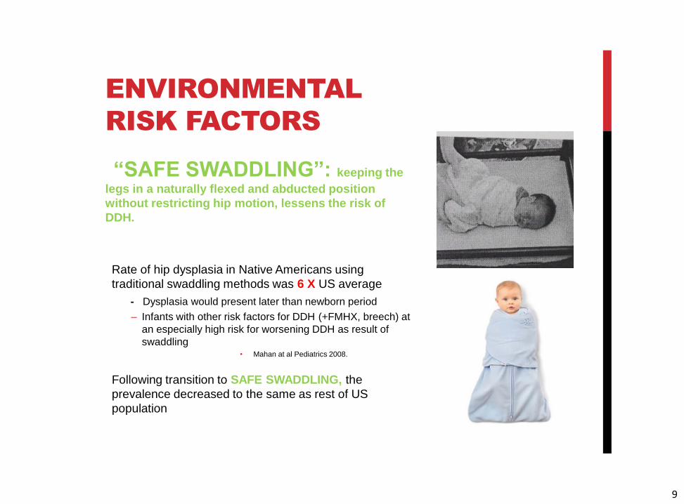
ENVIRONMENTAL
RISK FACTORS
“SAFE SWADDLING”: keeping the
legs in a naturally flexed and abducted position
without restricting hip motion, lessens the risk of
DDH.
Rate of hip dysplasia in Native Americans using
traditional swaddling methods was 6 X US average
- Dysplasia would present later than newborn period
– Infants with other risk factors for DDH (+FMHX, breech) at
an especially high risk for worsening DDH as result of
swaddling• Mahan at al Pediatrics 2008.
Following transition to SAFE SWADDLING, the
prevalence decreased to the same as rest of US
population

10
NATURAL HISTORY IN
INFANCY
88% mild dysplasia and instability noted in first few weeks resolve by 8 weeks of age.
• Majority of early clinically unstable hips will stabilize (without treatment)• 60 % in the first week• 88% in 2 months
• Barlow TG Proc R Soc Med 1963
• Majority of ultrasound findings normalize (without treatment)• 90% normalized by 9 weeks of age
• Marks et al. JBJS Br 1994
• The small percentage which do not resolve may sublux, dislocate or remain located but retain dysplastic features.

11
NATURAL HISTORY
DURING CHILDHOOD
•Limited abduction
•Leg length discrepancy
•Limp
• Untreated hip dislocation
– Rarely a cause of pain in childhood
– Generally does not delay ambulation
• Altered hip abductor mechanics -> Trendelenberg/waddling gait
» Weinstein, CORR 1987

12
NATURAL HISTORY
INTO ADULTHOOD
•Subluxation may not be as well
tolerated as dislocation
– patients may develop pain, degenerative
arthritis develops from excessive cartilage
contact pressure.
•Dislocation
– Femoral head cartilage in contact with bone of
pelvis can effect arthritic changes in 4th decade
– Knee pain, back pain, limp from leg length
discrepancy
» Weinstein CORR 1987
Wedge and Waslenko JBJS
1979

13
INFANT SCREENING
FOR DDH
THE EARLIER A DISLOCATED HIP IS DETECTED, THE
SIMPLER AND MORE EFFECTIVE IS THE TREATMENT.
ORTOLANI ( reducing dislocated hip) Maneuver developed in 1930’s
BARLOW( dislocating an unstable hip) Maneuver developed in 1950’s
Ultrasound as diagnostic tool popularized by Graf in 1980’s

14
SCREENING
RECOMMENDATIONS
AAP Clinical Practice Guideline, 2000
Careful clinical examination of all babies at birth and at well-child checks during first year. If positive, referral to orthopedist.
If clinical results are negative or equivocally positive, risk factors may be considered.
Selective ultrasound screening at age 4-6 weeks, ( or xray at 4 months if ultrasound not available) is recommended for babies with risk factors:
• Females born breech 120/1000
• Females with positive family history 44/1000
And consider ( strongly)
• Males born breech 26/1000
15
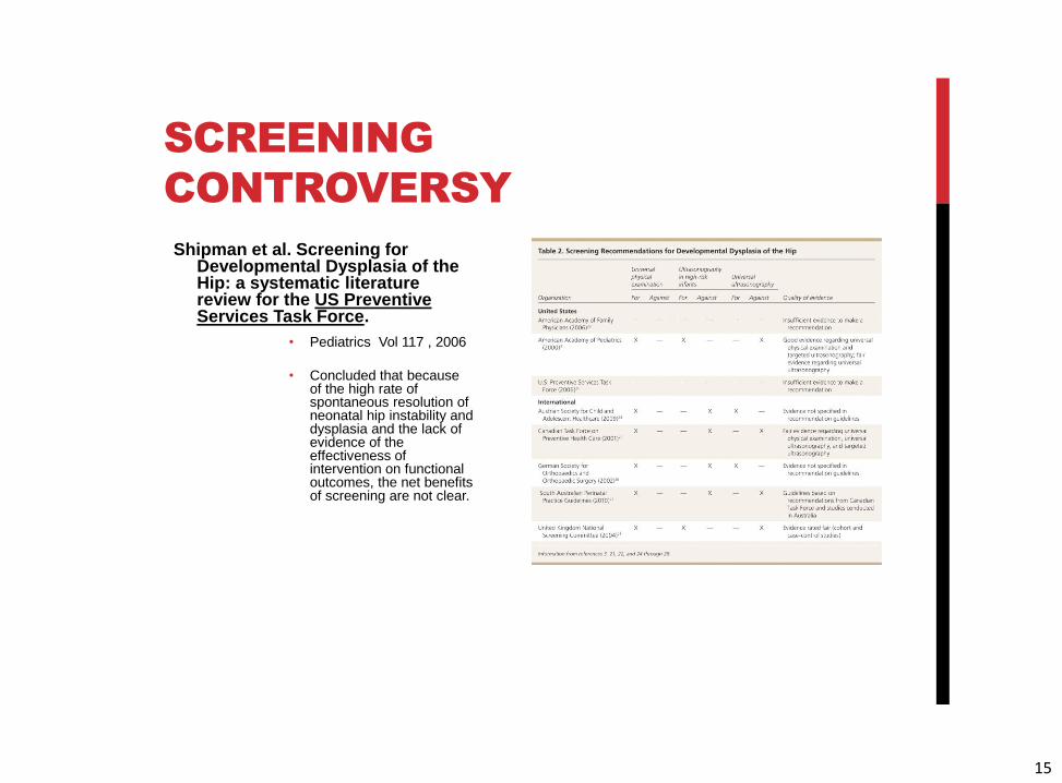
SCREENING
CONTROVERSY
Shipman et al. Screening for Developmental Dysplasia of the Hip: a systematic literature review for the US Preventive Services Task Force.
• Pediatrics Vol 117 , 2006
• Concluded that because of the high rate of spontaneous resolution of neonatal hip instability and dysplasia and the lack of evidence of the effectiveness of intervention on functional outcomes, the net benefits of screening are not clear.

16
TO SCREEN OR NOT
TO SCREEN?
To Screen or Not to Screen? A Decision Analysis of the Utility of Screening for Developmental Dysplasia of the Hip
• Mahan S, Katz J, Kim YJ
• JBJS 2009 91 (7) 1705-19
Used decision analysis techniques to compare 3 strategies:
• 1)No screening for DDH,
• 2)universal screening of newborns with both physical examination and ultrasonography,
• 3)universal screening with physical examination but only selective use of ultrasonography for neonates considered to be at high risk.
Conclusion:
• “The optimum strategy, associated with the highest probability of having a non arthritic hip at age 60 years, was to screen all neonates for hip dysplasia with a physical examination and to use ultrasonography selectively for infants who are at high risk.”
17
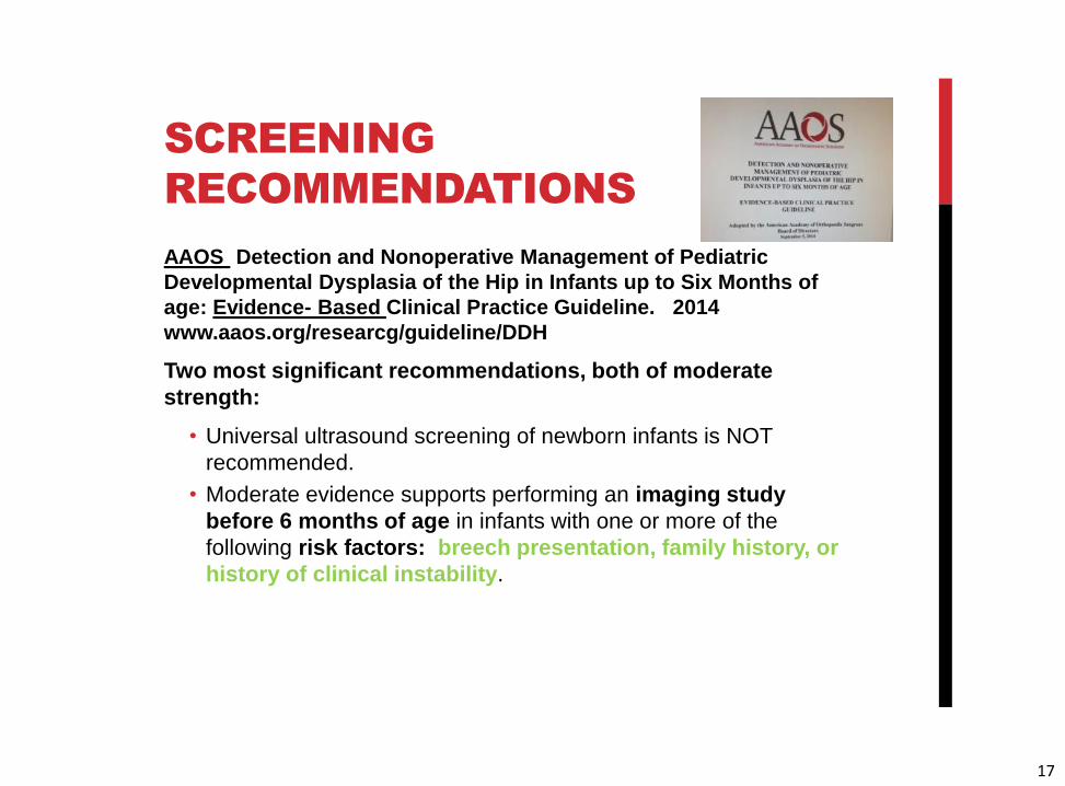
SCREENING
RECOMMENDATIONS
AAOS Detection and Nonoperative Management of Pediatric
Developmental Dysplasia of the Hip in Infants up to Six Months of
age: Evidence- Based Clinical Practice Guideline. 2014
www.aaos.org/researcg/guideline/DDH
Two most significant recommendations, both of moderate
strength:
• Universal ultrasound screening of newborn infants is NOT
recommended.
• Moderate evidence supports performing an imaging study
before 6 months of age in infants with one or more of the
following risk factors: breech presentation, family history, or
history of clinical instability.

18
SCREENING
RECOMMENDATIONS
What qualifies as “BREECH”?
1) Does short term breech matter?
2) Is breech in later stages more important?
3) What if the fetus has been turned before delivery?
4) Do males with breech have lower risk of DDH?
What qualifies as “POSITIVE FAMILY HISTORY” ?
1) First degree relatives only?
2) Specificity and severity of the diagnosis and treatment
Answers to these questions are not clear in the literature.

19
SCREENING
RECOMMENDATIONS
ADDITIONAL QUESTIONS: BREECH
1) After successful version?
Risk of Developmental dysplasia of the hip in breech
presentation : the effect of successful external cephalic version
Lambeek et al. BJOG 2012
498 breech pregnancies. Attempted version, 36% success.
Overall incidence of DDH in the cohort was 8% ( 40 infants)
The incidence of DDH requiring treatment among infants born after successful
version was 2.8%, compared with 9.3 % born after failed version attempt.
Successful version results in lower rate of DDH but high enough to still
recommend following routine breech screening program.

20
SCREENING
RECOMMENDATIONS
ADDITIONAL QUESTIONS: BREECH
2) After elective C-section?
Elective Ceasarean section is associated with a reduction in
developmental dysplasia of the hip in term breech infants
Lowry et al JBJS 2005
Cohort of 941 breech infants: 756 delivered via C section ( 515 prelabor, 241 intra-partum)
and 85 via vaginal delivery
Incidence of DDH was 3.69% following pre labor C-section,
6.64% following intra-partum C-section,
8.11% after vaginal delivery
Conclusion: These results demonstrate a significantly lower incidence of DDH in term
singleton breech births delivered by elective, pre-labor C –section and suggest that labor and
delivery influence hip stability in predisposed infants.

21
SCREENING
RECOMMENDATIONS
ADDITIONAL QUESTIONS: TWINS AND MULTIPLE BIRTHS
Should all twins and multiple births undergo ultrasound examination for developmental dysplasia of the hip? Barr et al Bone Joint J 2013
990 multiple birth compared to 25,246 singleton births
Incidence of DDH in multiple births was not higher than singleton p=0.8939
Developmental Dysplasia of the Hip in Twins: The importance of Mechanical Factors in the Etiology of DDH Pellegrin et al JPO 2010
105 pairs of twins : 48 cephalic –breech, 35 cephalic- cephalic, 22 breech-breech
Compared to control group 1 of 274 singleton infants cephalic presentation and control group 2 48 singleton infants breech presentation
Found a higher incidence of DDH in both control groups 3.4 % vs 0 % in the twin groups.
Proposed that the footling breech ( knees flexed and hips flexed) more common in twins is less detrimental to hip development that the frank breech presentation which is more common in singleton births.

22
FOLLOW UP AFTER
SCREENING
• Is ultrasound screening for DDH in babies born breech sufficient?– J Child Orthop (2010) 4: 3-8
– Imrie M, Scott V, Stearns P, Bastrom T, Mubarak S
300 infants with breech presentation (36% boys)• 34 pt with clinically unstable hips -> tx
• 266 clinically stable hips – 27 %(73 pts) abnl screening u/s at 6 weeks -> tx
» 73% (193 pts) normal screening u/s at 6 weeks of age
• 62/193 (32%) lost to follow up
• 93/131 (71%) nl clinical exams and xrays -> d/c’d from clinic
• 38/131 (29%) dx’d dysplasia based on clinical and radiographic findings and tx initiated with pavlikharness

23
FOLLOW UP AFTER
DIAGNOSIS
Radiographic Follow up of DDH in Infants: Are X-rays
Necessary After Normalized Ultrasound?
J Pediatr Orthop 2014
Sarkissian EJ, Sankar WN, Zhu X, Wu CH, Flynn JM
115 infants with DDH who has achieved both normal ultrasonographic and clinical examinations at 3.1
+/- 1.1 months of age.
At 6 months of age, 17% of all infants demonstrated radiographic signs of acetabular dysplasia.
Of infants left untreated, (n = 106), 33 % had dysplasia on subsequent radiographs at 12.5 +/-1.2
months of age.
Conclusions : The notable incidences of radiographic dysplasia after previous DDH normalization,
may warrant radiographic follow up in this population of infants through at least walking age to allow
timely diagnosis and early intervention of residual acetabular dysplasia.

24
RADIATION RISK
Routine hip xray requires a
dose of 0.001 m SV
(millisieverts)
By comparison, normal
background radiation from
one’s natural surroundings is
about 3 m SV/year.

25
SUMMARY
• Early diagnosis of DDH -> shorter duration of treatment, more reliable treatment, decreased risk of complication.
• Careful, routine clinical screening of every infant at each well child visit until established walking is recommended.
• Selective radiographic screening evaluations of at- risk infants ( breech presentation, + fam hx, positive clinical exam) is recommended.
• Importance of prevention with healthy hip positioning, especially in at risk infants.

26
RESOURCE
International Hip Dysplasia Institute http://hipdysplasia.org

27
THANK YOU FOR
YOUR ATTENTION
28
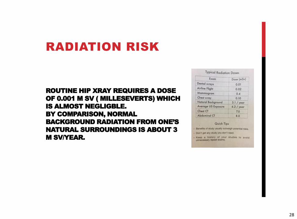
ROUTINE HIP XRAY REQUIRES A DOSE
OF 0.001 M SV ( MILLESEVERTS) WHICH
IS ALMOST NEGLIGBLE.
BY COMPARISON, NORMAL
BACKGROUND RADIATION FROM ONE’S
NATURAL SURROUNDINGS IS ABOUT 3
M SV/YEAR.
RADIATION RISK

29
TAKE HOME POINTS
WHAT DO I DO?
If clinical exam is stable, selective ultrasound screening for those infants with risk factors.
Best done when baby is 6 or more weeks old.
If documented/reported) breech, at any point, including boys, preemies, multiples, I treat as breech and get screening hip ultrasound at 6-8 weeks of age; age adjusted for the preemies.
Review with parents data about breech and concern for residual radiographic abnormalities. Recommend xray at 6 months. If family is xray averse, I offer repeat u/s at ? 5 and ½ months.
Advise parents of children with at risk hips to use safe hip techniques for swaddling, carriers and car seats.

30
SCREENING
RECOMMENDATIONS
ADDITIONAL QUESTIONS: PREMATURE BREECH
Breech preterm infants are at risk of developmental dysplasia
of the hip. Quan et al. J Paediatr Child Health 2013
Group 1 - 129 breech preterm infants ( no fam hx, oligo, or LGA)
ultrasound screening. 3/129 or 2.3% were positive for DDH
Group 2 - 163 breech term infants were screened. 3/163 or 1.8 %
positive for DDH.
Conclusion: Preterm infants born in the breech position appear to
have similar incidence of DDH to term infants and thus require
similar screening guidelines.

31
GOAL
•A reasonable goal for the primary care provide should be to
prevent hip subluxation or dislocation by 6 months of age
using periodic examination. Schwend et al. Ped Clin N Am 2014

32
THE CRUX
•Despite normal newborn and infant hip examination, a late
onset hip dislocation still occurs in approximately 1 in 5000
infants, as well as dysplasia in young adults.
•Despite all current methods of screening for DDH, most
young adults ( 92%) with dysplasia who require hip
arthroplasty are not detected at birth. Schwend et al Ped Clin
N Am 2014
•Well performed physical examination in infants is the most
important means of detecting instability; however dysplasia
is currently undetectable at birth for many adults.