Biomechanics of Developmental Dysplasia of the Hip - An ...
105
University of Central Florida University of Central Florida STARS STARS Electronic Theses and Dissertations, 2004-2019 2015 Biomechanics of Developmental Dysplasia of the Hip - An Biomechanics of Developmental Dysplasia of the Hip - An engineering study of closed reduction utilizing the Pavlik harness engineering study of closed reduction utilizing the Pavlik harness for a range of subtle to severe dislocations in infants. for a range of subtle to severe dislocations in infants. Victor Huayamave University of Central Florida Part of the Mechanical Engineering Commons Find similar works at: https://stars.library.ucf.edu/etd University of Central Florida Libraries http://library.ucf.edu This Doctoral Dissertation (Open Access) is brought to you for free and open access by STARS. It has been accepted for inclusion in Electronic Theses and Dissertations, 2004-2019 by an authorized administrator of STARS. For more information, please contact [email protected]. STARS Citation STARS Citation Huayamave, Victor, "Biomechanics of Developmental Dysplasia of the Hip - An engineering study of closed reduction utilizing the Pavlik harness for a range of subtle to severe dislocations in infants." (2015). Electronic Theses and Dissertations, 2004-2019. 1137. https://stars.library.ucf.edu/etd/1137
Transcript of Biomechanics of Developmental Dysplasia of the Hip - An ...

University of Central Florida University of Central Florida
STARS STARS
Electronic Theses and Dissertations, 2004-2019
2015
Biomechanics of Developmental Dysplasia of the Hip - An Biomechanics of Developmental Dysplasia of the Hip - An
engineering study of closed reduction utilizing the Pavlik harness engineering study of closed reduction utilizing the Pavlik harness
for a range of subtle to severe dislocations in infants. for a range of subtle to severe dislocations in infants.
Victor Huayamave University of Central Florida
Part of the Mechanical Engineering Commons
Find similar works at: https://stars.library.ucf.edu/etd
University of Central Florida Libraries http://library.ucf.edu
This Doctoral Dissertation (Open Access) is brought to you for free and open access by STARS. It has been accepted
for inclusion in Electronic Theses and Dissertations, 2004-2019 by an authorized administrator of STARS. For more
information, please contact [email protected].
STARS Citation STARS Citation Huayamave, Victor, "Biomechanics of Developmental Dysplasia of the Hip - An engineering study of closed reduction utilizing the Pavlik harness for a range of subtle to severe dislocations in infants." (2015). Electronic Theses and Dissertations, 2004-2019. 1137. https://stars.library.ucf.edu/etd/1137
BIOMECHANICS OF DEVELOPMENTAL DYSPLASIA OF THE HIP – AN
ENGINEERING STUDY OF CLOSED REDUCTION UTILIZING THE PAVLIK HARNESS
FOR A RANGE OF SUBTLE TO SEVERE DISLOCATIONS IN INFANTS
by
VICTOR A. HUAYAMAVE
B.S. University of Central Florida, 2008
M.S. University of Central Florida, 2010
A dissertation submitted in partial fulfillment of the requirements
for the degree of Doctor of Philosophy
in the Department of Mechanical and Aerospace Engineering
in the College of Engineering and Computer Science
at the University of Central Florida
Orlando, Florida
Spring Term
2015
Major Professor: Alain J. Kassab

ii
© 2015 by VICTOR A. HUAYAMAVE

iii
ABSTRACT
Developmental Dysplasia of the Hip (DDH) is an abnormal condition where hip joint
dislocation, misalignment, or instability is present in infants. Rates of incidence of DDH in
newborn infants have been reported to vary between 1 and 20 per 1000 births, making it the most
common congenital malformation of the musculoskeletal system. DDH early detection and
treatment is critical to avoid the use of surgical treatment in infants and to prevent future
complications such as osteoarthritis in adult life. To this day several non-surgical treatments
involving the use of harnesses and braces have been proposed to treat DDH in infants, with the
Pavlik harness being the current non-surgical standard used to treat DDH at early stages. Although
the Pavlik harness has been proven to be successful treating subtle dislocations, severe dislocations
do not always reduce. Until now the use of the harness remains an empirical method, and its
effectiveness often depends on physician expertise or trial-error procedures; thus a clear guideline
has not been established to determine the best optimal harness configuration to treat both subtle
and severe dislocations. The goal of this dissertation is to understand the connection between
reductions for subtle and severe dislocations and passive muscle forces and moments generated
while the harness is used during treatment.
While the understanding of DDH biomechanics will provide a valuable clinically
applicable approach to optimize and increase harness success rate, it is not without its difficulties.
This research has created and developed a three-dimensional based on patient-specific geometry
of an infant lower limb. The kinematics and dynamics of the lower limb were defined by modeling
the hip, femur, tibia, fibula, ankle, foot, and toe bones. The lines of action of five (5) adductor
muscles, namely, the Adductor Brevis, Adductor Longus, Adductor Magnus, Pectineus, and

iv
Gracilis were identified as mediators of reduction and its mechanical behavior was characterized
using a passive response. Four grades (1-4) of dislocation as specified by the International Hip
Dysplasia Institute (IHDI) were considered, and the computer model was computationally
manipulated to represent physiological dislocations. To account for proper harness modeling, the
femur was restrained to move in an envelope consistent with its constraints.
The model of the infant lower limb has been used to analyze subtle and severe dislocations.
Results are consistent with previous studies based on a simplified anatomically-consistent
synthetic model and clinical reports of very low success of the Pavlik harness for severe
dislocations. Furthermore the findings on this work suggest that for severe dislocations, the use of
the harness could be optimized to achieve hyperflexion of the lower limb leading to successful
reduction for cases where the harness fails.
This approach provides three main advantages and innovations: 1) the used of patient-
specific geometry to elucidate the biomechanics of DDH; 2) the ability to computationally
dislocate the model to represent dislocation severity; and 3) the quantification of external forces
needed to accomplish reduction for severe dislocations. This study aims to offer a practical solution
to effective treatment that draws from engineering expertise and modeling capabilities and also
draws upon medical input. The findings of this work will lay the foundation for future optimization
of non-surgical methods critical for the treatment of DDH.

v
To my family whose unconditional love and support always motivate me to set higher targets.
This work could have not been possible without your continuous encouragement. To Alina for
her patience, encouragement, and support provided throughout the dissertation process. Little
Emma Victoria, I promise to make up for all the weekends I could not spent with you while I was
working on my dissertation. Last but not least, to all my friends who inspired and encouraged me
along the way.

vi
ACKNOWLEDGMENTS
Special thanks to:
Professor Alain Kassab for supporting me throughout my journey towards a Ph.D. degree
and sharing his enthusiasm in the exciting field of biomechanics inspiring my love of research.
The academic opportunities, professional opportunities, and mentorship provided by Dr. Kassab
will certainly have a major impact in my future endeavors and I am anxiously looking forward to
apply everything I learned during these years at while working with the Computational Mechanics
Lab in my professional career. I will forever be thankful for your advice.
Professor Eduardo Divo for introducing me to numerical research early in my
undergraduate career. Dr. Divo’s technical guidance has enabled me to successfully pursue an
academic and research career. Additionally, I will be forever grateful to him for sharing his
business vision and expertise. His professionalism and friendship will continue to influence my
work in years to come.
Dr. Charles Price and the International Hip Dysplasia Institute for providing the inspiration
and supporting our work at national and international conferences. I will always remember our
Italy research trip since it gave us the opportunity to come face to face with the history of
physicians who were the pioneers in the area of developmental dysplasia of the hip.
Professors Seetha Raghavan and Faissal Moslehy for serving on my committee and
advising the work I developed on this dissertation.
All the students part of the Development Dysplasia of the Hip group at the University of
Central Florida. As a group we have accomplished great milestones and the work I am presenting
here would have not been possible without your input.

vii
TABLE OF CONTENTS
LIST OF FIGURES ............................................................................................... ix
LIST OF TABLES ................................................................................................ xii
INTRODUCTION ........................................................................... 1
1.1 State of the Art Problem ........................................................................................ 1
1.2 Hip Joint Motion and Hip Dislocations................................................................. 3
1.3 Statement of Purpose ............................................................................................. 5
1.4 Thesis Overview .................................................................................................... 5
BIOMECHANICS OF DEVELOPMENTAL DYSPLASIA OF
THE HIP ................................................................................................................... 7
2.1 Developmental Dysplasia of the Hip .................................................................... 7
2.1.1 Terminology, Etiology, Pathoanatomy, and Pathophysiology ........................ 8
2.1.2 Diagnosis, Classification, and Treatment ...................................................... 13
2.1.3 Treatment with the Pavlik Harness ................................................................ 21
2.2 Lower-Limb Bone Biomechanics ....................................................................... 24
MECHANICAL BEHAVIOR AND MODELING OF
SKELETAL MUSCLES ........................................................................................29
3.1 Skeletal Muscle Anatomical Structure and Physiology ...................................... 31
3.2 Skeletal Muscle Path Representation .................................................................. 36

viii
3.2.1 Straight-line Skeletal Muscle Path Representation ........................................ 36
3.2.2 Centroid Model of Skeletal Muscle Path ....................................................... 39
3.2.3 Modeling of Curved and Wrapping Skeletal Muscles ................................... 40
3.3 Mechanical Modeling of Muscles ....................................................................... 42
3.3.1 Active Muscle Behavior ................................................................................ 45
3.3.2 Passive muscle Behavior................................................................................ 47
METHODOLOGY .......................................................................50
4.1 Lower Limb Anatomical Reconstruction ............................................................ 51
4.2 Lower Limb Anatomical Assembly .................................................................... 55
4.3 Lower Limb Skeletal Muscle Modeling.............................................................. 57
........................................................ 60
4.5 Hip Joint Lubrication .......................................................................................... 62
4.6 Lower Limb Calibration Model .......................................................................... 63
RESULTS ......................................................................................68
5.1 Grade 3 Dislocation Results ................................................................................ 69
5.2 Grade 4 Dislocation Results ................................................................................ 71
CONCLUSION..............................................................................74
APPENDIX: PUBLICATIONS, PRESENTATIONS AND AWARDS ...........77
LIST OF REFERENCES ......................................................................................80
ix
LIST OF FIGURES
Figure 1-1:Anatomical Dislocation Classification (a) Normal Hip, (b) Subluxated Hip, and (c)
Dislocated Hip [4] ........................................................................................................................... 3
Figure 1-2: Anatomical Hip and Knee Joint Motions ..................................................................... 4
Figure 2-1: Representation of a Normal Socket (Top) and Shallow Socket (Bottom) [23] ........... 9
Figure 2-2: Proportional Changes of the Femoral Head Covered by the Acetabulum [23] ......... 10
Figure 2-3: Swaddling in African (Left), Chinese (Center), and Eskimo (Right) Groups [5] ...... 11
Figure 2-4: Swaddling in Italian (Left) and Native Indian (Right) Groups [5] ............................ 12
Figure 2-5: Ortolani and Barlow Maneuvers [32] ........................................................................ 14
Figure 2-6: Limited Abduction Left Infant Hip [32] .................................................................... 15
Figure 2-7: Radiographic Measuring Parameters for Severity Classification [36]....................... 16
Figure 2-8: International Hip Dysplasia Dislocation Grades [36] ................................................ 17
Figure 2-9: Hips Placed in Flexion and Abduction ...................................................................... 18
Figure 2-10: Developmental Dysplasia of the Hip Treatment Mapping in the First 6 Months of
Life [4]. ......................................................................................................................................... 20
Figure 2-11: Typical Pavlik Harness Configuration ..................................................................... 21
Figure 2-12: Relationship Between Age and Treatment Success [49]. ........................................ 22
Figure 2-13: Effect of Graf Type on Treatment Results [49]. ...................................................... 23
Figure 2-14: Forces Exerted on a Loaded Hip while Walking [53, 56] ....................................... 25
Figure 2-15: Changes in the Shape of the Femoral Head in Relation to Age. From Left to Right:
21-week fetus, 30-week fetus, Premature Newborn, 2 Years, 4 Years [23]. ................................ 25
Figure 2-16: Murphy’s Method for Measuring Femoral Anteversion Angle [57] ....................... 26

x
Figure 2-17: Femoral Anteversion Classification: Normal (Left), Increased Angle (Center),
Retroversion (Right) [58]. ............................................................................................................. 27
Figure 2-18: Femoral Anteversion in Relation to Age: (1) Hip with Developmental Dysplasia of
the Hip and (N) Healthy Hips [59]. .............................................................................................. 27
Figure 3-1: Borelli’s Classic Work on Muscle Mechanical Behavior [60]. ................................. 30
Figure 3-2. Myofibril and Sarcomere Architecture [61]. .............................................................. 32
Figure 3-3: Muscle Structural Elements [62]................................................................................ 33
Figure 3-4: Lower Limb Muscles Fiber Length and Physiological Cross-Sectional Areas [65].. 34
Figure 3-5: Hip Musculature Elastic Straight-Line String Model [68] ......................................... 37
Figure 3-6: Two-Dimensional, Geometric Representation of Elbow Joint Using a SLMPR of the
Brachialis (BRA) and the Brachioradialis (BRD) [69]. ................................................................ 38
Figure 3-7: Centroid-Line Representation of Gluteus Medius and Femur [72] ........................... 40
Figure 3-8: Psoas and Gluteus Maximus Muscle-Path Representation Using Curved Segments
Wrapped Around Multiple Surfaces [73]. .................................................................................... 41
Figure 3-9: Hill-Type Muscle Model ............................................................................................ 43
Figure 3-10: Muscle Tension-Length Curves Exhibiting Active and Passive Tension Behavior 45
Figure 3-11: Generalized Relationship between Muscle Length, Force, and Velocity during Muscle
Activation [86]. ............................................................................................................................. 46
Figure 3-12: Fung’s Biological Tissue Non-Linear Behavior [93]. ............................................. 48
Figure 4-1: Superposition of a Reconstructed Model of a Fourteen-Year Old Female (left) with a
model Rendered From CT-scans of a Six-Month Old Female Infant [95]. .................................. 52
Figure 4-2: Spherical Head in Neonates: Computational Model (Left) and Dissection (Right). . 53

xi
Figure 4-3: Femoral Anteversion in Children with Developmental Dysplasia of the Hip [101]. 54
Figure 4-4: Illustration of 50° Anteversion Angle in 3D Computational Model (Left) and a
Dissected Femur Showing Anteversion Angle (Right). ............................................................... 54
Figure 4-5: Reconstructed Lower Limb of 10-Week Old Female Infant. .................................... 55
Figure 4-6: Anterior View of Adductor Muscles Origin and Insertion Points [103]. ................... 56
Figure 4-7: Hip Dislocation: Healthy Hip (Left), Grade 3 (Center), and Grade 4 (Right). .......... 56
Figure 4-8: Ball and Socket Hip Joint Frictionless Model. .......................................................... 63
Figure 4-9: Ten-Week Old Female Infant Passive Force versus Stretch for Calibrated Model. .. 64
Figure 4-10: Adductor Brevis Force-Stretch Comparison between Ten-Week Old Female Infant
and Young Adult Model. .............................................................................................................. 65
Figure 4-11: Calibrated Model: a) Contact Forces (Single Headed Arrow) and Muscle Lines of
Action (Double Headed Arrow) on XZ-Plane, b) Contact Force (Single Headed Arrow) and
Muscle Lines of Action (Double Headed Arrow) on XY-Plane. .................................................. 66
Figure 5-1: a) Grade 1 and b) Grade 2 Anatomical dislocations. ................................................. 68
Figure 5-2: Grade 3 Anatomical Dislocation. ............................................................................... 69
Figure 5-3: Grade 4 Dislocation on Ten-Week Old Female Infant (XZ Plane). .......................... 71
Figure 5-4: Muscle Force Contribution Comparison for Pectineus and Brevis. ........................... 73
Figure 5-5: Muscle Force Contribution Comparison for Adductor Longus, Adductor Magnus and
Gracilis. ......................................................................................................................................... 73
Figure 6-1: Grade 4 Dislocation on Ten-Week Old Female Infant (YZ Plane). .......................... 74
Figure 6-2: Reduction by Driving Femoral Head Posterior to the Acetabulum in an Inferior
Direction. ...................................................................................................................................... 75

xii
LIST OF TABLES
Table 2-1: International Hip Dysplasia Institute (IHDI) Classification using Radiographic
Measurements [36]........................................................................................................................ 16
Table 4-1: Lower Extremity Mass Distribution. ........................................................................... 57
Table 4-2: Scaled Ten-Week Old Female Infant Physiologic Cross-Sectional Areas Using
Adductor Brevis Shape Factor [113]. ........................................................................................... 60
Table 4-3: Friction Coefficients for Common Engineering Materials.......................................... 62
Table 4-4: Muscle Force and Moment Contribution at Equilibrium Configuration. .................... 67
Table 5-1: Muscle Force and Moment Contribution on Grade 3 Dislocation. ............................. 70
Table 5-2: Muscle Force and Moment Contribution on Grade 4 Dislocation. ............................. 72

1
INTRODUCTION
Using a computational approach, the work developed in this dissertation investigates the
mechanical effects of developmental dysplasia of the hip in infants, which typically manifests as
a misalignment of the hip joint. To this end, a patient-specific anatomical model has been created
to unravel the biomechanics of developmental dysplasia of the hip. Simulation-based
computational research using patient-specific anatomy has proven to be a powerful tool in the field
of biomedical engineering. It has been suggested that by using such state of the art methods
physicians could avoid surgery. This can account for a 70% reduction in blood stream infections.
Additionally, using simulation training intervention provides a 7:1 rate of return [1]. Moreover,
translational research has shown that using simulation-based techniques in obstetric simulation
accounts for a 40% reduction in malpractice cases [2]. Thus, using such methods will improve
decision-making to diagnose and provide customized treatment.
1.1 State of the Art Problem
At earlier stages of infant development, the hip joint is susceptible to displacements. Subtle
and severe dislocations are usually detected by careful clinical examination. Identification of the
condition and treatment during the neonatal period is advised to prevent the progression of
subluxation into severe dislocations. Ultimately, early treatment enables proper growth of the hip
joint to reduce incidences of dysplasia. Going undetected or without treatment, developmental
dysplasia of the hip could progress to deformation, lost function, and eventual osteoarthritis [3].
Screening is often controversial, and in order to clearly define our problem, is necessary to
understand the pathophysiology of developmental dysplasia of the hip. Classical assessment

2
methods of developmental dysplasia of the hip treatment include physical, radiological imaging,
and ultrasound techniques.
Early treatment is always needed to avoid surgery in newborns and infants, since invasive
surgery complications resulting from hip reduction might lead to femoral head osteonecrosis and
redislocation. In infants up to six months of age, the Pavlik harness is the standard and preferred
orthopaedic device utilized worldwide to non-surgically correct developmental dysplasia of the
hip in infants. Long-term studies have shown the Pavlik harness has a 95% success ratio for subtle
dysplasia and subluxations, while for severe dislocations, this success ratio reduces drastically.
The harness’ effectiveness often depends on physician expertise and trial-error procedures.
Currently no study has yet been carried out to characterize and quantify the mechanics of
developmental dysplasia of the hip utilizing the Pavlik harness to understand its failure under
severe dislocations. Accepting the published rate of long-term disability caused by unsuccessful
treatment of developmental dysplasia of the hip with passive corrective devices such as the Pavlik
harness, and the lack of factual, concise guidelines regulating their use and construction, along
with no assertive methods or procedure to determine whether successful treatment will result, other
than mere chance, this dissertation will use engineering fundamentals to determine the mechanics
governing the operation of the Pavlik harness in order to bring to light conclusive determinants to
its mechanism of action to devise case-specific methods to actively vector the femoral head to its
proper concentric position in the acetabulum, thereby decreasing the incidence of disability at
cause of unsuccessful treatment of development dysplasia of the hip.
At present, there are no quantitative tools to evaluate muscle forces and moments
developed in hip dislocations. Therefore, computer models can assist in developing a framework

3
to understand the biomechanical response of developmental dysplasia of the hip. This computer
model will be able to simulate the complex interaction between muscles and bones. Furthermore,
results of this study could be applied in a clinical setting and serve as an effective approach to
optimize the use of the Pavlik harness.
1.2 Hip Joint Motion and Hip Dislocations
The purpose of this brief section is to provide the reader with a visual aid of anatomical
terms that will be used on this dissertation. Figure 1-1 shows different grades of developmental
dysplasia of the hip, where (a) represents a normal hip where the labrum envelops the head and
deepens the acetabulum, (b) represents a subluxated hip where the head lies eccentrically in the
acetabulum deforming it, and (c) represents a dislocated hip where the cartilaginous acetabular
margin deforms and the labrum may invert and obstruct reduction [4].
Figure 1-1:Anatomical Dislocation Classification (a) Normal Hip, (b) Subluxated Hip, and
(c) Dislocated Hip [4]
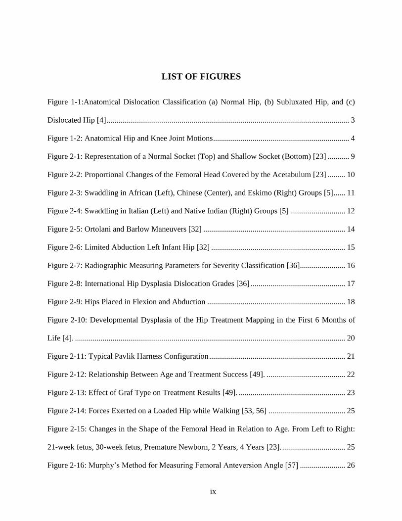
Figure 1-2 exhibits hip joint and knee motion terminology that will be used on this
dissertation to define functionality and ranges of motion of the joints.
4
Figure 1-2: Anatomical Hip and Knee Joint Motions

5
1.3 Statement of Purpose
The principal goal of this dissertation is to understand the connection between reductions
of subtle and severe dislocations, passive muscle forces and moments generated while the Pavlik
harness is used during treatment. By understanding the kinematics and dynamics of developmental
dysplasia of the hip, the use of orthosis to treat this condition can potentially be optimized and
improved. To this end, the concept of personalized pediatrics will be implemented to develop a
physical model of the lower limb of a ten-week old female infant. Next, adductor muscles will be
modeled in the lower limb as action/reaction forces exhibiting passive behavior. Finally, the
kinematics and dynamics will be analyzed using the Rigid Body Dynamics Method. With the use
of a patient-specific model, subtle and severe dislocations presented in infants can be successfully
elucidated. The outcome of the current dissertation will quantify the biomechanics of
developmental dysplasia of the hip in infants, and will also suggest means to optimize the use of
the Pavlik harness. In addition, results of this study will provide guidance to develop innovative
new medical devices to successfully treat developmental dysplasia of the hip in the future.
Moreover, the methodology used on this dissertation can be effectively implemented in several
conditions where fractures, dislocations, and separations in the skeleton may occur. Such methods
provide the flexibility of dislocating the hip joint, which to our knowledge, has not be done yet to
investigate and optimize orthopedic rehabilitation.
1.4 Thesis Overview
The work developed on this dissertation is captured and arranged in six chapters. Following
the introductory chapter, Chapter 2 presents an overview of developmental dysplasia of the hip
6
and hip biomechanics. Chapter 3 will then complement the groundwork started in Chapter 2 by
providing details of skeletal muscle mechanics and describes related work on this field. In addition,
Chapter 2 and Chapter 3 outline some of the major assumptions of this dissertation. The
information on these two chapters is also detailed enough aiming to develop an understanding on
readers without previous knowledge and experience in the field of bone and muscle mechanics.
Following this background information, the specific features and methodology used to develop the
patient-specific model of the ten-week old female infant will be presented in Chapter 4. To explain
the particulars of the physics-based anatomical model, Chapter 4 is divided into several
subsections. These subsections include the following subjects: the anatomical reconstruction; the
anatomical assembly; the utilization of a passive muscle model; the implementation of the Rigid
Body Dynamics Method; the lubrication of the hip joint; the anatomical model calibration; and the
different dislocations studied. Chapter 5 presents the forces and moments that were quantified by
elucidating the effects of the Pavlik harness on subtle and severe dislocations. Finally, Chapter 6
discusses the findings of this dissertation and summarizes the contributions. Moreover, this final
chapter will include final remarks on future research directions related to improve the usage of the
Pavlik harness and other orthosis.

7
BIOMECHANICS OF DEVELOPMENTAL DYSPLASIA OF
THE HIP
In order to understand the biomechanics of developmental dysplasia of the hip, a
reasonable understanding of the condition is needed. To this end, this chapter presents two
sections: section 2.1 will review and present detailed information of the etiology, pathoanatomy,
pathophysiology, diagnosis, classification, and treatment of developmental dysplasia of the hip. A
proper background knowledge of this condition will help to provide correct assumptions to
effectively tackle an anatomical model that exhibits the mechanics of developmental dysplasia of
the hip. Finally section 2.2 will present the biomechanics of the lower limb which aims to explain
the mechanical factors behind this common condition and to provide means of proper mechanical
modeling of the bony structure.
2.1 Developmental Dysplasia of the Hip
In general developmental dysplasia of the hip is an abnormal condition where hip joint
dislocation, misalignment, and instability are present in infants. It has been stated that “congenital
dislocation of the hip represents one of the most important and challenging congenital
abnormalities of the musculoskeletal system” [5] and ”in its severest form, developmental
dysplasia of the hip is one the most common congenital malformations” [6]. It has been reported
that clinical hip instability in newborn infants ranges from 1.6 to 28.5 per 1,000 [6] and 6 out of
every 1,000 will require treatment [7]. Studies had also shown that developmental dysplasia of the
hip was responsible for 29% of primary hip replacements in people up to age 60 years [8].
Although the condition may present after birth, most cases originate during fetal development,

8
thus it is commonly referred to as Congenital Hip Dysplasia (CHD). CHD is typically discovered
during physical examination at birth and some common signs of dislocations are: asymmetrical
hip abduction or asymmetrical limb lengths, hip click sounds, limited range of motion, and pain.
Therefore, it is crucial to diagnose hip dislocations timely postnatally, and expedite the onset of
treatment. Successful hip reductions in all cases must be achieved during the first few months of
life as the anatomy is still in its developmental phase. In all cases, the success of the treatment is
inversely related to the age at which treatment is begun, with success rates remaining the highest
when the treatment is begun immediately after birth or within the first month, and declining after
9 months of age [9].
2.1.1 Terminology, Etiology, Pathoanatomy, and Pathophysiology
Formerly known as congenital hip dislocation (CDH) [10] or hip dysplasia, the
terminology used to describe hip displacements or dislocations is controversial. The term
“congenital’ was previously used to describe dislocations or displacements due to genetics or
mechanical factors affecting the fetus in utero and the term “dysplasia” was used to refer to
irregular development of the femoral head, acetabulum, or both after the neonatal period [11].
Additionally, physicians in the past have favored the used of the term “congenital dysplasia’
instead of the term “congenital dislocation” [12]. This preference in terminology was based on the
fact that dysplasia better describes the morphological alteration of the hip. In addition, “dysplasia”
refers to cases where there is partial contact between the acetabulum and the femoral head
(subluxation) and cases where there is no contact between the acetabulum and the femoral head
(dislocation). The latest term, developmental dysplasia of the hip was adopted to define

9
displacements or dislocations regardless of nature or time when the abnormal condition first
appeared. Developing an understanding of the etiology, pathoanatomy, and pathophysiology of
developmental dysplasia of the hip is relevant to the study of this dissertation. Thus, this section
will present some of the factors that have been suggested to be main contributors of hip
displacements or dislocations before, during, and after the neonatal period.
Early experiments have shown that it is possible to dislocate the hip in an embryo for the
first time at 11 weeks old [13]. It has been suggested that there are critical periods during which
the hip of a fetus is at risk of dislocation: the 12th week, the 18th week, and the final 4 weeks of
gestation [14]. Additionally, packaging abnormalities at earlier stages of pregnancy may limit
movement in the utero leading to dislocations. Experimental results have also suggested that
breech presentation [5, 15], female sex [16-18], oligohydramnios [19, 20] , and primiparity [21]
are major risk factors. Furthermore, the risk is believed to increase when a breech-presenting child
is delivered vaginally rather than by caesarean section [3, 22]. Dislocation vulnerability before
birth is known to be caused mainly by the acetabular-capacity and depth relative to the femoral
head. Experimental studies have suggested that the acetabulum becomes shallow as birth
approaches [23]. This depth difference between a normal hip and a shallow hip affects the articular
range of hip motion as seen in Figure 2-1.
Figure 2-1: Representation of a Normal Socket (Top) and Shallow Socket (Bottom) [23]

10
Additionally, experimental results have indicated that the coverage provided by the
acetabulum to the femoral head decreases drastically before birth as seen in Figure 2-2. This
finding in particular provides a possible explanation for dislocations or displacements in the
infantile hip caused by the reduced contact between the femoral head and the acetabulum before
and during the neonatal period. This type of dislocation is known to be the most severe form of
developmental dysplasia of the hip since it will cause abnormal development of the lower limb.
Figure 2-2: Proportional Changes of the Femoral Head Covered by the Acetabulum [23]
Contrastingly, anatomic experiments in Africans have shown that the acetabulum remains
deeper before birth providing more coverage to the femoral head. Thus, confirming the
demographics of developmental dysplasia of the hip where incidence is almost nonexistent in

11
African neonates [24]. This particular study has lead clinicians to perform anatomical experiments
to understand how geographic and ethnic factors influence developmental dysplasia of the hip in
infants. It has been suggested that racial and geographical incidence is mainly caused by
positioning of the neonate. Particularly, studies had being focused on various swaddling methods
around the world. Reports have indicated that swaddling during the first months of life may
increase dislocation risk based on usage configuration. For instance, it is known that Africans,
Chinese, and Eskimos groups swaddle newborn infants to maintain hips in abduction and flexion
as seen in Figure 2-3. Such configuration mimics the ones found in typical non-surgical
harness/braces used to treat developmental dysplasia of the hip. Moreover, incidences recorded on
these groups have been reported to be remarkably low.
Figure 2-3: Swaddling in African (Left), Chinese (Center), and Eskimo (Right) Groups [5]
Conversely; for Italian and Native Indian groups, who swaddle newborn infants
maintaining the hip in extension and adduction as seen in Figure 2-4, incidences are remarkably

12
high. Furthermore, swaddling is controversial since its use has been linked not only to
developmental dysplasia of the hip but also to sudden infant death syndrome (SIDS) [25].
Figure 2-4: Swaddling in Italian (Left) and Native Indian (Right) Groups [5]
Furthermore, fully dislocated hips may present well developed false acetabulum and
bilaterality [26]. A severed dislocated hip with a well develop acetabulum will have a great impact
in adult life by increasing the risk to develop degenerative joint diseases.
For cases presented at birth, Barlow reported that one in sixty infants will show sign of
instability and 12% will require treatment [27]. Several cases have been reported where hip
appeared to be normal at birth but dislocated during the postnatal period [28, 29]. Incidences after
birth are believed to be caused mainly by postnatal positioning. Within the newborn population
affected with any type of instability, some may be reduced without treatment, some develop may
develop subluxation and some may progress to a dislocation.
13
2.1.2 Diagnosis, Classification, and Treatment
Physicians in the past believed that an examination of a newborn will successfully provide
a diagnoses for a dislocated hip. However recent studies have determined some types of
dislocations go by undetected at birth and some may occur in the neonatal period or later. The U.S.
Preventive Services Task Force has also stated that: “evidence is insufficient to recommend routine
screening for developmental dysplasia of the hip in infants as a means to prevent adverse
outcomes” [30]. Nonetheless, physical examination is recommended during regular infants check-
ups. As presented in the last section developmental dysplasia of the hip could appear at any point
after birth and is of paramount importance to keep track of hip joint development. A simple visual
way to detect dislocations in newborns is by looking for asymmetry patterns in the lower limb of
a neonate. Physical examination is always performed in the neonatal period and two of the most
common clinical test are the Ortolani [31] and Barlow [27] maneuvers. The Ortolani and Barlow
maneuvers, have become the “gold standard” techniques to asses hip stability on 3 months old
infants or younger. The Ortolani procedure is performed by gently abducting the leg with the hips
flexed at 90° and applying an upward force through the greater trochanter as seen in Figure 2-5.
An Ortolani positive test will feature a palpable “clunk” sensation representing the reduction of
the dislocated hip. In general, a positive Ortolani test indicates a reducible dislocation while a
negative Ortolani test indicates an irreducible dislocation. On the other hand, the Barlow procedure
is performed by adducting the leg with the hips flexed at 90° and gently pushing the knee as seen
in Figure 2-5. A Barlow positive test will feature a palpable “clunk” sensation and will force the
femoral head out of the acetabulum confirming hip dislocation.

14
Figure 2-5: Ortolani and Barlow Maneuvers [32]
From 3 to 12 months the use of both maneuvers is not recommended due to lack ligaments
and capsular laxity. For these older infants, a visual check is performed by the pediatric physician
by placing the infant supine on a firm table and then abducting both hips at the same time to check
for any asymmetries as shown in Figure 2-6.

15
Figure 2-6: Limited Abduction Left Infant Hip [32]
Radiographic evaluation is the standard whenever it comes to diagnoses. However, it is
known that during the neonatal period is difficult to interpret radiographs due to cartilaginous
regions where the femoral head-acetabulum correlation cannot be determined. Graf proposed the
use of ultrasound [33] to diagnose developmental dysplasia of the hip and suggested the use of
four sub-types of displacements or dislocations. However, it has been reported clinical ultrasound
usage may provide false-positive results [34]. Tonnis presented a method to classify developmental
dysplasia of the hip severity by assessing the location of the ossific nucleus using radiographs [35].
Additionally, the International Hip Dysplasia Institute has developed a new radiographic
classification using the Hilgenreiner’s line (H-line), Perkin’s line (P-line), Diagonal line (D-line),
and H-point as defined is Table 2-1 and shown in Figure 2-7.

16
Table 2-1: International Hip Dysplasia Institute (IHDI) Classification using Radiographic
Measurements [36]
Radiographic Measurement Description
Hilgenreiner’s line
(H-line)
The single line drawn through the top of the
triradiate radiate cartilage bilaterally
Perkin’s line
(P-line)
Perpendicular line from the superolateral
margin of the acetabulum
Diagonal’s line
(D-line)
45 degree line from the junction of H-line and
P-line
H-Point Midpoint of the superior margin of the ossified
metaphysis
Figure 2-7: Radiographic Measuring Parameters for Severity Classification [36]
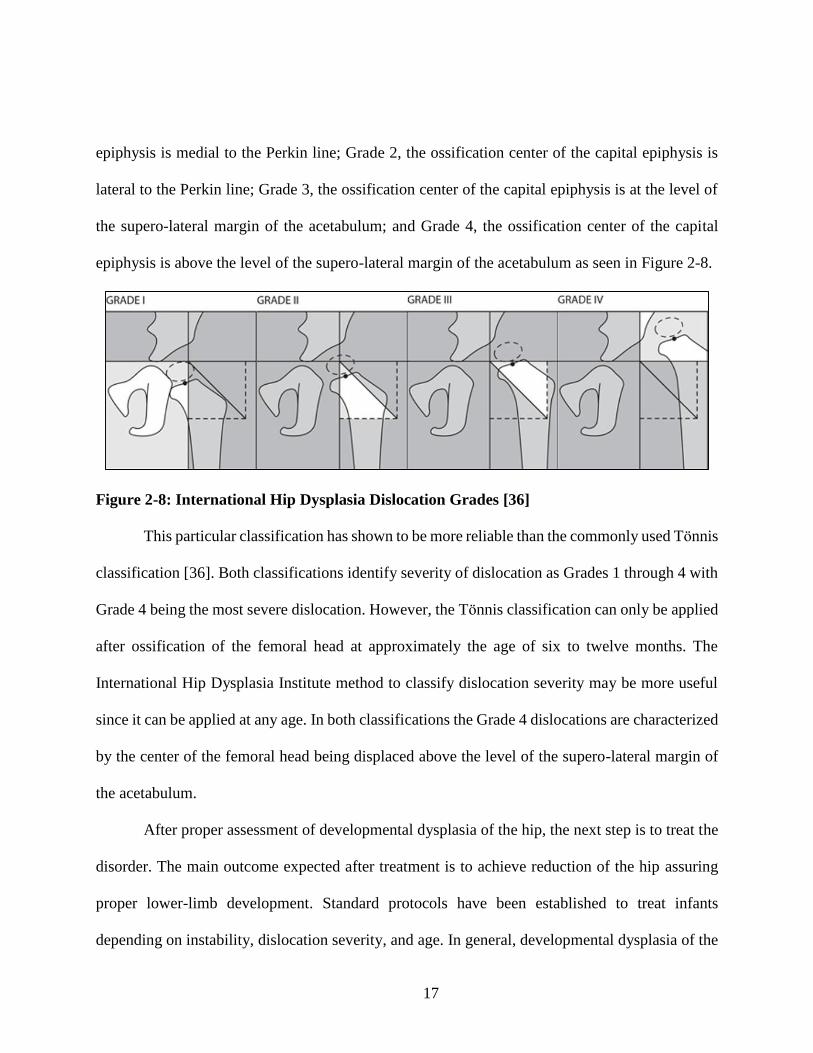
The International Hip Dysplasia classification uses the position of the proximal femoral
metaphysis to define the severities as follows: Grade 1, the ossification center of the capital
17
epiphysis is medial to the Perkin line; Grade 2, the ossification center of the capital epiphysis is
lateral to the Perkin line; Grade 3, the ossification center of the capital epiphysis is at the level of
the supero-lateral margin of the acetabulum; and Grade 4, the ossification center of the capital
epiphysis is above the level of the supero-lateral margin of the acetabulum as seen in Figure 2-8.
Figure 2-8: International Hip Dysplasia Dislocation Grades [36]
This particular classification has shown to be more reliable than the commonly used Tonnis
classification [36]. Both classifications identify severity of dislocation as Grades 1 through 4 with
Grade 4 being the most severe dislocation. However, the Tonnis classification can only be applied
after ossification of the femoral head at approximately the age of six to twelve months. The
International Hip Dysplasia Institute method to classify dislocation severity may be more useful
since it can be applied at any age. In both classifications the Grade 4 dislocations are characterized
by the center of the femoral head being displaced above the level of the supero-lateral margin of
the acetabulum.
After proper assessment of developmental dysplasia of the hip, the next step is to treat the
disorder. The main outcome expected after treatment is to achieve reduction of the hip assuring
proper lower-limb development. Standard protocols have been established to treat infants
depending on instability, dislocation severity, and age. In general, developmental dysplasia of the

18
hip could be treated using non-surgical and surgical treatment methods. A non-surgical treatment
method is recommended for infants less than 6 months of age when it is known infant ligaments
are lax, and the acetabular labra comprise malleable and elastic cartilage. Such methods aim to
accomplish reduction by maintaining the hips in flexion and abduction as seen in Figure 2-9. By
analyzing muscle forces exerted in flexion and abduction, this particular positioning may explain
mechanical reduction of the hip. The line of action generated by this posture may have a favorable
effect towards reduction in subtle dislocations when the femoral head lies near the acetabulum rim.
Contrastingly, if the femoral head lies posterior to the acetabulum, the line of action may contribute
to entrapment of the femoral head aggravating the already dislocated hip. These techniques involve
the use of harness or braces such as the Pavlik harness [37], the Hoffman-Daimler brace [38], the
Von Rosen splint [39], the Frejka pillow, Spica cast, semirigid hip orthosis [40] and traction
methods, to name a few.
Figure 2-9: Hips Placed in Flexion and Abduction
19
Surgical treatment methods include the use of closed reduction, open reduction, pelvic
osteotomy, and femoral osteotomy procedures. Osteotomy procedures are performed in children
older than 2 years old and involve the reshaping of either the pelvis or the femur to accomplish
reduction. Closed reduction is a minimally invasive procedure, commonly used between the ages
of 3 and 24 month, where the hip is physically manipulated to enable reduction using real-time x-
ray viewing. When unsuccessful, a closed reduction is usually followed by an open reduction
procedure where surgery is performed in the infant. The surgery is commonly performed in infants
at 6 months of age and older and the main purpose is to remove obstructing tissue that may prevent
reduction of the hip. A cast is often needed by the open reduced hip to guarantee hip alignment
and development. Avascular necrosis, which affects blood supply to the bones, has been reported
to frequently appear after open reduction procedures in about 60% of surgical reduced hips [41].
Thus, non-surgically methods are preferred instead of open surgery reductions that may lead to
complications such as femoral osteonecrosis and redislocation. Figure 2-10 presents a typical
mapping for treatment of an infant before 6 months of age showing possible procedures as a
function of age and dislocation severity. Some features reported due to dislocations in children
after birth are: evident asymmetry of thigh creases, difficulty in crawling, limping, walking on
tiptoe, and dragging one leg [6]. In adolescence, patients with developmental dysplasia of the hip
may experiment discomfort or pain when walking. If left untreated, this condition could progress
into diseases such as osteoarthritis, osteonecrosis, ochronosis, and hemochromatosis, to name a
few.

20
Figure 2-10: Developmental Dysplasia of the Hip Treatment Mapping in the First 6 Months
of Life [4].

21
2.1.3 Treatment with the Pavlik Harness
Based on dislocation severity, the Pavlik harness, which is a standard non-surgical
treatment method designed to maintain the hips in abduction and flexion simultaneously [9, 42] as
seen in Figure 2-11, might be used to avoid surgery and bring the femoral head back to the
acetabulum (reduction). The Pavlik harness has become the standard to treat developmental
dysplasia of the hip during the first six months since it enables spontaneous mechanical reduction
without rigid fixation and it also is a cheaper alternative compared to other harnesses or braces.
Figure 2-11: Typical Pavlik Harness Configuration
To improve the use of the Pavlik harness, Ramsey [42] proposed a safety zone, avoiding
adduction less than 35°, which will further severe dislocation, or abductions bigger than 75°, which
may increase the risk of avascular necrosis. Regardless of using this type of protocol; the harness
is not always effective, and its effectiveness often depends on physician expertise, or trial-error
procedures. Treatment success with the harness is inversely related to initial dislocation severity,
22
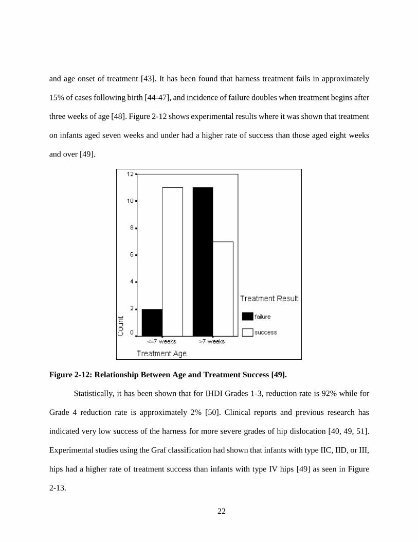
and age onset of treatment [43]. It has been found that harness treatment fails in approximately
15% of cases following birth [44-47], and incidence of failure doubles when treatment begins after
three weeks of age [48]. Figure 2-12 shows experimental results where it was shown that treatment
on infants aged seven weeks and under had a higher rate of success than those aged eight weeks
and over [49].
Figure 2-12: Relationship Between Age and Treatment Success [49].
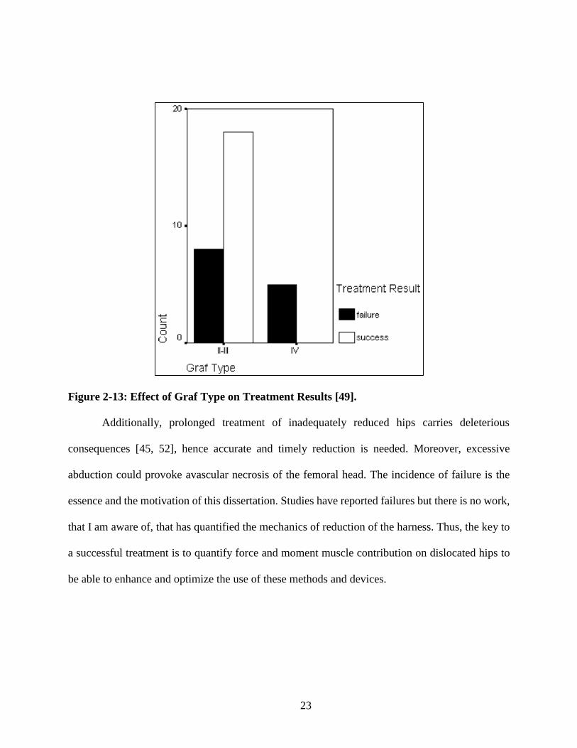
Statistically, it has been shown that for IHDI Grades 1-3, reduction rate is 92% while for
Grade 4 reduction rate is approximately 2% [50]. Clinical reports and previous research has
indicated very low success of the harness for more severe grades of hip dislocation [40, 49, 51].
Experimental studies using the Graf classification had shown that infants with type IIC, IID, or III,
hips had a higher rate of treatment success than infants with type IV hips [49] as seen in Figure
2-13.
23
Figure 2-13: Effect of Graf Type on Treatment Results [49].
Additionally, prolonged treatment of inadequately reduced hips carries deleterious
consequences [45, 52], hence accurate and timely reduction is needed. Moreover, excessive
abduction could provoke avascular necrosis of the femoral head. The incidence of failure is the
essence and the motivation of this dissertation. Studies have reported failures but there is no work,
that I am aware of, that has quantified the mechanics of reduction of the harness. Thus, the key to
a successful treatment is to quantify force and moment muscle contribution on dislocated hips to
be able to enhance and optimize the use of these methods and devices.
24
2.2 Lower-Limb Bone Biomechanics
Mechanically, the hip joint represents a ball and socket joint where the connection between
the femoral head and the acetabulum in the pelvis provides stability in the human body. Acting in
concert with their corresponding skeletal muscles, the hip joint enables locomotion providing a
wide range of motion. Extensive work has been done to quantify forces and moments during
routinely physical activities. Specifically, we are interested in covering the hip joint physiology
and pathology factors that will affect the biomechanics of the lower limb when developmental hip
dysplasia is present.
The biomechanics of the lower limb has been studied in the past to understand
abnormalities such as osteoarthritis and to plan orthopaedic procedures using analytical and
imaging techniques [53-56]. Maquet, based on Pauwels’ theories [56], studied hip dysplasia
mechanics on adults and children while walking [53]. He used the center of gravity of the mass of
the body supported by the hip (Ss), the force exerted by the mass of the body supported by the joint
(K), the lever arm of K (h’), the force exerted by the abductor muscles (M), and the lever arm of
M (h) to find the force transmitted across the joint (R) as seen in Figure 2-14. Additionally, he
looked at different cases of dislocation and observed that besides the muscular imbalance caused
by dislocations, the articular pressure in the hip increases and could potentially increase the risk to
develop osteoarthritis. Bombelli followed a similar approach using a radiographic analysis [55].
He suggested that mechanical forces exerted in the hip joint can be estimated using radiographs
and comparing deviation of force magnitudes and directions from healthy hips to assess different
types of osteoarthritis. Altogether, these studies indicate that hip mechanics are governed by the
anatomical architecture of the hip joint bones and the response of the skeletal muscles.

25
Figure 2-14: Forces Exerted on a Loaded Hip while Walking [53, 56]
The femoral head shape has been identified as one of the contributors that could potentially
affect the biomechanics of the hip joint. However, anatomic studies have shown that the shape of
the femoral head is nearly hemispherical around the time of birth as seen in Figure 2-15. These
results will allow us later on to use the assumption that for neonates less than twelve week old the
femoral head remains spherical during hip joint locomotion.
Figure 2-15: Changes in the Shape of the Femoral Head in Relation to Age. From Left to
Right: 21-week fetus, 30-week fetus, Premature Newborn, 2 Years, 4 Years [23].

26
Figure 2-16
Figure 2-16: Murphy’s Method for Measuring Femoral Anteversion Angle [57]

27
Figure 2-17
Figure 2-17: Femoral Anteversion Classification: Normal (Left), Increased Angle (Center),
Retroversion (Right) [58].
Figure 2-18
Figure 2-18: Femoral Anteversion in Relation to Age: (1) Hip with Developmental Dysplasia
of the Hip and (N) Healthy Hips [59].

28
Bones are composed of cortical bone, trabecular bone, and cancellous bones. All these
regions within the bone are critical for accurate modeling of hip mechanics. However, for the work
developed in this dissertation, mechanical properties of the different regions of the bone are not
accounted for and therefore not discussed further. Although relevant for biomechanics study, it is
assumed that deformations in infant bones are very small compared to the deformations of skeletal
muscles. Thus, bones are modeled as rigid bodies. In continuum mechanics, physical bodies are
classified as either rigid or deformable. A deformable body will present elastic behavior and a rigid
body, which is a mathematical and physical idealization, will not deform and will maintain a
constant distant between any two points within the body. This idealization will hold its validity for
very small deformations relative to the object size.
Having described hip biomechanics and developmental dysplasia of the hip, the next
chapter will introduce the mechanics of skeletal muscles. In conjunction, Chapter 2 and 3 should
provide sufficient information to successfully construct a neonate model to elucidate the
biomechanics of developmental dysplasia of the hip.

29
MECHANICAL BEHAVIOR AND MODELING OF
SKELETAL MUSCLES
The previous chapter provided essential details of lower limb bone biomechanics, but in
order to properly elucidate the biomechanics of developmental hip dysplasia, a modest
understanding of muscle mechanics is needed. After all, within the human body, the kinematics
and dynamics of locomotion are governed by muscle responses. In the human body, muscle tissues
are categorized in three types: skeletal, cardiac, and smooth. Skeletal muscles or “voluntary
muscles”, which provide global mobility by acting in concert, are bundles of muscle tissue
controlled by the neuro-central system and are attached to the bony skeleton. Cardiac muscle,
often referred as “involuntary muscle”, is only found in the heart and it is fascinating in nature
since it is the only tissue that can contract without stimulation of the neuro-central system. Smooth
muscle, also referred as “involuntary muscle”, is found in the walls of certain organs and their
function is to force continua through biological channels. The work developed on this dissertation
to model the biomechanics of developmental dysplasia of the hip mainly deals with the forces and
moments developed by muscles in the lower limb. Thus, this chapter will be focused on the
analysis and study of skeletal muscles in the lower limb of the human body.
The biomechanics of the skeletal muscles has been studied and explored by several authors
using novel approaches. Muscle biomechanics has been explored since the Renaissance when
Giovanni Borelli [60] started investigating animal movement using machine analogies and
mathematics as seen in Figure 3-1. For instance, Borelli linked heart behavior to the one observed
in a piston allowing him to derive the elastic behavior in arteries. It is important to acknowledge
that studying muscle mechanics has various limitations mainly due to the complexity of the

30
muscle-tendon unit. There are a lot of variables and limitations, which has made researches take
particular approaches when it comes to the understanding of muscle mechanics. Vast research has
been done in the area of muscle mechanics, but these studies are scattered and most of it has not
been fully organized yet.
Figure 3-1: Borelli’s Classic Work on Muscle Mechanical Behavior [60].
The purpose of this section is to provide a comprehensive analysis of muscle functionality
and its structure based on previous research. Section 3.1 addresses muscle anatomy and the
significance of mechanical features within skeletal muscles and also emphasizes skeletal muscle
functionality. Section 3.2 will present various methods that have been used to realistically capture
the mapping of a muscle path. Finally; section 3.3 will present the mechanical modeling of muscles
and specifically it will introduce the use of Hill’s muscle model, which has become the standard

31
for studies of muscle biomechanics. In addition, the last section of this chapter will also establish
the basis to accurately reproduce skeletal muscle behavior action under passive tension. Muscle
passive behavior, commonly known as relaxation, has been treated using distinct constitutive
models developed which will be emphasized in the last section of this chapter. By understanding
the behavior of relaxed muscles, scientists have been able to successfully simulate complex muscle
systems and study muscle processes associated to muscle damage and/or fatigue, as well as muscle
pathologies. Skeletal passive muscle response is of paramount importance on this dissertation, as
it will be explained later on this chapter, and precise force and moment quantification is needed to
elucidate the biomechanics of developmental dysplasia of the hip.
3.1 Skeletal Muscle Anatomical Structure and Physiology
This section will present biomechanically relevant anatomical features of skeletal muscles,
which are necessary to understand force generation and movement in the human body.
Mechanically, muscles can be considered biological motors which exert force and produce
mechanical work, where the coupling of muscles and tendons is modeled as force transmitters and
shock absorbers. The architecture of skeletal muscle exhibits important characteristics at multiple
scales and can be studied at the microscopic and macroscopic level. In general muscles are made
up of fiber bundles, called fascicles, and muscle fibers are muscle cells made up of small myofibrils
which contain filaments.
At the microscopic level muscle fibrils or myofibrils, which are composed of serially
connected elements called sarcomeres, represent the main mechanism of total muscle contraction.
A sarcomere is the smallest contractile unit and is composed of a thin and a thick filament as shown

32
in Figure 3-2. Muscle contraction is then caused trough a chemical reaction produced by
stimulation of the neuro-central system initializing the cross-bridge cycle, with the heads of the
myosin (thick filament) connecting to the actin (thin filament). Later on this chapter, the
mechanism of voluntary contraction will be explained to expose the concept of activation in
skeletal muscles.
Figure 3-2. Myofibril and Sarcomere Architecture [61].
At the macroscopic level, the muscle fibers arrangement in skeletal muscles is responsible
for loads that are mechanically transferred in the musculoskeletal system. In general this
arrangement is composed of skeletal muscle tissue, nervous tissue, blood, and connective tissue.
Particularly, connective tissue, which is made of collagen, adds the needed strength to keep
muscles together. The network of connective tissue in the skeletal muscle is comprised of the
epimysium, the perimysium and fascicle, the endomysium, and the fascia. Additionally, two other
relevant connective tissues are the tendon, which is known to store potential energy and connects
the skeletal muscle to the bone, and the aponeurosis, which connects external tendon to muscle

33
fiber and connective elements within the muscle fiber. A general representation of the muscle
elements arrangement and its architecture is provided in Figure 3-3. Macroscopic modeling of
skeletal muscle lays the foundation of the work developed on this dissertation as it can describe
skeletal muscle response in a relaxed state.
Figure 3-3: Muscle Structural Elements [62].
Defining muscle attachments also has mechanical relevance in force and moment
generation. Muscle attachments can be defined either as origins or insertions and mechanically
they can be model as points, lines, or surfaces. The immovable end of the muscle is designated as
the origin and the movable end as the insertion. During skeletal muscle contraction, the insertion
pulls towards the origin. For the lower limb, origins are located in the pelvis and insertion in the
femur and tibia.

34
Another important muscle characteristic required for the studies on this dissertation is the
definition of a physiological cross-sectional area (PCSA). The significance of this property lies in
the fundamental fact that PCSA is directly proportional to the force generated in the muscle unit
as it represents an area orthogonal to the fibers direction. Common sense tell us this area changes
along muscle path and estimating its value might represent a difficult task. However, the PCSA
can be estimated by capturing all the cross-sectional areas along the muscle length. Experimental
anatomical studies have verified that a representative PCSA [63, 64] can be estimated using the
total muscle volume and the fiber length applying the following expression:
𝑃𝐶𝑆𝐴 =
𝑀𝑢𝑠𝑐𝑙𝑒 𝑉𝑜𝑙𝑢𝑚𝑒
𝐹𝑖𝑏𝑒𝑟 𝐿𝑒𝑛𝑔𝑡ℎ (1)
Overall, muscle attachments, PCSA and muscle fiber length dictates the biomechanics of
skeletal muscles since fiber length is proportional to muscle excursion while physiological cross-
sectional area is proportional to maximum muscle force as seen in Figure 3-4.
Figure 3-4: Lower Limb Muscles Fiber Length and Physiological Cross-Sectional Areas [65].
35
Muscle mechanics have been studied in vivo, in situ, and in vitro; but in most cases not all
measurements needed could be obtained due to several limitations [66]. For instance, it is very
difficult to maintain a muscle’s biological life when they are isolated from the body for longs
periods of time. In the past, extensive experimental work has been performed on frogs and toads
mainly due to amphibians’ characteristics, which allows to keep their mechanical properties for
several hours when place in a physiological solution. It is also well known amphibian’s muscle
mechanical properties are similar to human muscles but at the same time similarities are limited.
Therefore in order to validate similarities and dissimilarities of amphibian models, experiments on
human muscles are needed. Engineers and scientists have also assumed muscles possess
mechanical properties similar to common composite materials used in engineering applications.
Using this unique approach, muscles could then be modeled as a fiber-reinforced composites [67]
where muscle fibers are made of high tensile strength material and are embedded in another
material called matrix. As explained before, adjoining connective tissues hold muscles together.
Therefore the connective tissue could be considered the muscle matrix and the muscle fiber could
be considered the fibers in the matrix.
To summarize, this section have provided a detailed grasp of skeletal muscle architecture-
functionality and its relevance on the study of muscle mechanics at the microscopic and
macroscopic level. The basic familiarity we have created until this point will allow us to link this
knowledge to the next two sections where skeletal muscle path representation and mechanical
models of skeletal muscles are discussed.

36
3.2 Skeletal Muscle Path Representation
To accurately quantify force and moment transmission in muscles, it is imperative to map
paths that will properly represent lines of action. In addition, muscle path provides necessary
information relevant to study of muscle mechanics such as muscle moment arm and muscle length.
This section provides a comprehensive analysis of several methods which has been developed and
used to generate muscle path representation. Namely, the straight-line representation, the centroid
muscle representation, and the curved and wrapping muscle representation are presented in this
section to understand the benefits, challenges, and limitations of each method. All the different
approaches presented have been theoretically and experimentally validated for various
musculoskeletal architectures. Additionally, implementation of these techniques should be based
on muscle geometry and its anatomical landmarks. Once a muscle path has been established,
muscle mechanical behavior could be modeled using kinematics and dynamic analysis.
3.2.1 Straight-line Skeletal Muscle Path Representation
This particular method allows to join the geometric centers of muscle attachments areas,
namely the origin and insertion points, using a straight-line. The straight-line muscle path
representation (SLMPR) allows for straightforward estimation of muscle moment arms and forces
using classic methods of vector analysis [66]. The SLMPR is without a doubt the easiest way to
model a muscle path but at the same time it has its limitations. There are specific sets of skeletal
muscles where the straight-line approach can be implemented since some skeletal muscles may
wrap around bones, diverging from a straight-line of action behavior. Dostal and Andrews [68]
have stablished proper guidelines to accurately define skeletal muscles in the lower limb, where

37
the SLMPR can be successfully applied. Their experiment showed for several ranges of hip joint
angles in the lower limb, various skeletal muscles can be modeled using the SLMPR. Their
benchmark experiment modeled skeletal muscles as fixed elastic strings attached to their origin
and insertion attachment points as seen in Figure 3-5. The interaction between strings with bones
and/or other strings was then closely observed to determine valid ranges where the straight-line
approach will yield to accurate force and moment estimation. Furthermore, this experiment
reported three important outcomes: (1) it provided hip joint angles values where the SLMPR is
valid, (2) it showed skeletal muscle SLMPR limits depend on hip joint angle, and (3) it presented
skeletal muscles that should never be modeled using a SLMPR at any hip joint angle.
Figure 3-5: Hip Musculature Elastic Straight-Line String Model [68]
When applicable, the SLMPR has been used to predict how muscle arm moment, which is
defined by the perpendicular distance from a joint center to its respective muscle line of action,
varies relative to the shortest distance between the muscle line of action and the joint center. Figure
3-6 shows a geometric representation of the elbow joint of the brachialis (BRA) and the
brachioradialis (BRD) where a triangle is formed by muscle’s origin and insertion points and the

38
joint center (JC). The triangle formed by both the BRA and BRD clearly shows that the maximum
value of moment arm (ma) is the shorter distance (Ds) between the joint center and a muscle’s
attachment points on the bone. Based on this simple two-dimensional model of the elbow joint, it
has been proved theoretically and experimentally that a closed relation exists between the shortest
attachment and muscle moment arm [69, 70].
Figure 3-6: Two-Dimensional, Geometric Representation of Elbow Joint Using a SLMPR of
the Brachialis (BRA) and the Brachioradialis (BRD) [69].
This study shows that muscle length on its own is not a good predictor for peak moment
arm and instead Ds values and joint dimension should be used to properly quantify it. Furthermore
Ds could be also used to properly normalize and scale available experimental muscle moments arm
data obtained from different human specimens.

39
3.2.2 Centroid Model of Skeletal Muscle Path
Modeling skeletal muscles using a muscle centroid path provides a better geometrical
representation of muscle’s line of action but at the same time its implementation requires intrinsic
parameters which are not easy to obtain. The method can be successfully implemented by
arranging the centers of muscle cross-sectional areas, obtained from a number of segments along
the muscle, to form a geometrical path. This procedure however requires cross-sectional
information of muscles, which could be obtained from imaging or experimental procedures. The
challenge is to use intervals small enough to accurately map the geometrical muscle line of action.
As explained previously, the effective application of the centroid technique yields to a better
representation of muscle’s lines of action but it also comes with limitations. For instance once a
path is mapped, the centroid muscle path represents a given muscle length, meaning a new centroid
muscle path is defined by changes in muscle length or joint motion. Additionally, it is known that
centroid muscle path representation neglects muscle pennation angle, which is a critical value
needed to precisely quantify forces and moments of some muscles where pennation is relevant.
Several experiments have been performed on skeletal muscles to tabulate muscle centroid data and
to show numerical differences between the centroid path technique and the SLMPR [71]. Figure
3-7 shows experimental data obtained for the gluteus medius using the muscle centroid approach
where it is visible, that for this particular muscle the SLMPR will not yield to accurate muscle
force and moment quantification.

40
Figure 3-7: Centroid-Line Representation of Gluteus Medius and Femur [72]
3.2.3 Modeling of Curved and Wrapping Skeletal Muscles
Within the musculoskeletal system, there are muscles that are curved and muscles that wrap
around anatomical constraints such as bones or other muscles as shown in Figure 3-8.

41
Figure 3-8: Psoas and Gluteus Maximus Muscle-Path Representation Using Curved
Segments Wrapped Around Multiple Surfaces [73].
Similar to the centroid muscle path, curved muscle path representation requires additional
parameters besides the standard required definition of origin and insertion points. Curved muscles
could be accurately mapped by using straight-line or curve segments joined through a collection
of specific geometrical data points. However, the modeling of the curve muscles’ line of action is
strenuous due to muscle length changes during concentric contraction. On the other hand,
elucidating muscle wrapping is a challenging task as it adds the mechanical interaction of muscles
with its surroundings. To account for muscle wrapping, simple one dimensional models have been

42
developed to define muscle line of action as it connects to the bone [74]. In addition, numerous
novel mathematical methods and imaging techniques have been proposed and used to account for
intricate muscle wrapping [54, 73, 75]. Particularly, these methods are computationally expensive
and often call for optimization techniques [76] to improve the time required to refine wrapping
objects. Once wrapping path has been established, the kinematics of wrapping muscle are easy to
compute and could be resolved using classical kinematics theory. The dynamics, however, become
laborious as muscle contact with other anatomical landmarks adds complexity to the mechanism.
Thus, mechanical contact definition is of paramount importance to quantify muscle forces and
moments. For instance, modeling friction becomes a critical constituent on muscle wrapping as it
will considerably affect the dynamics of the problem. Modeling such contact constraints is not
always a straightforward procedure since most of these physiological behavior have not been
quantified mechanically.
3.3 Mechanical Modeling of Muscles
Once muscle paths are developed, muscle mechanical behavior can be analyzed using well
known mechanical models such as the classic Hill’s model [77]. Hill developed this lumped-
parameter idealized force generator model based on experimental observations on isolated muscle
fibers. His simplistic approach has remained as the standard technique to analyze muscle
dynamics. The model uses three elements; a contractive element (CE), a series element (SE), and
a passive element (PE) to estimate total muscle force generation (FM) as shown in Figure 3-9.

43
Figure 3-9: Hill-Type Muscle Model
The contractile element (CE) models muscle activation, mainly driven by forces developed
at the sarcomere level during cross-bridge formation and is characterized by tension-length and
force-velocity muscle behavior. The cross-bridge formation mechanism, which is the interaction
between the actin and the myosin heads due to neuromuscular excitation, are beyond the scope of
this dissertation and therefore not explained in detail on this chapter. However, vast information
can be found in the literature describing the biomechanics of muscle contraction at the molecular
level [65, 78-80]. Ultimately, the contractile element allows muscle shortening when activated and
enables free shortening or lengthening when no activation is present.
In general, the series element (SE) models the compliance of tendons, aponeurosis, and
myofilaments. This compliance enables rapid change of the muscle from an inactive to an active
state. In addition, the SE acts as an energy storing mechanism. The properties of the SE yields to
non-linear spring behavior and hence the spring representation in Hill’s model. Considering the

44
nature of the series connection between the SE and CE, the shortening of the CE will always cause
a stretch of the SE to keep the total muscle length. This particular scenario can be observed during
isometric contraction.
The parallel element (PE) models the passive force exerted by connective tissue such as
epimysium, perimysium, endomysium, and fascia. The PE is arranged in parallel with the SE and
CE and similar to the SE, the PE’s passive response exhibits non-linear behavior. When activation
is not present and a muscle is stretched, the PE is exclusively responsible for the muscle passive
behavior. Chapter 4 will reveal how this passive response is relevant to the study of developmental
dysplasia of the hip.
Once the contribution of each the components in Hill’s circuit has been estimated, total
muscle force can be computed using the force exerted by each of the elements:
𝑭𝑴 = 𝑭𝑷𝑬 + 𝑭𝑺𝑬
𝑭𝑪𝑬 = +𝑭𝑺𝑬
(2)
The classic Hill model is controversial and there is vast discussion on the appropriate
utilization of the circuit. Specifically, the literature has debated its limitations, simplicity, and
complexity [81]. To this end, muscle models have been developed on a case basis where
assumptions are adequate to represent muscle behavior and match experimental observations.
Therefore, studies have been performed to yield modified Hill muscle models such as
dimensionless lumped models and viscoelastic models [82-85]. Hill models have suggested that
muscles generally develop two types of forces: active and passive. The contractile element
contributes to muscle active tension, while the series element and the passive element contribute

45
to the muscle passive tension. Subsequently, for a fully activated muscle the tension-length will
include a passive and an active component as shown in Figure 3-10.
Figure 3-10: Muscle Tension-Length Curves Exhibiting Active and Passive Tension Behavior
3.3.1 Active Muscle Behavior
Muscle activation, which causes voluntary movement, represents a complex phenomenon.
It all starts with a signal sent from the neuro-central system to skeletal muscles in order to achieve
lengthening or shortening of muscles. As a rule of thumb, activation force generation decreases
when shortening speed increases or vice versa. Thus, muscle tension during activation depends on
two parameters: length and velocity. Analyzing force generation during regular contraction cycles
represents a challenge due to the simultaneous changes of length and velocity. However,
experimental studies have been performed under controlled conditions to analyze muscle active

46
tension at constant velocities and muscle active tension at constant lengths [65, 86]. Obviously,
analyzing muscle activation using this approach represents unrealistic conditions but looking
closely one can superposition both functions to develop a force relationship for length and velocity
as seen in Figure 3-11. Although relevant for the study of muscle biomechanics, muscle activation
is not utilized in the development of this dissertation and therefore is briefly explained in this
section. Chapter 4 will reveal that hip reduction often occurs during relaxation, meaning passive
muscle tension, besides gravity, is the sole mechanical force exerted in skeletal muscles.
Figure 3-11: Generalized Relationship between Muscle Length, Force, and Velocity during
Muscle Activation [86].

47
3.3.2 Passive muscle Behavior
When muscle activation is not present, skeletal muscle lengthening occurs mainly by the
stretching of passive fibers. It is known that “when a relaxed skeletal muscle is stretched beyond
its resting length, it behaves as a deformable body; that is, it deforms and provides passive
resistance to the stretch. The resistance does not require metabolic energy and, hence, is called
passive” [66]. Furthermore, Hill’s model explicitly shows that the contractile element does not
contribute to muscle forces developed in a passive state influenced mainly by the response of the
series and the parallel elastic components [87, 88]. Research studies have suggested passive
tension force is mainly due to connective tissue, namely the endomysium, the perimysium, and the
epimysium but some authors suggests that most of the contribution might come from the myofibers
[89] and some other studies have suggested passive tension contribution is due to stretching of the
fascia [90].
Quantification of passive muscle response is critical to the study of both passive and active
skeletal muscle behavior. Measuring activation forces directly from skeletal muscles represents a
difficult task and has been attempted in the past without success. To overcome this obstacle, a
straightforward and conservative approach has been suggested involving the use of total muscle
force and passive force [91]. Such fundamental framework has been established using Equation
(2) to estimate activation force contribution by subtracting total muscle force from passive force.
Passive muscle response has a soft tissue mechanical behavior similar to the found in all
biological tissues. Furthermore, the physics underlining passive muscle response have shown its
behavior does not obey Hooke’s law and therefore muscle, and living soft tissue in general, will
yield to non-linear deformations. Biological experimental numerical results [92, 93], as seen in
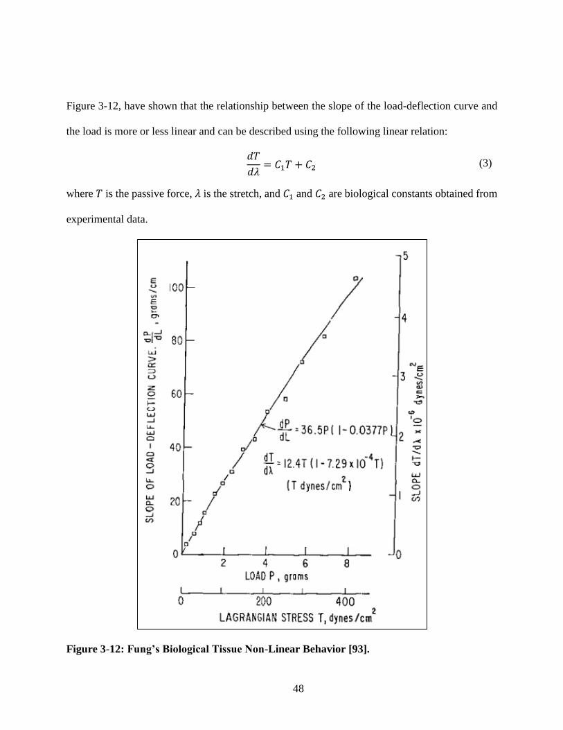
48
Figure 3-12, have shown that the relationship between the slope of the load-deflection curve and
the load is more or less linear and can be described using the following linear relation:
𝑑𝑇
𝑑𝜆= 𝐶1𝑇 + 𝐶2 (3)
where 𝑇 is the passive force, 𝜆 is the stretch, and 𝐶1 and 𝐶2 are biological constants obtained from
experimental data.
Figure 3-12: Fung’s Biological Tissue Non-Linear Behavior [93].

49
Moreover Equation (3) can be integrated, using conservative boundary conditions, to yield
a non-linear exponential relationship between the applied load and stretch:
𝑇 =
𝐶2𝐶1(𝑒𝐶1(𝜆−1) − 1) (4)
Equation (4), describes the standard exponential relationship that has been commonly used
to describe soft connective tissue mechanical behavior. For the purpose of this work the
aforementioned relationship will be used to describe a musculo-tendon actuator driven by the
responses of the parallel and the series elastic elements. The information provided in this chapter
should be sufficient not only to understand the biomechanics of skeletal muscles but also to
implement such models based on application. Chapter 4 will provide details and assumptions for
accurate muscle modeling of the skeletal muscles in infants affected with developmental dysplasia
of the hip.

50
METHODOLOGY
The study of muscle mechanics is currently governed by biological experiments and
mechanical models. While traditional approaches have used average and scaled anatomical data,
it is important to recognize that one of the most important and challenging limitations of analyzing
the human musculature, and the human body in general, is the fact that no individual has an
identical anatomy. The kinematic and dynamics of locomotion strongly depends on mechanical
interaction of the muscles with nearby bones, other muscles, and/or other connective tissues.
Therefore, the most important task is to define morphometry and anatomical landmarks of bones
and muscles accurately, which means there is a critical need to define the anatomical
characteristics of muscles and bones, such as origins, insertions, and moment arms. For that
purpose, a realistic model is needed to accurately investigate the biomechanics of the lower limb
model. Additionally, it is known that developmental dysplasia of the hip is more common in
females than males, with a female incidence of about ninety percent. Therefore, a ten-week old
female infant was selected for the biomechanical studies on this dissertation. The three-
dimensional model of the pelvis-femur-lower limb of the representative ten-week old female infant
was generated based on the composite of CT-scans and MRIs of a six-month and a fourteen-year
old female, as well as the Visible Human Project with the aid of anthropomorphic scaling of
anatomical landmarks.
This chapter presents details of a successful patient-specific model reconstruction, which
was used to investigate the forces and moments occurring on dislocated hips using the Pavlik
harness. To that end, this chapter will introduce the following sections: section 4.1 will present the
reconstruction of an idealized and representative patient-specific lower limb of a ten-week old

51
female infant; section 4.2 will explain how the reconstructed anatomy was assembled; section 4.3
will describe the implementation of passive muscle models in lower limbs; section 4.4 will provide
a brief description of the Rigid Body Dynamics approach used to elucidate the kinematics and
dynamics of lower limbs; section 4.5 will justify the use of a frictionless model on the hip joint;
and section 4.6 will propose an innovative approach to calibrate the anatomical model to account
for infant lower limb architecture.
4.1 Lower Limb Anatomical Reconstruction
The anatomical lower limb model was reconstructed using the aid of medical segmentation
packages: Mimics and 3-matic (Materialise Inc., Plymouth, MI). The lower extremity was
composed of the hip, femur, tibia, fibula, ankle, foot, and toe bones. It must be mentioned that
symmetry was assumed along the sagittal plane, which divides the body into right and left portions
(x-y plane). Thus only the right lower limb was modeled.
Due to the presence of large cartilaginous zones, the MRIs and CT-scans of a neonate offer
a challenge in the reconstruction of the complete anatomy based on medical segmentation software
alone. Moreover, retrospective MRIs and CT-scans of a subject with developmental dysplasia of
the hip focus mainly on the hip region, and therefore, do not provide the required geometric
information of the rest of the anatomy. As such, a model was constructed from the composite of
Ct-scans of a six-month and fourteen-year old female, as well as the Visible Human Project with
the aid of anthropomorphic scaling of anatomical landmarks in order to assemble the ten-week old
infant model. It is also well-known that bone growth is not proportional in all directions [94], thus,
anisotropic parameters were used for all of the lower limb bones.

52
For hip reconstruction, the starting point was to obtain rough bone geometry contours from
CT-scans of a six-month old female infant and then superimpose it on well-defined bone geometry
obtained from high quality CT-scan data of a fourteen-year old female [95]. Both the six-month
old female infant and the fourteen-year old female were superimposed so the iliac spines and
acetabuli were aligned as shown in Figure 4-1.
Computational frameworks for simulating muscle mechanics are currently available [96-
98], but unfortunately, hip dislocations cannot be simulated using these commercial and research
packages.
Figure 4-1: Superposition of a Reconstructed Model of a Fourteen-Year Old Female (left)
with a model Rendered From CT-scans of a Six-Month Old Female Infant [95].
In addition, the superposition technique accounted for the hip widening that occurs in
females during puberty. After successful reconstruction, the model of the six-month old female
infant was scaled to that of a ten-week old female infant by the matching infant orthopedic data
found in Hensinger’s Standards in Pediatric Orthopedics [99]. The final pelvic anisotropic scaling
factors were determined to be: 0.35 in the x-direction, 0.3 in the y-direction, and 0.35 in the z-
direction. The following final measurements were reported for the acetabulum: acetabulum depth

53
was approximately 7.9 mm; acetabular depth-width ratio (ADR) was 45%; and acetabulum
diameter was approximately 17.5 mm.
High-quality Ct-scans and MRIs from the Visible Human Project [100] were used to
reconstruct the femur, tibia, fibula, ankle, foot, and toe bones. The geometry was reconstructed
using the superposition approach used for the pelvis. Scaling parameters for all the other bones of
the lower extremity were found to be: 0.23 in the x-direction, 0.22 in the y-direction, and 0.25 in
the z-direction. The femoral head had a diameter of 14 mm and was assumed to be perfectly
spherical as observed during the infant dissection shown in Figure 4-2.
Figure 4-2: Spherical Head in Neonates: Computational Model (Left) and Dissection (Right).
The femoral anteversion angle also plays a critical role on hip reduction since it will affect
muscle force and moment contribution while the Pavlik Harness is in use. This particular angle
defines the difference between the axis of the femoral neck and the transchondylar axis of the knee
as explained in chapter 2. This angle ranges typically from 30° to 40°, but can reach up to 100° as
seen in Figure 4-3.
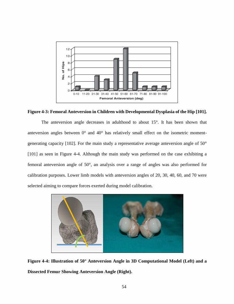
54
Figure 4-3: Femoral Anteversion in Children with Developmental Dysplasia of the Hip [101].
The anteversion angle decreases in adulthood to about 15°. It has been shown that
anteversion angles between 0° and 40° has relatively small effect on the isometric moment-
generating capacity [102]. For the main study a representative average anteversion angle of 50°
[101] as seen in Figure 4-4. Although the main study was performed on the case exhibiting a
femoral anteversion angle of 50°, an analysis over a range of angles was also performed for
calibration purposes. Lower limb models with anteversion angles of 20, 30, 40, 60, and 70 were
selected aiming to compare forces exerted during model calibration.
Figure 4-4: Illustration of 50° Anteversion Angle in 3D Computational Model (Left) and a
Dissected Femur Showing Anteversion Angle (Right).

55
4.2 Lower Limb Anatomical Assembly
After anatomic reconstruction of the lower limbs. Solidworks was used to assemble all
components as seen in Figure 4-5. To assemble the femur to the pelvis origins and orientations in
both bones were specified as defined in the literature [68].
Figure 4-5: Reconstructed Lower Limb of 10-Week Old Female Infant.
The origin of the inertial Cartesian reference frame was located at the center of the
acetabulum for the hip and at the center of the femoral head for the femur. Five adductor muscles
were identified as mediating muscles during close reductions: (1) Pectineus, (2) Adductor Brevis,
(3) Adductor Longus, (4) Adductor Magnus, and (5) Gracilis. The Adductor Magnus is a large
triangular muscle with extensive femoral insertion; consequently, it is commonly represented by
three effective components: (a) Adductor Magnus Minimus, (b) Adductor Magnus Middle, and (c)
Adductor Magnus Posterior. Origin and insertion points were defined using the anisotropic scaling
parameters previously mentioned on coordinate points obtained in experiments by Dostal and
Andrews on the lower extremity as seen in Figure 4-6. For this study, musculoskeletal tissue is

56
modeled using a straight-line muscle path representation since lines of action do not intersect in
the range of motion of interest, allowing to use classical methods of vector analysis [68].
Figure 4-6: Anterior View of Adductor Muscles Origin and Insertion Points [103].
Once the model was assembled, the hip was able to be computationally dislocated to match
physiological dislocations according to the dislocation grade under investigation as seen in Figure
4-7.
Figure 4-7: Hip Dislocation: Healthy Hip (Left), Grade 3 (Center), and Grade 4 (Right).
To account for proper weight distribution of muscles attached to the lower limbs,
calculations of the total body mass were based on a ten-week old female infant at the 50th length-

57
for-age percentile [104] as seen in Table 4-1. Centers of mass were also determined, thus achieving
accurate load and moment distribution in the model [105]. Gravity acts as the sole external driving
load in the dynamics model to be described later. Moreover, the constraints assigned to the three-
dimensional model serve to restrain the motion within the envelope realized with the Pavlik
harness during Grade 1-4 studies to constraint the knees in a hyperflexed configuration for Grade
4 studies.
Table 4-1: Lower Extremity Mass Distribution.
Total Body Mass (BM): 7.26 kg
Limb %BM Mass (kg)
Thigh 9.45 0.69
Leg 4.2 0.30
Foot 1.35 0.10
Full leg 1.5 1.09
4.3 Lower Limb Skeletal Muscle Modeling
To model the response of lower limbs muscles, proper assumptions are needed to
effectively elucidate the biomechanics of developmental dysplasia of the hip. Chapters 2 and 3
laid the foundations needed for understanding of developmental dysplasia of the hip biomechanics
and this section will use the background information previously presented to develop a realistic
muscle model for the adductors. It has been documented that reductions of developmental
dysplasia of the hip with the Pavlik harness occur passively with muscle relaxation in deep sleep
[106, 107]. In recent clinical studies, sleep was induced to attain muscle relaxation, and, in flexion
and abduction, towels adjusted to the appropriate height were placed under the knees to avoid

58
forcible abduction and it was clinically observed that under mild sedation and in natural abduction
and flexion, the spontaneous reduction with the Pavlik harness is reproduced [108]. This study in
particular further justifies the passive muscle model to be developed in this section. Chapter 3
revealed that skeletal muscles in general produce two kind of forces, active and passive, which
sum to compose muscle total force. The active component is due to the contraction of muscle fibers
that generate force and bear load, while passive components are due to stretching of muscle fibers,
the basal lamina, and the muscle fascia [109]. As previously explained, without the presence of
neuromuscular activation, muscle fibers and tendons are often considered mechanically in series
in skeletal muscle and its structural arrangement will yield similar forces in both. It is important to
acknowledge muscles and tendons, as well as connective tissue in general, display time-dependent
behavior. Thus connective tissue will present viscoelastic behavior and should be analyzed with
this concept in mind. Moreover, passive response is characterized as hyperelastic.
Chapter 3 presented well-known passive models [110, 111], which are extensively used for
biological soft connective tissue based on the experimental observation that such passive tissue
exhibits a fairly linear relationship between the stiffness and muscle stretch leading to an
exponential behavior of the force-stretch curve. A novel Hill-based model [82, 84] had been
proposed to estimate passive muscle force by normalizing it using a peak isometric muscle force
as shown in Equation (5):
𝐹𝑃𝐸 =
𝐹
𝐹𝑜𝑀 (5)
where is 𝐹𝑃𝐸 the normalized passive muscle force, 𝐹𝑜𝑀 is the peak isometric muscle force, 𝐹 and
is the total muscle force. It is also been reported that peak isometric force is proportional to its
physiologic cross-sectional area (PCSA) as seen in Equation (6):

59
𝐹𝑜𝑀 = 𝐶 (𝑃𝐶𝑆𝐴) (6)
where C is a biological constant and has a value of 25 N/cm2 [84] and was estimated to scale
physiologic cross-sectional areas that were measured in young adult cadavers to match moment
curves measured on young subjects. Furthermore, it has been reported [112] that 𝐹𝑃𝐸 can be
expressed as:
𝐹𝑃𝐸 =
𝑒𝐾𝑃𝐸(𝜆−1)/𝜀𝑜
𝑀− 1
𝑒𝐾𝑃𝐸− 1
(7)
where 𝐾𝑃𝐸 is an exponential shape factor with a value of 4.0 obtained from OpenSim (National
Center for Simulation in Rehabilitation Research, Palo Alto, CA) [96], 𝜀𝑜𝑀 is the passive muscle
strain with a value of 0.60 for young adults, and 𝜆 is the optimal muscle-fiber length (stretch).
Moreover, the stretch can be written as 𝜆 = 𝐿 𝐿𝑜⁄ with 𝐿 being the deformed length, and 𝐿𝑜being
the initial muscle length at rest, measured at 20° abduction and 120° flexion for newborns. This
flexion and adduction ‘neutral’ configuration accounts for the fact newborns maintain a posture
similar to the fetal positioning for the first few months after birth with the hips flexed in toward
the belly and wide apart. This initial muscle length at rest is also multiplied by a specified muscle
pre-stretch for the lower extremity muscles and its value is usually between 20-30%. By combining
Equations (5), (6), and (7), an expression can then be derived to obtain passive muscle force as
function of stretch as seen in Equation (8).
𝐹 = 𝑃𝐶𝑆𝐴[𝑎(𝑒𝑏(𝜆−1) − 1)]
{
𝑎 =𝐶
𝑒𝐾𝑃𝐸− 1
= 0.466𝑁
𝑐𝑚2
𝑏 =𝐾𝑃𝐸
𝜀𝑜𝑀= 6.667
(8)

60
To estimate physiologic cross sectional areas (PCSA), MRI data for the adductor brevis of
a ten-week old month female was used. A PCSA of 0.41 cm2 was computed by measuring the
volume of the adductor brevis and dividing it by its average length. A shape factor was then defined
as the ratio between an adult PCSA and the ten-week old female infant PCSA by using a PCSA of
11.52 cm2 of an adult’s adductor brevis [113]. The adductor brevis shape factor of 0.0036 was then
used to scale experimentally measured physiologic cross-sectional areas of the rest of the
adductors.
Table 4-2: Scaled Ten-Week Old Female Infant Physiologic Cross-Sectional Areas Using
Adductor Brevis Shape Factor [113].
f = 0.036 Friederich PCSA (cm2) TWOFI scaled PCSA (cm2)
Pectineus 9.03 0.321
Adductor Brevis 11.52 0.410
Adductor Longus 22.73 0.809
Adductor Magnus Minimus 25.52 0.908
Adductor Magnus Middle 18.35 0.653
Adductor Magnus Posterior 16.95 0.603
Gracilis 3.73 0.133
To elucidate the biomechanics of developmental dysplasia of the hip, a rigid-body
dynamics model was used. Thus, the lower limbs bones were assigned a rigid-body behavior. Such
assumption is proven to be valid since muscle forces developed in infants, especially under passive
tension, do not contribute significantly to their elastic deformation. On the contrary, muscle

61
response is relevant and its behavior is modeled using the straight-line approach explained in
Chapter 3. Moreover, its viscoelasticity and non-linearity are accounted for in the exponential
model previously presented.
Numerical rigid body dynamics was carried out using the ADAMS dynamics motion
analysis in Solidworks. The governing differential equations of motion provided in Equation (9) :
𝑀 �� + 𝛽𝜙𝑞
𝑇 − 𝐴𝑇𝐹 (𝑞, ��) = 0 (9)
are numerically solved subject to the natural constraints 𝜙 (𝑞, 𝑡) = 0 . The system mass matrix
is 𝑀, the vector of generalized coordinates for the displacements is 𝑞, the Lagrange multiplier
adjoining the gradient constraints 𝜙𝑞 is 𝛽, the set of configuration and applied motion constraints
is denoted by 𝜙, the matrix 𝐴𝑇 projects the applied force in the 𝑞 direction, and 𝐹 is the vector of
applied forces. The governing equations of dynamics described in Equation (9) are Newton’s laws
of motion augmented via a Lagrange multiplier by constraints. The force vector 𝐹 is in general a
function of displacement, linear in the case of a Hookean solid or non-linear as in our case as in
Equation (9), and a function of the time-derivative of the displacements in case of the presence of
damping. In this study, the Gear Stiff (GSTIFF) integrator was used, the Jacobian was updated at
each time-step, 25 sub-level iterations were taken per time-step, and the initial, minimum, and
maximum integrator step sizes were set to 1 x 10⁻⁴, 1 x 10⁻⁷, and 1 x 10⁻² respectively. Custom
functions were written in Solidworks for each of the muscles studied using the model described in
Equation (8) with their respective physiologic cross-sectional areas.

62
4.5 Hip Joint Lubrication
Physiologically, the hip joint is considered a synovial joint on which the bone ends are
covered by a slippery tissue called articular cartilage. Such joint bears diverse mechanical loads
during physical activities. Depending on activity, forces exerted in the hip joint can be modest or
large in magnitude. In humans, these actions can go from standing or sleeping to strenuous motions
such as running or jumping. Thanks to the complex lubrication mechanism in the hip joint, the
articular cartilage presents little wear regardless of physical activity and intensity under normal
circumstances. Such mechanism acts under low coefficients of friction, which for the articular
cartilage can vary from 0.002 to 0.04 [114]. These values can be then compared to common
engineering materials, as seen on Table 4-3, to develop an understanding of their impact in the
infant hip joint model.
Table 4-3: Friction Coefficients for Common Engineering Materials.
Materials Friction Coefficient
Articular Cartilage Cartilage, Tissue, Bones 0.002 - 0.04
Ice (0°C) Ice (0° C) 0.01 – 0.1
Diamond Diamond 0.1
Wood Wood 0.25 - 0.5
Steel Steel 0.8
Glass Glass 0.9 - 1.0
Rubber Rubber 1.16
Silver Silver 1.4

63
Ultimately, the hip joint in the ten-week old female infant can be modeled mechanically as
a multi-axial ball and socket that creates a frictionless bearing point as shown in Figure 4-8.
Moreover, for the present analysis the contact between surfaces was assumed to be frictionless
using a restitution coefficient of zero.
Figure 4-8: Ball and Socket Hip Joint Frictionless Model.
4.6 Lower Limb Calibration Model
Since the passive muscle model previously developed was specifically based on a young
adult, the constants a and b in Equation (9) needed to be recalibrated for a ten-week old female
infant and also to stiffen the exponential function to account for ligaments that start acting at the
limit of ranges of motion, namely, the iliofemoral, pubofemoral and ischiofemoral ligaments which
help to stabilize the hip joint. The muscle model was calibrated by calculating the factors that
allowed to precisely replicate the clinical observation that equilibrium is attained when infants are

64
supine and the hips are placed in abduction at 80° and 90° flexion. That is, in this position, the
passive action of the muscles holds the legs in place. Constants a and b were tuned in this model
until the equilibrium configuration was achieved by only using muscle passive tensions. For the
model with a femoral anteversion angle of 50°, constant a was found to be 0.0337 N/cm2 and b
was found to be 13.95. It is important to mention that there is no unique set of values for a and b.
However there is a minimum value requirement for b, which is needed to stiffen the curve in order
to achieve physically realistic equilibrium at 80° abduction. The constants a and b are inversely
related; while b needs a minimum value to allow the model to achieve realistic muscle behavior,
a, which is used to find final equilibrium, is found through iteration once b has been established.
The passive muscle force behavior for our calibrated ten-week old female infant model is presented
in Figure 4-9 where it observed that the exponential behavior becomes very pronounced after a
lambda (λ) value of about 1.35.
Figure 4-9: Ten-Week Old Female Infant Passive Force versus Stretch for Calibrated Model.

65
Figure 4-10 also shows a comparison of the force-stretch behavior between the ten-week old
female infant calibrated model and the young adult of the adductor brevis where it is clearly seen
our calibrated model stiffens faster once it get close to the limits of ranges of motion as expected.
After a close comparison between all the adductor muscles, it is found that all muscles display the
same behavior as the brevis.
Figure 4-10: Adductor Brevis Force-Stretch Comparison between Ten-Week Old Female
Infant and Young Adult Model.
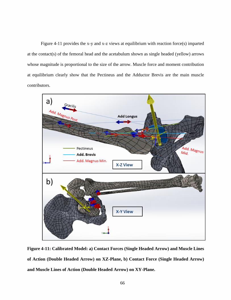
66
Figure 4-11 provides the x-y and x-z views at equilibrium with reaction force(s) imparted
at the contact(s) of the femoral head and the acetabulum shown as single headed (yellow) arrows
whose magnitude is proportional to the size of the arrow. Muscle force and moment contribution
at equilibrium clearly show that the Pectineus and the Adductor Brevis are the main muscle
contributors.
Figure 4-11: Calibrated Model: a) Contact Forces (Single Headed Arrow) and Muscle Lines
of Action (Double Headed Arrow) on XZ-Plane, b) Contact Force (Single Headed Arrow)
and Muscle Lines of Action (Double Headed Arrow) on XY-Plane.

67
Table 4-4: Muscle Force and Moment Contribution at Equilibrium Configuration.
λ
(Stretch)
Total Force
(N)
X-Moment
(Nm)
Y-Moment
(Nm)
Z-Moment
(Nm)
Pectineus 1.710 45.84 0.2834 0.3328 0.0738
Adductor
Brevis 1.523 15.01 0.1353 0.1342 -0.0165
Adductor
Longus 1.422 1.35 0.0138 0.0028 -0.0006
Adductor
Magnus
Minimus
1.392 1.83 0.0189 0.0013 -0.0014
Adductor
Magnus
Middle
1.316 0.84 0.0085 -0.0013 -0.0012
Adductor
Magnus
Posterior
1.229 0.33 0.0031 -0.0030 -0.0007
Gracilis 1.357 0.33 0.0040 0.0016 -0.0010

68
RESULTS
The lower extremity model of the ten-week old female infant was used to quantify force
and moment generation of the adductor muscles. It is known from clinical observation that
reduction is successful whenever the Pavlik harness is used on Grade 1 and Grade 2 dislocations
[40], thus these two grades were analyzed but values are not reported.
Figure 5-1 Figure 5-1
Figure 5-1: a) Grade 1 and b) Grade 2 Anatomical dislocations.

69
Force and moment contribution of the adductor muscles while the Pavlik harness is being
used are reported for Grade 3 and Grade 4. Results provided insight of their contribution to
reduction of developmental dysplasia of the hip on severe Grade 3 and Grade 4 dislocations
whenever the Pavlik harness is used and gravity is acting.
5.1 Grade 3 Dislocation Results
Figure 5-2
Table 5-1
Figure 5-2: Grade 3 Anatomical Dislocation.

70
Table 5-1: Muscle Force and Moment Contribution on Grade 3 Dislocation.
Grade 3
Dislocation
λ
(Stretch)
Total Force
(N)
X-Moment
(Nm)
Y-Moment
(Nm)
Z-Moment
(Nm)
Pectineus 2.009 2967.04 17.4055 20.1682 4.6945
Adductor
Brevis 1.658 105.68 0.9486 0.8630 -0.0649
Adductor
Longus 1.588 14.46 0.1479 0.0152 0.0052
Adductor
Magnus
Minimus
1.560 20.52 0.2135 -0.0082 0.0041
Adductor
Magnus
Middle 1.414 3.49 0.0359 -0.0083 -0.0026
Adductor
Magnus
Posterior 1.281 0.71 0.0068 -0.0071 -0.0011
Gracilis 1.417 0.77 0.0093 0.0032 -0.0020
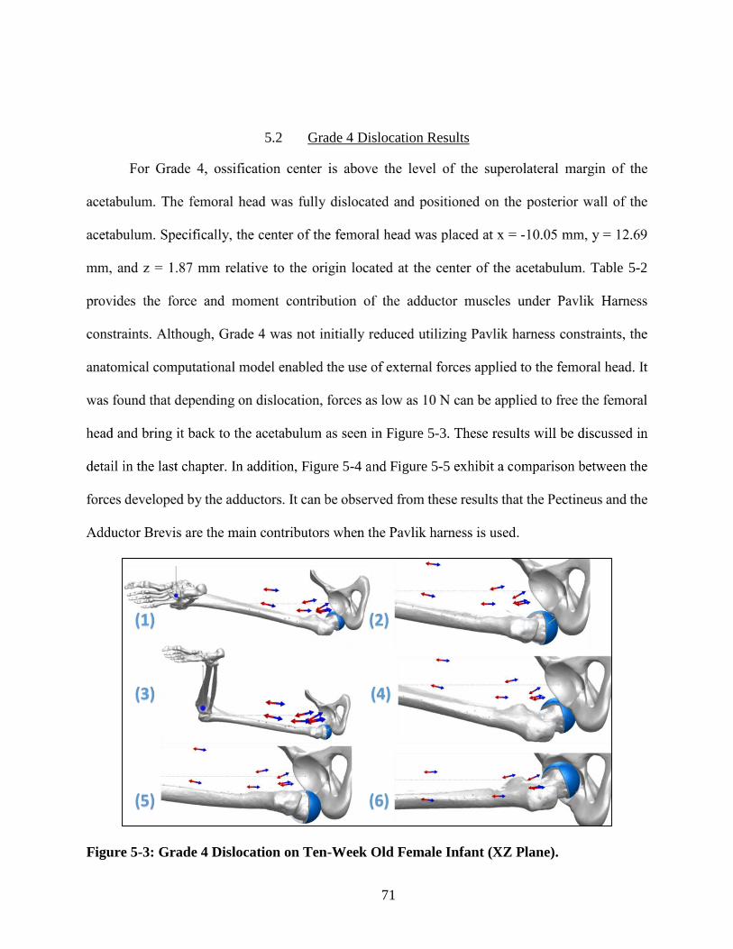
71
5.2 Grade 4 Dislocation Results
Table 5-2
Figure 5-3
Figure 5-4 Figure 5-5
Figure 5-3: Grade 4 Dislocation on Ten-Week Old Female Infant (XZ Plane).

72
Table 5-2: Muscle Force and Moment Contribution on Grade 4 Dislocation.
Grade 4
Dislocation
λ
(Stretch)
Total Force
(N)
X-Moment
(Nm)
Y-Moment
(Nm)
Z-Moment
(Nm)
Pectineus 2.153 2907.02 7.1963 12.1199 -0.3056
Adductor
Brevis 1.729 79.65 0.4442 0.4848 -0.0833
Adductor
Longus 1.667 11.78 0.0693 -0.0121 0.0175
Adductor
Magnus
Minimus
1.640 16.62 0.0974 -0.0394 0.0314
Adductor
Magnus
Middle 1.460 2.15 0.0162 -0.0075 0.0020
Adductor
Magnus
Posterior 1.299 0.39 0.0033 -0.0040 0.0001
Gracilis 1.432 0.42 0.0044 0.0014 -0.0009
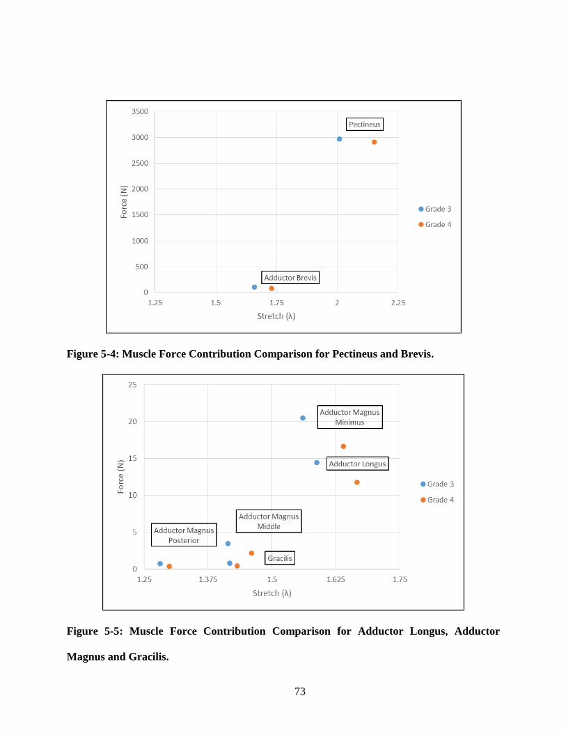
73
Figure 5-4: Muscle Force Contribution Comparison for Pectineus and Brevis.
Figure 5-5: Muscle Force Contribution Comparison for Adductor Longus, Adductor
Magnus and Gracilis.

74
CONCLUSION
As expected, Grades 1 and 2 were successfully reduced. This is consistent with previous
studies based on a synthetic model [51] as well as from clinical observation [40]. Grade 3 was
successfully reduced while Grade 4 dislocation was not. However, Grade 4 dislocation reduction
was achieved by an external rotation of the lower extremity followed by a semi-circular path taken
by the femoral head along the posterior wall of the acetabulum along the ischial plane, and the
subsequent entrance into the acetabulum via the acetabular notch as seen in Figure 6-1.
Figure 6-1: Grade 4 Dislocation on Ten-Week Old Female Infant (YZ Plane).
This path of reduction is generally consistent with Papadimitriou’s clinical studies [38].
These studies indicate methods to accomplish reduction by avoiding the surgical release of
adductor muscles. Papadimitriou’s clinical results showed reduction occur in three sub-phases: (1)

75
the femoral head travels from the grade 4 dislocated position arriving at the ischial tuberosity, (2)
the femoral head rides over the ischial tuberosity to the obturator foramen, and (3) the femoral
head departs from the obturator foramen an enters the acetabulum via the acetabular notch,
achieving reduction as explained in Figure 6-2. Moreover, Papadimitriou’s studies showed
successful reduction of severe dislocation by using this alternative hyperflexion procedure which
uses the Hoffmann-Daimler harness keeping the hip flexed at 120°.
Figure 6-2: Reduction by Driving Femoral Head Posterior to the Acetabulum in an Inferior
Direction.
In summary, a patient-derived geometry was successfully reconstructed to model the right
lower extremity of a ten-week old female infant. The model developed on this dissertation allows

76
for the detailed study of the biomechanics of developmental dysplasia of the hip and its treatment
with the Pavlik harness. Results suggest muscle force contributions on hip reduction are functions
of the severity of hip dislocation. Additionally, findings strongly indicate muscle tensions
contribute to reduction with the harness for subluxated and intermediate dislocations (Grades 1-
3). It is also notable that the force contributions of the Pectineus and Adductor Brevis favor
reduction significantly in Grades 1-3 while it disfavors reduction in Grade 4. The latter
dislocations, which have been considered irreducible by most physicians, could be reduced by
using a hyperflexion method. Further optimization of the Pavlik harness configuration may help
improve the effectiveness of treatment of developmental dysplasia of the hip. This multi-physics
computational approach is consistent with clinical observations reported in the literature and may
potentially provide physicians means to determine modifications to evaluate treatment that could
lead to new insights for non-surgical management of developmental dysplasia of the hip by passive
reduction with the Pavlik harness or in combination with other orthosis.
In addition, the work developed on this dissertation leaves the door open to include some
of the neglected assumptions to improve the lower limb computational model. For instance, the
absence of the abductor muscles, which may influence total force and moment contribution can be
included in the future. Moreover, the deformation of the non-ossified regions could be successfully
quantified by implementing a finite element method. Lastly, it is important to mention that the
functionality and design of the harness has not been modified in more than half a century. The
force and moment quantification method developed on this dissertation will not only optimize its
use but it will certainly lead to future development of a new harness based on patient-specific
information.

77
APPENDIX: PUBLICATIONS, PRESENTATIONS AND AWARDS
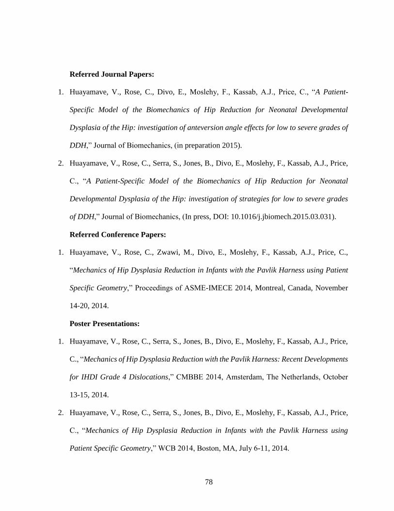
78
Referred Journal Papers:
1. Huayamave, V., Rose, C., Divo, E., Moslehy, F., Kassab, A.J., Price, C., “A Patient-
Specific Model of the Biomechanics of Hip Reduction for Neonatal Developmental
Dysplasia of the Hip: investigation of anteversion angle effects for low to severe grades of
DDH,” Journal of Biomechanics, (in preparation 2015).
2. Huayamave, V., Rose, C., Serra, S., Jones, B., Divo, E., Moslehy, F., Kassab, A.J., Price,
C., “A Patient-Specific Model of the Biomechanics of Hip Reduction for Neonatal
Developmental Dysplasia of the Hip: investigation of strategies for low to severe grades
of DDH,” Journal of Biomechanics, (In press, DOI: 10.1016/j.jbiomech.2015.03.031).
Referred Conference Papers:
1. Huayamave, V., Rose, C., Zwawi, M., Divo, E., Moslehy, F., Kassab, A.J., Price, C.,
“Mechanics of Hip Dysplasia Reduction in Infants with the Pavlik Harness using Patient
Specific Geometry,” Proceedings of ASME-IMECE 2014, Montreal, Canada, November
14-20, 2014.
Poster Presentations:
1. Huayamave, V., Rose, C., Serra, S., Jones, B., Divo, E., Moslehy, F., Kassab, A.J., Price,
C., “Mechanics of Hip Dysplasia Reduction with the Pavlik Harness: Recent Developments
for IHDI Grade 4 Dislocations,” CMBBE 2014, Amsterdam, The Netherlands, October
13-15, 2014.
2. Huayamave, V., Rose, C., Serra, S., Jones, B., Divo, E., Moslehy, F., Kassab, A.J., Price,
C., “Mechanics of Hip Dysplasia Reduction in Infants with the Pavlik Harness using
Patient Specific Geometry,” WCB 2014, Boston, MA, July 6-11, 2014.

79
3. Huayamave, V., Divo, E., Kassab, A.J., “An Optimization Approach for Hip Dysplasia
using the Pavlik Harness,” SHPE 2011, Anaheim, CA, October 26-30, 2011.
Invited Talks:
1. Huayamave, V., “Mechanics of Hip Dysplasia Reduction in Infants,” SHPE 2014, Detroit,
MI, November 5- November 9, 2014.
2. Huayamave, V., and Rose, C., “Mechanics of Hip Dysplasia Reduction in Infants with the
Pavlik Harness using Patient Specific Geometry,” POSNA 2014, Hollywood, CA, April
30- May 3, 2014.
Awards:
1. McKnight Dissertation Fellowship, Florida Education Fund (FEF). 2014
2. ASME International Mechanical Engineering Congress and Exposition (IMECE).
Technical Paper Competition Award. 2014.

80
LIST OF REFERENCES
1. Cohen, E.R., J. Feinglass, J.H. Barsuk, C. Barnard, A. O'Donnell, W.C. McGaghie, and
D.B. Wayne, Cost savings from reduced catheter-related bloodstream infection after
simulation-based education for residents in a medical intensive care unit. Simulation in
healthcare, 2010. 5(2): p. 98-102.
2. Gardner, R., T.B. Walzer, R. Simon, and D.B. Raemer, Obstetric simulation as a risk
control strategy: course design and evaluation. Simulation in Healthcare, 2008. 3(2): p.
119-127.
3. Bleck, E.E. and K.A.B. Michael, Children's orthopaedics and fractures. 2010: Springer
Science & Business Media.
4. Benson, M., J. Fixsen, M. Macnicol, and K. Parsch, Children’s orthopaedics and fractures.
2010: Springer.
5. Salter, R.B., Etiology, pathogenesis and possible prevention of congenital dislocation of
the hip. Canadian Medical Association Journal, 1968. 98(20): p. 933.
6. Dezateux, C. and K. Rosendahl, Developmental dysplasia of the hip. The Lancet, 2007.
369(9572): p. 1541-1552.
7. Bialik, V., G.M. Bialik, S. Blazer, P. Sujov, F. Wiener, and M. Berant, Developmental
Dysplasia of the Hip: A New Approach to Incidence. Pediatrics, 1999. 103(1): p. 93-99.
8. Furnes, O., S.A. Lie, B. Espehaug, S.E. Vollset, L.B. Engesaeter, and L.I. Havelin, Hip
disease and the prognosis of total hip replacements: a review of 53 698 primary total hip
replacements reported to the Norwegian Arthroplasty Register 1987-99. Journal of Bone
& Joint Surgery, British Volume, 2001. 83B(4): p. 579-586.

81
9. Weinstein, S.L., S.J. Mubarak, and D.R. Wenger, Developmental Hip Dysplasia and
Dislocation Part II. The Journal of Bone and Joint Surgery, 2003. 85(10): p. 2024-2035.
10. Weinstein, S.L., Natural history of congenital hip dislocation (CDH) and hip dysplasia.
Clinical orthopaedics and related research, 1987. 225: p. 62-76.
11. Coleman, S.S., Congenital dysplasia and dislocation of the hip. 1978: Mosby.
12. Ortolani, M., Congenital hip dysplasia in the light of early and very early diagnosis.
Clinical Orthopaedics And Related Research, 1976(119): p. 6-10.
13. Watanabe, R.S., Embryology of the human hip. Clinical orthopaedics and related research,
1974. 98: p. 8-26.
14. Stanisavljevic, S., Diagnosis and treatment of congenital hip pathology in the newborn.
1964: Williams & Wilkins Co.
15. Wilkinson, J.A., Prime factors in the etiology of congenital dislocation of the hip. Journal
of Bone & Joint Surgery, British Volume, 1963. 45(2): p. 268-283.
16. Bjerkreim, I., Congenital dislocation of the hip joint in Norway. Acta Orthopaedica, 1974.
45(S157): p. 1-88.
17. Wynne-Davies, R., A family study of neonatal and late-diagnosis congenital dislocation of
the hip. Journal of medical genetics, 1970. 7(4): p. 315.
18. Coleman, S.S., Congenital dysplasia of the hip in the Navajo infant. Clinical orthopaedics
and related research, 1968. 56: p. 179-194.
19. Dunn, P., Perinatal observations on the etiology of congenital dislocation of the hip.
Clinical orthopaedics and related research, 1976. 119: p. 11-22.

82
20. Dunn, P., Congenital dislocation of the hip (CDH): necropsy studies at birth. Proceedings
of the Royal Society of Medicine, 1969. 62(10): p. 1035.
21. Wynne-Davies, R., Acetabular dysplasia and familial joint laxity: two etiological factors
in congenital dislocation of the hip A review of 589 patients and their families. Journal of
Bone & Joint Surgery, British Volume, 1970. 52(4): p. 704-716.
22. Chan, A., K.A. McCaul, P.J. Cundy, E.A. Haan, and R. Byron-Scott, Perinatal risk factors
for developmental dysplasia of the hip. Archives of Disease in Childhood-Fetal and
Neonatal Edition, 1997. 76(2): p. F94-F100.
23. Ráliš, Z. and B. McKibbin, Changes in shape of the human hip joint during its development
and their relation to its stability. Journal of Bone & Joint Surgery, British Volume, 1973.
55(4): p. 780-785.
24. Skirving, A. and W. Scadden, The African neonatal hip and its immunity from congenital
dislocation. Journal of Bone & Joint Surgery, British Volume, 1979. 61(3): p. 339-341.
25. Blair, P.S., P. Sidebotham, C. Evason-Coombe, M. Edmonds, E.M. Heckstall-Smith, and
P. Fleming, Hazardous cosleeping environments and risk factors amenable to change:
case-control study of SIDS in south west England. Bmj, 2009. 339: p. b3666.
26. Crawford, A.H., C.T. Mehlman, and R.W. Slovek, The fate of untreated developmental
dislocation of the hip: long-term follow-up of eleven patients. Journal of Pediatric
Orthopaedics, 1999. 19(5): p. 641.
27. Barlow, T., Early diagnosis and treatment of congenital dislocation of the hip. Journal of
Bone & Joint Surgery, British Volume, 1962. 44(2): p. 292-301.

83
28. Bialik, V., J. Fishman, J. Katzir, and M. Zeltzer, Clinical assessment of hip instability in
the newborn by an orthopedic surgeon and a pediatrician. Journal of Pediatric
Orthopaedics, 1986. 6(6): p. 703-705.
29. Tredwell, S.J. and H.M. Bell, Efficacy of neonatal hip examination. Journal of Pediatric
Orthopaedics, 1981. 1(1): p. 61-65.
30. USPSTF, Screening for developmental dysplasia of the hip: recommendation statement.
Journal of the American Academy of Pediatrics, 2006. 117(3): p. 898-902.
31. Ortolani, M., La lussazione congenita dell'anca: nuovi criteri diagnostici e profilattico-
correttivi. 1948: Cappelli.
32. Aronsson, D.D., M.J. Goldberg, T.F. Kling, and D.R. Roy, Developmental dysplasia of the
hip. Pediatrics, 1994. 94(2): p. 201-208.
33. Graf, R., New possibilities for the diagnosis of congenital hip joint dislocation by
ultrasonography. Journal of Pediatric Orthopaedics, 1983. 3(3): p. 354-359.
34. Yousefzadeh, D. and J. Ramilo, Normal hip in children: correlation of US with anatomic
and cryomicrotome sections. Radiology, 1987. 165(3): p. 647-655.
35. Tonnis, D., Congenital hip dislocation--avascular necrosis: necrosis of the femoral head
as a complication of different conservative and operative methods of reduction in
congenital dislocation of the hip: collective statistics ed. D. Tonnis and G. Anders. 1982:
Thieme-Stratton ; Stuttgart ; New York : Georg Thieme Verlag.
36. Narayanan, U., K. Mulpuri, W.N. Sankar, N. Clarke, H. Hosalkar, and C.T. Price,
Reliability of a New Radiographic Classification for Developmental Dysplasia of the Hip.
Journal of pediatric orthopedics, 2014.

84
37. Pavlik, A., Die funktionelle Behandlungsmethode mittels Riemenbügel als Prinzip der
konservativen Therapie bei angeborener Hüftgelenksverrenkung der Säuglinge. Z Orthop,
1957. 89: p. 341-352.
38. Papadimitriou, N.G., A. Papadimitriou, J.E. Christophorides, T.A. Beslikas, and P.K.
Panagopoulos, Late-Presenting Developmental Dysplasia of the Hip Treated with the
Modified Hoffmann-Daimler Functional Method. Journal of Bone and Joint Surgery, 2007.
89(-): p. 1258-1268.
39. Von Rosen, S., Diagnosis and treatment of congenital dislocation of the hip joint in the
new-born. Journal of Bone & Joint Surgery, British Volume, 1962. 44(2): p. 284-291 %@
2049-4394.
40. Uras, I., O.Y. Yavuz, M. Uygun, H. Yldrm, and M. Kömürcü, The efficacy of semirigid hip
orthosis in the delayed treatment of developmental dysplasia of the hip. Journal of Pediatric
Orthopaedics B, 2014. 23(4): p. 339-342.
41. Malvitz, T.A. and S.L. Weinstein, Closed reduction for congenital dysplasia of the hip.
Functional and radiographic results after an average of thirty years. The Journal of Bone
& Joint Surgery, 1994. 76(12): p. 1777-1792.
42. Ramsey, P.L., S. Lasser, and G.D. MacEwen, Congenital dislocation of the hip. Use of the
Pavlik harness in the child during the first six months of life. The Journal of Bone and Joint
Surgery, American volume, 1976. 58(7): p. 1000-1004.
43. Graf, R., Hip Sonography: Diagnosis and Management of Infant Hip Dysplasia, ed. H.U.
Helilman. 2006, New York: Springer.

85
44. Kitoh, H., M. Kawasumi, and N. Ishiguro, Predictive Factors for Unsuccessful Treatment
of Developmental Dysplasia of the Hip by the Pavlik Harness. Journal of Pediatric
Orthopaedics 2009. 29(6): p. 552-557.
45. Mubarak, S., S. Garfin, R. Vance, B. McKinnon, and D. Sutherland, Pitfalls in the use of
the Pavlik harness for treatment of congenital dysplasia, subluxation, and dislocation of
the hip. The Journal Of Bone And Joint Surgery, American Volume, 1981. 63(8): p. 1239-
1248.
46. Viere, R.G., J.G. Birch, J.A. Herring, J.W. Roach, and C.E. Johnston, Use of the Pavlik
harness in congenital dislocation of the hip. An analysis of failures of treatment. The
Journal of Bone and Joint Surgery, 1990. 72(2): p. 238-244.
47. Weinstein, S.L., S.J. Mubarak, and D.R. Wenger, Developmental Hip Dysplasia and
Dislocation Part I. The Journal of Bone & Joint Surgery, 2003. 85(9): p. 1824-1832.
48. Harding, M.G.B., H.T. Harcke, J.R. Bowen, J.T. Guille, and J. Glutting, Management of
Dislocated Hips with Pavlik Harness Treatment and Ultrasound Monitoring. Journal of
Pediatric Orthopaedics, 1997. 17(2): p. 189-198.
49. Atalar, H., U. Sayli, O.Y. Yavuz, I. Uraş, and H. Dogruel, Indicators of successful use of
the Pavlik harness in infants with developmental dysplasia of the hip. International
Orthopaedics, 2007. 31(2): p. 145-150.
50. Grill, F., H. Bensahel, J. Canadell, P. Dungl, T. Matasovic, and T. Vizkelety, The Pavlik
Harness in the Treatment of Congenital Dislocating Hip: Report on a Multicenter Study of
the European Paediatric Orthopaedic Society. Journal of Pediatric Orthopaedics, 1988.
8(1): p. 1-8.

86
51. Ardila, O.J., E.A. Divo, F.A. Moslehy, G.T. Rab, A.J. Kassab, and C.T. Price, Mechanics
of hip dysplasia reductions in infants using the Pavlik harness: A physics-based
computational model. Journal of Biomechanics, 2013. 46(9): p. 1501-1507.
52. Rombouts, J.-J. and A. Kaelin, Inferior (obturator) dislocation of the hip in neonates. A
complication of treatment by the Pavlik harness. Journal of Bone and Joint Surgery, British
Volume, 1992. 74(5): p. 708-710.
53. Maquet, P., Biomechanics of Hip Dysplasia. Acta Orthopaedica Belgica, 1999. 65(3): p.
302-314.
54. Arnold, A.S., S. Salinas, D.J. Asakawa, and S.L. Delp, Accuracy of muscle moment arms
estimated from MRI‐based musculoskeletal models of the lower extremity. Computer Aided
Surgery, 2000. 5(2): p. 108-119.
55. BOMBELLI, R., R.F. SANTORE, and R. POSS, Mechanics of the Normal and
Osteoarthritic Hip A New Perspective. Clinical Orthopaedics and Related Research, 1984.
182: p. 69-78.
56. Pauwels, F., Biomechanics of the normal and diseased hip. 1976: Springer Science &
Business Media.
57. Murphy, S.B., S.R. Simon, P.K. Kijewski, R.H. Wilkinson, and N.T. Griscom, Femoral
anteversion. The Journal of Bone & Joint Surgery, 1987. 69(8): p. 1169-1176.
58. Cibulka, M.T., Determination and significance of femoral neck anteversion. Physical
therapy, 2004. 84(6): p. 550-558.
87
59. Fabry, G.U.Y., G.D. Macewen, and A.R. Shandsjr, Torsion of the Femur A FOLLOW-UP
STUDY IN NORMAL AND ABNORMAL C0NDITI0NS. The Journal of Bone & Joint
Surgery, 1973. 55(8): p. 1726-1738.
60. Borelli, G.A., J. Bernoulli, N. Elinger, and C.J. a Jesu, De motu animalium. 1743: Apud
Petrum Gosse.
61. Marieb, E.N. and K. Hoehn, Human anatomy & physiology. 2007: Pearson Education.
62. Shier, D., J. Butler, and R. Lewis, Hole’s Human Anatomy. 1996: McGraw-Hill.
63. Alexander, R.M. and A. Vernon, The dimensions of knee and ankle muscles and the forces
they exert. Journal of Human Movement Studies, 1975. 1(1): p. 115-123.
64. Fukunaga, T., R. Roy, F. Shellock, J. Hodgson, M. Day, P. Lee, H. Kwong‐Fu, and V.
Edgerton, Physiological cross‐sectional area of human leg muscles based on magnetic
resonance imaging. Journal of Orthopaedic Research, 1992. 10(6): p. 926-934.
65. Lieber, R.L., Skeletal muscle structure, function, and plasticity. 2009: Lippincott Williams
& Wilkins Baltimore, MD:.
66. Zatsiorsky, V.M. and B.I. Prilutsky, Biomechanics of skeletal muscles. 2012, Champaign,
IL: Human Kinetics.
67. Huijing, P.A., Muscle as a collagen fiber reinforced composite: a review of force
transmission in muscle and whole limb. Journal of Biomechanics, 1999. 32(4): p. 329-345.
68. Dostal, W.F. and J.G. Andrews, A three-dimensional biomechanical model of hip
musculature. Journal of Biomechanics, 1981. 14(11): p. 803-812.

88
69. Murray, W.M., T.S. Buchanan, and S.L. Delp, Scaling of peak moment arms of elbow
muscles with upper extremity bone dimensions. Journal of biomechanics, 2002. 35(1): p.
19-26.
70. Van Zuylen, E., A. Van Velzen, and J.D. van der Gon, A biomechanical model for flexion
torques of human arm muscles as a function of elbow angle. Journal of Biomechanics,
1988. 21(3): p. 183-190.
71. Jensen, R.H. and D.T. Davy, An investigation of muscle lines of action about the hip: A
centroid line approach vs the straight line approach. Journal of Biomechanics, 1975. 8(2):
p. 103-110.
72. Jensen, R.H. and W.K. Metcalf, A systematic approach to the quantitative description of
musculo-skeletal geometry. Journal Of Anatomy, 1975. 119(Pt 2): p. 209-221.
73. Blemker, S.S. and S.L. Delp, Three-dimensional representation of complex muscle
architectures and geometries. Annals of biomedical engineering, 2005. 33(5): p. 661-673.
74. Gao, F., M. Damsgaard, J. Rasmussen, and S.T. Christensen, Computational method for
muscle-path representation in musculoskeletal models. Biological Cybernetics, 2002.
87(3): p. 199-210.
75. Tsuang, Y.H., G.J. Novak, O.D. Schipplein, A. Hafezi, J.H. Trafimow, and G.B.J.
Andersson, Trunk muscle geometry and centroid location when twisting. Journal of
Biomechanics, 1993. 26(4–5): p. 537-546.
76. Gatti, C.J. and R.E. Hughes, Optimization of muscle wrapping objects using simulated
annealing. Annals of biomedical engineering, 2009. 37(7): p. 1342-1347.

89
77. Hill, A., The heat of shortening and the dynamic constants of muscle. Proceedings of the
Royal Society of London B: Biological Sciences, 1938. 126(843): p. 136-195.
78. Ebashi, S. and M. Endo, Calcium and muscle contraction. Progress in biophysics and
molecular biology, 1968. 18: p. 123-183.
79. Huxley, H., The mechanism of muscular contraction. Science, 1969. 164(3886): p. 1356-
1366.
80. Geeves, M.A. and K.C. Holmes, Structural Mechanism of Muscle Contraction Annual
review of biochemistry, 1999. 68(1): p. 687-728.
81. Winters, J.M., Hill-based muscle models: a systems engineering perspective, in Multiple
muscle systems. 1990, Springer. p. 69-93.
82. Zajac, F.E., Muscle and tendon: properties, models, scaling, and application to
biomechanics and motor control. Critical reviews in biomedical engineering, 1988. 17(4):
p. 359-411.
83. Chen, D.T. and D. Zeltzer, Pump it up: Computer animation of a biomechanically based
model of muscle using the finite element method. Vol. 26. 1992: ACM.
84. Delp, S.L., Surgery Simulation: A Computer Graphics System to Analyze and Design
Musculoskeletal Reconstructions of the Lower Limb, in Department of Mechanical
Engineering. 1990, Stanford, California: Department of Mechanical Engineering.
85. Martins, J., E. Pires, R. Salvado, and P. Dinis, A numerical model of passive and active
behavior of skeletal muscles. Computer methods in applied mechanics and engineering,
1998. 151(3): p. 419-433.

90
86. Friden, J. and R.L. Lieber, Structural and mechanical basis of exercise-induced muscle
injury. Medicine and science in sports and exercise, 1992. 24(5): p. 521-530.
87. Hill, A., The series elastic component of muscle. Proceedings of the Royal Society of
London. Series B-Biological Sciences, 1950. 137(887): p. 273-280.
88. Fung, Y.-C., Mathematical representation of the mechanical properties of the heart
muscle. Journal of biomechanics, 1970. 3(4): p. 381-404.
89. Magid, A. and D. Law, Myofibrils bear most of the resting tension in frog skeletal muscle.
Science, 1985. 230(4731): p. 1280-1282.
90. Stecco, C., A. Porzionato, V. Macchi, C. Tiengo, A. Parenti, R. Aldegheri, V. Delmas, and
R. De Caro, A histological study of the deep fascia of the upper limb. Italian Journal of
Anatomy and Embryology, 2006. 111(2): p. 105-110.
91. Gajdosik, R.L., Passive extensibility of skeletal muscle: review of the literature with
clinical implications. Clinical biomechanics, 2001. 16(2): p. 87-101.
92. Snyder, R.W., Large deformation of isotropic biological tissue. Journal of biomechanics,
1972. 5(6): p. 601-606.
93. Fung, Y., Elasticity of soft tissues in simple elongation. American Journal of Physiology--
Legacy Content, 1967. 213(6): p. 1532-1544.
94. Rho, J.-Y., L. Kuhn-Spearing, and P. Zioupos, Mechanical properties and the hierarchical
structure of bone. Medical engineering & physics, 1998. 20(2): p. 92-102.
95. Snethen, K.G., A Computed Tomography-Based Model of the Infant Hip Anatomy for
Dynamic Finite Element Analysis of Hip Dysplasia Biomechanics. 2013, University of
Central Florida Orlando, Florida.

91
96. Delp, S.L., F.C. Anderson, A.S. Arnold, P. Loan, A. Habib, C.T. John, E. Guendelman,
and D.G. Thelen, OpenSim: open-source software to create and analyze dynamic
simulations of movement. Biomedical Engineering, IEEE Transactions on, 2007. 54(11):
p. 1940-1950.
97. Delp, S.L. and J.P. Loan, A computational framework for simulating and analyzing human
and animal movement. Computing in Science & Engineering, 2000. 2(5): p. 46-55.
98. Arnold, E.M., S.R. Ward, R.L. Lieber, and S.L. Delp, A model of the lower limb for
analysis of human movement. Annals of Biomedical Engineering, 2010. 38(2): p. 269-279.
99. Hensinger, R.N., Standards in Pediatric Orthopedics. 1986, New York: Raven Press.
100. NIH, National Institute of Health and US National Library of Medicine, "The Visible
Human Project," http://www.nlm.nih.gov/research/visible/visible_human.html. 2009.
101. Sankar, W.N., C.O. Neubuerger, and C.F. Moseley, Femoral anteversion in developmental
dysplasia of the hip. Journal of Pediatric Orthopaedics, 2009. 29(8): p. 885-888.
102. Delp, S.L., A.V. Komattu, and R.L. Wixson, Superior displacement of the hip in total joint
replacement: Effects of prosthetic neck length, neck‐stem angle, and anteversion angle on
the moment‐generating capacity of the muscles. Journal of Orthopaedic Research, 1994.
12(6): p. 860-870.
103. Kelley, L.L. and C. Petersen, Sectional anatomy for imaging professionals. 2013: Elsevier
Health Sciences.
104. CDC, Centers for Disease and Prevention, "Birth to 24 months: Girls Length-for-age
percentiles and Weight-for-age percentiles,"
http://www.cdc.gov/growthcharts/who_charts.htm 2009.

92
105. Drillis, R. and R. Contini, Body Segment Parameters. 1966, New York University School
of Engineering and Science: New York.
106. Iwasaki, K., Treatment of congenital dislocation of the hip by the Pavlik harness.
Mechanism of reduction and usage. The Journal of Bone and Joint Surgery, 1983. 65(6):
p. 760-7.
107. Suzuki, S., Reduction of CDH by the Pavlik harness. Spontaneous reduction observed by
ultrasound. Journal of Bone and Joint Surgery, British Volume, 1994. 76-B(3): p. 460-462.
108. Fukiage, K., N. Yamasaki, and S. Suzuki, Visualization of developmental dysplasia of the
hip in infants by using three-dimensional magnetic resonance imaging. Journal of Pediatric
Orthopaedics B, 2014. 23(2): p. 107-111.
109. Hill, A.V., Is Relaxation an Active Process? Proceedings of the Royal Society of London.
Series B, Biological Sciences, 1949. 136: p. 420-435.
110. Hill, A.V., First and last experiments in muscle mechanics. 1970: University Press
Cambridge.
111. Fung, Y.-C., Stress-strain-history relations of soft tissues in simple elongation.
Biomechanics: its foundations and objectives, 1972. 7(-): p. 181-208.
112. Thelen, D.G., Adjustment of muscle mechanics model parameters to simulate dynamic
contractions in older adults. Journal of biomechanical engineering, 2003. 125(1): p. 70-
77.
113. Friederich, J.A. and R.A. Brand, Muscle fiber architecture in the human lower limb.
Journal of Biomechanics, 1990. 23(1): p. 91-95.
114. Cowin, S.C. and S.B. Doty, Tissue mechanics. 2007: Springer Science & Business Media.