Development of Targeted Near-Infrared Imaging Agents for … · Small Molecule Therapeutics...
13
Small Molecule Therapeutics Development of Targeted Near-Infrared Imaging Agents for Prostate Cancer Xinning Wang 1 , Steve S. Huang 2 , Warren D.W. Heston 3 , Hong Guo 4 , Bing-Cheng Wang 4 , and James P. Basilion 1 Abstract Prostate cancer is the most common noncutaneous malignancy affecting men in North America. Radical prostatectomy remains a definitive treatment for prostate cancer. However, prostate surgeries are still performed "blindly" with the extent of tumor infiltration past the margins of the surgery only being determined postoperatively. An imaging modality that can be used during surgery is needed to help define the tumor margins. With its abundant expression in prostate cancer, prostate-specific membrane antigen (PSMA) is an ideal target for detection of prostate cancer. The purpose of this study was to develop PSMA-targeted near- infrared (NIR) optical imaging probes for intraoperative visualization of prostate cancer. We synthesized a high- affinity PSMA ligand (PSMA-1) with low molecular weight and further labeled it with commercially available NIR dyes IRDy800 and Cy5.5. PSMA-1 and PSMA-1–NIR conjugates had binding affinities better than the parent ligand Cys-CO-Glu. Selective binding was measured for each of the probes in both in vitro and in vivo studies using competitive binding and uptake studies. Interestingly, the results indicated that the pharmacokinetics of the probes was dependent of the fluorophore conjugated to the PSMA-1 ligand and varied widely. These data suggest that PSMA-targeted probes have the potential to be further developed as contrast agents for clinical intraoperative fluorescence-guided surgery. Mol Cancer Ther; 13(11); 2595–606. Ó2014 AACR. Introduction Prostate cancer is the most diagnosed cancer among men in the United States. Approximately 233,000 new diagnoses and 29,480 deaths from prostate cancer are projected in 2014 among men in the United States (1). An estimated 91% of prostate cancers detected at initial screenings are clinically localized and these patients are candidates for radical prostatectomies. However, surgery fails to halt the disease in approximately 20% of the patients who undergo radical prostatectomy and can be associated with comorbidities (2–4). The main challenge with radical prostatectomy is that it is difficult for sur- geons to assess invasion of prostate cancer during surgery because it is often microscopic and "invisible" to the surgeon during the procedure. Therefore, the entire gland is removed, the extent of infiltrative disease only being revealed postoperatively by pathologic assessment of the resected tissues. Consequently, approximately 20% of prostatectomies do not achieve complete resections (pos- itive margins identified postoperatively by pathology) resulting in more than 60% recurrence of the disease in those patients (2, 4). There is an urgent need to develop a technology that will improve the success rate for prosta- tectomies and simultaneously reduce surgery-related morbidities in localized cancers. Particularly useful would be an imaging technique that could be correlated with a relevant tumor biomarker. Among the markers of prostate cancer, prostate-specific membrane antigen (PSMA) is the most well-established, highly specific prostate epithelia cell membrane antigen known. PSMA is a type II membrane protein with a molecular weight at about 110 kDa, originally identified from the human prostate cancer line LNCaP by Horosze- wicz and colleagues (5–7). It is highly expressed in most prostate cancers; its expression increases progressively in higher grade cancers, metastatic diseases, and castration- resistant prostate cancer (6–11). In addition, PSMA has also been found in the neovasculature of almost all solid tumors (9, 12, 13). Unlike other prostate-specific antigens, PSMA is not secreted and is membrane bound (9). These properties make PSMA an attractive extracellular target for imaging and therapy. The only FDA-approved imag- ing agent for targeting PSMA in prostate cancer is Pros- taScint. It consists of a murine antibody 7E11 labeled with 111 In (14). However, the antibody 7E11 binds to the 1 Department of Radiology and NFCR Center for Molecular Imaging, Case Western Reserve University, Cleveland, Ohio. 2 Department of Nuclear Medicine, Cleveland Clinic, Cleveland, Ohio. 3 Department of Cancer Biol- ogy, Cleveland Clinic, Cleveland, Ohio. 4 Departments of Medicine, Phar- macology and Oncology, MetroHealth Campus, Case Western Reserve University School of Medicine, Cleveland, Ohio. Note: Supplementary data for this article are available at Molecular Cancer Therapeutics Online (http://mct.aacrjournals.org/). Corresponding Author: James P. Basilion, Department of Radiology, Case Western Reserve University, 11100 Euclid Avenue, Wearn Building B-42, Cleveland, OH 44106. Phone: 216-983-3246; Fax: 216-844-4987; E-mail: [email protected] doi: 10.1158/1535-7163.MCT-14-0422 Ó2014 American Association for Cancer Research. Molecular Cancer Therapeutics www.aacrjournals.org 2595 on March 29, 2020. © 2014 American Association for Cancer Research. mct.aacrjournals.org Downloaded from Published OnlineFirst September 19, 2014; DOI: 10.1158/1535-7163.MCT-14-0422
Transcript of Development of Targeted Near-Infrared Imaging Agents for … · Small Molecule Therapeutics...
Small Molecule Therapeutics
Development of Targeted Near-Infrared Imaging Agents forProstate Cancer
Xinning Wang1, Steve S. Huang2, Warren D.W. Heston3, Hong Guo4, Bing-Cheng Wang4, andJames P. Basilion1
AbstractProstate cancer is the most common noncutaneous malignancy affecting men in North America. Radical
prostatectomyremainsadefinitive treatment forprostate cancer.However,prostate surgeries are still performed
"blindly" with the extent of tumor infiltration past the margins of the surgery only being determined
postoperatively. An imaging modality that can be used during surgery is needed to help define the tumor
margins. With its abundant expression in prostate cancer, prostate-specific membrane antigen (PSMA) is an
ideal target for detection of prostate cancer. The purpose of this study was to develop PSMA-targeted near-
infrared (NIR) optical imagingprobes for intraoperative visualization of prostate cancer.We synthesized a high-
affinity PSMA ligand (PSMA-1) with low molecular weight and further labeled it with commercially available
NIRdyes IRDy800andCy5.5. PSMA-1andPSMA-1–NIRconjugateshadbindingaffinities better than theparent
ligand Cys-CO-Glu. Selective binding was measured for each of the probes in both in vitro and in vivo studies
using competitive binding and uptake studies. Interestingly, the results indicated that the pharmacokinetics of
the probes was dependent of the fluorophore conjugated to the PSMA-1 ligand and varied widely. These data
suggest that PSMA-targeted probes have the potential to be further developed as contrast agents for clinical
intraoperative fluorescence-guided surgery. Mol Cancer Ther; 13(11); 2595–606. �2014 AACR.
IntroductionProstate cancer is the most diagnosed cancer among
men in the United States. Approximately 233,000 newdiagnoses and 29,480 deaths from prostate cancer areprojected in 2014 among men in the United States (1). Anestimated 91% of prostate cancers detected at initialscreenings are clinically localized and these patients arecandidates for radical prostatectomies. However, surgeryfails to halt the disease in approximately 20% of thepatients who undergo radical prostatectomy and can beassociated with comorbidities (2–4). The main challengewith radical prostatectomy is that it is difficult for sur-geons to assess invasion of prostate cancer during surgerybecause it is often microscopic and "invisible" to thesurgeon during the procedure. Therefore, the entire gland
is removed, the extent of infiltrative disease only beingrevealed postoperatively by pathologic assessment of theresected tissues. Consequently, approximately 20% ofprostatectomies do not achieve complete resections (pos-itive margins identified postoperatively by pathology)resulting in more than 60% recurrence of the disease inthose patients (2, 4). There is an urgent need to develop atechnology that will improve the success rate for prosta-tectomies and simultaneously reduce surgery-relatedmorbidities in localized cancers. Particularly usefulwould be an imaging technique that could be correlatedwith a relevant tumor biomarker.
Among themarkers of prostate cancer, prostate-specificmembrane antigen (PSMA) is the most well-established,highly specific prostate epithelia cell membrane antigenknown. PSMA is a type II membrane protein with amolecular weight at about 110 kDa, originally identifiedfrom the human prostate cancer line LNCaP by Horosze-wicz and colleagues (5–7). It is highly expressed in mostprostate cancers; its expression increases progressively inhigher grade cancers, metastatic diseases, and castration-resistant prostate cancer (6–11). In addition, PSMA hasalso been found in the neovasculature of almost all solidtumors (9, 12, 13). Unlike other prostate-specific antigens,PSMA is not secreted and is membrane bound (9). Theseproperties make PSMA an attractive extracellular targetfor imaging and therapy. The only FDA-approved imag-ing agent for targeting PSMA in prostate cancer is Pros-taScint. It consists of a murine antibody 7E11 labeledwith 111In (14). However, the antibody 7E11 binds to the
1Department of Radiology and NFCR Center for Molecular Imaging, CaseWestern Reserve University, Cleveland, Ohio. 2Department of NuclearMedicine, Cleveland Clinic, Cleveland, Ohio. 3Department of Cancer Biol-ogy, Cleveland Clinic, Cleveland, Ohio. 4Departments of Medicine, Phar-macology and Oncology, MetroHealth Campus, Case Western ReserveUniversity School of Medicine, Cleveland, Ohio.
Note: Supplementary data for this article are available at Molecular CancerTherapeutics Online (http://mct.aacrjournals.org/).
Corresponding Author: James P. Basilion, Department of Radiology,Case Western Reserve University, 11100 Euclid Avenue, Wearn BuildingB-42, Cleveland, OH 44106. Phone: 216-983-3246; Fax: 216-844-4987;E-mail: [email protected]
doi: 10.1158/1535-7163.MCT-14-0422
�2014 American Association for Cancer Research.
MolecularCancer
Therapeutics
www.aacrjournals.org 2595
on March 29, 2020. © 2014 American Association for Cancer Research. mct.aacrjournals.org Downloaded from
Published OnlineFirst September 19, 2014; DOI: 10.1158/1535-7163.MCT-14-0422
intracellular domain of PSMA and therefore is not acces-sible for viable cells. A second-generation antibody,J591, which binds to the extracellular domain of PSMA,has been radiolabelled with 111In, 90Y, and 177Lu and hasdemonstrated excellent binding characteristics andtumor-to-background signals in clinical trials with meta-static and castration-resistant prostate cancers (12, 15–19).A recent study has shown that 89Zr-J591 can identifyintraprostatic tumor foci in patientswith localized disease(18). The major disadvantages of antibodies are the slowtarget recognition and background clearance in an appro-priate time frame for diagnostic imaging and reducedutility for image-guided surgical approaches. The firstsmall-molecule–based PSMA targeting imaging agentwas reported in 2005 by Humblet and colleagues (20).Since then many small molecular PSMA-targeting imag-ing agents have been developed for single-photon emis-sion tomography (SPECT), PET, and optical imaging(21–30). Among these imaging agents, the urea-based18F-DCFBC (25, 31, 32), 123I-MIP-1072 (26), and 123I-MIP-1095 (33) have entered into clinical trials and shown theability to detect both bone and soft-tissue metastases inpatients with prostate cancer.
Over the past decade, optical imaging has emerged as areal-time, sensitive, and noninvasive modality for visu-alization, localization, and measurement of bioactivemolecules in vivo. It would be ideal to have an imagingagent selectively targeted to tumor lesions for best imag-ing contrast and diagnostic accuracy in vivo. This can beachieved by conjugation of receptor ligands to opticalprobes. The objective of this study is to develop PSMA-targeted near-infrared (NIR) imaging probes that can helpdefine extraprostatic extensionof prostate cancer andhelpdifferentiate tumor margins during surgery, improvingprostatectomies. Recently, we have created a stable deriv-ative of RBI-1033 (a nucleotide-based PSMA receptorligand), EE’Amc-Ahx-dEdEdEG with increased negativecharge and demonstrated its utility as a PET imagingagent (34). Here, we synthesized a similar high-affinityPSMA ligand and conjugated it to different NIR fluoro-phores. Our results demonstrate that these probes canbind efficiently and selectively to PSMA and that thefluorophores significantly affect the pharmacokineticbehavior of the PSMA–NIR conjugates. Overall, ouragents have the potential to be further developed fordiagnosis and image-guided surgery for prostate cancer.
Materials and MethodsGeneral
(S)-2-(3-((S)-5-amino-1-carboxypentyl)ureido)pentane-dioic acid (Cys-CO-Glu) was custom made by BachemBioscience Inc. H-Glu(OtBu)-OtBu was purchased fromBachem Bioscience Inc. Fmoc-Rink Amide MBHA resin,Fmoc-(D)Glu(OtBu)-OH, Fmoc-Lys(Mtt)-OH, Fmoc-Ahx-OH, and 2-(6-chloro-1H-benzotriazole-1-yl)-1,1,3,3-tetra-methylaminium hexafluorophosphate (HCTU) were pur-chased fromPeptides International Inc. Fmoc-Glu-a-OtBu
(Glu’) was from Novabiochem. Fmoc-Amc-OH was fromABX Advanced Biochemical Compounds. All the otherchemicals were purchased from Sigma-Aldrich Inc.
Synthesis of Glu-CO-Glu’-Amc-Ahx-Glu-Glu-Glu-Lys-NH2 (PSMA-1)
PSMA-1 was synthesized manually using standardFmoc chemistry. Generally, peptide was synthesized at0.2 mmol scale starting fromC-terminal Fmoc-rink amideMBHA resin. Fmoc deprotection at each cycle was carriedout using 20% piperidine in DMF. Coupling reactionswere carried out using 3.3 equivalents of Fmoc aminoacids in DMF activatedwith 3.3 equivalents of HCTU and5 equivalents of diisopropylethylamine (DIPEA) in DMF.These steps were repeated each time with an amino acidadded. After the peptide sequence Fmoc-Glu’-Amc-Ahx-Glu-Glu-Glu-Lys(Mtt) was built on the resin, the Fmocgroup of N-terminal amino acid Glu’ was deprotected by20% piperidine. Then, a chloroform solution containing 3equivalents ofH-Glu(OtBu)-OtBu mixed with 2.5 equiva-lents of DIPEA was prepared. The solution is then addedslowly to 0.25 equivalents triphosgene in chloroform over10 minutes at room temperature. After 15-minute incu-bation, the reaction mixture was mixed with Glu’-Amc-Ahx-Glu-Glu-Glu-Lys on rink amide resin preswollen inchloroform with 2.5 equivalents of DIPEA. After thereaction was complete, the resin was washed with DMFand then dichloromethane and dried. The peptide wascleaved from resin by TFA/water/triisopropylsilane(950:25:25). The cleaved peptide was purified by prepar-ative high-performance liquid chromatography (HPLC).The productswere ascertained by high-resolutionmatrix-assisted laser desorption/ionization mass (MALDI-MS)spectra from an Applied Biosystem 4800 MALDI TOF/TOF Analyzer using positive ion mode. Retention timewas 18.6 minutes. MALDI-MS: C48H74N10O20, 1,087.5(found); 1,087.1 (calculated).
Synthesis of PSMA-1–IR800Coupling of PSMA-1 to IRDye800cwNHS ester (Li-Cor
Biosciences) was performed in DMF. Basically, 100 nmolof PSMA-1 was dissolved in 100 mL of DMF, to which50 nmol of IRDye800cw NHS ester in DMF was added.The reaction was carried out at room temperature for 3hours. The crude product was then purified by prepara-tive HPLC. Yield: 67%. Retention time: 23.4 minutes.MALDI-MS: C92H126N12O34S4, 2,071.8 (found); 2,072.3(calculated)
Synthesis of PSMA-1–Cy5.5Thecompoundwas synthesizedusing the samemethod
as the synthesis of PSMA-1–IR800 using Cy5.5 NHS ester(Lumiprobe Life Science Solutions). Yield: 73%. Retentiontime: 39.4 minutes. MALDI-MS: C86H115N12O2, 1,651.7(found); 1,651.8 (calculated).
HPLC was performed on a Shimadzu HPLC systemequipped with a SPD-20A prominence UV/visible detec-tor and monitored at a wavelength at 220 nm for PSMA-1
Wang et al.
Mol Cancer Ther; 13(11) November 2014 Molecular Cancer Therapeutics2596
on March 29, 2020. © 2014 American Association for Cancer Research. mct.aacrjournals.org Downloaded from
Published OnlineFirst September 19, 2014; DOI: 10.1158/1535-7163.MCT-14-0422
or 254 nm for PSMA-1–IR800 and PSMA-1–Cy5.5. Pre-parative HPLC was achieved using Luna 5m C18(2) 100Acolumn (250mm� 10mm� 5 mm; Phenomenex) at a flowrate of 3.0 mL/min. Analytical HPLC was performedusing an analytical Luna 5mC18(2) 100A column (250mm� 4.6 mm � 5 mm; Phenomenex) at a flow rate of 1.0mL/min. The gradient used was 5% to 55% acetonitrileagainst 0.1% trifluoroacetic acid over 30minutes and then55% acetonitrile for another 15 minutes.
Cell cultureRetrovirally transformed PSMA-positive PC3pip cells
and transfection control PC3flu cells were obtained fromDr. Michel Sadelain in 2000 (Laboratory of Gene Transferand Gene Expression, Gene Transfer and Somatic CellEngineering Facility, Memorial-Sloan Kettering CancerCenter, New York, NY). The 2 cell lines were last checkedand authenticated byWestern blotting in 2014. No geneticauthentication was performed. Cells were grown at 37�Cand 5% CO2 under a humidified atmosphere. Cells weremaintained in RPMI-1640 medium supplemented (Invi-trogen Life Technology) with 2 mmol/L L-glutamine and10% FBS.
Partition coefficient (log P)Determination of log P was performed by the "shake-
flask method." To a solution containing 500 mL of octanoland 500 mL of PBS (pH 7.4), 10 mL of 1 mmol/L PSMA-1–NIR was added. The resulting solution was vortexed andcentrifugedat 3,000 rpm for 10minutes.Aliquots of 100mLwere removed from the octanol and the saline phase. Theabsorbance of each layer was measured at 780 nm forPSMA-1–IR800 or 680 nm for PSMA-1–Cy5.5. Log P wascalculated as the average log ratio value of the absorbancein the octanol fraction and PBS fraction from 3 samples.
Competitive binding assayThe assay was carried out as previously reported (35).
Briefly, PC3pip cells (5 � 105) were incubated with dif-ferent concentrations of ligands in the presence of12 nmol/L N-[N-[(S)-1,3-dicarboxypropyl]carbamoyl]-S-[3H]-methyl-L-cysteine (GE Healthcare Life Sciences) ina total volume of 300 mL for 1 hour at 37�C. The mixturewas centrifuged at 3,000� g for 5 minutes at 4�C and thenwashed 3 times with 500 mL of cold PBS. Finally, 4 mL ofEcoLume cocktail (MP Biomedicals) was added, andradioactivity was counted by scintillation counter. Theconcentration required to inhibit 50% of binding is deter-mined (IC50) by GraphPad Prism 3.0.
In vitro cellular uptake studiesPC3pip and PC3flu cells were plated on coverslips at
about 70% confluency. After incubating overnight to pro-mote adherence, cells were treated with 1 mmol/L ofPSMA-1–Cy5.5 or PSMA-1–IR800. After incubation forvarious times (5minutes, 30minutes, 1 hour, and 4hours),cells were washed 3 times with PBS, fixed with 4% para-formaldehyde, counterstained with 40,6-diamidino-2-
phenylindole (DAPI), mounted with Fluor-Mount aque-ous mounting solution, sealed with nail polish, andobserved using Leica DM4000B fluorescence microscopy(LeicaMicrosystem Inc.). Blocking experiments were per-formed by coincubation of PC3pip and PC3flu cells with 1mmol/L of PSMA-1–Cy5.5 or PSMA-1–IR800 and 10mmol/L of Cys-CO-Glu for 4 hours.
Mouse tumor xenograft modelsAll animal procedures were performed according to
Institutional Animal Care and Use Committee (IACUA)-approved protocols. Animalswere fed on a special rodentdiet (Harlan Laboratories, Inc.) to reduce auto fluores-cence. For flank tumor xenografts, 6- to 8-week-old athy-mic nude mice were implanted subcutaneously with 1 �106 of PSMA-negative PC3flu and PSMA-positive PC3pipcells in 100 mL Matrigel under the right and left upperchests, respectively. Animals were observed every otherday until tumors reached at about 10 mm in diameter.Orthotopic implantation of prostate cancer was carried aspreviously described (36). Briefly, 6- to 8-week-old maleathymic nude mice were first anesthetized by intraperi-toneal injection of 200mLof 5mg/mLketamine/3mg/mLxylazine solution in 0.9% saline. The lower abdomen wasopen to expose the dorsolateral prostate, to which 10 to 20mL cell suspension in PBS (5� 107 cells/mL) was injected.The incision in the abdominal wall was closed. After 4weeks, animals were ready for experimentation.
In vivo NIR imaging studiesImaging was performed with the aid of the Maestro In
Vivo Imaging System (Perkin-Elmer) with each mousereceiving 1 nmol of NIR probe in PBS through tale veininjection. Imaging was performed at different time pointsusing the appropriate filter set (deep red filter set forPSMA-1–IR800 and yellow filter set for PSMA-1–Cy5.5).During imaging, the temperature of imaging bed wasadjusted to 37�C. Mice received inhalation of isofluoranethrough a nose cone attached to the imaging bed. Micewere imaged over 5 days postinjection, after which, themice were sacrificed by cervical dislocation and tissuessuch as liver, kidneys, tumors, heart, and bladder wereharvested for ex vivo imaging. Fluorescent moleculartomographic (FMT) images were obtained using theFMT2500 Device (Perkin-Elmer), and 3-dimensionalreconstructions of fluorescent signalswere acquiredusingthe accompanying software, TrueQuant.Quantification offluorescent signals was obtained by calibration of PSMA-1–IR800 and PSMA-1–Cy5.5 using the 780 and 680 nmchannels, respectively. To block the binding of PSMA-1–NIR in mice, mice were coinjected with 1 nmol of PSMA-1–NIR probes and 100 nmol of ZJ-MCC-Ahx-YYYG, ananalogue of PSMA-1with similar binding affinity butwithno optical probe attached (34). Mice were imaged by theMaestro Imaging System and FMT for up to 24 hours. Fororthotopic mouse models, mice were imaged by the Mae-stro Imaging System at 4 hours after tail vein injection of 1nmol of PSMA-IR800 or 24 hours after tail vein injection of
Optical Imaging of Prostate Cancer
www.aacrjournals.org Mol Cancer Ther; 13(11) November 2014 2597
on March 29, 2020. © 2014 American Association for Cancer Research. mct.aacrjournals.org Downloaded from
Published OnlineFirst September 19, 2014; DOI: 10.1158/1535-7163.MCT-14-0422

1 nmol of PSMA-1–Cy5.5. After the completion of theoptical imaging, themouse was euthanized, the abdomenwasopened to expose the tumor, and themousewas againimaged. Finally, tumor was harvest for ex vivo imaging.
Statistical analysisTo compare the data obtained fromFMT, t testwasused
to analyze the data using Excel.
ResultsChemistry
All compoundswere characterized byMALDI-TOFMSto confirm the structure (Supplementary Figs. S1–S3).PSMA-1 (Fig. 1) contains 3 D-glutamic acid residues tomimic the negative charges on the phosphate backbone ofRBI1033 (the D-isomer was selected to improve the mole-cule’s in vivo stability). A lysine was introduced at the C-terminal end of the ligand allowing future coupling witheither IRDye800cw or Cy5.5. Both dyes are NIR emittingdyes and can avoid the natural background fluorescenceinterference of biomolecules, providing a high contrastbetween target andbackground tissues. The attachment ofIRDye800 toPSMA-1 shifted itsHPLC retention time from18.6 to 23.4minutes,whereas the attachment of PSMA-1 to
Cy5.5 shifted the retention time further to 39.4 minutes(Supplementary Figs. S1–S3). The hydrophobicity of the2 PSMA-1–NIR probes was determined by their logP values. PSMA-1–IR800 had a log P value at �2.14 �0.17, and PSMA-1–Cy5.5 had a logP value at�1.02� 0.23.Therefore, PSMA-1–Cy5.5 is more hydrophobic thanPSMA-1–IR800, which concurred with the longer HPLCretention time of PSMA-1–Cy5.5 than PSMA-1–IR800.
Competition binding studiesTo determine the binding affinity of the newly synthe-
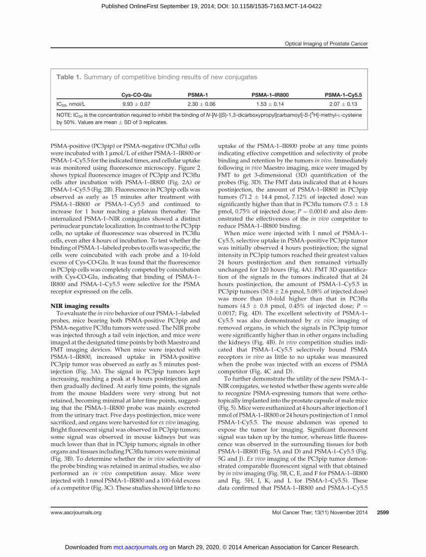
sized ligands, we performed competition binding studies(35). The results summarized in Table 1 show that therationally designedPSMA-1has a binding affinity 4.3-foldbetter (IC50 ¼ 2.30 nmol/L) than the parent ligand Cys-CO-Glu (IC50 ¼ 9.93 nmol/L). Interestingly, inclusion ofIR800 further improved the IC50 of PSMA-1–IR800 to 1.53nmol/L; in contrast, introduction of Cy5.5 did notshowmuch effect to the binding affinity of PSMA-1–Cy5.5(IC50 ¼ 2.07 nmol/L).
In vitro cellular uptake resultsTo determine whether these new imaging probes
would result in cellular binding and labeling of PSMA-expressing cells, we performed in vitro uptake studies.
Figure 1. Structures of PSMA ligands and PSMA–NIR conjugates. D-isomers of glutamic acid were used in PSMA-1 to provide better in vivo stability.IRDye800 and Cy5.5 are conjugated to PSMA-1 through the g-NH2 group of C-terminal lysine.
Wang et al.
Mol Cancer Ther; 13(11) November 2014 Molecular Cancer Therapeutics2598
on March 29, 2020. © 2014 American Association for Cancer Research. mct.aacrjournals.org Downloaded from
Published OnlineFirst September 19, 2014; DOI: 10.1158/1535-7163.MCT-14-0422
PSMA-positive (PC3pip) or PSMA-negative (PC3flu) cellswere incubatedwith 1 mmol/L of either PSMA-1–IR800 orPSMA-1–Cy5.5 for the indicated times, and cellular uptakewas monitored using fluorescence microscopy. Figure 2shows typical fluorescence images of PC3pip and PC3flucells after incubation with PSMA-1–IR800 (Fig. 2A) orPSMA-1–Cy5.5 (Fig. 2B). Fluorescence in PC3pip cells wasobserved as early as 15 minutes after treatment withPSMA-1–IR800 or PSMA-1–Cy5.5 and continued toincrease for 1 hour reaching a plateau thereafter. Theinternalized PSMA-1–NIR conjugates showed a distinctperinuclearpunctate localization. In contrast to the PC3pipcells, no uptake of fluorescence was observed in PC3flucells, even after 4 hours of incubation. To test whether thebindingofPSMA-1–labeledprobes to cellswasspecific, thecells were coincubated with each probe and a 10-foldexcess of Cys-CO-Glu. It was found that the fluorescencein PC3pip cells was completely competed by coincubationwith Cys-CO-Glu, indicating that binding of PSMA-1–IR800 and PSMA-1–Cy5.5 were selective for the PSMAreceptor expressed on the cells.
NIR imaging resultsTo evaluate the in vivo behavior of our PSMA-1–labeled
probes, mice bearing both PSMA-positive PC3pip andPSMA-negative PC3flu tumorswere used. TheNIR probewas injected through a tail vein injection, and mice wereimaged at the designated timepoints by bothMaestro andFMT imaging devices. When mice were injected withPSMA-1–IR800, increased uptake in PSMA-positivePC3pip tumor was observed as early as 5 minutes post-injection (Fig. 3A). The signal in PC3pip tumors keptincreasing, reaching a peak at 4 hours postinjection andthen gradually declined. At early time points, the signalsfrom the mouse bladders were very strong but notretained, becoming minimal at later time points, suggest-ing that the PSMA-1–IR800 probe was mainly excretedfrom the urinary tract. Five days postinjection, mice weresacrificed, and organs were harvested for ex vivo imaging.Bright fluorescent signal was observed in PC3pip tumors;some signal was observed in mouse kidneys but wasmuch lower than that in PC3pip tumors; signals in otherorgans and tissues includingPC3flu tumorswereminimal(Fig. 3B). To determine whether the in vivo selectivity ofthe probe binding was retained in animal studies, we alsoperformed an in vivo competition assay. Mice wereinjectedwith 1 nmol PSMA-1–IR800 and a 100-fold excessof a competitor (Fig. 3C). These studies showed little to no
uptake of the PSMA-1–IR800 probe at any time pointsindicating effective competition and selectivity of probebinding and retention by the tumors in vivo. Immediatelyfollowing in vivo Maestro imaging, mice were imaged byFMT to get 3-dimensional (3D) quantification of theprobes (Fig. 3D). The FMT data indicated that at 4 hourspostinjection, the amount of PSMA-1–IR800 in PC3piptumors (71.2 � 14.4 pmol, 7.12% of injected dose) wassignificantly higher than that in PC3flu tumors (7.5 � 1.8pmol, 0.75% of injected dose; P ¼ 0.0014) and also dem-onstrated the effectiveness of the in vivo competitor toreduce PSMA-1–IR800 binding.
When mice were injected with 1 nmol of PSMA-1–Cy5.5, selective uptake in PSMA-positive PC3pip tumorwas initially observed 4 hours postinjection; the signalintensity in PC3pip tumors reached their greatest values24 hours postinjection and then remained virtuallyunchanged for 120 hours (Fig. 4A). FMT 3D quantifica-tion of the signals in the tumors indicated that at 24hours postinjection, the amount of PSMA-1–Cy5.5 inPC3pip tumors (50.8 � 2.6 pmol, 5.08% of injected dose)was more than 10-fold higher than that in PC3flutumors (4.5 � 0.8 pmol, 0.45% of injected dose; P ¼0.0017; Fig. 4D). The excellent selectivity of PSMA-1–Cy5.5 was also demonstrated by ex vivo imaging ofremoved organs, in which the signals in PC3pip tumorwere significantly higher than in other organs includingthe kidneys (Fig. 4B). In vivo competition studies indi-cated that PSMA-1–Cy5.5 selectively bound PSMAreceptors in vivo as little to no uptake was measuredwhen the probe was injected with an excess of PSMAcompetitor (Fig. 4C and D).
To further demonstrate the utility of the new PSMA-1–NIR conjugates, we testedwhether these agents were ableto recognize PSMA-expressing tumors that were ortho-topically implanted into the prostate capsule ofmalemice(Fig. 5).Micewere euthanized at 4hours after injection of 1nmol of PSMA-1–IR800 or 24hours postinjection of 1 nmolPSMA-1-Cy5.5. The mouse abdomen was opened toexpose the tumor for imaging. Significant fluorescentsignal was taken up by the tumor, whereas little fluores-cence was observed in the surrounding tissues for bothPSMA-1–IR800 (Fig. 5A and D) and PSMA-1–Cy5.5 (Fig.5G and J). Ex vivo imaging of the PC3pip tumor demon-strated comparable fluorescent signal with that obtainedby in vivo imaging (Fig. 5B, C, E, and F for PSMA-1–IR800and Fig. 5H, I, K, and L for PSMA-1–Cy5.5). Thesedata confirmed that PSMA-1–IR800 and PSMA-1–Cy5.5
Table 1. Summary of competitive binding results of new conjugates
Cys-CO-Glu PSMA-1 PSMA-1–IR800 PSMA-1–Cy5.5
IC50, nmol/L 9.93 � 0.07 2.30 � 0.06 1.53 � 0.14 2.07 � 0.13
NOTE: IC50 is the concentration required to inhibit the binding of N-[N-[(S)-1,3-dicarboxypropyl]carbamoyl]-S-[3H]-methyl-L-cysteineby 50%. Values are mean � SD of 3 replicates.
Optical Imaging of Prostate Cancer
www.aacrjournals.org Mol Cancer Ther; 13(11) November 2014 2599
on March 29, 2020. © 2014 American Association for Cancer Research. mct.aacrjournals.org Downloaded from
Published OnlineFirst September 19, 2014; DOI: 10.1158/1535-7163.MCT-14-0422

0 min 15 min 30 min 1 h 4 h 4 h
0 min 15 min 30 min 1 h 4 h 4 h
(+10X blocker)
PSMA-1–
IR800
DAPI
Merge
PC3pip
(PSMA+)
PSMA-1–
IR800
Merge
PC3flu
(PSMA–)DAPI
A
PSMA-1–
Cy5.5
DAPI
Merge
PC3pip
(PSMA+)
PSMA-1–
Cy5.5
Merge
PC3flu
(PSMA–)DAPI
B(+10X blocker)
Figure 2. In vitro cellular uptake results of PSMA-1–IR800 and PSMA-1–Cy5.5. PSMA-positive PC3pip cells and PSMA-negative cells PC3flu cells oncoverslips were incubated with no probe (0 minutes, A and B) or 1 mmol/L of PSMA-1–IR800 (A) or 1 mmol/L of PSMA-1–Cy5.5 (B) for 15 minutes, 30 minutes,1 hour, and 4 hours. The nucleus was stained by DAPI (false color blue), and uptake of PSMA-1–IR800 and PSMA-1–Cy5.5 was assessed by fluorescencemicroscopy (false color red). Specificity of PSMA-1–NIR conjugates to PSMA was evaluated by incubation of PC3pip and PC3flu cells with 1 mmol/L ofPSMA-1–NIR conjugates and 10 mmol/L of Cys-CO-Glu, last column in each panel. Signal in PC3pip cells was significantly competed by Cys-CO-Glu,suggesting that the binding of PSMA-1–IR800 and PSMA-1–Cy5.5 to PSMA is specific. Images are taken at 40�. Representative images are shown from3 independent experiments.
Wang et al.
Mol Cancer Ther; 13(11) November 2014 Molecular Cancer Therapeutics2600
on March 29, 2020. © 2014 American Association for Cancer Research. mct.aacrjournals.org Downloaded from
Published OnlineFirst September 19, 2014; DOI: 10.1158/1535-7163.MCT-14-0422

can selectively recognize and bind to orthotopic PC3piptumors.
DiscussionThe purpose of this study was to develop PSMA-tar-
geted NIR imaging agents to help identify prostatetumors. We have synthesized a urea-based PSMA ligand(PSMA-1) and identified 2NIRmolecular imaging probesfor noninvasive selective detection of tumors expressingPSMAin live animals. The ligandPSMA-1 reportedhere isa newurea-based PSMA ligand rationally designed on thebasis of our previous structure–activity relationship stud-ies of a 2-5A–based PSMA analogue, RBI1033 (35, 37).PSMA-1 demonstrated improved binding affinity (IC50 ¼2.30 nmol/L) to PSMAreceptor comparedwith the parentligand Cys-CO-Glu (IC50 ¼ 9.93 nmol/L) as determinedby a competition binding assay.A major concern of conjugation of a bulky dye to the
ligand is that thedyemight significantly interfere betweenthe interaction of the ligand and receptor, causing loss ofbinding affinity. This, however, was not the case for thesecompounds. PSMA-1–IR800 and PSMA-1–Cy5.5 showed
comparable or even improved binding affinity to thePSMA receptor compared with the unlabeled PSMA-1itself.Wehypothesize that thefluorophore conjugated to along peptide linker of glutamate may exploit both the S1and S10 regions of the PSMA-binding site by positioningthe large fluorophores outside of the 20 A long substrate-binding pocket (S1) of PSMA (38, 39) reducing any sterichindrance and increasing binding affinity by exploitingthe glutamate-binding region. In vitro cellular uptakeexperiments with PSMA-1–IR800 and PSMA-1–Cy5.5showed that it can selectively bind to and be taken upby PSMA-expressing PC3pip cells but not by cells that donot express PSMA, that is, PC3flu cells. In the presence ofexcess amount of Cys-CO-Glu, the binding of PSMA-1–IR800 and PSMA-1–Cy5.5 to PC3pip cells was competed,indicating the binding is specific to the PSMA receptor. Ithas been found that the PSMA or PSMA–antibody com-plex undergoes internalization through clathrin-coatedpits, closely resembling that of transferrin receptor inter-nalization pathway, a receptor with which the PSMAreceptor has a high degree of homology (40). Our resultsshowed that our PSMA-1–NIR probes are internalizedinto the cells forming a punctate accumulation in the
PSMA-1–IR800, in vivo
PSMA-1–IR800 + 100XZJ-MCC-Ahx-YYYG FMT quantification of PSMA-1–IR800
Time after injection
PC3pip
PC3flu
PC3flu (+ 100X blocker)
PC3pip (+ 100X blocker)
PS
MA
-1–I
R80
0 in
tum
ors
(pm
ol/g
)
PSMA-1–IR800, ex vivoB&W Preimage 5 min 30 min 1 h
6 h
B&WC D
A B
Preimage 5 min 30 min 1 h
2 h 4 h 6 h 8 h 24 h
8 h 24 h 48 h 72 h 96 h 120 h
2 h 4 h
Dis
play
key
:
00.
050.
12
0 00.
002
0.
004
0.00
76
0.05
0.12
Dis
play
key
:
Dis
play
key
:
9080
7060504030
20
100
0 m
in5
min
30 m
in 1 h
2 h
4 h
6 h
8 h
24 h
48 h
72 h
96 h
120
h
Figure 3. Imaging of PSMA-1–IR800 in mice bearing flank PC3pip and PC3flu tumors. A, in vivo Maestro imaging of a typical mouse treated withPSMA-1–IR800. Mice received 1 nmol of PSMA-1–IR800 via tail vein injection and then were imaged at the designated times. Representative images areshown of n ¼ 5. B, ex vivo imaging of mice organs at 120 hours postinjection of PSMA-1–IR800. The fluorescent signal in PC3pip tumor was significantlyhigher than in other organs. C, in vivo Maestro imaging of mice injected with 1 nmol of PSMA-1–IR800 and 100 nmol of a selective PSMA receptor–bindingmolecule, ZJ-MCC-Ahx-YYYG (34). Images are on the same scale as in A. Blockade of fluorescent uptake in PC3pip tumorswas observed. D, FMT3Dquantification of PSMA-1–IR800 in PC3pip and PC3flu tumors from the mice used in A and C. Values represent mean � SD of 5 animals.
Optical Imaging of Prostate Cancer
www.aacrjournals.org Mol Cancer Ther; 13(11) November 2014 2601
on March 29, 2020. © 2014 American Association for Cancer Research. mct.aacrjournals.org Downloaded from
Published OnlineFirst September 19, 2014; DOI: 10.1158/1535-7163.MCT-14-0422

perinuclear region, suggesting that PSMA is localized tothe recycling endosome (41). The internalization of ourlow molecular weight ligand PSMA-1 also suggests thatPSMA-1 may serve as a putative ligand for the PSMAreceptor to substitute for antibodies not only for imagingbut also for targeted therapeutic approaches, creatingoptions such as the use of toxin or drug conjugatestargeted to cancerous cells.
In in vivo experiments, both PSMA-1–IRdye800 andPSMA-1–Cy5.5 showed excellent binding selectivity toPSMA-positive PC3pip tumors with more than a 10-folddifferential between PC3pip and PC3flu tumors. Interest-ingly, the 2 probes showed distinctively different phar-macokinetic behaviors (Supplementary Fig. S4). Theamount of PSMA-1–IR800 reached its highest levels inPSMA-positive PC3pip tumors at 4 hours postinjection,whereas it took PSMA-1–Cy5.5 24 hours to reach its high-est amount in PC3pip tumors. PSMA-1–IR800 waswashed out relatively rapidly from the tumor, clearing
in 24 to 120 hours. In contrast, in vivo studies with PSMA-1–Cy5.5 demonstrated that the probe remained tumorassociated and virtually unchanged 5 days after admin-istration. To understand this, we compared the structureof eachfluorophore in the PSMA-1–NIRprobes,which areotherwise virtually identical. IRDye800 contains 3 morenegatively charged sulfate groups than Cy5.5. Therefore,PSMA-1–Cy5.5 is less polar and much more hydrophobic(log P ¼ �1.02) than IRDye800 (log P ¼ �2.14), and thisdifference may be responsible for the vastly differentpharmacokinetics of the 2 PSMA-1–NIR probes. Otherstudies where fluorophores of intermediate polarity toIRDye800 and Cy5.5 were conjugated to PSMA-1 showedstill different in vivo kinetics (data not shown), suggestingthat polarity of the fluorophore might dramatically influ-ence pharmacokinetics of the agents. The phenomenonthat a particular NIR dye can affect the pharmacokineticsof a conjugated probe was also reported by Chen andcolleagues (30). They observed that more hydrophobic
PSMA-1–Cy5.5,in vivo PSMA-1–Cy5.5,ex vivoB&W Preimage 5 min 30 min 1 h 2 h 4 h
6 h 8 h 24 h 48 h 72 h 96 h 120 h
PSMA-1–Cy5.5 + 100XZJ-MCC-Ahx-YYYG
B&W Preimage 5 min 30 min 1 h
2 h 4 h 6 h 8 h 24 h
FMT quantification of PSMA-1–Cy5.5
Time after injection
PC3pip
PC3flu
PC3flu (+ 100X blocker)
PC3pip (+ 100X blocker)
PS
MA
-1–C
y5.5
in tu
mor
(pm
ol/g
)
DC
A B
Dis
play
key
:
00.
050.
100.
145
Dis
play
key
:
00.
050.
100.
145
90
80
70
60
50
40
30
20
10
0
0 m
in5
min
30 m
in 1 h
2 h
4 h
6 h
8 h
24 h
48 h
72 h
96 h
120
h
Figure 4. Imaging of PSMA-1–Cy5.5 in mice bearing flank PC3pip and PC3flu tumors. A, in vivoMaestro imaging of mice treated with PSMA-1–Cy5.5. Micereceived 1 nmol of PSMA-1–Cy5.5 through tail vein injection and then were imaged at the indicated times. Representative images of n ¼ 5 mice are shown.Selective uptake in PC3pip tumors was observed. Highest PC3pip tumor uptake was observed 24 hours postinjection. B, ex vivo imaging of mice organs at120 hours postinjection of PSMA-1–Cy5.5. The fluorescent signal in PC3pip tumor was significantly higher than in other organs. C, in vivoMaestro imagingof mice injected with 1 nmol of PSMA-1–Cy5.5 and 100 nmol of a selective PSMA receptor–binding molecule, ZJ-MCC-Ahx-YYYG (34). Images areon the same scale as in A. Blockade of fluorescent uptake in PC3pip tumors was observed. D, FMT 3D quantification of PSMA-1–Cy5.5 in PC3pip and PC3flutumors from mice used in A and C. Values represent mean � SD of 5 animals.
Wang et al.
Mol Cancer Ther; 13(11) November 2014 Molecular Cancer Therapeutics2602
on March 29, 2020. © 2014 American Association for Cancer Research. mct.aacrjournals.org Downloaded from
Published OnlineFirst September 19, 2014; DOI: 10.1158/1535-7163.MCT-14-0422

probe has longer retention time in the body, consistentwith our findings.To further demonstrate that binding of our PSMA-1–
NIR probes are specific for PSMA in vivo, competitionstudies were conducted by coinjection into the mice bothPSMA-1–NIRs and a 100-fold excess of ZJ-MCC-Ahx-YYYG, a high affinity binder to the PSMA receptor (34).We first tried these studies using Cys-CO-Glu or PSMA-1butwereunable to achieve effective competitionusingupto a 1,000-fold excess of Cys-CO-Glu or PSMA-1. This islikely due to the rapid clearance from the bodymeasuredfor Cys-CO-Glu (25) which may also be the case for theunderivatized PSMA-1. We therefore conducted the in
vivo competition studies using the more hydrophobicligand ZJ-MCC-Ahx-YYYG. Using this agent, we wereable to measure strong and significant displacement ofthe PSMA-1–NIRs (Figs. 3C and D and 4C and D). Forexample, in competition studieswith PSMA-1–Cy5.5 anda 100-fold excess of ZJ-MCC-Ahx-YYYG, the amount ofPSMA-1–Cy5.5 in PC3pip tumor decreased from 50.8 �2.6 pmol/g to 5.4 � 5.74 pmol/g (P ¼ 0.0018), demon-strating that approximately 90% of PSMA-1–Cy5.5 inPC3pip tumors was competed. In contrast, the amountof PSMA-1–Cy5.5 in PC3flu tumors remained unchanged(4.27 � 0.98 pmol/g compared with 4.51 � 0.77 pmol/g,P ¼ 0.454). Similar results were obtained for the
PC3pip tumor
PC3pip tumor
Liver
Stomach Spleen
Kidneys
Heart
Lung
PC3pipBladder
PC3pip tumor
Liver
Stomach Spleen
Kidneys
Heart
Lung
PC3pip
Bladder
PC3pip tumor
A B C
D E F
G H I
J K L
PSMA-1–IR800
PSMA-1–Cy5.5
Figure 5. PSMA-1–NIR probes can selectively target orthotopic PSMA-positive PC3pip tumors as shown by Maestro images. Mice received 1 nmol ofPSMA–IR800 (A–F) or 1 nmol of PSMA-1–Cy5.5 (G–L) via a tail vein injection. Mice were sacrificed at 4 hours postinjection of PSMA-1–IR800, the abdomenopened to expose the tumor, and both black and white images (A) and fluorescent images (D) were taken. Organs were then harvested for ex vivo images(B and E) and finally tumors were imaged separately ex vivo (C and F). Mice that were administered PSMA-1–Cy5.5 were sacrificed at 24 hours postinjection;the abdomen was opened to expose tumor and then imaged. Both black and white images (G) and fluorescent images (J) were taken, organs wereharvested for imaging (H andK), and finally tumorswere imaged separately ex vivo (I and L). Pictures are representative images of 4mice for each probe. Brightfluorescent signal was observed in PC3pip tumor.
Optical Imaging of Prostate Cancer
www.aacrjournals.org Mol Cancer Ther; 13(11) November 2014 2603
on March 29, 2020. © 2014 American Association for Cancer Research. mct.aacrjournals.org Downloaded from
Published OnlineFirst September 19, 2014; DOI: 10.1158/1535-7163.MCT-14-0422

competition studies with PSMA-1–IR800, in which morethan 95% PSMA-1–IR800 in PC3pip tumors was dis-placed (P¼ 0.0003). These results again suggest that highPC3pip tumor uptake of the PSMA-1–NIRs is due toPSMA-specific binding.
Since the discovery of PSMA, a wide variety of imagingagents targeting PSMA have been reported. Amajority ofthem are radiopharmaceuticals, but noninvasive opticalagents are also presented. Humblet and colleagues syn-thesized the first phosphoramidate-IRDye78 (GPI-78)conjugate with a binding affinity at Ki ¼ 9 nmol/L, butGPI-78 was cleared too fast from the body, and imaginghad to be performed 20 seconds postinjection resulting invery low tumor signals (20). Liu and colleagues reported aCy5.5-labeled phosphoramidate peptidomimetic PSMAligand Cy5.5-CTT-54.2 with IC50 at 0.55 nmol/L (42). Thisprobedemonstrated the ability to specifically label PSMA-positive prostate cancer cells, but no further in vivo imag-ing data were reported. Nakajima and colleagues synthe-sized an activatable J591–ICG conjugate with high spec-ificity to PSMA; however, it took 2 days to be sufficientlyactivated and taken up by the tumor (43).
Recently, Chen and colleagues reported high PSMA-specific uptake in vivo with 800CW-2-, 800CW-3-, Cy7-2-,and Cy7-3–labeled probes (30), however, a direct com-parison of binding affinity of these agents with our com-pounds cannot be made as Ki of these conjugates forNAALADase activity of thePSMAreceptorwere reportedinstead of binding affinity. In this work, Chen and col-leagues reported imaging, uptake, and biodistributionresults up to 24 hours postinjection for optical imagingprobes and noted that the linker chemistry and the fluor-ophore both can affect the pharmacokinetics of the probes.In our study, we measured the uptake of the conjugatesduring an elongated time course and demonstrated sig-nificant differences in probe pharmacokinetics that werefluorophore-dependent and long-lived. Given the largeimpact of the fluorophore on the pharmacokinetics ofthese agents, full pharmacokinetic studies are importantfor the determination of the best way to use the imagingagents. Overall our probes show favorable pharmacoki-netic behavior, fast tumor accumulation, and rapid clear-ance compared with the antibody-based J591–ICG conju-gate and suggest a future potential of developing theseagents for optical imaging of prostate cancer.
In cancer surgery, it is of utmost importance to exactlyidentify the extent of malignancy because the presence orabsence of tumor cells after surgical removal is a decisionfactor in determining the therapeutic approach and thesuccess of said therapy. Radiological techniques such asX-ray, CT, and MRI have been considered in assistingsurgery, but these are not very useful for intraoperativeapplication. While SPECT (44, 45) is possible to be usedintraoperatively, patients and the surgeons will beexposed to the danger of ionizing radiation. In contrast,fluorescent imaging can provide real-time imaging dur-ing surgery, improving detection of tumors tissues andmore radical removal of tumor tissues without radiation
exposure. Indocyanine green (ICG) is one of the firstfluorescent dyes tested for intraoperative application inglioma surgery (46). It has also shown promise in intrao-perative sentinel lymph node mapping (47). The combi-nation of optical imaging technologies with tumor-target-ing strategies can shift the paradigm of surgical oncologicimaging, offering the unique opportunity to intraopera-tively detect and quantify tumor growth and intra-abdominal spread. Notably, fluorescent imaging probeshave recently been successfully applied for the intrao-perative detection of ovarian cancer. Recently, NatureMedicine describes the first use of intraoperative, tumor-specific, folate receptor–targeted fluorescence imaging tohighlight the precise position of small groups of cancercells in women with ovarian cancer, thereby allowing thesurgeon to carefully excise these small groups of cells (48).
For prostate cancer removal, a modality to guide sur-gery will be extremely useful due to the complicatedstructure of theprostate gland.Theprostate is surroundedby many nerves and sphincter urethrae muscle fibers,which control different excretory and erectile functions. Ithas been shown that significant side effects can result fromradical surgery, including genitourinary, gastrointestinal,and sexual dysfunction (49). The ideal fluorescent imag-ing agent for intraoperative use should be able to helpsurgeons (i) accurately define tumor spread and deter-mine the aggressiveness of the surgical intervention inreal-time, for example, determine whether the cavernousnerves need to be removed or could be spared duringsurgery and (ii) avoid leaving behind the cancerous tissuethat is commonly associated with pathologically positivesurgical margins. We have tried an IRDye800-labeledPSMA-targeting conjugate on mice bearing PC3pip flanktumors using a Da Vinci system for robotic surgery, andnegative surgical margins were obtained for all excisedPC3pip tumors (50) underscoring the potential impact ofthis technology. The 2 probes reported here have pro-longed uptake in PSMA-positive tumors, especiallyPSMA-1–Cy5.5, and therefore may be more suitable forcombination with the Da Vinci System intraoperativeimage-guided surgery. In the future, wewill combine thistechnology with targeted therapeutics that will allow"cleanup" of cancerous tissues thatmight not be accessibleto the surgeons’ scalpel.
ConclusionIn summary, a peptide-based highly negatively char-
ged PSMA ligand was designed and 2 PSMA-targetingNIR probes were synthesized on the basis of the newligand. The molecules were evaluated in vitro and in vivo.Bothprobesdemonstratedhighbinding affinity and selec-tivity for PSMA on PC3pip tumors. Our data suggestedthat the 2 NIR probes reported here have the ability toeffectively distinguish between PSMA-expressing andnonexpressing tumors and other tissues. They have thepotential to aid in the diagnosis of prostate cancer. Impor-tantly, it may also have the potential to reshape standard
Wang et al.
Mol Cancer Ther; 13(11) November 2014 Molecular Cancer Therapeutics2604
on March 29, 2020. © 2014 American Association for Cancer Research. mct.aacrjournals.org Downloaded from
Published OnlineFirst September 19, 2014; DOI: 10.1158/1535-7163.MCT-14-0422

prostatectomies. In the future, probes like these willenable surgeons to identify extracapsular disease, whichis currently invisible during prostatectomy, decidewhether surgical removal is possible, allow discrimina-tion between diseased, normal, and neural tissues pre-venting significant morbidities and result in improvedpatient outcome.
Disclosure of Potential Conflicts of InterestNo potential conflicts of interest were disclosed.
Authors' ContributionsConception and design: X. Wang, S.S. Huang, J.P. BasilionDevelopment ofmethodology:X.Wang,H.Guo, B.-C.Wang, J.P. BasilionAcquisition of data (provided animals, acquired and managed patients,provided facilities, etc.): X. WangAnalysis and interpretation of data (e.g., statistical analysis, biostatis-tics, computational analysis): X. Wang, J.P. Basilion
Writing, review, and/or revision of themanuscript: X.Wang, S.S. Huang,W.D.W. Heston, J.P. BasilionAdministrative, technical, or material support (i.e., reporting or orga-nizing data, constructing databases): H. Guo, J.P. BasilionStudy supervision: J.P. Basilion
AcknowledgmentsThe authors thank the laboratory of Dr. John Crabb at the Cole Eye
Institute, Cleveland Clinic (Cleveland, OH) for assistance with massspectrometry.
Grant SupportThe study was supported by the National Foundation of Cancer
Research to J.P. Basilion.The costs of publication of this article were defrayed in part by the
payment of page charges. This article must therefore be hereby markedadvertisement in accordance with 18 U.S.C. Section 1734 solely to indicatethis fact.
Received May 16, 2014; revised August 27, 2014; accepted September 2,2014; published OnlineFirst September 19, 2014.
References1. Siegel R, Ma J, Zou Z, Jemal A. Cancer statistics, 2014. CA Cancer J
Clin 2014;64:9–29.2. Theiss M, Wirth MP, Manseck A, Frohmuller HG. Prognostic signifi-
cance of capsular invasion and capsular penetration in patients withclinically localized prostate cancer undergoing radical prostatectomy.Prostate 1995;27:13–7.
3. SwansonGP, Lerner SP. Positivemargins after radical prostatectomy:implications for failure and role of adjuvant treatment. Urol Oncol2013;31:531–41.
4. Wright JL, Dalkin BL, True LD, Ellis WJ, Stanford JL, Lange PH, et al.Positive surgical margins at radical prostatectomy predict prostatecancer specific mortality. J Urol 2010;183:2213–8.
5. Horoszewicz JS, Kawinski E, Murphy GP. Monoclonal antibodies to anewantigenicmarker in epithelial prostatic cells and serumof prostaticcancer patients. Anticancer Res 1987;7:927–35.
6. Israeli RS, Miller WH Jr, Su SL, Powell CT, Fair WR, Samadi DS, et al.Sensitive nested reverse transcription polymerase chain reactiondetection of circulating prostatic tumor cells: comparison of pros-tate-specific membrane antigen and prostate-specific antigen-basedassays. Cancer Res 1994;54:6306–10.
7. Tasch J, Gong M, Sadelain M, Heston WD. A unique folate hydrolase,prostate-specific membrane antigen (PSMA): a target forimmunotherapy? Crit Rev Immunol 2001;21:249–61.
8. Mannweiler S, Amersdorfer P, Trajanoski S, Terrett JA, King D, MehesG. Heterogeneity of prostate-specific membrane antigen (PSMA)expression in prostate carcinoma with distant metastasis. PatholOncol Res 2009;15:167–72.
9. Troyer JK, BeckettML,Wright GL Jr. Detection and characterization ofthe prostate-specificmembrane antigen (PSMA) in tissue extracts andbody fluids. Int J Cancer 1995;62:552–8.
10. Ross JS, SheehanCE, Fisher HA, Kaufman RP Jr, Kaur P, Gray K, et al.Correlation of primary tumor prostate-specific membrane antigenexpression with disease recurrence in prostate cancer. Clin CancerRes 2003;9:6357–62.
11. Wang X, Yin L, Rao P, Stein R, Harsch KM, Lee Z, et al. Targetedtreatment of prostate cancer. J Cell Biochem 2007;102:571–9.
12. Chang SS, Reuter VE, HestonWD, Bander NH, Grauer LS, Gaudin PB.Five different anti-prostate-specific membrane antigen (PSMA) anti-bodies confirm PSMA expression in tumor-associated neovascula-ture. Cancer Res 1999;59:3192–8.
13. ChangSS,O'Keefe DS, BacichDJ, Reuter VE, HestonWD,Gaudin PB.Prostate-specific membrane antigen is produced in tumor-associatedneovasculature. Clin Cancer Res 1999;5:2674–81.
14. Wynant GE,MurphyGP,Horoszewicz JS,Neal CE, Collier BD,MitchellE, et al. Immunoscintigraphy of prostatic cancer: preliminary resultswith 111In-labeled monoclonal antibody 7E11-C5.3 (CYT-356). Pros-tate 1991;18:229–41.
15. Bander NH. Technology insight: monoclonal antibody imaging ofprostate cancer. Nat Clin Pract Urol 2006;3:216–25.
16. Bander NH, Milowsky MI, Nanus DM, Kostakoglu L, Vallabhajosula S,Goldsmith SJ. Phase I trial of 177lutetium-labeled J591, a monoclonalantibody to prostate-specific membrane antigen, in patients withandrogen-independent prostate cancer. J Clin Oncol 2005;23:4591–601.
17. Bander NH, Trabulsi EJ, Kostakoglu L, Yao D, Vallabhajosula S,Smith-Jones P, et al. Targeting metastatic prostate cancerwith radiolabeled monoclonal antibody J591 to the extracellulardomain of prostate specific membrane antigen. J Urol 2003;170:1717–21.
18. Osborne JR, Green DA, Spratt DE, Fareedy SB, Robinson BD, BeattieBJ, et al. A prospective pilot study of (89)Zr-J591/prostate specificmembrane antigen positron emission tomography in men with local-ized prostate cancer undergoing radical prostatectomy. J Urol2013;191:1439–45.
19. Tagawa ST, Milowsky MI, Morris M, Vallabhajosula S, Christos P,Akhtar NH, et al. Phase II study of Lutetium-177-labeled anti-prostate-specific membrane antigen monoclonal antibody J591 for metastaticcastration-resistant prostate cancer. Clin Cancer Res 2013;19:5182–91.
20. Humblet V, Lapidus R, Williams LR, Tsukamoto T, Rojas C, Majer P,et al. High-affinity near-infrared fluorescent small-molecule contrastagents for in vivo imaging of prostate-specificmembrane antigen. MolImaging 2005;4:448–62.
21. Banerjee SR, FossCA,CastanaresM,MeaseRC, ByunY, Fox JJ, et al.Synthesis and evaluation of technetium-99m- and rhenium-labeledinhibitors of the prostate-specific membrane antigen (PSMA). J MedChem 2008;51:4504–17.
22. Banerjee SR, Pullambhatla M, Byun Y, Nimmagadda S, Green G, FoxJJ, et al. 68Ga-labeled inhibitors of prostate-specific membraneantigen (PSMA) for imaging prostate cancer. J Med Chem 2010;53:5333–41.
23. Chen Y, Dhara S, Banerjee SR, Byun Y, Pullambhatla M, Mease RC,et al. A low molecular weight PSMA-based fluorescent imaging agentfor cancer. Biochem Biophys Res Commun 2009;390:624–9.
24. Chen Y, Foss CA, Byun Y, Byun Y, Pullambhatla M, Mease RC, et al.Radiohalogenated prostate-specific membrane antigen (PSMA)-basedureas as imaging agents for prostate cancer. JMedChem2008;51:7933–43.
25. Foss CA, Mease RC, Fan H, Wang Y, Ravert HT, Dannals RF, et al.Radiolabeled small-molecule ligands for prostate-specific membraneantigen: in vivo imaging inexperimentalmodels of prostate cancer. ClinCancer Res 2005;11:4022–8.
26. Hillier SM, Kern AM, Maresca KP, Wang Y, Ravert HT, Dannals RF,et al. 123I-MIP-1072, a small-molecule inhibitor of prostate-specific
Optical Imaging of Prostate Cancer
www.aacrjournals.org Mol Cancer Ther; 13(11) November 2014 2605
on March 29, 2020. © 2014 American Association for Cancer Research. mct.aacrjournals.org Downloaded from
Published OnlineFirst September 19, 2014; DOI: 10.1158/1535-7163.MCT-14-0422

membrane antigen, is effective atmonitoring tumor response to taxanetherapy. J Nucl Med 2011;52:1087–93.
27. Hillier SM, Maresca KP, Femia FJ, Marquis JC, Foss CA, Nguyen N,et al. Preclinical evaluation of novel glutamate-urea-lysine analoguesthat target prostate-specific membrane antigen as molecular imagingpharmaceuticals for prostate cancer. Cancer Res 2009;69:6932–40.
28. Hillier SM,MarescaKP, LuG,Merkin RD,Marquis JC, ZimmermanCN,et al. 99mTc-labeled small-molecule inhibitors of prostate-specificmembrane antigen for molecular imaging of prostate cancer. J NuclMed 2013;54:1369–76.
29. Kelderhouse LE, Chelvam V, Wayua C, Mahalingam S, Poh S, Kular-atne SA, et al. Development of tumor-targeted near infrared probes forfluorescence guided surgery. Bioconjug Chem 2013;24:1075–80.
30. Chen Y, Pullambhatla M, Banerjee SR, Byun Y, Stathis M, Rojas C,et al. Synthesis and biological evaluation of low molecular weightfluorescent imaging agents for the prostate-specific membrane anti-gen. Bioconjug Chem 2009;23:2377–85.
31. Cho SY, Gage KL, Mease RC, Senthamizhchelvan S, Holt DP, Jeffrey-Kwanisai A, et al. Biodistribution, tumor detection, and radiationdosimetry of 18F-DCFBC, a low-molecular-weight inhibitor of pros-tate-specific membrane antigen, in patients with metastatic prostatecancer. J Nucl Med 2012;53:1883–91.
32. Mease RC, Dusich CL, Foss CA, Senthamizhchelvan S, Holt DP,Jeffrey-Kwanisai A, et al. N-[N-[(S)-1,3-Dicarboxypropyl]carbamoyl]-4-[18F]fluorobenzyl-L-cysteine, [18F]DCFBC: a new imaging probe forprostate cancer. Clin Cancer Res 2008;14:3036–43.
33. Barrett JA, Coleman RE, Goldsmith SJ, Vallabhajosula S, Petry NA,Cho S, et al. First-in-man evaluation of 2 high-affinity PSMA-avid smallmolecules for imaging prostate cancer. J Nucl Med 2013;54:380–7.
34. Huang SS, Wang X, Zhang Y, Doke A, Difilippo FP, Heston WD.Improving the biodistribution of PSMA-targeting tracers with a highlynegatively charged linker. Prostate 2014;74:702–13.
35. Wang X, Tian H, Lee Z, Heston WD. Structure-activity relationships of20,50-oligoadenylate analogue modifications of prostate-specificmembrane antigen (PSMA) antagonists. Nucleosides NucleotidesNucleic Acids 2012;31:432–44.
36. Petty A, Myshkin E, Qin H, Guo H, Miao H, Tochtrop GP, et al. A smallmolecule agonist of EphA2 receptor tyrosine kinase inhibits tumor cellmigration in vitro and prostate cancer metastasis in vivo. PLoS ONE2012;7:e42120.
37. Cramer H, Okicki JR, Rho T, Wang X, Silverman RH, Heston WD. 2-5Aligands–a new concept for the treatment of prostate cancer. Nucleo-sides Nucleotides Nucleic Acids 2007;26:1471–7.
38. Mesters JR, Henning K, Hilgenfeld R. Human glutamate carboxypep-tidase II inhibition: structures of GCPII in complex with two potent
inhibitors, quisqualate and2-PMPA.ActaCrystallogrDBiolCrystallogr2007;63:508–13.
39. Hlouchova K, Barinka C, Konvalinka J, Lubkowski J. Structural insightinto the evolutionary and pharmacologic homology of glutamatecarboxypeptidases II and III. FEBS J 2009;276:4448–62.
40. Ghosh A, Heston WD. Tumor target prostate specific membraneantigen (PSMA) and its regulation in prostate cancer. J Cell Biochem2004;91:528–39.
41. Rajasekaran SA, Anilkumar G, Oshima E, Bowie JU, Liu H, Heston W,et al. A novel cytoplasmic tail MXXXLmotif mediates the internalizationof prostate-specific membrane antigen. Mol Biol Cell 2003;14:4835–45.
42. Liu T, Wu LY, Hopkins MR, Choi JK, Berkman CE. A targeted lowmolecular weight near-infrared fluorescent probe for prostate cancer.Bioorg Med Chem Lett 2010;20:7124–6.
43. Nakajima T, Mitsunaga M, Bander NH, Heston WD, Choyke PL,Kobayashi H. Targeted, activatable, in vivo fluorescence imaging ofprostate-specificmembrane antigen (PSMA) positive tumors using thequenched humanized J591 antibody-indocyanine green (ICG) conju-gate. Bioconjug Chem 2011;22:1700–5.
44. Vermeeren L, ValdesOlmos RA,MeinhardtW, Bex A, van der Poel HG,Vogel WV, et al. Intraoperative radioguidance with a portable gammacamera: a novel technique for laparoscopic sentinel node localisationin urological malignancies. Eur J Nucl Med Mol Imaging 2009;36:1029–36.
45. Sanchez F, Fernandez MM, Gimenez M, Benlloch JM, Rodríguez-AlvarezMJ,García deQuir�osF, et al. Performance tests of twoportablemini gamma cameras for medical applications. Med Phys 2006;33:4210–20.
46. HaglundMM, Berger MS, Hochman DW. Enhanced optical imaging ofhuman gliomas and tumor margins. Neurosurgery 1996;38:308–17.
47. Colen RR, Kekhia H, Jolesz FA. Multimodality intraoperative MRI forbrain tumor surgery. Expert Rev Neurother 2010;10:1545–58.
48. van Dam GM, Themelis G, Crane LM, Harlaar NJ, Pleijhuis RG, KelderW, et al. Intraoperative tumor-specific fluorescence imaging in ovariancancer by folate receptor-alpha targeting: first in-human results. NatMed 2011;17:1315–9.
49. Talcott JA, Rieker P, Clark JA, Weeks JC, Beard CJ, Wishnow KI, et al.Patient-reported symptoms after primary therapy for early prostatecancer: results of a prospective cohort study. J Clin Oncol 1998;16:275–83.
50. Laydner H, Autorino R, Isac W, Khalifeh A, Panumatrassamee K,Kassab A, et al. Robotic retroperitoneal transvaginal natural orificetranslumenal endoscopic surgery (NOTES) nephrectomy: feasibilitystudy in a cadaver model. Urology 2012;81:1232–7.
Mol Cancer Ther; 13(11) November 2014 Molecular Cancer Therapeutics2606
Wang et al.
on March 29, 2020. © 2014 American Association for Cancer Research. mct.aacrjournals.org Downloaded from
Published OnlineFirst September 19, 2014; DOI: 10.1158/1535-7163.MCT-14-0422

2014;13:2595-2606. Published OnlineFirst September 19, 2014.Mol Cancer Ther Xinning Wang, Steve S. Huang, Warren D.W. Heston, et al. CancerDevelopment of Targeted Near-Infrared Imaging Agents for Prostate
Updated version
10.1158/1535-7163.MCT-14-0422doi:
Access the most recent version of this article at:
Material
Supplementary
http://mct.aacrjournals.org/content/suppl/2014/09/20/1535-7163.MCT-14-0422.DC1
Access the most recent supplemental material at:
Cited articles
http://mct.aacrjournals.org/content/13/11/2595.full#ref-list-1
This article cites 50 articles, 15 of which you can access for free at:
Citing articles
http://mct.aacrjournals.org/content/13/11/2595.full#related-urls
This article has been cited by 6 HighWire-hosted articles. Access the articles at:
E-mail alerts related to this article or journal.Sign up to receive free email-alerts
Subscriptions
Reprints and
To order reprints of this article or to subscribe to the journal, contact the AACR Publications Department at
Permissions
Rightslink site. Click on "Request Permissions" which will take you to the Copyright Clearance Center's (CCC)
.http://mct.aacrjournals.org/content/13/11/2595To request permission to re-use all or part of this article, use this link
on March 29, 2020. © 2014 American Association for Cancer Research. mct.aacrjournals.org Downloaded from
Published OnlineFirst September 19, 2014; DOI: 10.1158/1535-7163.MCT-14-0422