Near-infrared light-activated cancer cell targeting and ... delivery with aptamer modified...
19
Near-infrared light-activated cancer cell targeting and drug delivery with aptamer modified nanostructures Yu Yang 1 , Jingjing Liu 2 , Xiaoqi Sun 2 , Liangzhu Feng 2 , Wenwen Zhu 2 , Zhuang Liu 2 ( ), and Meiwan Chen 1 ( ) Nano Res., Just Accepted Manuscript • DOI: 10.1007/s12274-015-0898-4 http://www.thenanoresearch.com on September 10 2015 © Tsinghua University Press 2015 Just Accepted This is a “Just Accepted” manuscript, which has been examined by the peer-review process and has been accepted for publication. A “Just Accepted” manuscript is published online shortly after its acceptance, which is prior to technical editing and formatting and author proofing. Tsinghua University Press (TUP) provides “Just Accepted” as an optional and free service which allows authors to make their results available to the research community as soon as possible after acceptance. After a manuscript has been technically edited and formatted, it will be removed from the “Just Accepted” Web site and published as an ASAP article. Please note that technical editing may introduce minor changes to the manuscript text and/or graphics which may affect the content, and all legal disclaimers that apply to the journal pertain. In no event shall TUP be held responsible for errors or consequences arising from the use of any information contained in these “Just Accepted” manuscripts. To cite this manuscript please use its Digital Object Identifier (DOI®), which is identical for all formats of publication. Nano Research DOI 10.1007/s12274-015-0898-4
Transcript of Near-infrared light-activated cancer cell targeting and ... delivery with aptamer modified...

Nano Res
1
Near-infrared light-activated cancer cell targeting anddrug delivery with aptamer modified nanostructures
Yu Yang1, Jingjing Liu2, Xiaoqi Sun2, Liangzhu Feng2, Wenwen Zhu2, Zhuang Liu2 (), and Meiwan Chen1 () Nano Res., Just Accepted Manuscript • DOI: 10.1007/s12274-015-0898-4
http://www.thenanoresearch.com on September 10 2015
© Tsinghua University Press 2015
Just Accepted
This is a “Just Accepted” manuscript, which has been examined by the peer-review process and has been
accepted for publication. A “Just Accepted” manuscript is published online shortly after its acceptance,
which is prior to technical editing and formatting and author proofing. Tsinghua University Press (TUP)
provides “Just Accepted” as an optional and free service which allows authors to make their results available
to the research community as soon as possible after acceptance. After a manuscript has been technically
edited and formatted, it will be removed from the “Just Accepted” Web site and published as an ASAP
article. Please note that technical editing may introduce minor changes to the manuscript text and/or
graphics which may affect the content, and all legal disclaimers that apply to the journal pertain. In no event
shall TUP be held responsible for errors or consequences arising from the use of any information contained
in these “Just Accepted” manuscripts. To cite this manuscript please use its Digital Object Identifier (DOI®),
which is identical for all formats of publication.
Nano Research
DOI 10.1007/s12274-015-0898-4
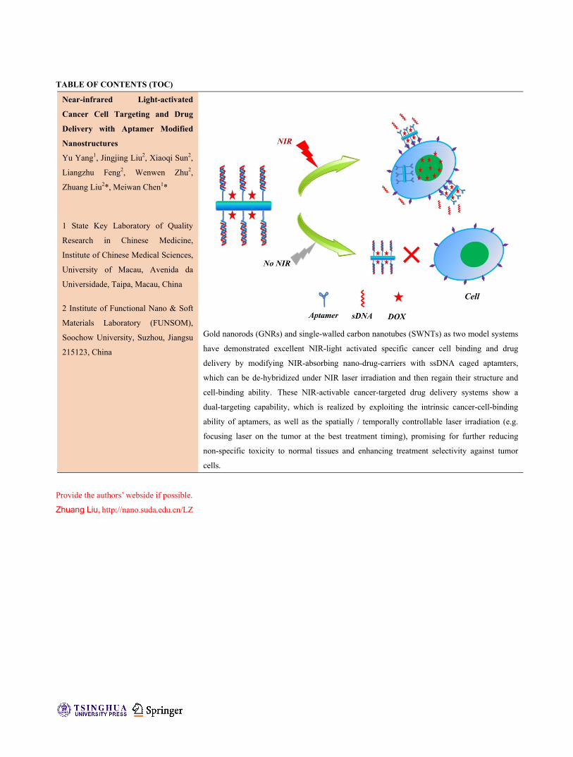
TABLE OF CONTENTS (TOC)
Near-infrared Light-activated
Cancer Cell Targeting and Drug
Delivery with Aptamer Modified
Nanostructures
Yu Yang1, Jingjing Liu2, Xiaoqi Sun2,
Liangzhu Feng2, Wenwen Zhu2,
Zhuang Liu2*, Meiwan Chen1*
1 State Key Laboratory of Quality
Research in Chinese Medicine,
Institute of Chinese Medical Sciences,
University of Macau, Avenida da
Universidade, Taipa, Macau, China
2 Institute of Functional Nano & Soft
Materials Laboratory (FUNSOM),
Soochow University, Suzhou, Jiangsu
215123, China
Gold nanorods (GNRs) and single-walled carbon nanotubes (SWNTs) as two model systems
have demonstrated excellent NIR-light activated specific cancer cell binding and drug
delivery by modifying NIR-absorbing nano-drug-carriers with ssDNA caged aptamters,
which can be de-hybridized under NIR laser irradiation and then regain their structure and
cell-binding ability. These NIR-activable cancer-targeted drug delivery systems show a
dual-targeting capability, which is realized by exploiting the intrinsic cancer-cell-binding
ability of aptamers, as well as the spatially / temporally controllable laser irradiation (e.g.
focusing laser on the tumor at the best treatment timing), promising for further reducing
non-specific toxicity to normal tissues and enhancing treatment selectivity against tumor
cells.
Provide the authors’ webside if possible.
Zhuang Liu, http://nano.suda.edu.cn/LZ


Near-infrared Light-activated Cancer Cell Targeting andDrug Delivery with Aptamer Modified Nanostructures
Yu Yang1, Jingjing Liu2, Xiaoqi Sun2, Liangzhu Feng2, Wenwen Zhu2, Zhuang Liu2(), Meiwan Chen1() 1 State Key Laboratory of Quality Research in Chinese Medicine, Institute of Chinese Medical Sciences, University of Macau, Avenida da Universidade, Taipa, Macau, China 2 Institute of Functional Nano & Soft Materials Laboratory (FUNSOM), Soochow University, Suzhou, Jiangsu 215123, China
Received: day month year
Revised: day month year
Accepted: day month year
(automatically inserted by
the publisher)
© Tsinghua University Press
and Springer-Verlag Berlin
Heidelberg 2014
KEYWORDS
NIR-activable, drug
delivery, aptamer, gold
nanorods, single-walled
carbon nanotubes
ABSTRACT
Stimuli-activated targeted delivery systems to provide highly accurate
treatment to tumors have received considerable attention in recent years.
Herein, we design a light-activable cancer targeting strategy that uses a
complementary DNA sequence to hybridize and mask sgc8 aptamers
conjugated on photothermal agents such as gold nanorods (GNRs) or
single-walled carbon nanotubes (SWNTs). Upon exposure to near-infrared
(NIR) laser, the localized photothermal heating on the surface of those
nano-agents would result in the dehybridization of the double strand DNA and
un-caging of the aptamer sequence to allow specific cancer cell targeting.
Utilizing doxorubicin (DOX) loaded SWNTs as a model system, cancer cell
targeted drug delivery that is activated by NIR light is realized. This work
demonstrates the concept of NIR-activable tumor-targeting delivery systems,
achieving controllable cancer cell binding to potentially enable highly specific
and efficient cancer therapy.
Nano Research
DOI (automatically inserted by the publisher)
Review Article/Research Article Please choose one

1. Introduction
In the past decade, many different types of
drug delivery systems have been explored, aiming
at increasing efficacy of therapeutic agents and
reducing toxicity to normal cells and tissues.1
Compared to the passive tumor targeting via
enhanced permeability and retention (EPR) effect,
the active tumor targeting can be achieved through
modifying targeting ligands including small
molecules2, peptides3, antibodies4 and aptamers5-7
on the surface of nanocarriers, enabling site-specific
targeting of tumor cells, tumor vasculatures, or
tumor microenvironment. Moreover, in order to
provide more accurate treatment to tumors and
improve the efficiency of anticancer therapy,
stimuli-activable targeted delivery systems based
on external physical stimuli including magnetic
field8, ultrasound9,10, temperature11 and light12 have
also been widely investigated13. Such strategies
would allow specific binding of drug carriers to
cancer cells only under certain physical stimuli
locally applied onto the tumor, to further enhance
treatment specificity and minimize non-specific
binding to normal tissues, some of which may
exhibit low levels of receptor expressions.14
Near-infrared (NIR) light, which exhibits little
phototoxicity and high tissue penetration ability,
has been extensively applied not only in biomedical
imaging15-17, but also in phototherapy of cancer18-22.
As a typical class of phototherapy, photothermal
therapy has been widely studied for efficient cancer
treatment through killing cancer cells by
light-generated hyperthermia.2,18,23,24 However, the
use of NIR light to activate cancer cell targeting of
nanoparticles via the photothermal effect has been
rarely explored except a recent work by Kohane et
al. 25 In that work, poly (NIPAAm-co-AAm), a
thermosensitive polymer, was used to cover the
target moieties conjugated on the surface of
silica-gold core-shell nanoparticles. After NIR laser
irradiation, the thermosensitive polymer was
constringed to allow the exposure of target ligands
for specific cell binding. However, photo-activated
cancer cell imaging and therapy has not yet been
demonstrated in their work.
Aptamers are oligonucleic acids which can
diversely and selectively bind to proteins, small
biological molecules and even cells, and have been
widely studied as targeting ligands for cancer
treatment with nanomolar even picamolar scale
dissociation constants (Kd).5-7,26-28 Herein, we
demonstrate a NIR-activable cancer cell targeting
strategy by conjugating aptamer which is caged by
their complimentary sequences, to the surface of
two types of commonly used photothermal agents,
gold nanorods (GNRs) and single-walled carbon
nanotubes (SWNTs). Upon NIR laser irradiation,
the effective photothermal heating localized on the
surface of GNRs or SWNTs could induce
de-hybridization of double-strand DNA and thus
uncaging of aptamer structures, which could then
recognize specific types of cancer cells. After
realizing NIR-activated specific cancer cell binding
with both GNRs and SWNTs, we further
demonstrate NIR-activated specific drug delivery
using SWNTs as a drug carrier for selective killing
of cancer cells under the control of NIR light. Our
strategy is promising for highly specific / selective
cancer treatment under the control of physical
stimuli.
2 Experimental
2.1 Materials.
HAuCl4 was purchased from Sigma-Aldrich
and doxorubicin (DOX) was purchased from Beijing
HuaFeng United Technology Co. Ltd.
Polylactide-poly (ethylene glycol) (PEG) was
obtained from Biomatrik Inc.
poly(maleicanhydride-alt-1-octadecene) (C18PMH),
1-Ethyl-3-(3-dimethylaminopropyl) carbodiimide
(EDC) were obtained from Sigma-Aldrich.
Thiol-functionalized sgc8 aptamer and
complementary single stranded DNA were
obtained from Takara Biotechnology (Dalian) Co.,

www.theNanoResearch.com∣www.Springer.com/journal/12274 | Nano Research
3Nano Res.
Ltd. Other chemicals were bought from Sinopharm
Group Co. Ltd.
2.2 Preparation of DNA modified GNRs.
GNRs were synthesized by the well-established
seed-mediated growth method 29 and purified by
washing with water for twice by centrifugation. To
confer them excellent physiological stability for
further experiments, the as-prepared GNRs were
coated with mPEG-SH by mixing 10 ml of 4 μM
GNRs with 2.0 μM mPEG-SH at room temperature
for 12 h. Afterwards, PEGylated GNRs (GNRs-PEG)
were collected by centrifugation, and stored at 4 oC
for further use. The molar concentrations of GNRs
were determined by their molar extinction
co-efficient at 808 nm (~1.02 × 10⁹ M−1cm−1).30
To prepare dsDNA, thiol-functionalized sgc8
aptamer in tris(hydroxymethyl)aminomethane-
ethylendiamintetraessigsäure (Tris-EDTA) buffer
was firstly heated at 95 oC for 2 min and then mixed
with its complementary ssDNA and annealed at 37
oC for 1 h. Thereafter, the as-prepared
thiol-functionalized dsDNA was incubated with
GNRs-PEG solution for 24 h under stirring, and
aged overnight using 0.2 M NaCl. Finally, DNA
modified GNRs complexes (GNRs-Apt/DNA) were
collected by centrifugation at 14800 rpm for 5 min
and stored at 4 oC for further use.
2.3 Functionalization of SWNTs with
C18PMH-PEG-NH2.
C18PMH-PEG-NH2 was synthesized according
to our previously used protocol.31 To prepare
SWNT-PEG-NH2, 1 mg of SWNTs were sonicated in
10 mL aqueous solution containing 10 mg of
C18PMH-PEG-NH2 for 60 min and then centrifuged
at 14800 rpm for 30 min to remove any precipitates,
yielding a black suspension of PEGylated SWNTs.
Then, the excess C18PMH-PEG-NH2 was removed
using a membrane filter with mean pore size of 100
nm. The molar concentrations of SWNTs were
determined by their molar extinction co-efficient at
808 nm (7.9 x 10⁶ M cm-1), with the estimated
average molecular weight of nanotubes to be 170
kDa.32
For aptamer conjugation, 8 mL as-prepared
SWNT-PEG-NH2 (300 nM) was mixed with 5.2 mg
of 4-(N-Maleimidomethyl)cyclohexane-1-carboxylic
acid 3-sulfo-N-hydroxysuccinimide ester sodium
salt (sulfo-SMCC) and stirred at room temperature
for 2 h. After removal of excess sulfo-SMCC using
an Amico centrifugal filter device with a molecular
weight cut off (MWCO) of 100 kDa, the obtained
SWNT-PEG-NH2 was mixed with dsDNA prepared
as aforementioned overnight in the presence of 0.l
mM tris(2-carboxyethyl)phosphine (TCEP) to break
disulfide bonds at 4 oC. Finally, the un-reacted
aptamer was removed with the Amico centrifugal
filter device.
2.4 Characterization
Transmission electron microscopy (TEM) images of
as-prepared GNRs and SWNTs were observed
using a transmission electron microscope (Philips
CM300) at an acceleration voltage of 200 kV.
UV-Vis-NIR absorption spectra were recorded with
a UV-Vis-NIR spectrophotometer (PerkinElmer
Lambda 750). Laser irradiation was carried out
using an optical-fiber-coupled power-tunable diode
laser (Hi-Tech Optoelectronics Co., Beijing, China)
Fluorescent emission spectra were obtained with
excitation at 490 nm using a FluoroMax-4
luminescent spectrometer (HORIBA JobinYvon
S.A.S).
2.5 Drug Loading
DOX loading onto different PEGylated SWNTs
was performed by mixing DOX with PEGylated
SWNTs (0.05 mg/mL) in PBS, (20 mM, pH8)
according to our previously developed procedure19.
For the DOX loading saturation experiment,
SWNT-PEG-Apt/DNA (0.05 mg/mL) was mixed
with various concentrations of DOX (0.1-0.4 mg/mL)
in PBS (20 mM, pH8) under stirring for 24 h. Excess

| www.editorialmanager.com/nare/default.asp
4 Nano Res.
unloaded DOX was removed using an Amico
centrifugal filter device with MWCO of 100 kDa
and washed at least three times with water until the
filtrate was free of color. Loading of DOX on other
SWNT-PEG solutions were implemented using the
same protocol.
2.6 Cell culture
Human leukemic lymphoblasts cells
(CCRF-CEM cells) were cultured using RPMI-1640
culture medium supplemented with 10 % fetal
bovine serum (FBS) and 1 % penicillin/streptomycin
at 37 °C within a humidified atmosphere containing
5 % CO2.
2.7 NIR light activated cellular uptake of
GNRs-Apt visualized by dark field imaging
The cellular uptake profiles of various GNRs
into CCRF-CEM cells were evaluated using an
Olympus BX51 optical microscopy with the
dark-field imaging setting. In brief, CCRF-CEM
cells were seeded in 12-well plate at a density of
1×10⁵ cells per well and then incubated with GNRs,
GNRs-Apt/DNA, and GNRs-Apt at 0.25 nM in
terms of GNRs. After being irradiated with an
808-nm NIR laser for 10 min at 0.5 W/cm2, the cells
were incubated for another 2 h. After that, the cells
were collected and washes with fresh cell culture
medium for 3 times followed by being imaged
using the aforementioned dark field imaging
system.
2.8 NIR light activated cellular uptake of
SWNTs-Apt visualized by a confocal laser
scanning microscopy
To explore the cellular uptake profile of various
SWNTs nanoconjugates under the NIR laser
irradiation, the amine groups of SWNT-PEG-NH2
were pre-labeled with FITC during aptamer
conjugation. CCRF-CEM cells were seeded in
12-well plates at a density of 1×10⁵ cells per well and
then treated with FITC-labeled SWNT-PEG,
SWNT-PEG-Apt/DNA, and SWNT-PEG-Apt at 50
nM in terms of SWNTs followed by being irradiated
with an 808-nm NIR laser at the power density of
0.5 W/cm2 for 10 min. Then, the cells were
incubated for 2 h and imaged under a confocal laser
scanning microscopy (Leica SP5II, German).
2.9 NIR-light activated targeted cell killing abilities
of DOX loaded SWNT-Apt nanocomplexes
The in vitro cytotoxicity of various DOX loaded
SWNT nanoconjugated was determined by a
standard methyl thiazolyl tetrazolium (MTT) assay.
In brief, CCRF-CEM cells (1×10⁴ cells per well) were
cultured in 96-well plates for 12 h. Then, the cells
were incubated with free DOX, SWNT-PEG+DOX,
SWNT-PEG-Apt/DNA+DOX, or
SWNT-PEG-Apt+DOX at a concentration of 5
g/mL in terms of DOX, as well as
SWNT-PEG-Apt/DNA and SWNT-PEG-Apt at the
same SWNTs concentration (50 nM) to their
corresponding counterparts. After being irradiated
with an 808-nm laser at a power density of 0.5
W·cm-2 for 10 min, the cells were incubated for 2 h
before being washed and transferred into fresh cell
culture for 24 h of re-incubation. After that, MTT
assay was conducted to determine relative cell
viabilities of different samples. 3. Results and Discussion
The synthesis of aptamer modified
photothermal nanostructures is illustrated in
Scheme 1. Sgc8 (5’-(SH)- TT TTT TTT TTT TTT
TTT TAT CTA ACT GCT GCG CCG CCG GGA
AAA TAC TGT ACG GTT AGA-3’)29, an aptamer
which has high affinity and selectivity to bind the
cell membrane protein tyrosine kinase-7 (PTK7),
was conjugated on the surface of photothermal
nanoparticles. Then, a single-strand DNA (ssDNA)
with the complementary sequence (5’-TCT AAC
CGT ACA GTA TTT T-3’) was added to mask the
sgc8 aptamer via the formation of double-stranded

www.theNanoResearch.com∣www.Springer.com/journal/12274 | Nano Research
5Nano Res.
DNA (dsDNA). Upon NIR laser irradiation, the heat
converted by NIR light would enable the
dehybridization of dsDNA and the subsequent
exposure of the sgc8 aptamer sequence, to allow for
specific cancer cell binding.
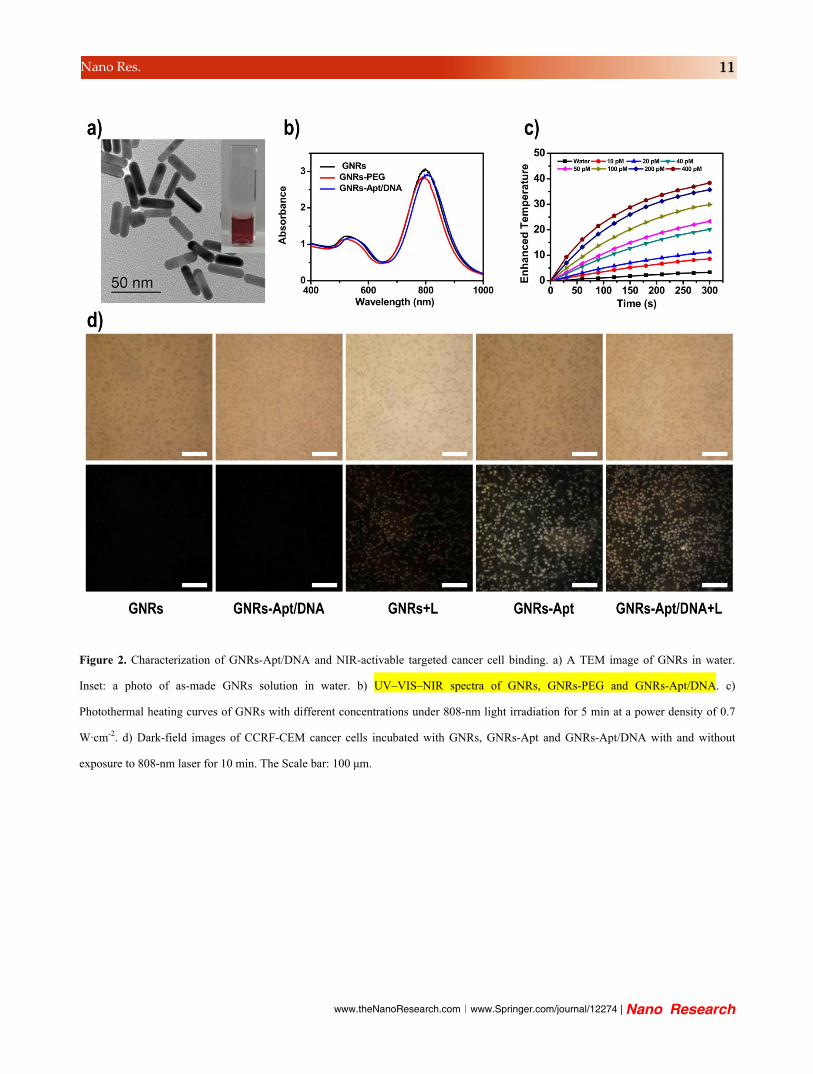
Gold nanorods (GNRs) were firstly used as a
photothermal agent in this experiment because of
their high efficiency of photothermal conversion as
well as convenient and adjustable surface
modification. GNRs were synthesized according to
the seed-mediated method.29,33-35 As shown in
Figure 2a&b, the aspect ratio of gold nanorods
(GNRs) was approximately 3 as revealed by
transmission electron microscope (TEM) imaging.
The strong surface plasmon resonance absorption
peak of GNRs in the NIR region was observed from
the UV-vis-NIR spectrum (Figure 2b). As expected,
GNRs displayed remarkable and
concentration-dependent photothermal effect under
irradiation by an 808-nm NIR laser, indicating that
they were effective photothermal agents (Figure 2c).
In the next step, thiol-terminated PEG was
conjugated on the surfaces of GNRs via Au-S bond
to reduce the agglomeration and cytotoxicity of
GNRs. Meanwhile, the thiol-terminated poly-T
chain linked with the 5’-end of sgc8 aptamer, which
could self-hybridize to form the special hairpin
structure, was also conjugated on the surface of
GNRs (GNRs-Apt) via Au-S bond36. Sgc8 aptamer
has high affinity to bind protein tyrosine kinase-7,
which is over-expressed on the membrane of
CCRF-CEM cells (human T lymphoblast cell).
Subsequently, ssDNA with the complementary
sequence was added to hybridize with sgc8
aptamer and mask / cage its cancer cell binding
ability.
GNRs could be imaged under dark-field
microscope due to their strong light scattering by
surface plasmonic resonance.37 Therefore, we
investigated the NIR-activated cancer cells binding
of GNRs-Apt/DNA complexes via dark-field
scattering imaging. As shown in Figure 2d, strong
scattered light signals were observed for
CCRF-CEM cells after incubation with GNRs-Apt
but for those incubated with plain GNRs,
suggesting the high affinity binding of GNRs-Apt to
CCRF-CEM cells. Subsequently, followed by adding
of complementary sequence, the GNRs-Apt was
hybridized with its complementary sequence and
the recognition capability between CCRF-CEM cells
and GNRs-Apt/DNA was vanished. As the result,
rather weak scattered light signals were observed
for CCRF-CEM cells incubated with
GNRs-Apt/DNA (Figure 2d). Interestingly, with the
irradiation of 808 nm laser illuminating at the
power intensity of 0.5 W·cm-2 for 10 min, strong
scattered light signals were observed for cells
incubated with GNRs-Apt/DNA complexes. This
phenomenon could be explained by that GNRs
could convert absorbed NIR light into heat, and
then trigger the de-hybridization of dsDNA by
increasing the local temperature to be above the Tm
of this dsDNA, so as to expose the sgc8 aptamer for
cancer cell targeting. In the meanwhile, weak
signals were also observed when CCRF-CEM cells
were incubated with GNRs under 808 nm NIR laser
because of the photothermally enhanced cellular
uptake of nanoparticles19.
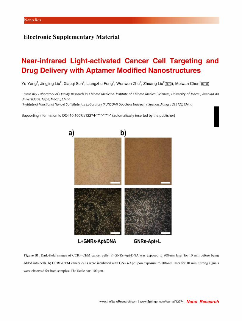
To further prove the enhanced cell binding of
GNRs-Apt/DNA on cells under NIR laser was
indeed owing to the de-hybridization of dsDNA,
GNRs-Apt/DNA complexes were irradiated by the
808-nm NIR laser (0.5 W·cm-2 for 10 min) first before
incubation with CCRF-CEM cells. As shown in
Supporting Figure S1, the strong scattered light
signals from CCRF-CEM cells evidenced that the
photothermal effect could induce dsDNA
dehybridization to release complementary sequence,
exposing sgc8 aptamers to allow binding with cells.
Overall, GNRs-Apt/DNA complexes exhibit NIR
light-controllable cancer cell binding ability, which
may be utilized to enable high efficient cancer
therapy and reduce the normal tissues toxicity.
In order to utilize NIR light activated cancer

| www.editorialmanager.com/nare/default.asp
6 Nano Res.
cells targeting to achieve light-controlled cancer
therapy, SWNTs, a commonly used photothermal
agent and drug nanocarrier38,39, was used as another
nano-platform in addition to GNRs in this work.
Firstly, amine-terminated polyethylene glycol
(PEG)-grafted poly (maleic
anhydride-alt-1-octadecene) (C18PMH-PEG-NH2)
polymer was used to modify the surface of SWNTs
to enhance their biocompatibility and prevent their
agglomeration in aqueous solutions. The same
thiol-terminated poly-T chain linked the 5’-end of
sgc8, was introduced on the surface of SWNTs via a
bi-functional linker. TEM image revealed the length
of SWNT-PEG-aptamer conjugates
(SWNT-PEG-Apt) to be in the range from 100 to 300
nm (Figure 3a). With strong NIR absorbance,
SWNTs could also serve as an effective
photothermal agent under NIR laser exposure
(Figure 3b).
Subsequently, a commonly used anti-cancer drug,
doxorubicin (DOX), was loaded onto the outer-wall
of SWNTs via π-π stacking and hydrophobic
interaction owing to their sp2-bonded carbon
surface and high surface area. As shown in figure
3c, the UV-vis-NIR absorption spectra of
SWNT-PEG-DOX complexes demonstrated the
special absorption of DOX at ~490 nm was
enhanced with the increasing of mass of added
DOX, indicating DOX could be effectively loaded
on SWNTs. DOX loaded SWNTs (SWNT-PEG+DOX)
with a mass ratio of 1:1 was used in our
experiments.
We next would like to evaluate the concept of
NIR-activated cancer cell targeting with
SWNT-PEG-Apt/DNA. Since the fluorescence of
DOX was quenched after loading on the surface of
SWNTs (Supporting Figure S2), a fluorescent dye
(FITC) was used to label PEGylated SWNTs to
enable confocal fluorescence imaging of cells. In the
experiment, CCRF-CEM cancer cells were incubated
with SWNT-PEG, SWNT-PEG-Apt, or
SWNT-PEG-Apt/DNA, all of which were
pre-labeled by FITC. Confocal fluorescence images
of those cells were performed with or without
exposing to NIR light. As expected, strong
fluorescence signals were detected from
CCRF-CEM cancer cells with incubation of
SWNT-PEG-Apt but not for those incubated with
SWNT-PEG (Figure 3d). Similar to data with gold
nanorods, after the aptamer was caged by the
complementary ssDNA, the cell binding of
SWNT-PEG-Apt/DNA was greatly reduced. By
comparison, obvious enhanced fluorescence was
seen from CCRF-CEM cancer cells incubated with
SWNT-PEG-Apt/DNA exposed to the NIR laser,
because SWNTs could efficiently convert NIR light
into heat to dehybridize the dsDNA and un-cage
the sgc8 aptamers for cells binding. As expected
and consistent to previous findings (Figure 1d),40
the mild photothermal heating of SWNT-PEG
incubated cells would also slightly enhanced the
cellular uptake of SWNTs, but to a degree much
lower than that of cells incubated with
SWNT-PEG-Apt in dark, or SWNT-PEG-Apt/DOX
with NIR exposure. Those results were further
confirmed by quantitative flow cytometry data.
SWNT-PEG-Apt/DNA under laser irradiation
showed the highest level of cellular binding /
uptake (Supporting Figure S3), owing not only to
the NIR-activated cancer cell binding, but also to
the photothermally enhanced cellular uptake of
nanotubes.19,40
At last, NIR-activated targeted cell killing
ability was demonstrated by evaluating the relative
cell viabilities of CCRF-CEM cells treated by free
DOX, SWNT-PEG+DOX, SWNT-PEG-Apt/DNA,
SWNT-PEG-Apt/DNA+DOX, SWNT-PEG-Apt and
SWNT-PEG-Apt+DOX in the presence or absence of
NIR laser irradiation using the standard MTT assay.
As shown in Figure 4, our results indicated that
SWNT-PEG-Apt and SWNT-PEG-Apt/DNA would
not induce obvious cell death even after 10-min
laser irradiation at 0.5 W/cm2, indicating that
PEGylated SWNTs are not toxic to cells and the

www.theNanoResearch.com∣www.Springer.com/journal/12274 | Nano Research
7Nano Res.
mild heating effect induced by SWNTs would be
tolerable by cells, consistent with our previously
published results. 40,41 Under the tested condition
(relatively low drug concentration and short
incubation time), free DOX showed rather limited
cancer cell killing efficiency regardless of laser
irradiation, so as SWNT-PEG+DOX in dark. The
mild photothermal heating of SWNT-PEG+DOX
could slightly enhance its toxicity against cancer
cells, likely owing to the mild hyperthermia
enhanced endocytosis of nano-carriers. Interesting,
SWNT-PEG-Apt/DNA+DOX, while being non-toxic
to cells in dark, showed rather effective cancer cell
killing ability in the presence of NIR light, which
could uncage the cell-binding aptamer sequence to
allow for the specific binding of drug-loaded
nanotubes with cells. As a positive control,
SWNT-PEG-Apt+DOX without caging aptamer
with the base-pairing ssDNA exhibited high
cytotoxicity to cells under both dark and laser
irradiation conditions.
Our results in this work demonstrate excellent
NIR-light activated specific cancer cell binding and
drug delivery by modifying NIR-absorbing
nano-drug-carriers with caged aptamters. In
comparison with commonly explored conventional
cancer targeting strategies, such NIR-activable
cancer-targeted drug delivery systems show a
dual-targeting capability, which is realized by
exploiting the intrinsic cancer-cell-binding ability of
aptamers, as well as the spatially / temporally
controllable laser irradiation (e.g. focusing laser on
the tumor at the best treatment timing), promising
for further reducing non-specific toxicity to normal
tissues and enhancing treatment selectivity against
tumor cells.
4. Conclusion
In summary, our work using GNRs and SWNTs
as two model systems have demonstrated excellent
NIR-light activated specific cancer cell binding and
drug delivery by modifying NIR-absorbing
nano-drug-carriers with ssDNA caged aptamters,
which can be de-hybridized under NIR laser
irradiation and then regain their structure and
cell-binding ability. In comparison with commonly
explored conventional cancer targeting strategies,
such NIR-activable cancer-targeted drug delivery
systems show a dual-targeting capability, which is
realized by exploiting the intrinsic
cancer-cell-binding ability of aptamers, as well as
the spatially / temporally controllable laser
irradiation (e.g. focusing laser on the tumor at the
best treatment timing), promising for further
reducing non-specific toxicity to normal tissues and
enhancing treatment selectivity against tumor cells.
Acknowledgements
This work was partially supported by the National
Natural Science Foundation of China (51222203,
51132006), the National “973” Program of China
(2011CB911002, 2012CB932601), a Jiangsu Natural
Science Fund for Distinguished Young Scholars, the
Macao Science and Technology Development Fund
(062/2013/A2) and the Research Fund of the
University of Macau
(MYRG2014-00033-ICMS-QRCM,
MRG004/CMW/2014/ICMS).
Electronic Supplementary Material: Supplementary
material (data regarding dark-field images of
CCRF-CEM cells, Fluorescence emission spectra of
DOX before and after loading on the
SWNT-PEG-Apt/DNA, and Flow cytometry assay of
CCRF-CEM cells) is available in the online version of
this article at
http://dx.doi.org/10.1007/s12274-***-****-*
(automatically inserted by the publisher). References
(1) Allen, T. M.; Cullis, P. R.: Drug delivery systems: entering the mainstream. Science 2004, 303, 1818-1822.
(2) Cheng, L.; Yang, K.; Li, Y.; Chen, J.; Wang, C.; Shao, M.; Lee, S.-T.; Liu, Z.: Facile Preparation of Multifunctional Upconversion Nanoprobes for Multimodal Imaging and Dual-Targeted

| www.editorialmanager.com/nare/default.asp
8 Nano Res.
Photothermal Therapy. Angewandte Chemie 2011, 123, 7523-7528.
(3) Sun, X.; Wang, C.; Gao, M.; Hu, A.; Liu, Z.: Remotely Controlled Red Blood Cell Carriers for Cancer Targeting and Near ‐ Infrared Light ‐Triggered Drug Release in Combined Photothermal–Chemotherapy. Advanced Functional Materials 2015, 25, 2386-2394.
(4) Gearhart, P. J.; Johnson, N. D.; Douglas, R.; Hood, L.: IgG antibodies to phosphorylcholine exhibit more diversity than their IgM counterparts. 1981.
(5) Chu, T. C.; Marks, J. W.; Lavery, L. A.; Faulkner, S.; Rosenblum, M. G.; Ellington, A. D.; Levy, M.: Aptamer: toxin conjugates that specifically target prostate tumor cells. Cancer research 2006, 66, 5989-5992.
(6) Farokhzad, O. C.; Jon, S.; Khademhosseini, A.; Tran, T.-N. T.; LaVan, D. A.; Langer, R.: Nanoparticle-aptamer bioconjugates a new approach for targeting prostate cancer cells. Cancer research 2004, 64, 7668-7672.
(7) Kim, D.; Jeong, Y. Y.; Jon, S.: A drug-loaded aptamer− gold nanoparticle bioconjugate for combined CT imaging and therapy of prostate cancer. Acs Nano 2010, 4, 3689-3696.
(8) Oliveira, H.; Pérez-Andrés, E.; Thevenot, J.; Sandre, O.; Berra, E.; Lecommandoux, S.: Magnetic field triggered drug release from polymersomes for cancer therapeutics. J. Control. Release 2013, 169, 165-170.
(9) Hernot, S.; Klibanov, A. L.: Microbubbles in ultrasound-triggered drug and gene delivery. Advanced drug delivery reviews 2008, 60, 1153-1166.
(10) Geers, B.; Lentacker, I.; Sanders, N. N.; Demeester, J.; Meairs, S.; De Smedt, S. C.: Self-assembled liposome-loaded microbubbles: The missing link for safe and efficient ultrasound triggered drug-delivery. J. Control. Release 2011, 152, 249-256.
(11) Kim, C.; Lee, Y.; Kim, J. S.; Jeong, J. H.; Park, T. G.: Thermally triggered cellular uptake of quantum dots immobilized with poly (N-isopropylacrylamide) and cell penetrating peptide. Langmuir 2010, 26, 14965-14969.
(12) Hansen, M. B.; van Gaal, E.; Minten, I.; Storm, G.; van Hest, J. C.; Löwik, D. W.: Constrained and UV-activatable cell-penetrating peptides for intracellular delivery of liposomes. J. Control. Release 2012, 164, 87-94.
(13) Huang, Y.; Jiang, Y.; Wang, H.; Wang, J.; Shin, M. C.; Byun, Y.; He, H.; Liang, Y.; Yang, V. C.: Curb challenges of the “Trojan Horse” approach: smart strategies in achieving effective yet safe
cell-penetrating peptide-based drug delivery. Advanced drug delivery reviews 2013, 65, 1299-1315.
(14) Parker, N.; Turk, M. J.; Westrick, E.; Lewis, J. D.; Low, P. S.; Leamon, C. P.: Folate receptor expression in carcinomas and normal tissues determined by a quantitative radioligand binding assay. Anal. Biochem. 2005, 338, 284-293.
(15) Larson, D. R.; Zipfel, W. R.; Williams, R. M.; Clark, S. W.; Bruchez, M. P.; Wise, F. W.; Webb, W. W.: Water-soluble quantum dots for multiphoton fluorescence imaging in vivo. Science 2003, 300, 1434-1436.
(16) Yang, Y.; Xiang, K.; Yang, Y.-X.; Wang, Y.-W.; Zhang, X.; Cui, Y.; Wang, H.; Zhu, Q.-Q.; Fan, L.; Liu, Y.: An individually coated near-infrared fluorescent protein as a safe and robust nanoprobe for in vivo imaging. Nanoscale 2013, 5, 10345-10352.
(17) Welsher, K.; Liu, Z.; Sherlock, S. P.; Robinson, J. T.; Chen, Z.; Daranciang, D.; Dai, H.: A route to brightly fluorescent carbon nanotubes for near-infrared imaging in mice. Nature nanotechnology 2009, 4, 773-780.
(18) Cheng, L.; Wang, C.; Feng, L.; Yang, K.; Liu, Z.: Functional nanomaterials for phototherapies of cancer. Chem. Rev. 2014, 114, 10869-10939.
(19) Liu, J.; Wang, C.; Wang, X.; Wang, X.; Cheng, L.; Li, Y.; Liu, Z.: Mesoporous Silica Coated Single-WallednCarbon Nanotubes as a Multifunctional Light-Responsive Platform for Cancer Combination Therapy. Advanced Functional Materials 2015, 25, 384-392.
(20) Yavuz, M. S.; Cheng, Y.; Chen, J.; Cobley, C. M.; Zhang, Q.; Rycenga, M.; Xie, J.; Kim, C.; Song, K. H.; Schwartz, A. G.: Gold nanocages covered by smart polymers for controlled release with near-infrared light. Nature materials 2009, 8, 935-939.
(21) Cheng, L.; Liu, J.; Gu, X.; Gong, H.; Shi, X.; Liu, T.; Wang, C.; Wang, X.; Liu, G.; Xing, H.: PEGylated WS2 Nanosheets as a Multifunctional Theranostic Agent for in vivo Dual ‐ Modal CT/Photoacoustic Imaging Guided Photothermal Therapy. Advanced Materials 2014, 26, 1886-1893.
(22) Yan, H.; Teh, C.; Sreejith, S.; Zhu, L.; Kwok, A.; Fang, W.; Ma, X.; Nguyen, K. T.; Korzh, V.; Zhao, Y.: Functional mesoporous silica nanoparticles for photothermal‐controlled drug delivery in vivo. Angewandte Chemie International Edition 2012, 51, 8373-8377.
(23) Liu, T.; Wang, C.; Gu, X.; Gong, H.; Cheng, L.; Shi, X.; Feng, L.; Sun, B.; Liu, Z.: Drug Delivery with PEGylated MoS2 Nano‐sheets for Combined Photothermal and Chemotherapy of Cancer. Advanced Materials 2014 26, 3433-3440..
(24) Song, X.; Chen, Q.; Liu, Z.: Recent

www.theNanoResearch.com∣www.Springer.com/journal/12274 | Nano Research
9Nano Res.
advances in the development of organic photothermal nano-agents. Nano Research 2015, 8, 340-354.
(25) Barhoumi, A.; Wang, W.; Zurakowski, D.; Langer, R. S.; Kohane, D. S.: Photothermally Targeted Thermosensitive Polymer-Masked Nanoparticles. Nano letters 2014, 14, 3697-3701.
(26) Qiu, L.; Chen, T.; O�c�soy, I.; Yasun, E.; Wu, C.; Zhu, G.; You, M.; Han, D.; Jiang, J.; Yu, R.: A cell-targeted, size-photocontrollable, nuclear-uptake nanodrug delivery system for drug-resistant cancer therapy. Nano letters 2014, 15, 457-463.
(27) Liang, H.; Zhang, X.-B.; Lv, Y.; Gong, L.; Wang, R.; Zhu, X.; Yang, R.; Tan, W.: Functional DNA-containing nanomaterials: cellular applications in biosensing, imaging, and targeted therapy. Accounts of chemical research 2014, 47, 1891-1901.
(28) Zhu, Z.; Wu, C.; Liu, H.; Zou, Y.; Zhang, X.; Kang, H.; Yang, C. J.; Tan, W.: An Aptamer Cross‐Linked Hydrogel as a Colorimetric Platform for Visual Detection. Angewandte Chemie 2010, 122, 1070-1074.
(29) Wang, J.; Zhu, G.; You, M.; Song, E.; Shukoor, M. I.; Zhang, K.; Altman, M. B.; Chen, Y.; Zhu, Z.; Huang, C. Z.: Assembly of aptamer switch probes and photosensitizer on gold nanorods for targeted photothermal and photodynamic cancer therapy. ACS nano 2012, 6, 5070-5077.
(30) Zauner, W.: Technical Note 802: Photothermal Characteristics of Gold Nanorods. 2008.
(31) Wang, C.; Cheng, L.; Liu, Z.: Drug delivery with upconversion nanoparticles for multi-functional targeted cancer cell imaging and therapy. Biomaterials 2011, 32, 1110-1120.
(32) Kam, N. W. S.; O'Connell, M.; Wisdom, J. A.; Dai, H.: Carbon nanotubes as multifunctional biological transporters and near-infrared agents for selective cancer cell destruction. Proceedings of the National Academy of Sciences of the United States of America 2005, 102, 11600-11605.
(33) Jang, B.; Kim, Y. S.; Choi, Y.: Effects of Gold Nanorod Concentration on the Depth‐Related Temperature Increase During Hyperthermic Ablation. Small 2011, 7, 265-270.
(34) Terentyuk, G.; Panfilova, E.; Khanadeev, V.; Chumakov, D.; Genina, E.; Bashkatov, A.; Tuchin, V.; Bucharskaya, A.; Maslyakova, G.; Khlebtsov, N.: Gold nanorods with a hematoporphyrin-loaded silica shell for dual-modality photodynamic and photothermal treatment of tumors in vivo. Nano Research 2014, 7, 325-337.
(35) Bhaumik, J.; Mittal, A. K.; Banerjee, A.; Chisti, Y.; Banerjee, U. C.: Applications of
phototheranostic nanoagents in photodynamic therapy. Nano Research 2014, 8, 1373-1394.
(36) Qiu, L.; Chen, T.; Öçsoy, I.; Yasun, E.; Wu, C.; Zhu, G.; You, M.; Han, D.; Jiang, J.; Yu, R.; Tan, W.: A Cell-Targeted, Size-Photocontrollable, Nuclear-Uptake Nanodrug Delivery System for Drug-Resistant Cancer Therapy. Nano Letters 2015, 15, 457-463.
(37) Ding, H.; Yong, K.-T.; Roy, I.; Pudavar, H. E.; Law, W. C.; Bergey, E. J.; Prasad, P. N.: Gold nanorods coated with multilayer polyelectrolyte as contrast agents for multimodal imaging. The Journal of Physical Chemistry C 2007, 111, 12552-12557.
(38) Wang, F.; Liu, J.: Nanodiamond decorated liposomes as highly biocompatible delivery vehicles and a comparison with carbon nanotubes and graphene oxide. Nanoscale 2013, 5, 12375-12382.
(39) Samorì, C.; Ali-Boucetta, H.; Sainz, R.; Guo, C.; Toma, F. M.; Fabbro, C.; Da Ros, T.; Prato, M.; Kostarelos, K.; Bianco, A.: Enhanced anticancer activity of multi-walled carbon nanotube–methotrexate conjugates using cleavable linkers. Chemical Communications 2010, 46, 1494-1496.
(40) Tian, B.; Wang, C.; Zhang, S.; Feng, L. Z.; Liu, Z.: Photothermally Enhanced Photodynamic Therapy Delivered by Nano-Graphene Oxide. Acs Nano 2011, 5, 7000-7009.
(41) Feng, L.; Yang, X.; Shi, X.; Tan, X.; Peng, R.; Wang, J.; Liu, Z.: Polyethylene Glycol and Polyethylenimine Dual ‐ Functionalized Nano ‐Graphene Oxide for Photothermally Enhanced Gene Delivery. Small 2013, 9, 1989-1997.
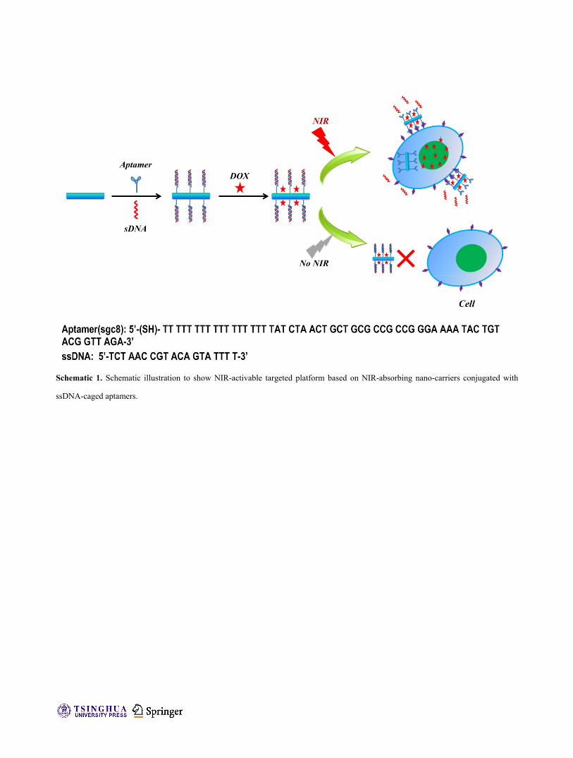
Schematic 1. Schematic illustration to show NIR-activable targeted platform based on NIR-absorbing nano-carriers conjugated with
ssDNA-caged aptamers.
www.theNanoResearch.com∣www.Springer.com/journal/12274 | Nano Research
11Nano Res.
Figure 2. Characterization of GNRs-Apt/DNA and NIR-activable targeted cancer cell binding. a) A TEM image of GNRs in water.
Inset: a photo of as-made GNRs solution in water. b) UV–VIS–NIR spectra of GNRs, GNRs-PEG and GNRs-Apt/DNA. c)
Photothermal heating curves of GNRs with different concentrations under 808-nm light irradiation for 5 min at a power density of 0.7
W·cm-2. d) Dark-field images of CCRF-CEM cancer cells incubated with GNRs, GNRs-Apt and GNRs-Apt/DNA with and without
exposure to 808-nm laser for 10 min. The Scale bar: 100 μm.

| www.editorialmanager.com/nare/default.asp
12 Nano Res.
Figure 3. Characterization of SWNT-PEG-Apt/DNA and NIR-activable targeted cancer cell binding. a) A TEM image of SWNT -PEG.
Inset: a photo of as-made SWNT-PEG solution in water. b) Photothermal heating curves of SWNT-PEG with different concentrations
irradiated with the 808-nm light for 5 min at a power density of 0.7 W·cm-2. c) UV–Vis–NIR spectra of SWNT-PEG-Apt/DNA loaded
with various concentrations of DOX. d) Laser scanning confocal microscope images of CCRF-CEM cells incubated with FITC-labeled
SWNT-PEG, SWNT-PEG-Apt and SWNT-PEG-Apt/DNA with and without exposure to 808-nm NIR light for 10 min. The Scale bar: 50
μm.

www.theNanoResearch.com∣www.Springer.com/journal/12274 | Nano Research
13Nano Res.
Figure 4. Relative viabilities of CCRF-CEM cells after various treatments. In this experiment, CCRF-CEM cells were incubated
with DOX, DOX loaded SWNT-PEG (SWNT-PEG+DOX), SWNT-PEG-Apt, DOX loaded SWNT-PEG-Apt
(SWNT-PEG-Apt+DOX), SWNT-PEG-Apt/DNA, and DOX loaded SWNT-PEG-Apt/DNA (SWNT-PEG-Apt/DNA+DOX) with and
without exposure to the 808-nm laser at 0.5 W/cm2 for 10 min. After further incubation for 2 h, cells were washed and transferred
into fresh cell culture for addition incubation for 24 h before the MTT assay.

www.theNanoResearch.com∣www.Springer.com/journal/12274 | Nano Research
Nano Res.
Electronic Supplementary Material
Near-infrared Light-activated Cancer Cell Targeting andDrug Delivery with Aptamer Modified Nanostructures
Yu Yang1, Jingjing Liu2, Xiaoqi Sun2, Liangzhu Feng2, Wenwen Zhu2, Zhuang Liu2(), Meiwan Chen1() 1 State Key Laboratory of Quality Research in Chinese Medicine, Institute of Chinese Medical Sciences, University of Macau, Avenida da Universidade, Taipa, Macau, China 2 Institute of Functional Nano & Soft Materials Laboratory (FUNSOM), Soochow University, Suzhou, Jiangsu 215123, China
Supporting information to DOI 10.1007/s12274-****-****-* (automatically inserted by the publisher)
Figure S1. Dark-field images of CCRF-CEM cancer cells. a) GNRs-Apt/DNA was exposed to 808-nm laser for 10 min before being
added into cells. b) CCRF-CEM cancer cells were incubated with GNRs-Apt upon exposure to 808-nm laser for 10 min. Strong signals
were observed for both samples. The Scale bar: 100 μm.

| www.editorialmanager.com/nare/default.asp
Nano Res.
Figure S2. Fluorescence emission spectra of DOX before and after loading on the SWNT-PEG-Apt/DNA . Excitation wavelength =
490 nm.
Figure S3. Flow cytometry assay of CCRF-CEM cells after different treatment.