Development Biology INTRODUCTION Ms. Lucky Juneja Lecturer, School of Biotechnology, DAVV.
30
Development Biology INTRODUCTION Ms. Lucky Juneja Lecturer, School of Biotechnology, DAVV
-
Upload
jeremy-newton -
Category
Documents
-
view
219 -
download
0
Transcript of Development Biology INTRODUCTION Ms. Lucky Juneja Lecturer, School of Biotechnology, DAVV.

Development Biology INTRODUCTION
Ms. Lucky JunejaLecturer, School of Biotechnology, DAVV
Aristotle: The first embryologist
Development: slow process of progressive change
Development of a multicellular organism begins with a single cell the fertilized egg, or zygote, which divides mitotically to produce all the cells of the body
Objectives of development:
. generates cellular diversity
. order within each generation
. it ensures the continuity of life from one generation to the next.

FEW TERMS:
Differentiation: A single cell, the fertilized egg, gives rise to hundreds of different cell type. This generation of cellular diversity is called differentiation.
Morphogenesis: differentiated cells are not randomly distributed. Rather, they are organized into intricate tissues and organs. This creation of ordered form is called morphogenesis.
Homologous structures are those organs whose underlying similarity arises from their being derived from a common ancestral structure. For example, the wing of a bird and the forelimb of a human are homologous.
Analogous structures are those whose similarity comes from their performing a similar function, rather than their arising from a common ancestor. Therefore, for example, the wing of a butterfly and the wing of a bird are analogous.
Abnormalities due to exogenous agents (certain chemicals or viruses, radiation, or hyperthermia) are called disruptions. The agents responsible for these disruptions are called teratogens (Greek, "monster-formers"), and the study of how environmental agents disrupt normal development is called teratology.

QUESTIONS ABOUT DEVELOPMENT:
The question of differentiation: Since each cell of the body (exceptions) contains the same set of genes, we need to understand how this same set of genetic instructions can produce different types of cells?
The question of morphogenesis: How can the cells form such ordered structures?
The question of growth: How do our cells know when to stop dividing? How is cell division so tightly regulated?
The question of reproduction: How are sperm and egg cells set apart to form the next generation, and what are the instructions in the nucleus and cytoplasm that allow them to function this way?
The question of evolution: How do changes in development create new body forms?
The question of environmental integration: How is the development of an organism integrated into the larger context of its habitat?

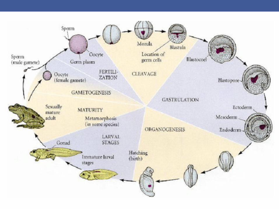
Circle of Life: The Stages of Animal Development
1. Immediately following fertilization, cleavage occurs. Cleavage is a series of extremely rapid mitotic divisions wherein the enormous volume of zygote cytoplasm is divided into numerous smaller cells. These cells are called blastomeres, and by the end of cleavage, they generally form a sphere known as a blastula. 2. After the rate of mitotic division has slowed down, the blastomeres undergo dramatic movements wherein they change their positions relative to one another. This series of extensive cell rearrangements is called gastrulation, and the embryo is said to be in the gastrula stage. As a result of gastrulation, the embryo contains three germ layers: the ectoderm, the endoderm, and the mesoderm.
3. The cells interact with one another and rearrange themselves to produce tissues and organs. This process is called organogenesis.

4.Germ cell: in many species a specialized portion of egg cytoplasm gives rise to cells that are the precursors of the gametes (the sperm and egg). The gametes and their precursor cells are collectively called germ cells, and they are set aside for reproductive function.
All the other cells of the body are called somatic cells.
5. In many species, the organism that hatches from the egg or is born into the world is not sexually mature. Indeed, in most animals, the young organism is a larva that may look significantly different from the adult. Larvae often constitute the stage of life that is used for feeding or dispersal. In many species, the larval stage is the one that lasts the longest, and the adult is a brief stage solely for reproduction.
Fertilization is the initiating step in development. The zygote, with its new genetic potential and its new arrangement of cytoplasm, now begins the production of a multicellular organism. Between these events of fertilization and the events of organ formation are two critical stages: cleavage and gastrulation.
Cleavage, a series of mitotic divisions whereby the enormous volume of egg cytoplasm is divided into numerous smaller, nucleated cells. These cleavage-stage cells are called blastomeres. In most species (mammals being the chief exception), the rate of cell division and the placement of the blastomeres with respect to one another is completely under the control of the proteins and mRNAs stored in the oocyte by the mother.
First the egg is divided in half, then quarters, then eighths, and so forth. This division of egg cytoplasm without increasing its volume is accomplished by abolishing the growth period between cell divisions (that is, the G1 and G2 phases of the cell cycle). Meanwhile, the cleavage of nuclei occurs at a rapid rate.

One consequence of this rapid cell division is that the ratio of cytoplasmic to nuclear volume gets increasingly smaller as cleavage progresses.
This decrease in the cytoplasmic to nuclear volume ratio is crucial in timing the activation of certain genes. For example, in the frog Xenopus laevis, transcription of new messages is not activated until after 12 divisions.
The transition from fertilization to cleavage is caused by the activation of mitosis promoting factor (MPF).
MPF continues to play a role after fertilization, regulating the biphasic cell cycle of early blastomeres.
Blastomeres generally progress through a cell cycle consisting of just two steps: M (mitosis) and S (DNA synthesis)
The MPF activity of early blastomeres is highest during M and undetectable during S.
What causes this cyclic activity of MPF? Mitosis-promoting factor contains two subunits. The large subunit is called cyclin B (component that shows a periodic behavior, accumulating during S and then being degraded after the cells have reached M) (Evans et al. 1983; Swenson et al. 1986). Cyclin B is often encoded by mRNAs stored in the oocyte cytoplasm, and if the translation of this message is specifically inhibited, the cell will not enter mitosis (Minshull et al. 1989). Cyclin B regulates the small subunit of MPF, the cyclin-dependent kinase.
This kinase activates mitosis by phosphorylating several target proteins, including histones, the nuclear envelope lamin proteins, and the regulatory subunit of cytoplasmic myosin.
This brings about chromatin condensation, nuclear envelope depolymerization, and the organization of the mitotic spindle. Without cyclin, the cyclin-dependent kinase will not function. The presence of cyclin is controlled by several proteins that ensure its periodic synthesis and degradation. In most species studied, the regulators of cyclin (and thus, of MPF) are stored in the egg cytoplasm. Therefore, the cell cycle is independent of the nuclear genome for numerous cell divisions

The embryo now enters the mid-blastula transition, in which several new phenomena are added to the biphasic cell divisions of the embryo. First, the growth stages (G1 and G2) are added to the cell cycle, permitting the cells to grow
Second, the synchronicity of cell division is lost, as different cells synthesize different regulators of MPF.
Third, new mRNAs are transcribed.
Cleavage is actually the result of two coordinated processes. The first of these cyclic processes is karyokinesis the mitotic division of the nucleus.
The second process is cytokinesis the division of the cell.
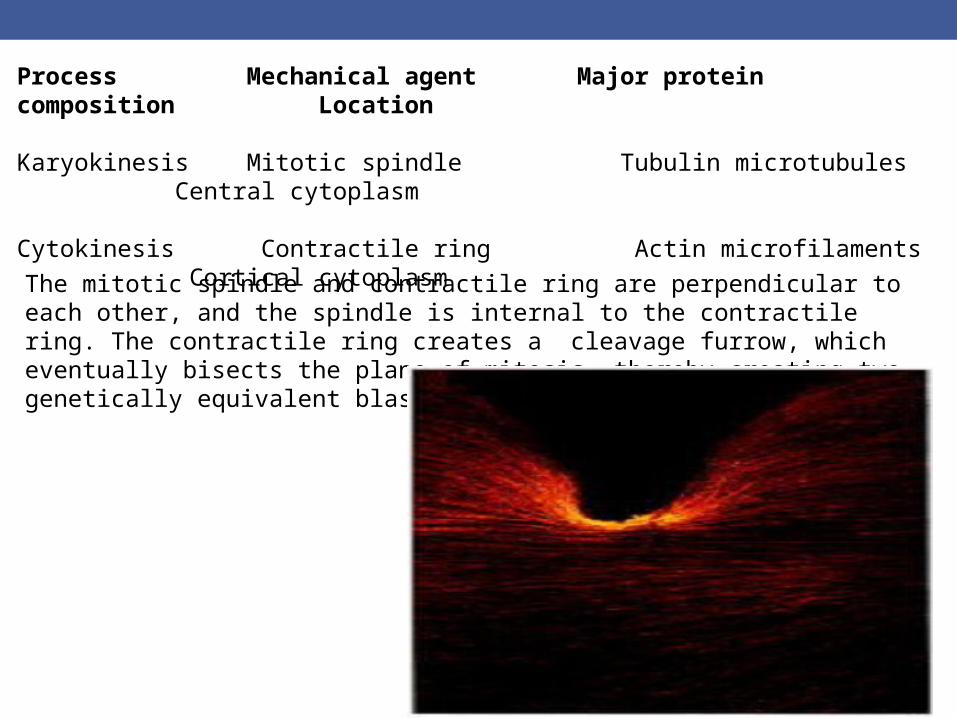
Process Mechanical agent Major protein composition Location Karyokinesis Mitotic spindle Tubulin microtubules Central cytoplasm Cytokinesis Contractile ring Actin microfilaments Cortical cytoplasmThe mitotic spindle and contractile ring are perpendicular to each other, and the spindle is internal to the contractile ring. The contractile ring creates a cleavage furrow, which eventually bisects the plane of mitosis, thereby creating two genetically equivalent blastomeres.
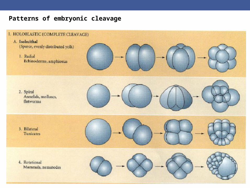
Patterns of embryonic cleavage

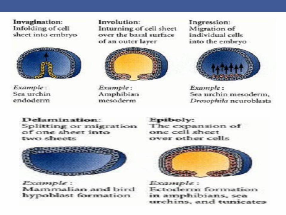
Gastrulation:Gastrulation is the process of highly coordinated cell and tissue movements whereby the cells of the blastula are dramatically rearranged.
The cells that will form the endodermal and mesodermal organs are brought inside the embryo, while the cells that will form the skin and nervous system are spread over its outside surface.
Thus, the three germ layers outer ectoderm, inner endoderm, and interstitial mesoderm are first produced during gastrulation. In addition, the stage is set for the interactions of these newly positioned tissues.
TYPES OF CELL MOMENTS DURING GASTRULATION:
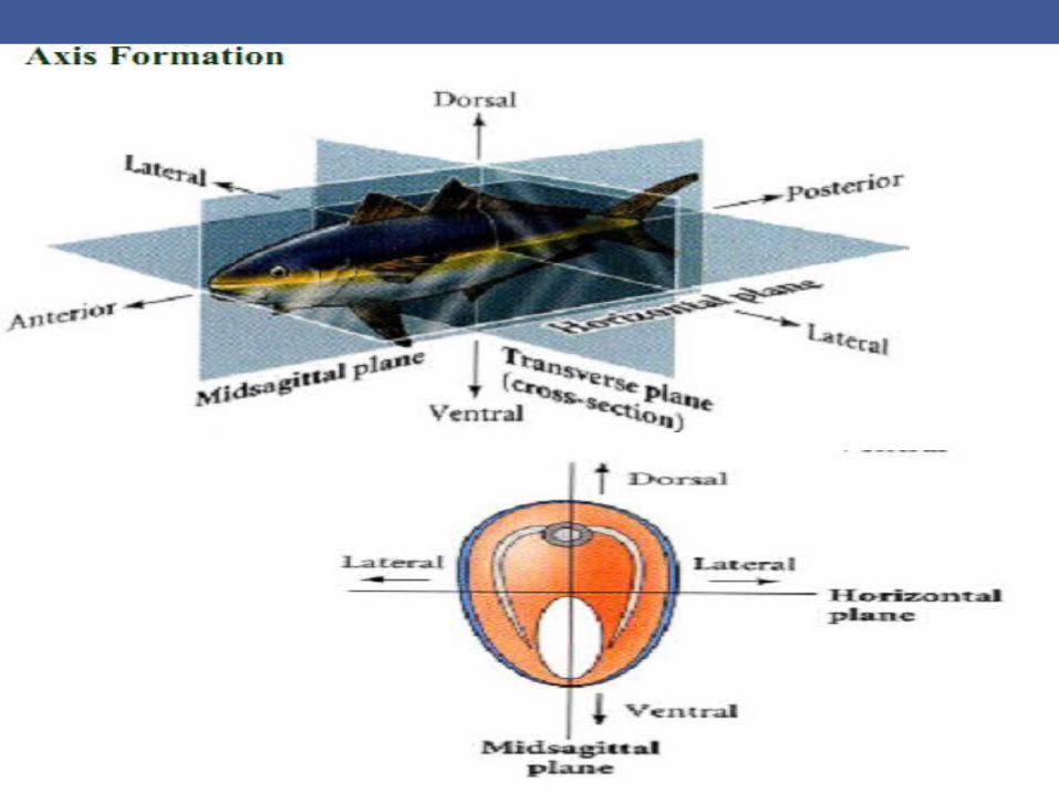
Cell Specification and Axis Formation :
Embryos must develop three very important axes that are the foundations of the body:the anterior-posterior axis, the dorsal-ventral axis, and the right-left axis.
The anterior-posterior (or anteroposterior) axis is the line extending from head to tail (or mouth to anus in those organisms that lack heads and tails).
The dorsal-ventral (dorsoventral) axis is the line extending from back (dorsum) to belly (ventrum). For instance, in vertebrates, the neural tube is a dorsal structure. In insects, the neural cord is a ventral structure.
The right-left axis is a line between the two lateral sides of the body.
DROSOPHILA DEVELOPMENT

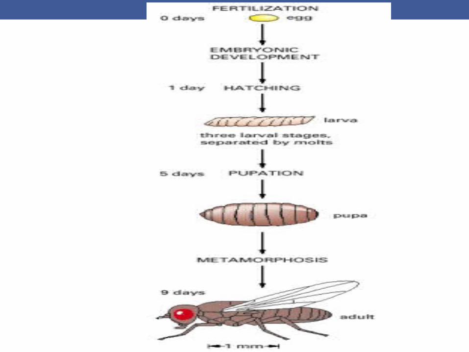
Embryonic development:Fertilization….Day 1: development ,embryo hatches out of the egg shell to become a larva
Day 2,03 and so : larva- three stages/instars, separated by moltingend of third stage, Pupa forms.
Inside a pupa a radical remodeling of the body - a process called metamorphosis
After Day 9:Adult fly/ Imago
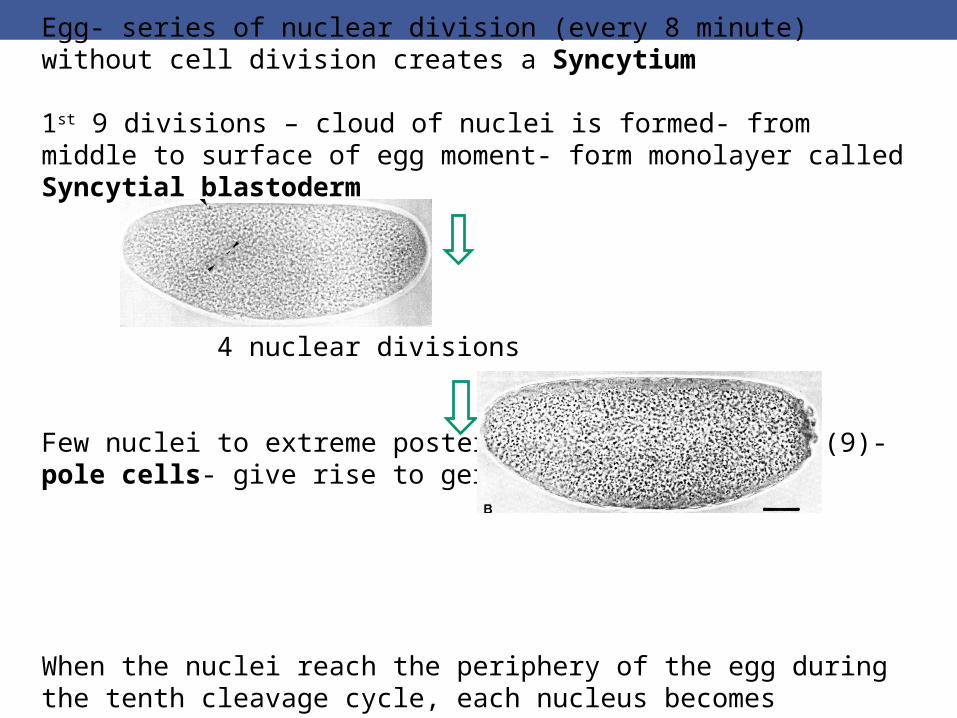
Egg- series of nuclear division (every 8 minute) without cell division creates a Syncytium
1st 9 divisions – cloud of nuclei is formed- from middle to surface of egg moment- form monolayer called Syncytial blastoderm 4 nuclear divisions
Few nuclei to extreme posterior end – few cycles (9)- pole cells- give rise to germ cells.
When the nuclei reach the periphery of the egg during the tenth cleavage cycle, each nucleus becomes surrounded by microtubules and microfilaments. The nuclei and their associated cytoplasmic islands are called energids. (up to 12th cycle)

Plasma membrane grow inward converting syncytial blastoderm into cellular blastoderm (13th cycle)
Cell division slows down, asynchronous, and transcription rate is increased.
Blastoderm fate map:

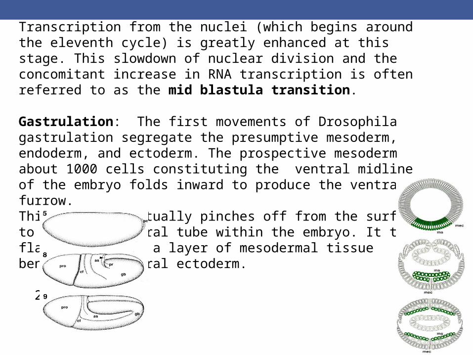
Transcription from the nuclei (which begins around the eleventh cycle) is greatly enhanced at this stage. This slowdown of nuclear division and the concomitant increase in RNA transcription is often referred to as the mid blastula transition.
Gastrulation: The first movements of Drosophila gastrulation segregate the presumptive mesoderm, endoderm, and ectoderm. The prospective mesoderm about 1000 cells constituting the ventral midline of the embryo folds inward to produce the ventral furrow. This furrow eventually pinches off from the surface to become a ventral tube within the embryo. It then flattens to form a layer of mesodermal tissue beneath the ventral ectoderm. 2:50 h
3:40 h
4:20 h

The prospective endoderm invaginates as two pockets at the anterior and posterior ends of the ventral furrow. The pole cells are internalized along with the endoderm. At this time, the embryo bends to form the cephalic furrow.
The ectodermal cells on the surface and the mesoderm undergo convergence and extension, migrating toward the ventral midline to form the germ band, a collection of cells along the ventral midline that includes all the cells that will form the trunk of the embryo.
The germ band extends posteriorly and, perhaps because of the egg case, wraps around the top (dorsal) surface of the embryo


While the germ band is in its extended position, several key morphogenetic processes occur: organogenesis, segmentation, and the segregation of the imaginal discs. Imaginal discs are those cells set aside to produce the adult structures.
In addition, the nervous system forms from two regions of ventral ectoderm. Neuroblasts differentiate from this neurogenic ectoderm within each segment (and also from the Non segmented region of the head ectoderm). Therefore, in insects like Drosophila, the nervous system is located ventrally.

Body plan of Drosophila:
HeadThree thoracic segments (T1 T2 T3)Eight abdominal segments (A1 to A8)
The first thoracic segment, has only legs; the second thoracic segment has legs and wings; and the third thoracic segment has legs and halteres (balancers).
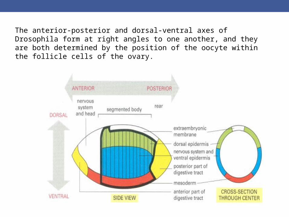
The anterior-posterior and dorsal-ventral axes of Drosophila form at right angles to one another, and they are both determined by the position of the oocyte within the follicle cells of the ovary.