
Development and validation of an oligonucleotide microarray to ...
11
BioMed Central Page 1 of 11 (page number not for citation purposes) BMC Microbiology Open Access Methodology article Development and validation of an oligonucleotide microarray to characterise ectomycorrhizal fungal communities Marlis Reich*, Annegret Kohler, Francis Martin and Marc Buée* Address: UMR 1136 INRA/Nancy Université Interactions Arbres/Microorganimes, INRA Nancy, 54280 Champenoux, France Email: Marlis Reich* - [email protected]; Annegret Kohler - [email protected]; Francis Martin - [email protected]; Marc Buée* - [email protected] * Corresponding authors Abstract Background: In forest ecosystems, communities of ectomycorrhizal fungi (ECM) are influenced by several biotic and abiotic factors. To understand their underlying dynamics, ECM communities have been surveyed with ribosomal DNA-based sequencing methods. However, most identification methods are both time-consuming and limited by the number of samples that can be treated in a realistic time frame. As a result of ongoing implementation, the array technique has gained throughput capacity in terms of the number of samples and the capacity for parallel identification of several species. Thus far, although phylochips (microarrays that are used to detect species) have been mostly developed to trace bacterial communities or groups of specific fungi, no phylochip has been developed to carry oligonucleotides for several ectomycorrhizal species that belong to different genera. Results: We have constructed a custom ribosomal DNA phylochip to identify ECM fungi. Specific oligonucleotide probes were targeted to the nuclear internal transcribed spacer (ITS) regions from 95 fungal species belonging to 21 ECM fungal genera. The phylochip was first validated using PCR amplicons of reference species. Ninety-nine percent of the tested oligonucleotides generated positive hybridisation signals with their corresponding amplicons. Cross-hybridisation was mainly restricted at the genus level, particularly for Cortinarius and Lactarius species. The phylochip was subsequently tested with environmental samples that were composed of ECM fungal DNA from spruce and beech plantation fungal communities. The results were in concordance with the ITS sequencing of morphotypes and the ITS clone library sequencing results that were obtained using the same PCR products. Conclusion: For the first time, we developed a custom phylochip that is specific for several ectomycorrhizal fungi. To overcome cross-hybridisation problems, specific filter and evaluation strategies that used spot signal intensity were applied. Evaluation of the phylochip by hybridising environmental samples confirmed the possible application of this technology for detecting and monitoring ectomycorrhizal fungi at specific sites in a routine and reproducible manner. Published: 24 November 2009 BMC Microbiology 2009, 9:241 doi:10.1186/1471-2180-9-241 Received: 1 June 2009 Accepted: 24 November 2009 This article is available from: http://www.biomedcentral.com/1471-2180/9/241 © 2009 Reich et al; licensee BioMed Central Ltd. This is an Open Access article distributed under the terms of the Creative Commons Attribution License (http://creativecommons.org/licenses/by/2.0 ), which permits unrestricted use, distribution, and reproduction in any medium, provided the original work is properly cited.
Transcript of Development and validation of an oligonucleotide microarray to ...

BioMed CentralBMC Microbiology
ss
Open AcceMethodology articleDevelopment and validation of an oligonucleotide microarray to characterise ectomycorrhizal fungal communitiesMarlis Reich*, Annegret Kohler, Francis Martin and Marc Buée*Address: UMR 1136 INRA/Nancy Université Interactions Arbres/Microorganimes, INRA Nancy, 54280 Champenoux, France
Email: Marlis Reich* - [email protected]; Annegret Kohler - [email protected]; Francis Martin - [email protected]; Marc Buée* - [email protected]
* Corresponding authors
AbstractBackground: In forest ecosystems, communities of ectomycorrhizal fungi (ECM) are influencedby several biotic and abiotic factors. To understand their underlying dynamics, ECM communitieshave been surveyed with ribosomal DNA-based sequencing methods. However, most identificationmethods are both time-consuming and limited by the number of samples that can be treated in arealistic time frame. As a result of ongoing implementation, the array technique has gainedthroughput capacity in terms of the number of samples and the capacity for parallel identificationof several species. Thus far, although phylochips (microarrays that are used to detect species) havebeen mostly developed to trace bacterial communities or groups of specific fungi, no phylochip hasbeen developed to carry oligonucleotides for several ectomycorrhizal species that belong todifferent genera.
Results: We have constructed a custom ribosomal DNA phylochip to identify ECM fungi. Specificoligonucleotide probes were targeted to the nuclear internal transcribed spacer (ITS) regions from95 fungal species belonging to 21 ECM fungal genera. The phylochip was first validated using PCRamplicons of reference species. Ninety-nine percent of the tested oligonucleotides generatedpositive hybridisation signals with their corresponding amplicons. Cross-hybridisation was mainlyrestricted at the genus level, particularly for Cortinarius and Lactarius species. The phylochip wassubsequently tested with environmental samples that were composed of ECM fungal DNA fromspruce and beech plantation fungal communities. The results were in concordance with the ITSsequencing of morphotypes and the ITS clone library sequencing results that were obtained usingthe same PCR products.
Conclusion: For the first time, we developed a custom phylochip that is specific for severalectomycorrhizal fungi. To overcome cross-hybridisation problems, specific filter and evaluationstrategies that used spot signal intensity were applied. Evaluation of the phylochip by hybridisingenvironmental samples confirmed the possible application of this technology for detecting andmonitoring ectomycorrhizal fungi at specific sites in a routine and reproducible manner.
Published: 24 November 2009
BMC Microbiology 2009, 9:241 doi:10.1186/1471-2180-9-241
Received: 1 June 2009Accepted: 24 November 2009
This article is available from: http://www.biomedcentral.com/1471-2180/9/241
© 2009 Reich et al; licensee BioMed Central Ltd. This is an Open Access article distributed under the terms of the Creative Commons Attribution License (http://creativecommons.org/licenses/by/2.0), which permits unrestricted use, distribution, and reproduction in any medium, provided the original work is properly cited.
Page 1 of 11(page number not for citation purposes)

BMC Microbiology 2009, 9:241 http://www.biomedcentral.com/1471-2180/9/241
BackgroundEctomycorrhizal (ECM) fungi form a mutualistic symbio-sis with tree roots and play key roles in forest ecosystems.In return for receiving nutrients and water from the soilvia the roots, they receive carbohydrates as photosynthatefrom their host plants [1]. As is the case for other soil fun-gal species, the composition of the ECM community isaffected by both biotic and abiotic factors; these includeclimate changes, seasons, soil micro-site heterogeneity,soil and litter quality, host tree species and forest manage-ment [2-6]. To describe in more detail the impact of envi-ronmental factors on community composition, long-term, year-round monitoring and a detailed spatialdescription of the community has to be carried out. How-ever, analyses are very often hindered by a limited samplenumber and by the ephemeral or cryptic lifestyle of thefungi [7,8].
Over the last fifteen years, PCR-based molecular methodsand DNA sequencing of nuclear and mitochondrial ribos-omal DNA have been used routinely to identify mycor-rhizal fungi [9]. However, these methods are time-consuming and are limited in the number of samples thatcan be treated in a realistic time frame [10]. With auto-mated molecular genotyping techniques, appropriateDNA databases [11] and a better knowledge of ITS varia-bility within fungal species [12], identification of fungaltaxa in environmental samples can now be expandedfrom the aforementioned methods to high-throughputmolecular diagnostic tools, such as phylochips [13]. Sofar, DNA arrays have been mainly used for genome-widetranscription profiling [14,15], but also for the identifica-tion of bacterial species from complex environmentalsamples [16] or for the identification of a few genera ofpathogenic fungi and Oomycetes [17,18]. Phylochipsmay comprise up to several thousand probes that targetphylogenetic marker genes, such as 16S rRNA in bacteriaor the internal transcribed spacer (ITS) region in fungi[19]; indeed, the latter is one of the most widely used bar-coding regions for fungi [20]. Phylochips have severaladvantages over traditional approaches, including higherspecificity, cost efficiency, rapid identification and detec-tion of target organisms, and the high numbers of samplesthroughput; therefore, they are increasingly used for thedetection of bacterial and pathogenic fungi [21,22]. In theECM fungal ecology field, the first application of ribos-omal DNA arrays was reported by Bruns and Gardes [23];they developed a specific phylochip (on nylon mem-branes) to detect Suilloid fungi. Recently, this approachhas also been used for truffle identification [24]. To thebest of our knowledge, no study has reported the con-struction and application of an ECM fungal phylochip todetect a large number of ECM fungal species that belongto various genera from environmental samples.
Here, we report the first application of a custom ribos-omal ITS phylochip to describe the community composi-tion of ECM fungi on roots. The phylochip carried specificoligonucleotides for 95 fungal species that belong to 25ECM fungal genera. The specificity of the oligonucleotideswas evaluated using ITS amplicons of known referencespecies. The method was then used to describe ECM fun-gal communities that were obtained from 30-year-oldspruce and beech plantations. To validate the phylochip,morphotyping and ITS sequencing of the ECM root tips,together with sequencing of ITS clone libraries, were car-ried out. We discuss the pros and cons of the phylochip incomparison to conventional approaches, and outline itspotential applications for environmental monitoring.
ResultsIdentification of ECM fungi from environmental samples by morphotyping/ITS sequencing and sequencing of ITS clone librariesBy combining morphotyping and ITS sequencing of indi-vidual ECM root tips, and sequencing of ITS clone librar-ies, 26 fungal species were identified on the roots of beechand spruce trees; these included 25 ECM fungi (Table 1).Rarefaction curves of clone library coverage nearlyreached a plateau, which indicated a near complete sam-pling of the ECM species in the soil samples that weretaken from under the beech and spruce. In order to detectonly one more species from spruce samples and a furthertwo species from beech samples, it would be necessary toincrease the sequencing effort two-fold (Additional file 1).The species richness was very similar for the two planta-tions, with 13 and 16 species being associated with spruceand beech, respectively; however, the community compo-sitions were clearly distinct. Only three ECM taxa werefound on the root tips of both hosts: Cenococcumgeophilum, Xerocomus pruinatus and Tomentellopsis submollis(Table 1). Sequencing of the ITS clone libraries or identi-fication of individual ECM morphotypes revealed similarfungal ECM profiles. Most fungi that were detected onspruce roots by sequencing of the ITS library were alsodetected by morphotyping (Additional file 2). Of thesemorphotypes, nine were also supported by sequencingthe ITS of individual morphotypes (Table 1). One taxonwas only identified with morphotyping and ITS-sequenc-ing of individual ECM morphotypes, and another wasidentified only by morphotyping. Overall, 9 of 13 taxa(69%) from the spruce roots were identified by bothmolecular methods. A total of 10 of 16 taxa (62.5%) fromthe beech roots were identified by both approaches.Sequencing of the ITS clone libraries resulted in the detec-tion of an additional two taxa. One of these was related toan unidentified endophyte, which was difficult to identifyby morphotyping alone as it is likely leaving inside theroot tissues (Table 1). A single taxon was identified onlyby the morphotyping/ITS sequencing approach, and three
Page 2 of 11(page number not for citation purposes)

BMC Microbiology 2009, 9:241 http://www.biomedcentral.com/1471-2180/9/241
taxa were identified only by morphotyping. Using ITS1Fand ITS4 primers [9] or NSI1/NLB4 [25], the ITS regionfrom six ECM morphotypes (Amanita rubescens, Inocybe sp1, Lactarius sp 1 + 2, Tomentella sp 1, Tomentellopsis submol-lis) were not amplified. The ITS regions from four fungi(A. rubescens, Lactarius sp 1 + 2, Tomentella sp 1) of thosesix morphotypes were also not amplified using the ITSclone library approach (Table 1). However, the use of thesecond primer pair, NSl1/NLB4, enabled the molecularbiological characterisation of four morphotypes (Pilo-derma sp., Sebacinaceae sp., Sebacina sp. and Pezizales sp.)that were not amplified with ITS1f/ITS4.
Specificity of designed oligonucleotidesThe specificity of the 95 designed oligonucleotides (Addi-tional file 3) was evaluated using PCR amplicons that
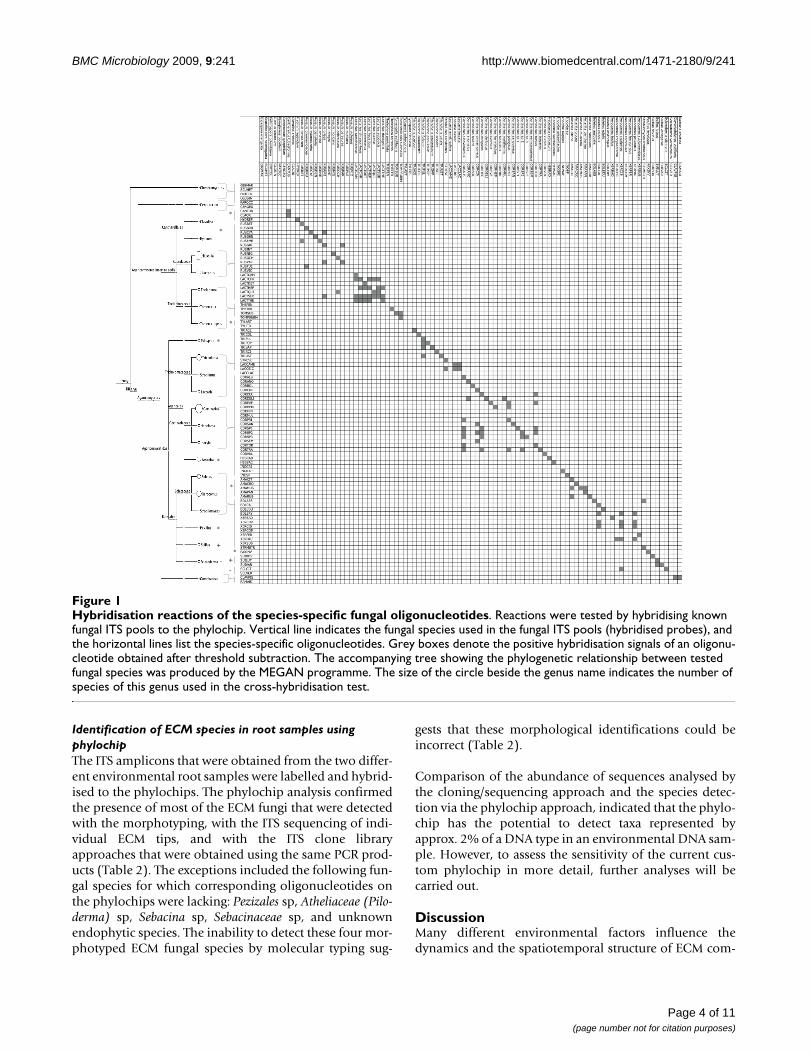
were generated from sporocarp tissues. PCR ampliconsmainly hybridised to the phylochip oligonucleotidesaccording to the expected patterns (Figure 1), and the pat-terns were highly reproducible in the replications con-ducted with each of the templates. The hybridisationsignal intensities ranged from -22 (background value) to44,835 units. Ninety-nine percent of the oligonucleotidestested generated positive hybridisation signals with theirmatching ITS. Cross-hybridisations were mainly observedwithin the Cortinarius and Lactarius species complex.Among the Boletaceae species, a few cross-hybridisationswere observed between the species that belonged to theBoletus and Xerocomus genera. Within the Amanita, Russulaor Tricholoma genus, rare cross-reactions occurred betweensingle sequences from closely related species.
Table 1: Fungal taxa identified on root tip samples from spruce and beech by sequencing of the ITS clone libraries of the pooled ECM tips and morphotyping/ITS sequencing of the individual ECM root tips.
Pooled ECM tipsITS cloning/ITS sequencing
Individual ECM tipsMorphotyping/ITS sequencing
Species name Acc. n° Identities (%)(Unite�/NCBI❍)
Acc. n° Identities (%)(Unite�/NCBI❍)
ECM from Picea abies:Thelephora terrestris EU427330.1 360/363 (100)❍ UDB000971 142/151 (94)�Cenococcum geophilum UDB002297 375/379 (98)� UDB002297 211/216 (97)�Clavulina cristata UDB001121 375/375 (100)� UDB 001121 281/289 (97)�Atheliaceae (Piloderma) sp AY097053.1 343/362 (94)❍ EU597016.1 612/624 (98)❍
Cortinarius sp 1 AJ889974.1 361/367 (98)❍ UDB002224 232/242 (95)�Xerocomus pruinatus UDB000018 348/351 (99)� UDB 000016 692/696 (99)�Tomentelopsis submollis AM086447.1 319/324 (98)❍ morphotyping onlyInocybe sp AY751555.1 249/266 (93)❍ morphotyping onlyXerocomus badius UDB000080 375/379 (98)� UDB000080 400/417 (95)�Tylospora asterophora UDB002469 353/354 (99)� UDB002469 591/594 (99)�Tylospora fibrillosa AF052563.1 405/408 (99)❍ AJ0534922.1 561/578 (97)❍
Sebacina sp not detected UDB000975 162/168 (96)�Lactarius sp 1 not detected morphotyping only
ECM from Fagus sylvatica:Pezizales sp UDB002381 28/28 (100)� DQ990873.1 602/646 (93)❍
Sebacinaceae sp EF619763.1 327/347 (94)❍ EF195570.1 495/497 (99)❍
Laccaria amethystina UDB002418 356/360 (98)� UDB002418 276/277 (99)�Endophyte AY268198.1 205/243 (84)❍ not detectedInocybe napipes UDB000017 292/294 (99)� UDB000017 148/155 (95)�Xerocomus pruinatus UDB000483 241/242 (99)� UDB000483 279/288 (96)�Cortinarius sp 2 UDB002410 416/437 (95)� UDB002410 227/239 (95)�Cortinarius sp 3 UDB002170 306/316 (96)� UDB002445 57/59 (96)�Cortinarius tortuosus UDB002164 279/284 (98)� not detectedRussula puellaris UDB000010 313/315 (99)� UDB000010 246/247 (99)�Tomentellopsis submollis UDB000198 272/273 (99)� UDB000198 224/228 (98)�Laccaria laccata UDB000104 322/327 (98)� UDB000769 283/283 (100)�Cenococcum geophilum not detected UDB002297 216/222 (97)�Amanita rubescens not detected morphotyping onlyLactarius sp 2 not detected morphotyping onlyTomentella sp not detected morphotyping only
For the sequence homology search, BLASTN was carried out with the NCBI (❍) and UNITE (�) databases. Accession numbers (Acc. n°) and identities are given.
Page 3 of 11(page number not for citation purposes)
BMC Microbiology 2009, 9:241 http://www.biomedcentral.com/1471-2180/9/241
Identification of ECM species in root samples using phylochipThe ITS amplicons that were obtained from the two differ-ent environmental root samples were labelled and hybrid-ised to the phylochips. The phylochip analysis confirmedthe presence of most of the ECM fungi that were detectedwith the morphotyping, with the ITS sequencing of indi-vidual ECM tips, and with the ITS clone libraryapproaches that were obtained using the same PCR prod-ucts (Table 2). The exceptions included the following fun-gal species for which corresponding oligonucleotides onthe phylochips were lacking: Pezizales sp, Atheliaceae (Pilo-derma) sp, Sebacina sp, Sebacinaceae sp, and unknownendophytic species. The inability to detect these four mor-photyped ECM fungal species by molecular typing sug-
gests that these morphological identifications could beincorrect (Table 2).
Comparison of the abundance of sequences analysed bythe cloning/sequencing approach and the species detec-tion via the phylochip approach, indicated that the phylo-chip has the potential to detect taxa represented byapprox. 2% of a DNA type in an environmental DNA sam-ple. However, to assess the sensitivity of the current cus-tom phylochip in more detail, further analyses will becarried out.
DiscussionMany different environmental factors influence thedynamics and the spatiotemporal structure of ECM com-
Hybridisation reactions of the species-specific fungal oligonucleotidesFigure 1Hybridisation reactions of the species-specific fungal oligonucleotides. Reactions were tested by hybridising known fungal ITS pools to the phylochip. Vertical line indicates the fungal species used in the fungal ITS pools (hybridised probes), and the horizontal lines list the species-specific oligonucleotides. Grey boxes denote the positive hybridisation signals of an oligonu-cleotide obtained after threshold subtraction. The accompanying tree showing the phylogenetic relationship between tested fungal species was produced by the MEGAN programme. The size of the circle beside the genus name indicates the number of species of this genus used in the cross-hybridisation test.
*
*
*
*
*
*
+
+
--
*
*
Page 4 of 11(page number not for citation purposes)

BMC Microbiology 2009, 9:241 http://www.biomedcentral.com/1471-2180/9/241
munities [26,27,5,4]. A better understanding of the mech-anisms underlying these dynamics will require year-roundECM monitoring at incrementally increased spatial reso-lutions. However, the limited number of samples that cancurrently be analysed hinders the use of molecularapproaches for large-scale studies. With the ongoingdevelopment of high-throughput molecular diagnostictools, such as DNA oligoarrays [19] and 454 pyrosequenc-ing [28], larger scale surveys (in terms of both the fre-quency and depth of analysis) of soil fungi are nowpossible. Ecologically relevant sample throughput in thein the 100 to 1000 range is now accessible. So far, phylo-chips have been used for the identification of bacteria
[29], viruses [30], and a few genera of closely related fun-gal species [18].
In the present study, we constructed a custom ribosomalDNA phylochip for the identification of ECM fungi thatwas based on the ITS1 and ITS2 regions. One of the greatadvantages of using ITS regions for oligonucleotide designis the high number of sequences that are available in pub-lic databases [12]. Furthermore, these regions are some ofthe most frequently used regions for the barcoding ofECM fungi [20], and compared to other possible barcod-ing regions, they show a high specificity at the specieslevel [31]. We designed a total of 95 oligonucleotides,
Table 2: Detection of fungal taxa from root tips of spruce and beech using different identification approaches.
Species name Morphotyping/ITS sequencingof individual ECM tips
ITS cloning/sequencing of ECM tip pools
Phylochip
samples from Picea abiesThelephora terrestris x x xCenococcum geophilum x x xClavulina cristata x x xAtheliaceae (Piloderma) sp x x no oligonucleotideCortinarius sp 1 x x xXerocomus pruinatus x x xTomentellopsis submollis morphotyping only x xInocybe sp morphotyping only x xXerocomus badius x x xTylospora asterophora x x xTylospora fibrillosa x x xSebacina sp x no oligonucleotideCortinarius sp 2 xRussula integra xCortinarius alboviolaceus xCortinarius traganus xAmanita muscaria xLactarius sp1 morphotyping only
ECM from Fagus sylvaticaPezizales sp x x no oligonucleotideSebacinaceae sp x x no oligonucleotideLaccaria amethystina x x xEndophyte sp. x no oligonucleotideInocybe napipes x x xXerocomus pruinatus x x xCortinarius sp 2 x x xCortinarius sp 3 x x xCortinarius tortuosus x xRussula puellaris x x xTomentellopsis submollis x x xLaccaria laccata x x xCenococcum geophilum x xCortinarius sp 1 xCortinarius hinnuleus xRussula integra xLaccaria bicolor xAmanita rubescens morphotyping onlyLactarius sp2 morphotyping onlyTomentella sp morphotyping only
Page 5 of 11(page number not for citation purposes)

BMC Microbiology 2009, 9:241 http://www.biomedcentral.com/1471-2180/9/241
from which 89 were species-specific for ECM fungal spe-cies. According to regular fruiting body surveys, these 89ECM species are the most common species to be found inthe long-term observatory of the Breuil-Chenue forestover the last ten years [32]. The ease with which high-quality species-specific oligonucleotides could be selected(mismatch in the middle of the designed oligonucleotide,without forming secondary structures), depended on thefungal genera. For example, the ITS sequences of Laccariaspecies showed only a few discriminative nucleotides thatwere spread as single nucleotide polymorphisms over theITS1 and ITS2 regions. Consequently, prior to synthesis,oligonucleotide sequences were screened in silico for thepresence of fortuitous similarities with fungal ITSsequences for which they were not designed.
The specificity of the spotted oligonucleotides was testedby hybridising ITS amplicons from reference species. Mostof the oligonucleotides exhibited the expected hybridisa-tion patterns (99% of the tested probes gave a positive sig-nal with their corresponding ITS amplicon). However,cross-hybridisation was observed and it accumulated par-ticularly in the genera Cortinarius or Lactarius that targetedother species in the same genus (Figure 1). With an esti-mated 2,000 spp. worldwide, Cortinarius is the most spe-cies-rich genus of mushroom-forming ECM fungi. Speciesdelimitation within this genus is often controversial [33].For these cryptic species, as for Lactarius or Inocybe species,the phylogenetic separation of species is ambiguous;indeed, most of these fungi have less than 3% intra-spe-cific variability in the ITS region of their nuclear ribos-omal DNA [34]. To keep cross-hybridisation low, we useda two-step data filtering process that involved: (i) accept-ing only spots with a significantly higher signal intensityvalue than the one obtained for the negative controls and,(ii) the requirement for a positive signal for at least four ofthe six replicates of one spot (see Methods). The hybridi-sation results were identical over the different replicates.
To test whether the current custom phylochip could beutilised in environmental studies that sought to describethe composition of an ECM community, ITS amplicons ofroot samples taken from beech and spruce plantationswere hybridised to the array. As the focus of the currentstudy was the validation of the phylochip, rather than anecological study of the whole ECM fungal communities ofthe two plantations, a total of only six soil cores wereused. The results of the phylochip were compared to theresults that were obtained from the morphotyping/ITS-sequencing of individual ECM morphotypes and thesequencing of ITS clone libraries. Provided that the corre-sponding oligonucleotides were included on the array, allspecies that were detected by cloning-sequencing couldalso be identified with the phylochip. As the correspond-ing oligonucleotides were lacking on the phylochip, spe-
cies belonging to the Atheliaceae, Sebacinaceae or Pezizaleswere not detected. Furthermore, the comparison of arraysignal intensity with ITS sequence frequency in the ITSclone library revealed the potential of the phylochip todetect taxa that were represented by approx. 2% of DNAtypes in the amplified DNA sample. However, the quanti-tative potential of this custom phylochip remains to befurther accessed as bias linked to the PCR amplificationcould take place. The phylochip also detected species thatwere not expected according to the results obtained fromthe use of the other two approaches. This could be due tocross-hybridisations and/or to the fact that these under-represented species in the community could not bedetected by the other approaches as the rarefaction curvesof the ITS library sequencing method did not reach a pla-teau (Additional file 1).
When compared to each other, both of the otherapproaches provided similar, but not identical, profiles ofthe ECM communities. Approximately 70% of the specieswere detected using either method individually (Table 1).For the beech sample, three species were detected only bymorphotyping as the PCR amplification of their DNAusing ITS1F/ITS4 and/or NSI1/NLB4 primer pairs failed.Tedersoo et al. [35] showed that PCR of ITS from severalECM species failed using these universal fungal rDNAprimers, and they stressed the need for additional taxon-specific PCR primers to be used for comprehensive geno-typing of ECM communities. One of the morphotypesdetected in the beech sample was a Lactarius species. In thesame root sample, a Pezizales species was found by ITS-sequencing and cloning/sequencing; this suggests a possi-ble co-colonisation of the ECM root tip [36]. ECM roottips can be colonised by more than one fungal taxon, bytwo different ECM species, or by one ECM species and anendophytic or parasitic species. Typically, these species areoverlooked by the use of only morphotyping, but they canbe detected by molecular biological approaches.
ConclusionIn this study, we demonstrated that identification of ECMfungi in environmental studies is possible using a customphylochip. The detection of most of the species by thephylochip was confirmed by two other widely used detec-tion methods. Although the possible application of thephylochip technique to other study areas is dependent onthe fungal species to be analysed, high-quality sequencesupport for several temperate and boreal forest ecosys-tems is found in databases such as UNITE [11]. For thenext generation of phylochips, we will add additional spe-cies-specific probes or use additional marker gene regionsin the probe design to overcome the small number ofobserved cross-hybridisations. In addition, we willincrease the number of specific oligonucleotides that arespotted onto the phylochip (up to 10,000) to adapt to the
Page 6 of 11(page number not for citation purposes)

BMC Microbiology 2009, 9:241 http://www.biomedcentral.com/1471-2180/9/241
taxonomic diversity found in soils at the study sites.Small-scale phylochips, so-called "boutique" arrays, suchas the one designed in this study, are a time-saving andcheap approach for monitoring specific fungal speciesover years and/or in several hundred of samples. At thepresent time, the detection of a single species with our cus-tom phylochip cost only one sixth of the price paid for thecloning/sequencing approach. The upscaling of detectablespecies on the phylochip (up to 10,000) will further lowerthe cost (by a factor of twenty). Thus, the phylochipapproach should be an attractive method for routine,accurate and reproducible monitoring of fungal specieson specific sites, in which a high sample throughput isrequired.
MethodsSite description and root samplingThe Breuil-Chenue experimental site is a temperate forestlocated in the Morvan Mountains (47°18'10"N,4°4'44"E, France) at 650 m. The parent rock is granite andthe soil is an alocrisol that is characterised by a pH rangingbetween 4 and 4.5, with moder type humus and micro-podzolisation features in the upper mineral horizon. In1976, a part of the original stand, composed mainly ofbeech (90% of the stems), oak and young birch on ahomogeneous soil type, was clear-cut. Subsequently,beech (Fagus sylvatica L.) and spruce (Picea abies (L.) H.Karsten) were planted separately in 20 m by 20 m adjacentstands [37].
Sampling of the root tips was performed in each stand(beech and spruce) in October 2007. A drill was used toobtain three soil cores (4 cm diameter × 10 cm depth)from each of the two treatments, along 18 m transects inthe middle of each of the two plantations. The distancebetween the soil cores was 6 m, and the samples were col-lected at distances of more than 0.5 m from the trees orthe stumps. Soil cores were immediately transported tothe laboratory in isotherm boxes and stored at 4°C.Within five days, the roots were manually separated fromthe adhering soil, gently washed, and then examinedunder a stereomicroscope at 40×. Morphological typing ofall of the ECM tips (approximately 50-250 tips per sam-ple) was performed according to Agerer [38].
ITS sequencingAn individual ECM root tip from each ECM morphotypewas selected for molecular characterisation by ITSsequencing. The remainders of the ECM root tips in eachsample were used for ITS amplification, cloning andsequencing, and phylochip analysis (Figure 2). The sam-ples were conserved at -20°C. DNA was extracted fromsingle ECM root tips, or from the pooled ECM tips, and itwas subjected to PCR amplification to produce a specificITS amplicon or a heterogeneous mixture of ITS sequences
(Figure 2), respectively. ITS amplicons from single tipswere directly sequenced. Heterogeneous mixtures ofsequences were either used to construct ITS clone librariesor used directly for phylochip hybridisation.
The ECM roots (up to 100 mg fresh weight depending onthe sample) were freeze-dried and ground in a ball millMM200 (Retsch®, Haan, Germany). Ground tissue wasresuspended in 400 μl AP1 buffer from the DNeasy PlantMini Kit (Qiagen, Courtaboeuf, France), and the DNA wasextracted according to the manufacturer's instructions.Purified DNA was solubilised in dH2O (~100 ng/μl) andstored at -80°C. The ITS was amplified as described inBuée et al. [5], using primers ITS1F and ITS4 [9] and/orNSI1 and NLB4 [25]. PCR products were purified using a96-well filtration system (MultiScreen-PCR plates, Milli-pore Corporation, MA, USA) and sequenced with ITS1Fand/or ITS4 primers and the Genome Lab DTCS QuickStart Kit (Beckman Coulter, Roissy CDG, France), using aCEQ 8000XL sequencer and the CEQ 8000 Genetic Anal-ysis System. ITS sequences were assembled with theSequencher program for Macintosh, version 4.1.2 (GeneCodes Corporation, Ann Arbor, MI, USA), when sharing ≥97.0% identity. To identify the ECM fungi, BlastN wasperformed using ITS sequences that are available in thefollowing public databases: NCBI http://www.ncbi.nlm.nih.gov/, UNITE http://unite.ut.ee/ andMycorWeb http://mycor.nancy.inra.fr/. ECM fungal mor-photypes were considered to be identified at the specieslevel when they shared ≥ 97% of their ITS region sequenceidentity with a sequence in these public databases [35].
Sporocarp collection and taxonomic identificationThree times per year, during the autumnal periods of 2004to 2007, fungal sporocarps of all epigeous fungi were sur-veyed at the Breuil-Chenue experimental site, and maturefungal fruiting bodies that exhibited all the characteristicsnecessary for an unequivocal identification, were col-lected. An expert mycologist, Jean Paul Maurice (GroupeMycologique Vosgien, 88300 Neufchâteau, France), usedtraditional mycological methods for taxonomic determi-nation of the sporocarps [39]. They were named accordingto the new "French Reference of Mycology" http://www.mycofrance.org. Samples were taken from the innercap tissue (50-100 mg) and ground using a ball mill MM200 (Retsch). DNA was extracted using the DNeasy PlantMini Kit (Qiagen, Courtaboeuf, France) following themanufacturer's instructions. The ITS regions were ampli-fied as described above, and they were used for hybridis-ing the phylochips to assess the specificity of the designedoligonucleotides (see below).
Cloning and sequencing of ITSPrior to cloning, the amplified ITS products that wereobtained from the bulk ECM tips of all soil cores were
Page 7 of 11(page number not for citation purposes)
BMC Microbiology 2009, 9:241 http://www.biomedcentral.com/1471-2180/9/241
pooled to obtain only two samples: one sample each forthe beech and spruce plantations. The amplified ITS frag-ments were cloned into Escherichia coli plasmids with theTOPO TA Cloning Kit, using the pCR®2.1-TOPO plasmidvector with a LacZα gene and One Shot DH5α chemicallycompetent Escherichia coli, according to the manufac-turer's instructions (Invitrogen, Cergy Pontoise Cedex,France). Seventy white recombinant colonies wereselected; they were cultured overnight in LB medium andthen frozen in glycerol at -80°C. Three microlitres of thesebacterial suspensions were used directly for PCR, amplify-ing the inserts with M13-F (5'-GTAAAACGACGGCCAG-3') and M13-R (5'-CAGGAAACAGCTATGAC-3') primers.PCR was performed using the following protocol: initialdenaturation at 94°C for 3 min, followed by 30 cycles of94°C for 1 min, 50°C for 30 s and 72°C for 3 min, witha final extension step at 72°C for 15 min. The PCR prod-ucts were purified with MultiScreen HTS™ PCR filter plates(Millipore, Molsheim, France). Sequencing was per-
formed with a CEQ 8000XL sequencer (as describedabove), in which the ITS1F and ITS4 primer pairs wereused to obtain sequences with lengths of up to 600 bp thatincluded the ITS1 region and part of the ITS2 region.Sequences were edited as described above. The sequencescan be accessed in public databases using the accessionnumber FN545289 - 545352. In addition, a rarefactionanalysis was performed to measure the proportion of theestimated diversity that could be reached by sequenceeffort using the freeware software Analytic Rarefaction ver-sion 1.3 http://www.uga.edu/strata/software/Software.html.
Design of specific ITS oligonucleotide probesTo design specific ITS oligonucleotide probes for 89 ECMspecies, 368 ITS sequences of 171 ECM fungal species(around 600 bp) were aligned with the MultAlin program[40]. To take into account intraspecific ITS variability andsequencing errors, several ITS sequences from a number of
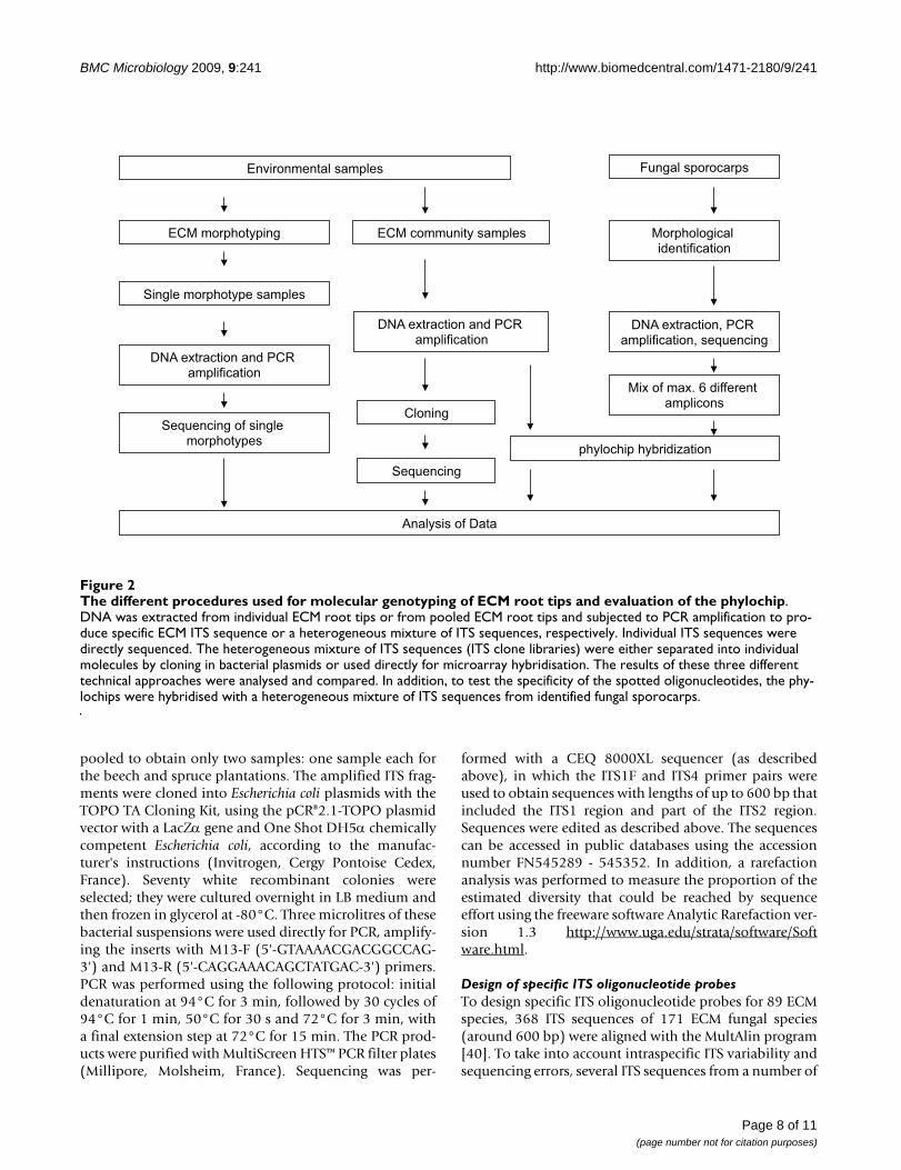
The different procedures used for molecular genotyping of ECM root tips and evaluation of the phylochipFigure 2The different procedures used for molecular genotyping of ECM root tips and evaluation of the phylochip. DNA was extracted from individual ECM root tips or from pooled ECM root tips and subjected to PCR amplification to pro-duce specific ECM ITS sequence or a heterogeneous mixture of ITS sequences, respectively. Individual ITS sequences were directly sequenced. The heterogeneous mixture of ITS sequences (ITS clone libraries) were either separated into individual molecules by cloning in bacterial plasmids or used directly for microarray hybridisation. The results of these three different technical approaches were analysed and compared. In addition, to test the specificity of the spotted oligonucleotides, the phy-lochips were hybridised with a heterogeneous mixture of ITS sequences from identified fungal sporocarps.
Environmental samples
ECM morphotyping ECM community samples
Single morphotype samples
DNA extraction and PCR amplification
DNA extraction and PCR amplification
Sequencing of single morphotypes
Cloning
phylochip hybridization
Sequencing
Analysis of Data
Fungal sporocarps
Mix of max. 6 different amplicons
Morphologicalidentification
DNA extraction, PCR amplification, sequencing
Page 8 of 11(page number not for citation purposes)

BMC Microbiology 2009, 9:241 http://www.biomedcentral.com/1471-2180/9/241
different species were used for the alignment. Singlenucleotide polymorphisms and indels were identified bymanual curation. The sequences, including the ITS1, 5.8Sand ITS2 regions of the nuclear rRNA genes, wereobtained from the public databases NCBI and UNITE.Perfectly matching oligonucleotides, 67 to 70 bases inlength, were designed for each ITS sequence within theITS1 or ITS2 regions. They were selected for optimal melt-ing temperatures (Tm; 75°C ± 2.5°C) and GC content(45-55%) using the AmplifX 1.37 software http://ifrjr.nord.univ-mrs.fr/AmplifX. To enhance specificity,oligonucleotides that had selective nucleotides located ina central position were favoured. The specificity of the oli-goprobes was first tested in silico by querying the oligonu-cleotide sequences against the UNITE and NCBIdatabases. An oligonucleotide was designed as a positivehybridisation control on the ITS region of Arabidopsis thal-iana. Five additional 62- to 70-mer oligonucleotides thatmatched the LSU region of the Glomeromycota were usedto measure the background signal resulting from unspe-cific hybridisation. To avoid cross-hybridisations withundescribed species or cryptic species, we did not use theITS region of untargeted fungal groups as a negative con-trol.
Spotting of glass slide microarray and hybridisation conditionsThe 95 species-specific oligonucleotides (see above) werespotted; one well was spotted with only hybridisationbuffer. Solutions of species-specific oligonucleotides wereadjusted to a concentration of 600 pM and printed in trip-licate by Eurofins, MWG/Operon (Cologne, Germany) onslide arrays with an activated epoxide surface. Oligonucle-otides were bound via their 5' ends on the coating layer ofthe glass surface (for details, see http://www.operon.com). Arrays were prehybridised using theOpArray Pre-Hyb solution (Eurofins, MWG/Operon)according to the manufacturer's instructions. PCR-gener-ated amplicons (maximal 30 ng/μl) were labelled withAlexa Fluor® 555 dye (Invitrogen, Cergy Pontoise, France)using the BioPrime® Plus Array CGH Indirect GenomicLabelling System Kit (Invitrogen) following the manufac-turer's instructions. After the last purification step,labelled amplicons were concentrated with a vacuum con-centrator centrifuge UNIVAPO 100 H (UNIEQUIP, Mar-tinsried, Germany), and then dissolved in 7 μl sterilewater. The sample hybridisation procedure followedRinaldi et al. [41] and is fully described in sample seriesGSM162978 in the GEO at NCBI http://www.ncbi.nlm.nih.gov/geo/. Slide arrays were scannedusing a GenePix 4000 B scanner (Axon-MolecularDevices, Sunnyvale, CA, USA) at a wavelength of 532 nmfor the Alexa Fluor 555 dye. Fluorescent images were cap-tured as TIFF files and the signal intensity was quantifiedby GenePix Pro 5.0 software (Axon-Molecular Devices).
Specificity of oligonucleotides and validation of the phylochipTo validate the specificity of the designed oligonucle-otides, PCR-amplified ITS fragments from the sporocarptissues of known fungal species were hybridised (Figure2). Prior to hybridisation, amplicons (5 ng/μl) from threeto six different ITS amplicons were mixed in a 1:1 ratio.Species-specific ITS within a mix were chosen based onthe in silico tested species phylogenetic distance (minimal30% bp differences were observed between the oligonu-cleotides of one species and the ITS sequences of the otherspecies in the mix). In total, 74 fungal species were probedvia the fungal amplicon mixes. The PCR product that wasamplified from the ITS region of Arabidopsis thaliana wasadded to all amplicon mixes (at a concentration of 5 ng/μl) as a positive hybridisation control. To test the possibleuse of this custom phylochip for describing ECM commu-nity composition in environmental samples, 10 μl of thePCR product that was amplified from the bulked ECMroot tips of beech and spruce was used (spiked with theamplicon of Arabidopsis thaliana). Six technical replicateswere carried out for each sample (three block replicationsper slide × two slides per sample). The results of the cross-hybridisation test are outlined in Figure 1. The ITS-basedcladogram was constructed for all tested fungal speciesusing the default setting of the MEGAN software (version3.0.2., [42]).
Array evaluationPrior to further analyses, spots exhibiting poor quality (forexample, as a result of the presence of dust) were flaggedand excluded from the analyses. Hybridisation qualitywas surveyed using the positive (oligonucleotides of Ara-bidopsis thaliana) and negative controls (five oligonucle-otides for the Glomeromycota (non-ECM species) and theone spot spotted with only hybridisation buffer) of eacharray. Data of the array were further used when (i) signalintensity values of the positive controls were within thegroup of oligonucleotides that showed the highest signalintensity values and (ii) the mean signal intensity value ofthe negative controls were a maximal 1.5% of the signalintensity with the highest value.
Individual spots were considered to be positive (speciespresent in the sample) if their signal intensity showed avalue that was five-fold higher than the averaged intensityvalue for all of the negative controls. Additionally, at leastfour of the six replicates per spot were required to generatea significant positive hybridisation. The threshold factorwas fixed to five-fold after evaluation of the results of thearrays that were hybridised with the known ampliconmixes derived from sporocarp tissues (see "Sporocarp col-lection" and "Specificity of oligonucleotides"). Using athreshold factor of "5" defined the minimal 90% of all
Page 9 of 11(page number not for citation purposes)

BMC Microbiology 2009, 9:241 http://www.biomedcentral.com/1471-2180/9/241
species in the amplicon mixes as positive and filteredmost false-positives (cross-hybridisation).
Authors' contributionsMR conceived and designed the array, set-up the clonelibrary, acquired, analysed, and interpreted the data anddrafted the manuscript. AK analysed and interpreted thearray data. FM conceived and directed the project anddrafted the manuscript. MB carried out the morphotypingand sequencing of the ECM root tips, drafted the manu-script and co-directed the project. All authors read andapproved the final manuscript.
Additional material
AcknowledgementsMR is supported by a Marie Curie PhD scholarship within the framework of the TraceAM programme. The array approach was partly funded by INRA, the European projects TraceAM and ENERGYPOPLAR, the Euro-pean Network of Excellence EVOLTREE, and the Typstat project (GIP ECOFOR). We would like to thank Dr. Melanie Jones (University of British Columbia Okanagan) for her critical reading of the manuscript and helpful comments. We also thank Christine Delaruelle (INRA-Nancy) for her tech-nical assistance with the ITS sequencing. Three anonymous referees are acknowledged for their valuable input and comments on the manuscript and on the development of the technique.
References1. Smith SE, Read DJ: Mycorrhizal Symbiosis 3rd edition. London: Aca-
demic Press; 2008. 2. Erland S, Taylor AFS: Diversity of Ecto-mycorrhizal Fungal
Communities in Relation to the Abiotic Environment. InMycorrhizal Ecology Edited by: van der Heijden M, Sanders I. Berlin,Heidelberg: MGA Springer-Verlag Berlin Heidelberg; 2002:163-200.
3. Rosling A, Landeweert R, Lindahl BD, Larsson KH, Kuyper TW, Tay-lor AFS, Finlay RD: Vertical distribution of ectomycorrhizalfungal taxa in a podzol soil profile. New Phytol 2003,159:775-783.
4. Koide RT, Shumway DL, Xu B, Sharda JN: On temporal partition-ing of a community of ectomycorrhizal fungi. New Phytol 2007,174:420-429.
5. Buée M, Vairelles D, Garbaye J: Year-round monitoring of diver-sity and potential metabolic activity of the ectomycorrhizalcommunity in a beech (Fagus sylvatica) forest subjected totwo thinning regimes. Mycorrhiza 2005, 15:235-245.
6. Ishida TA, Nara K, Hogetsu T: Host effects on ectomycorrhizalfungal communities: insight from eight host species in mixedconifer-broadleaf forests. New Phytol 2007, 174:430-440.
7. Hedh J, Samson P, Erland S, Tunlid A: Multiple gene genealogiesand species recognition in the ectomycorrhizal fungus Paxil-lus involutus. Mycol Res 2008, 112:965-975.
8. Horton TR, Bruns TD: The molecular revolution in ectomycor-rhizal ecology: peeking into the black-box. Mol Ecol 2001,10:1855-1871.
9. Gardes M, Bruns TD: ITS primers with enhanced specificity forbasidiomycetes - applications to the identification of mycor-rhizae and rusts. Mol Ecol 1993, 2:113-118.
10. Anderson IC: Molecular Ecology of Ectomycorrhizal FungalCommunities: New Frontiers. Molecular approaches to Soil, Rhizo-sphere and Plant Microorganism analysis 2006:183-192.
11. Kõljalg U, Larsson KH, Abarenkov K, Nilsson RH, Alexander IJ, Eber-hardt U, Erland S, Hoiland K, Kjøller R, Larsson E, Pennanen T, Sen R,Taylor AFS, Tedersoo L, Vralstad T, Ursing BM: UNITE: a databaseproviding web-based methods for the molecular identifica-tion of ectomycorrhizal fungi. New Phytol 2005, 166:1063-1068.
12. Nilsson RH, Kristiansson E, Ryberg M, Hallenberg N, Larsson KH:Intraspecific ITS variability in the Kingdom Fungi asexpressed in the International Sequence Databases and itsimplications for molecular species identification. Evol Bioinfor-matics 2008, 4:193-201.
13. Martin F, Slater H: New Phytologist - an evolving host for ecto-mycorrhizal research. New Phytol 2007, 174:225-228.
14. Le Quéré A, Schuetzenduebel A, Rajashekar B, Canbäck B, Hedh J,Erland S, Johannson T, Tunlid A: Divergence in gene expressionrelated to variation in host specificity of an ectomycorrhizalfungus. Mol Ecol 2004, 13:3809-3819.
15. Martin F, Aerts A, Ahrén D, Brun A, Duchaussoy F, Kohler A,Lindquist E, Salamov A, Shapiro HJ, Wuyts J, Blaudez D, Buée M,Brokstein P, Canbäck B, Cohen D, Courty PE, Coutinho PM, DanchinEGJ, Delaruelle C, Detter JC, Deveau A, DiFazio S, Duplessis S, Frais-sinet-Tachet L, Lucic E, Frey-Klett P, Fourrey C, Feussner I, Gay G,Gibon J, Grimwood J, Hoegger P, Jain P, Kilaru S, Labbé J, Lin YC, LeTacon F, Marmeisse R, Melayah D, Montanini B, Muratet M, Nehls U,Niculita-Hirzel H, Oudot-Le Secq MP, Pereda V, Peter M, QuesnevilleH, Rajashekar B, Reich M, Rouhier N, Schmutz J, Yin T, Chalot M,Henrissat B, Kües U, Lucas S, Peer Y Van de, Podila G, Polle A, PukkilaPJ, Richardson PM, Rouzé P, Sanders I, Stajich JE, Tunlid A, Tuskan G,Grigoriev I: The genome sequence of the basidiomycete fun-gus Laccaria bicolor provides insights into the mycorrhizalsymbiosis. Nature 2008, 452:88-92.
16. Cook KL, Sayler GS: Environmental application of array tech-nology: promise, problems and practicalities. Curr Opinion inBiotechnol 2003, 14:311-318.
17. Leinberger DM, Schumacher U, Autenrieth IB, Bachmann TT: Devel-opment of a DNA Microarray for detection and identifica-tion of fungal pathogens involved in invasive mycoses. J ClinMicrobiol 2005, 43:4943-4953.
18. Tambong JT, de Cock AWAM, Tinker NA, Lévesque CA: Oligonu-cleotide array for identification and detection of pythiumspecies. AEM 2006, 72:2691-2706.
19. Sessitsch A, Hackl E, Wenzl P, Kilian A, Kostic T, Stralis-Pavese N,Sandjong BT, Bodrossy L: Diagnostic microbial microarrays insoil ecology. New Phytol 2006, 171:719-736.
20. Seifert KA: Integrating DNA barcoding into the mycologicalsciences. Persoonia 2008, 21:162-166.
21. Peplies J, Lau SC, Pernthaler J, Amann R, Glockner FO: Applicationand validation of DNA microarrays for the 16S rRNA-basedanalysis of marine bacterioplankton. Envir Microbiol 2004,6:638-645.
Additional file 1Rarefied species accumulation curve of fungal species detected in ECM root tip samples of (A) spruce and (B) beech. Figures of the rarefaction curves of detected fungal species in ECM root tips of spruce and beech.Click here for file[http://www.biomedcentral.com/content/supplementary/1471-2180-9-241-S1.PDF]
Additional file 2Species described by morphotyping with description of observed mor-photypes according to Agerer (1987-2001). List of all ECM species detected by morphotyping and detailed description of their morphotypes.Click here for file[http://www.biomedcentral.com/content/supplementary/1471-2180-9-241-S2.PDF]
Additional file 3Sequences of the 95 species-specific oligonucleotides. List of sequences of the 95 designed species-specific oligonucleotides.Click here for file[http://www.biomedcentral.com/content/supplementary/1471-2180-9-241-S3.PDF]
Page 10 of 11(page number not for citation purposes)
http://www.ncbi.nlm.nih.gov/entrez/query.fcgi?cmd=Retrieve&db=PubMed&dopt=Abstract&list_uids=8180733
http://www.ncbi.nlm.nih.gov/entrez/query.fcgi?cmd=Retrieve&db=PubMed&dopt=Abstract&list_uids=8180733

BMC Microbiology 2009, 9:241 http://www.biomedcentral.com/1471-2180/9/241
Publish with BioMed Central and every scientist can read your work free of charge
"BioMed Central will be the most significant development for disseminating the results of biomedical research in our lifetime."
Sir Paul Nurse, Cancer Research UK
Your research papers will be:
available free of charge to the entire biomedical community
peer reviewed and published immediately upon acceptance
cited in PubMed and archived on PubMed Central
yours — you keep the copyright
Submit your manuscript here:http://www.biomedcentral.com/info/publishing_adv.asp
BioMedcentral
22. Lievens B, Brouwer M, Vanachter ACRC, Lévesque CA, CammueBPA, Thomma BPHJ: Design and development of a DNA arrayfor rapid detection and identification of multiple tomato vas-cular wilt pathogens. FEMS Microbioloy Letters 2003, 223:113-122.
23. Bruns TD, Gardes M: Molecular tools for the indentification ofectomycorrhizal fungi - taxon specific oligonucleotideprobes for suilloid fungi. Mol Ecol 1993, 2:233-242.
24. El Karkouri K, Murat C, Zampieri E, Bonfante P: Identification ofITS sequence motifs in truffles: a first step toward their DNAbarcoding. AEM 2007, 73:5320-5330.
25. Martin KJ, Rygiewicz RT: Fungal-specific PCR primers devel-oped for analysis of the ITS region of environmental DNAextracts. BMC Microbiol 2005, 5:28.
26. Dickie IA, Xu B, Koide T: Vertical niche differentiation of ecto-mycorrhizal hyphae in soil as shown by T-RFLP analysis. NewPhytol 2002, 156:527-535.
27. Genney DR, Anderson IC, Alexander IJ: Fine-scale distribution ofpine extomycorrhizas and their extrametrical mycelium.New Phytol 2005, 170:381-390.
28. Rothberg JM, Leamon JH: The development and impact of 454sequencing. Nature Biotechnol 2008, 26:1117-1124.
29. Volokhov DA, Rasooly K, Chumakov K, Rasooly A: Identification ofListeria species by microarray based assay. J Clin Microbiol 2002,40:4720-4728.
30. Townsend MB, Dawson ED, Mehlmann M, Smagala JA, Dankbar DM,Moore CL, Smith CB, Cox NJ, Kuchta RD, Rowlen KL: Experimen-tal Evaluation of the FluChip Diagnostic Microarray for Influ-enza Virus Surveillance. J Clin Microbiol 2006, 44:2863-2871.
31. Vialle A, Feau N, Allaire M, Didukh M, Martin F, Moncalvos JM, Hame-lin RC: Evaluation of mitochondrial genes as DNA barcodefor basidiomycota. Mol Ecol Resources 2009, 9:99-113.
32. Buée M, Courty PE, Le Tacon F, Garbaye J: Écosystèmes fores-tiers: Diversité et fonction des champignons. Biofutur 2006,268:42-45.
33. Frøslev TG, Jeppesen T, Læssøe T, Kjøller R: Molecular phyloge-netics and delimitation of species in Cortinarius section Calo-chroi (Basidiomycota, Agaricales) in Europe. Mol PhylogeneticsEvol 2007, 44:217-227.
34. Smith ME, Douhan GW, Rizzo DM: Ectomycorrhizal communitystructure in a xeric Quercus woodland based on rDNAsequence analysis of sporocarps and pooled roots. New Phytol2007, 174:847-863.
35. Tedersoo L, Kõljalg U, Hallenberg N, Larsson KH: Fine scale distri-bution of ectomycorrhizal fungi and roots across substratelayers including coarse woody debris in a mixed forest. NewPhytol 2003, 159:153-165.
36. Prévost A, Pargney JC: Comparaison des ectomycorhizesnaturelles entre le hêtre (Fagus sylvatica) et 2 lactaires (Lac-tarius blennius var viridis et Lactarius subdulcis). I. Cara-ctéristiques morphologiques et cytologiques. Ann Sci For 1995,52:131-146.
37. Ranger J, Andreux J, Bienaimé SF, Berthelin J, Bonnaud P, Boudot JP,Bréchet C, Buée M, Calmet JP, Chaussod R, Gelhaye D, Gelhaye L,Gérard F, Jaffrain J, Lejon D, Le Tacon F, Léveque J, Maurice JP, MerletD, Moukoumi J, Munier-Lamy C, Nourrisson G, Pollier B, Ranjard L,Simonsson M, Turpault MP, Vairelles D, Zeller B: Effet des substi-tutions d'essence sur le fonctionnement organo-minéral del'écosystème forestier, sur les communautés microbienneset sur la diversité des communautés fongiques mycorhi-ziennes et saprophytes (cas du dispositif expérimental deBreuil-Morvan). In Proceedings of the Final Report of contract INRA-GIP Ecofor 2001-24, no. INRA 1502A Champenoux: INRA BEF Nancy;2004.
38. Agerer R: Colour atlas of ectomycorrhizae Munich: Einhorn-Verlag Edu-ard Dietenberger; 1987.
39. Courtecuisse R: Mushrooms of Britain and Europe London: Harper Col-lins; 2000.
40. Corpet F: Multiples sequence alignment with hierarchicalclustering. Nucl Acids Res 1988, 16:10881-10890.
41. Rinaldi C, Kohler A, Frey P, Duchaussoy F, Ningre N, Couloux A,Wincker P, Le Thiec D, Fluch S, Martin F, Duplessis S: Transcriptprofiling of poplar leaves upon infection with compatible andincompatible strains of the foliar rust Melampsora larici-pop-ulina. Plant Physiol 2007, 144:347-366.
42. Huson DH, Auch AF, Qi J, Schuster SC: MEGAN Analysis ofMetagenomic Data. Genome Research 2007, 17:377-386.
Page 11 of 11(page number not for citation purposes)
http://www.ncbi.nlm.nih.gov/entrez/query.fcgi?cmd=Retrieve&db=PubMed&dopt=Abstract&list_uids=7513242
http://www.ncbi.nlm.nih.gov/entrez/query.fcgi?cmd=Retrieve&db=PubMed&dopt=Abstract&list_uids=7513242
http://www.ncbi.nlm.nih.gov/entrez/query.fcgi?cmd=Retrieve&db=PubMed&dopt=Abstract&list_uids=7513242
http://www.ncbi.nlm.nih.gov/entrez/query.fcgi?cmd=Retrieve&db=PubMed&dopt=Abstract&list_uids=2849754