Developing New Diagnostic Tests for Human African ... New Diagnostic Tests for Human African...
20
Current status and future plans Developing New Diagnostic Tests for Human African Trypanosomiasis foundation for innovative new diagnostics
-
Upload
phungtuyen -
Category
Documents
-
view
220 -
download
1
Transcript of Developing New Diagnostic Tests for Human African ... New Diagnostic Tests for Human African...

Current status and future plans
Developing New Diagnostic Tests for Human African Trypanosomiasis
foundationfor innovative new diagnostics

FIND’s purpose
FIND addresses the market failures that result in the lack of appropriate diagnostic tools in disease-endemic countries. Our expertise is in the development of innovative, field-adapted diagnostics that have significant impact in low-resource settings. We demonstrate how improved diagnostics save lives and resources, and we provide technical support for their introduction and implementation in national health systems. The process requires bringing together knowledge of the needs on the ground with technological know-how, and a sound understanding of the intricacies of developing diagnostics.
Enrolment of participants in a clinical trial on a diagnostic test for sleeping sickness.
Contents
Overview of FIND . . . . . . . . . . . . . . . . . . . . . . . . . . . . . . . . . . . . . . . . . . . . . . . . . . . . . . . . . . . . . . 2
The HAT & OND programme . . . . . . . . . . . . . . . . . . . . . . . . . . . . . . . . . . . . . . . . . . . . . . . . . 4
Projects on diagnostics for HAT . . . . . . . . . . . . . . . . . . . . . . . . . . . . . . . . . . . . . . . . . . . 5
Problem and scope . . . . . . . . . . . . . . . . . . . . . . . . . . . . . . . . . . . . . . . . . . . . . . . . . . . . . . . . . . . 5
Tests for screening . . . . . . . . . . . . . . . . . . . . . . . . . . . . . . . . . . . . . . . . . . . . . . . . . . . . . . . . . . . . 5
Tests for confirming cases . . . . . . . . . . . . . . . . . . . . . . . . . . . . . . . . . . . . . . . . . . . . . . . . . . . . 7
Miniature anion exchange centrifugation technique (mAECT) . . . . . . . . . . . . . 7
Fluorescence microscopy . . . . . . . . . . . . . . . . . . . . . . . . . . . . . . . . . . . . . . . . . . . . . . . . . 8
LED FM on whole blood, and blood after lysis and concentration . . . . . . . . 8
Molecular detection . . . . . . . . . . . . . . . . . . . . . . . . . . . . . . . . . . . . . . . . . . . . . . . . . . . . . . . 10
Tests for staging and confirmation of cure . . . . . . . . . . . . . . . . . . . . . . . . . . . . . . . . . . 11
Biological samples to support test development . . . . . . . . . . . . . . . . . . . . . . . . . . . 13
Funding . . . . . . . . . . . . . . . . . . . . . . . . . . . . . . . . . . . . . . . . . . . . . . . . . . . . . . . . . . . . . . . . . . . . . . 13
Agreements with partners . . . . . . . . . . . . . . . . . . . . . . . . . . . . . . . . . . . . . . . . . . . . . . . . . 13
Publications . . . . . . . . . . . . . . . . . . . . . . . . . . . . . . . . . . . . . . . . . . . . . . . . . . . . . . . . . . . . . . . . . . . 14

2 Human Afr ican Trypanosomias is
FIND was established in 2003 to bring diagnos-tic solutions to societies where treatable diseases are rampant and where poverty and poor health are closely intertwined. The diagnostics available in disease-endemic countries are often not well adapted or too costly for use in the field. FIND addresses these challenges by developing diagnostic tests that are field-adapted and affordable. During the past 10 years, we have been developing diag-nostic approaches that have been proven in prin-ciple, transforming them into effective products for identifying tuberculosis. Strong partnerships with academia, public and private research institutes,
and industry have increased the scope of activities which now includes diagnostics for human African trypanosomiasis (HAT), malaria, HIV/AIDS, leish-maniasis and Chagas disease.
Since 2006, FIND has also been working to upgrade healthcare systems and laboratory standards, so that the new tools can be rolled out in low-resource settings. The impact and effectiveness of these improved diagnostic tests is measured through a continuous flow of information and feedback from the field.
Overview of FIND
CEO
BD SARMO CFO CSO CMO HRM SOO
C r o s s - c u t t i n g f u n c t i o n s
Leadership team members
Exec. Assist
STAFF: BD – Business Development; SARMO – Senior Advocacy & Resource Mobilization Officer; CFO – Chief Financial Officer; CSO – Chief Scientific Officer; CMO – Chief Medical Officer; HR&AM – Human Resources & Administration Manager; SOO – Senior Operating OfficerOTHER: HAT – Human African Trypanosomiasis; OND – Other neglected diseases; AFS – Acute febrile syndrome; SEA – South-East Asia
Contracts & BD • Advocacy
• ResourceMobilizazion
• Communications
• Staff
• Consultants
• Administration
• Finance
• Legal Affairs
• Logistics
• IT
• Field officesIndia & Uganda
• Science &Technology
• Opstream
• Programmes
• Acting TBHead
• Clinical Trials
• DownstreamProgrammes
• Quality Mgmt & ISO Certification
• Project Mgmt
• Doc. Controlling
• Grant Mgmt
• Regulatory Affairs
TB
HAT & OND
Malaria & AFS
Downstream Programmes
TB Diagnostics Implementation
Lab Systems Strengthening
TB Programme India & SEA
TB Programme Uganda
Figure 1: The FIND Organogram

3Overv iew of F IND
The organization’s business model (figure 1) provides an environment for maximizing available expertise across diseases and technologies.
FIND’s strategy is to focus on developing diagnostic technology platforms that can be applied to several diseases, thereby reducing product development costs, time and risk, while building on our experience in
facilitating efficient introduction of tests and training of health personnel. The diagnostic tools we work on are intended to improve patient care at an individual level, as well as disease control at a population level. The level of FIND’s involvement in development of a technology is dictated by the stage the technology is at on the value chain (figure 2), starting from proof-of-concept to early implementation.
FIND’s funding base includes, among others, the Bill & Melinda Gates Foundation, UNITAID, the Global Fund to fight AIDS, tuberculosis and malaria,
the governments of the Netherlands, Germany and the United Kingdom, WHO-TB Reach, USAID, and the UBS Optimus Foundation.
Feasibility Development Evaluation Demonstration Evidence for scale-up
and Scale-up
WHO
New diagnostic test prototypedeveloped and technicalspecifications validated
New diagnostic test fully developed(design lock) and manufacturingprocess validated
Diagnostic accuracy and analyticalperformance of new product validatedin clinical trials
Performance and operational characteristicsvalidated during uncontrolled routine use inprogrammatic settings
Implementation in routine diagnostic services under qualityassurance followed by roll-out in high-endemic countries,market surveillance and impact assessment
STAG/WHOEndorsement into
global policy
Figure 2: FIND’s basic diagnostics development value chain, which can be adapted depending on the disease
4 Human Afr ican Trypanosomias is
Human African trypanosomiasis (HAT) was the first neglected tropical disease (NTD) to be included in FIND’s portfolio in 2006. The programme was renamed
HAT & other neglected diseases (HAT & OND) after the addition of leishmaniasis in 2010 and Chagas disease in 2012.
The HAT & OND programme
Status of various projects implemented under the HAT & OND programme
Human African Trypanosomiasis (HAT; sleeping sickness)
a) A test for screening populations that are at risk of infection
i. Further to the successful evaluation of the performance of a prototype 1st generation rapid diagnostic test (RDT) using
native trypanosome antigens on more than 14,000 individuals in Angola, the Democratic Republic of the Congo (DRC)
and Central African Republic (CAR), the SD BIOLINE HAT test was launched in December 2012. Demonstration studies
to further evaluate the performance of the test and determine its cost effectiveness in various diagnostic algorithms
are ongoing.
ii. Development of a 2nd generation RDT using recombinant antigens, which could be effective for both Trypanosoma
brucei gambiense and T.b. rhodesiense HAT, is going on.
b) A test for confirming cases of HAT
i. The mini-anion exchange centrifugation technique (mAECT) was improved and its production successfully transferred
to the DRC.
ii. Evaluation of an LED fluorescence microscope has been completed in the DRC and demonstration studies are going on.
iii. Evaluation of a molecular test based on loop-mediated isothermal amplification of DNA (LAMP) is going on in the DRC
and Uganda.
c) A test to determine if HAT patients have brain disease, and to monitor treatment
– Development of an RDT for staging and monitoring treatment by measuring neopterin and/or CXCL13 in CSF
is in process.
Leishmaniasis
a) ELISA test for parasite antigens in urine
An ELISA assay for urinary antigens using polyclonal antibodies is being developed in collaboration with DNDi, Kalon
Biologicals (UK) and the Royal Tropical Institute (KIT, Netherlands). The test, to be used for case detection and monitoring
therapy, is performing well.
b) LAMP test to detect parasite DNA
This test is being developed as part of expansion of the LAMP platform, in collaboration with Eiken, KIT and the Institute
of Primate Research (IPR, Kenya). Prototype reagents are under evaluation.
Chagas Disease
LAMP test for congenital Chagas disease
Development of the test was initiated in January 2012, in collaboration with INGEBI-CONICET and a Colombian
multi-disciplinary team. Feasibility studies and optimization of sample preparation are going on.
5The HAT & OND programme
projects oN DIagNostIcs For Hat
problem and scope
Human African trypanosomiasis (HAT) is a disease of poor rural communities caused by extracellular protozoan parasites of the genus Trypanosoma. In early or Stage 1 infection when parasites are in the blood and lymphatic system, treatment is relatively safe and cheap. During this phase, however, clinical signs are not suggestive of HAT, and diagnostic tests have problems of sensitivity and specificity. Many cases, therefore, remain undetected and with time, parasites invade the brain, resulting in late or Stage 2 disease. Treatments of Stage 2 HAT are either lengthy and expensive, and difficult to administer, or cause poten-tially fatal side effects. Currently, tests to determine the stage of disease are non-specific and insensitive.
FIND and partners are developing and implement-ing tests for early and accurate diagnosis and staging of HAT patients, in order to ensure safe treatment. Other tests for early detection of treatment failure will ensure proper re-treatment, reduced transmis-sion and accelerated control of the disease.
tests for screening
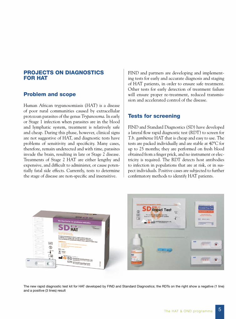
FIND and Standard Diagnostics (SD) have developed a lateral flow rapid diagnostic test (RDT) to screen for T.b. gambiense HAT that is cheap and easy to use. The tests are packed individually and are stable at 40°C for up to 25 months; they are performed on fresh blood obtained from a finger prick, and no instrument or elec-tricity is required. The RDT detects host antibodies to infection in populations that are at risk, or in sus-pect individuals. Positive cases are subjected to further confirmatory methods to identify HAT patients.
The new rapid diagnostic test kit for HAT developed by FIND and Standard Diagnostics; the RDTs on the right show a negative (1 line) and a positive (3 lines) result

6 Human Afr ican Trypanosomias is
The new RDT is an alternative to the card aggluti-nation test for trypanosomiasis (CATT), the primary screening tool used by control programmes in areas where T.b. gambiense HAT is endemic.
Current status1st generation test – Field evaluation of the proto-type has been successfully completed on more than 14,000 individuals at sites in Angola, Central African Republic (CAR) and the Democratic Republic of the Congo (DRC). Job Aids and a manual for train-ing health workers on proper use of the new RDT (in English and French) have also been made.
The SD HAT BIOLINE test was launched at a work-shop hosted by the Ministry of Health of the Dem o-cratic Republic of the Congo on 6th December 2012 in Kinshasa. Demonstration studies to further evalu-ate the performance of the test and determine its cost-effectiveness in various diagnostic algorithms are ongoing.
2nd generation test – The 1st generation RDT is made using native antigens generated from pathogenic trypanosomes, and is not applicable for T.b. rhodesiense HAT. Efforts are therefore being made by FIND and SD to develop a 2nd generation
The study monitor discusses a point with the principal investigator (right) during clinical evaluation of the new rapid diagnostic test for sleeping sickness in a village in the Democratic Republic of the Congo.
The blood sample taken from a finger prick is introduced into the sample well of the rapid diagnostic test. The results of the test are read after only 15 minutes.
Children follow activities during active screening of a community in the Democratic Republic of the Congo, when clinical trials on the new rapid test for HAT were being carried out.
A technician takes blood after a finger prick during clinical evaluation of the new rapid diagnostic test for sleeping sickness in rural Democratic Republic of the Congo.

7The HAT & OND programme
RDT using recombinant antigens. Such a test would be easier and cheaper to manufacture, thus reducing the cost of the final product. Eight recombinant antigens (GM6, 4 ISGs and 3 VSGs) are being investigated.
Antigen detection test – A test that detects parasite antigens is a better indicator of infection than one that detects host antibodies, and could also be useful for monitoring treatment. The challenge has been in the identification of target antigens, since the ones on the surface of the parasite are variable or mutable. Current efforts are focused on using nanobodies, small camel antibodies that are particularly thermostable and that can penetrate the parasite surface coat and detect invariable antigens. Work carried out in col-laboration with the Flanders Interuniversity Institute for Biotechnology (VIB) at the University of Brussels has identified a number of promising nanobodies. Their potential in development of a test is being determined in collaboration with SD.
Future plans• Conductdemonstrationstudiesof
the 1st generation RDT• Rolloutthe1stgenerationRDTin
collaboration with partners• Developa2ndgenerationRDTbasedon
recombinant antigens• Developa1stgenerationantigendetection
test based on nanobodies
tests for confirming cases
Demonstration of parasites in body fluids is the most accurate way to confirm HAT, and is critical in order to avoid the risk of exposing healthy suspects to poten-tially dangerous therapy. For most T. b. rhodesiense patients, parasitaemia is usually high enough for para-sites to be seen by direct examination of blood under a light microscope. However, the number of parasites
in T.b. gambiense infections is often below the limit of detection of the most sensitive methods that are in clinical use. In the cerebrospinal fluid (CSF), parasites exist in very low numbers, and have to be concen-trated before they can be viewed. The most sensitive methods for detecting trypanosomes in blood that are in clinical use include the mini anion exchange centrifugation technique (mAECT) and the capillary tube centrifugation (CTC) method, while the modi-fied single centrifugation (MSC) technique is used to detect trypanosomes in CSF. Although CTC is widely used due to its simplicity and low cost, widespread use of mAECT has been constrained by the high costs associated with it, and by the need for a cold chain.
Miniature anion exchange centrifugation technique (maect)The mAECT is carried out in two stages, including chromatography of samples, then concentration of the eluate by low speed centrifugation and viewing under low magnification microscopy. Similarly with MSC, CSF is collected from a lumbar puncture and centrifuged before viewing. Both mAECT and MSC were, until 2006, manufactured at the Institute of Tropical Medicine (ITM) in Belgium and then shipped to endemic coun-tries – a process that was untenable due to the associ-ated high costs. A partnership between FIND, the ITM and the Institut National de Recherche Biomédicale (INRB) in DRC not only improved components of both mAECT and MSC, but by 2008 had successfully transferred their production to Kinshasa, DRC.
Current status Production of mAECT and MSC is done at the INRB. The largest percentage of kits produced is used locally in the DRC, mainly in clinical trials.
Future plans• Explorecheaperandmorepracticalmethodsof
separating parasites
8 Human Afr ican Trypanosomias is
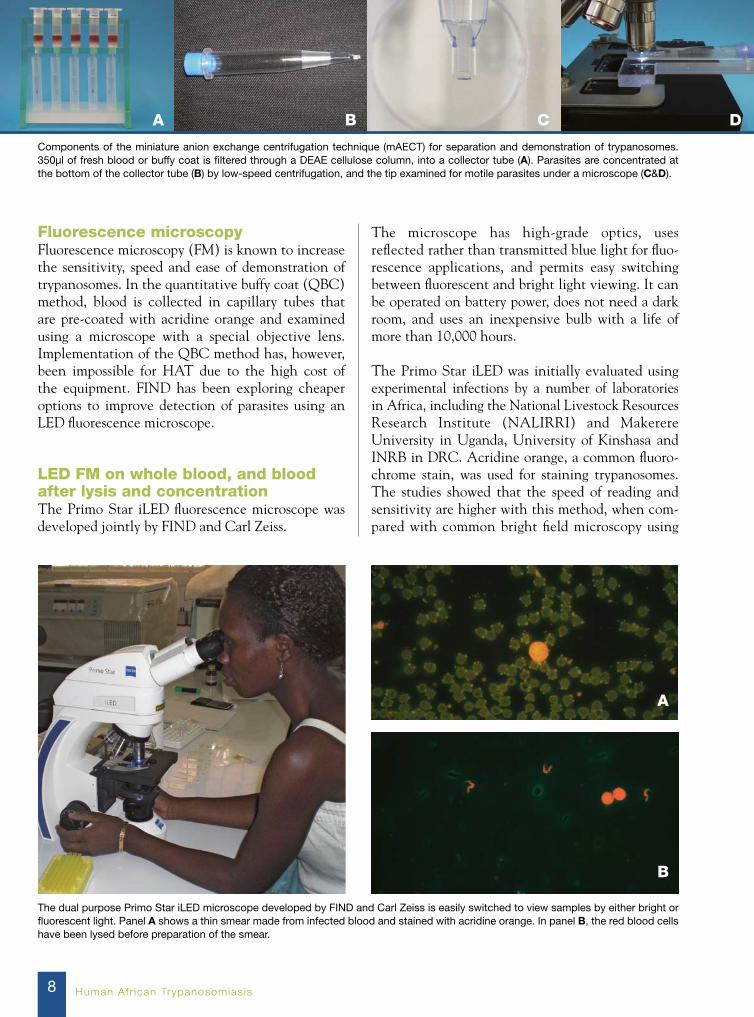
Fluorescence microscopyFluorescence microscopy (FM) is known to increase the sensitivity, speed and ease of demonstration of trypanosomes. In the quantitative buffy coat (QBC) method, blood is collected in capillary tubes that are pre-coated with acridine orange and examined using a microscope with a special objective lens. Implementation of the QBC method has, however, been impossible for HAT due to the high cost of the equipment. FIND has been exploring cheaper options to improve detection of parasites using an LED fluorescence microscope.
LeD FM on whole blood, and blood after lysis and concentrationThe Primo Star iLED fluorescence microscope was developed jointly by FIND and Carl Zeiss.
The microscope has high-grade optics, uses reflected rather than transmitted blue light for fluo-rescence applications, and permits easy switching between fluorescent and bright light viewing. It can be operated on battery power, does not need a dark room, and uses an inexpensive bulb with a life of more than 10,000 hours.
The Primo Star iLED was initially evaluated using experimental infections by a number of laboratories in Africa, including the National Livestock Resources Research Institute (NALIRRI) and Makerere University in Uganda, University of Kinshasa and INRB in DRC. Acridine orange, a common fluoro-chrome stain, was used for staining trypanosomes. The studies showed that the speed of reading and sensitivity are higher with this method, when com-pared with common bright field microscopy using
Components of the miniature anion exchange centrifugation technique (mAECT) for separation and demonstration of trypanosomes. 350µl of fresh blood or buffy coat is filtered through a DEAE cellulose column, into a collector tube (A). Parasites are concentrated at the bottom of the collector tube (B) by low-speed centrifugation, and the tip examined for motile parasites under a microscope (C&D).
The dual purpose Primo Star iLED microscope developed by FIND and Carl Zeiss is easily switched to view samples by either bright or fluorescent light. Panel A shows a thin smear made from infected blood and stained with acridine orange. In panel B, the red blood cells have been lysed before preparation of the smear.
a B c D
a
B

9The HAT & OND programme
Giemsa staining. The diagnostic capacity of the method is significantly enhanced when staining of samples is preceded by lysis of red blood cells using ammonium chloride or commercial lysis buffer.
Current statusClinical evaluation of the LED microscope using the red blood cell lysis and acridine orange stain-ing methods has been completed at several sites in DRC and Uganda. The trial in the DRC was done in collaboration with the national HAT control programme and the SwissTPH, while the one in Uganda was done in collaboration with Makerere University and Lwala hospital. Based on the excellent results from these studies, the Ministry of Health of DRC approved the use of these new methods for routine diagnosis of HAT on 7th December 2012.
Future plans• Demonstration/earlyimplementationofLED
microscopy in diagnosis of HAT
Molecular detectionDetection of trypanosome DNA from body fluids of a HAT patient could be a significant improvement on parasitological examination. Loop-mediated iso-thermal amplification (LAMP) of DNA is a promising molecular technique that shows high sensitivity and specificity.
The test amplifies target DNA at a constant tempera-ture, meaning that it can be carried out with minimal equipment. Furthermore, positive samples fluoresce under LED light. The test can be performed by staff with minimal experience in molecular biology.
Carl Zeiss has developed a plastic box that enables easy and safe transportation of the microscope during active screening programmes (C&D). Panel E shows a technician operating an LED fluorescence microscope using solar energy in eastern Democratic Republic of the Congo. (Photos C&D are courtesy of Carl Zeiss AG)
The LoopampTM LF-160 incubator (A) used to perform the HAT LAMP reaction. After extraction of DNA, the LAMP reaction is kept at a constant temperature of 65°C for 40 minutes. The results are visualized under LED light (B). PC- Positive control, NC- Negative control. (Photo on the right is courtesy of E. Matovu)
c D eD
a B

10 Human Afr ican Trypanosomias is
In 2008, FIND signed a development agreement with Eiken Chemical Company, Ltd. (Japan), the owner of the patent rights to LAMP, and completed develop-ment of a LAMP kit for HAT in 2011. The reagents for LAMP are dried on the inside part of the cap of the reaction tube, which can be stored at room temperature. When test samples are added, the tubes are heated at constant temperature in an incubator, and the results read visually using LED light. The test can be performed on blood, either fresh or dried on filter papers, or buffy coat, or microscopy slides.
Current statusClinical evaluation of the HAT LAMP kit is being carried out at multiple sites in the DRC and Uganda. Prospects of using the same test in drug screen-ing studies, and for monitoring treatment, are also being explored.
Future plans• Demonstration/earlyimplementationstudies• Cost-effectivenessstudiestoguidenovel
strategies for introduction of LAMP in diagnosis of HAT
A technician takes blood from a finger prick during clinical evaluation of the new LAMP test for sleeping sickness in Bandundu province, Democratic Republic of the Congo.
11The HAT & OND programme
tests for staging and confirmation of cure
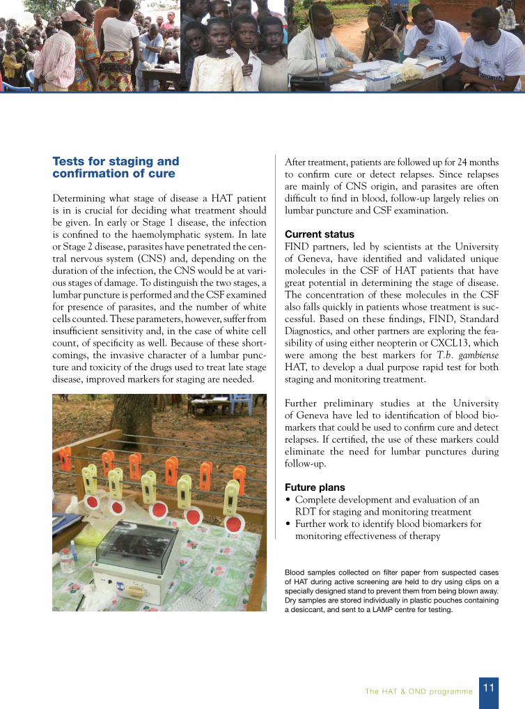
Determining what stage of disease a HAT patient is in is crucial for deciding what treatment should be given. In early or Stage 1 disease, the infection is confined to the haemolymphatic system. In late or Stage 2 disease, parasites have penetrated the cen-tral nervous system (CNS) and, depending on the duration of the infection, the CNS would be at vari-ous stages of damage. To distinguish the two stages, a lumbar puncture is performed and the CSF examined for presence of parasites, and the number of white cells counted. These parameters, however, suffer from insufficient sensitivity and, in the case of white cell count, of specificity as well. Because of these short-comings, the invasive character of a lumbar punc-ture and toxicity of the drugs used to treat late stage disease, improved markers for staging are needed.
After treatment, patients are followed up for 24 months to confirm cure or detect relapses. Since relapses are mainly of CNS origin, and parasites are often difficult to find in blood, follow-up largely relies on lumbar puncture and CSF examination.
Current statusFIND partners, led by scientists at the University of Geneva, have identified and validated unique molecules in the CSF of HAT patients that have great potential in determining the stage of disease. The concentration of these molecules in the CSF also falls quickly in patients whose treatment is suc-cessful. Based on these findings, FIND, Standard Diagnostics, and other partners are exploring the fea-sibility of using either neopterin or CXCL13, which were among the best markers for T.b. gambiense HAT, to develop a dual purpose rapid test for both staging and monitoring treatment.
Further preliminary studies at the University of Geneva have led to identification of blood bio-markers that could be used to confirm cure and detect relapses. If certified, the use of these markers could eliminate the need for lumbar punctures during follow-up.
Future plans• Completedevelopmentandevaluationofan
RDT for staging and monitoring treatment• Furtherworktoidentifybloodbiomarkersfor
monitoring effectiveness of therapy
Blood samples collected on filter paper from suspected cases of HAT during active screening are held to dry using clips on a specially designed stand to prevent them from being blown away. Dry samples are stored individually in plastic pouches containing a desiccant, and sent to a LAMP centre for testing.

12 Human Afr ican Trypanosomias is
A technician applies blood from a participant on a filter paper at a rural health centre in the Democratic Republic of the Congo during clinical evaluation of LAMP for the diagnosis of HAT. The sample is left to dry at ambient temperature, then stored in a plastic pouch containing a desiccant, before being sent to a LAMP centre for testing.
A technician reads the results of LAMP at a rural hospital in Uganda, where the test is being evaluated for the diagnosis of T.b. rhodesiense HAT.

13The HAT & OND programme
Biological samples to support test development
A critical obstacle in development of improved assays for HAT is access to carefully collected and stored biological materials.
Current statusFIND and the World Health Organization (WHO) have addressed this problem by establishing a HAT specimen bank, which is owned by the WHO. The Institut Pasteur in Paris hosts the WHO specimen bank, storing samples and dispatching them to research institutions and other users after approval by an Exit Committee of WHO.
Additional specimen collections are available at the Institute of Tropical Neurology (INT) in Limoges France, and Makerere University in Uganda, for use on FIND-supported activities. The number of samples in these collections is increasing as new clinical studies are carried out.
FuNDINg
The current funders for HAT diagnostics at FIND include the Bill & Melinda Gates Foundation, EU-FP7 Programme, DFID (UK), UBS Optimus Foundation and the German government.
agreeMeNts wItH partNers
Most activities with our partners are based on con-tractual agreements. FIND has, in addition to such agreements, signed a Memorandum of Understand-ing with the following strategic organizations, among others:• WorldHealthOrganization• PanAfricanTsetseandTrypanosomiasis
Eradication Campaign (PATTEC)• MédecinsSansFrontières–Spain
14 Human Afr ican Trypanosomias is
1. Tiberti N., Matovu E., Hainard A., Enyaru J.C., Lejon V., Robin X., Turck N., Ngoyi D.M., Krishna S., Bisser S., Courtioux B., Büscher P., Kristensson K., Ndung'u J.M., and Sanchez J.C. (2013). New biomarkers for stage determination in Trypanosoma brucei rhodesiense sleeping sickness patients. Clinical and Translational Medicine http://www.clintransmed.com/content/2/1/1.
2. Tiberti N., Lejon V., Hainard A., Courtioux B., Robin X., Turck N., Kristensson K., Matovu E., Enyaru J.C., Ngoyi D.M., Krishna S., Bisser S., Ndung'u J.M., Büscher P. and Sanchez J.C. (2013) Neopterin is a cerebrospinal fluid marker for treatment outcome evaluation in patients affected by Trypanosoma brucei gambiense sleeping sickness. PLoS Neglected Tropical Diseases 7(2): e2088. doi:10.1371/journal.pntd.0002088
3. Dancet E.A.F., Brannstrom M., Brasky K., Chai D., Chan A.W.S., Conn P.M., Else J., Falconer H., Fazleabas A.T., Farah I.O., Goddeeris B.M., Golos T.G., Hau J., Hearn J.P., Kariuki T.M., Kyama C.M., Lebovic D.I., Mwenda J.M., Ndung’u J., Nyachieo A., Parker J., Slayden Ov.D., Stouffer R.L., Strauss J.F., Taylor H.S., Vanderpoel S., Westergaard J.G., Zelinski M., D’Hooghe T.M. (2012). The role of scientists and clinicians in raising public support for animal research in reproductive biology and medicine. Biology of Reproduction 112.105908
4. Franco JR., Simarro PP., Diarra A., Ruiz-Postigo JA. and Jannin JG. (2012). The human African trypanosomiasis
specimen biobank: a necessary tool to support research of new diagnostics. PLoS Neglected Tropical Diseases 6(6): e1571. doi:10.1371/ journal.pntd.0001571
5. Tiberti N., Hainard A., Lejon V., Courtioux B., Matovu E., Enyaru J.C., Robin X, Turck N., Kristensson K., Ngoyi D.M., Vatunga G M.L., Krishna S., Büscher P., Bisser S., Ndung'u J. M. and Sanchez J-C. (2012). Neopterin for the staging and follow-up of sleeping sickness patients: evidence from a multi-centric cohort. PLoS One 7(7) e40909.
6. Matovu E., Kazibwe A.J., Mugasa C.M., Ndung’u J.M. and Njiru Z.K. (2012). Towards point-of-care diagnostic and staging tools for human African trypanosomiasis. Journal of Tropical Medicine: Article ID 340538. doi:10.1155/2012/340538
7. Biéler S., Matovu E., Mitashi P., Ssewannyana E., Karhemere Bi Shamamba S., Bessell P.R. and Ndung’u J.M. (2012). Improved detection of Trypanosoma brucei by lysis of red blood cells, concentration and LED fluorescence microscopy. ACTA Tropica 121(2): 135-140.
8. Tiberti N., Hainard A., Lejon V., Robin X., Ngoyi D.M., Turck N., Matovu E., Enyaru J., Ndung’u J.M., Scherl A., Dayon L., Sanchez J.C. (2010). Discovery and verification of osteopontin and beta-2-microglobulin as promising markers for staging human African trypanosomiasis. Molecular and Cellular Proteomics 9:2783–2795.
Publications

15Publ icat ions
9. Hainard, A., Tiberti, N., Robin, X., Ngoyi, D.M., Matovu, E., Enyaru J.C.K., Müller, M., Turck, N., Ndung’u, J.M., Lejon, V., and Sanchez, J.C. (2010). Matrix metalloproteinase-9 and intercellular adhesion molecule 1 are powerful staging markers for human African trypanosomiasis. Tropical Medicine & International Health 16:119–126.
10. Ndung’u JM., Bieler S. and Roscigno G. (2010). “Piggy-backing” on diagnostic platforms brings hope to neglected diseases: the case of sleeping sickness. PLoS Neglected Tropical Diseases 4(5): e715. doi:10.1371/ journal.pntd.0000715.
11. Molyneux, D., Ndung’u J.M. and Maudlin I. (2010). Controlling sleeping sickness – “when will they ever learn?” PLoS Neglected Tropical Diseases 4(5): e609. doi:10.1371/ journal.pntd.0000609
12. Gibson W., Nemetschke L. and Ndung'u J. (2010). Conserved sequence of the TgsGP gene in Group 1 Trypanosoma brucei gambiense. Infection, Genetics and Evolution, 10:453-458.
13. Gray S.A., Weigel K.M., Büscher P., Miller K.D., Baird C., Ndung’u J.M., and Cangelosi G.A. (2010). Flow cytometry-based methods for assessing soluble scFv activities and detecting antigens in solution. Journal of Biotechnology and Bioengineering 105(5): 973-981.
14. Büscher, P. Mumba Ngoyi, D. Kaboré, J. Lejon, V. Robays, J. Jamonneau, V. Bebronne,
N. Van der Veken, W. Biéler, S. (2009). Improved models of mini anion exchange centrifugation technique (mAECT) and modified single centrifugation (MSC) for sleeping sickness diagnosis and staging. PLoS Neglected Tropical Diseases 3(11): e471. doi:10.1371/journal.pntd.0000471
15. Ndung’u J.M. and Magez, S (2009). Exploiting nanobodies in diagnosis and treatment of African trypanosomiasis. In: Research Reviews, EU Parliament Magazine, Issue 9 of May 2009.
16. Hainard, A., Tiberti, N., Robin, X., Lejon, V., Ngoyi, D.M., Matovu, E., Enyaru, J.C., Fouda, C., Ndung’u, J.M., Lisacek, F., Müller, M., Turck, N., and Sanchez, J. (2009). A combined CXCL10, CXCL8 and H-FABP panel for the staging of human African trypanosomiasis patients. PLoS Neglected Tropical Diseases 3: e459.

16 Human Afr ican Trypanosomias is
FIND provides the necessary expertise, capacity and facilities needed to drive the R&D process, through partnerships with academia, industry and endemic country governments. We also help create the necessary conditions for widespread implementation of these tools through collection of evidence, sharing knowledge and technology transfer.
Viewing the results of a HAT LAMP test in Eastern DRC.
foundationfor innovative new diagnostics

Partnering for better diagnosis for all

foundationfor innovative new diagnostics
Avenue de Budé 16 • 1202 Geneva • Switzerland • Tel.: +41 (0)22 710 05 90 • Fax: +41 (0)22 710 05 99 wwww.finddiagnostics.org
© FIND 2013
Production and editing: FIND Communications
Authors: HAT & OND team
Design: www.latitudesign.com
Printer: NBmedia
All photos except three are courtesy of Joseph M. Ndung’u