Detection of Protein–Protein Interactions Through Vesicle...
21
Copyright Ó 2009 by the Genetics Society of America DOI: 10.1534/genetics.109.101162 Detection of Protein–Protein Interactions Through Vesicle Targeting Jacob H. Boysen,* Saranna Fanning,* ,† Justin Newberg, ‡ Robert F. Murphy ‡,§, ** ,†† and Aaron P. Mitchell* ,††,1 *Department of Microbiology and Institute of Cancer Research, Columbia University, New York, New York 10032, † Department of Microbiology, University College Cork, Cork, Ireland, ‡ Center for Bioimage Informatics and Department of Biomedical Engineering, Carnegie Mellon University, Pittsburgh, Pennsylvania 15213, § Lane Center for Computational Biology and Department of Machine Learning, Carnegie Mellon University, Pittsburgh, Pennsylvania 15213, **External Fellow, Freiburg Institute for Advanced Studies, University of Freiburg, 79104 Freiburg, Germany and †† Department of Biological Sciences, Carnegie Mellon University, Pittsburgh, Pennsylvania 15213 Manuscript received January 26, 2009 Accepted for publication March 19, 2009 ABSTRACT The detection of protein–protein interactions through two-hybrid assays has revolutionized our understanding of biology. The remarkable impact of two-hybrid assay platforms derives from their speed, simplicity, and broad applicability. Yet for many organisms, the need to express test proteins in Saccharomyces cerevisiae or Escherichia coli presents a substantial barrier because variations in codon specificity or bias may result in aberrant protein expression. In particular, nonstandard genetic codes are characteristic of several eukaryotic pathogens, for which there are currently no genetically based systems for detection of protein– protein interactions. We have developed a protein–protein interaction assay that is carried out in native host cells by using GFP as the only foreign protein moiety, thus circumventing these problems. We show that interaction can be detected between two protein pairs in both the model yeast S. cerevisiae and the fungal pathogen Candida albicans. We use computational analysis of microscopic images to provide a quantitative and automated assessment of confidence. T HE ability to detect protein–protein interactions rapidly and systematically has driven our under- standing of gene function by implicating new proteins in key biological processes and by defining interpathway communication mechanisms (Cusick et al. 2005; Parrish et al. 2006). The clear value of protein–protein interaction information has prompted the development of many different genetic and biochemical approaches to test for interaction (Berggard et al. 2007). Genetic approaches, such as the two-hybrid assay (Fields and Song 1989), facilitate large-scale screening so that diverse protein pairs and growth conditions can be sampled (Parrish et al. 2006; Tarassov et al. 2008). Two hybrid assays are typically carried out in surrogate hosts Saccharomyces cerevisiae or Escherichia coli, which use the universal genetic code. However, many organisms use nonstandard genetic codes (Knight et al. 2001), making surrogate hosts unwieldy for heterologous protein expression. This issue can be overcome with a native host-based protein interaction assay, such as we describe here. The interaction assay that we present is built upon properties of the highly conserved endosomal sorting complex required for transport (ESCRT). ESCRT com- prises 10 subunits, including Snf7/Vps32, that are transiently associated with the cytoplasmic face of endocytic vesicles (reviewed in Hurley and Emr 2006). ESCRT is dissociated through the action of the Vps4 ATPase; Vps4 defects cause accumulation of ESCRT-containing vesicles called class E compartments (Babst et al. 1997, 1998; Obita et al. 2007). The assay is a test for reassignment of fusion protein localization. One fusion protein has an N-terminal segment chosen by the investigator (Yfg1 protein) fused to the ESCRT subunit Vps32. The Yfg1-Vps32 fusion serves as a bait protein that is targeted to endocytic vesicle surfaces. The second fusion protein has another N-terminal segment chosen by the investigator (Yfg2 protein) fused to GFP. The Yfg2-GFP fusion serves as a prey protein whose targeting to endocytic vesicles depends upon interaction with the bait. The assay is conducted in a vps4 mutant host strain (Babst et al. 1998), which promotes vesicular accumulation of Vps32. A positive interaction between the fusion proteins results in the targeting of GFP to vesicles, yielding bright punctate signals when cells are viewed with fluorescence microscopy. Microscopic images are analyzed through computational methods to arrive at a confidence value for the interaction. Because the assay system captures protein complexes on vesicle surfaces, we refer to it as the vesicle capture interaction (VCI) assay. Supporting information is available online at http://www.genetics.org/ cgi/content/full/genetics.109.101162/DC1. 1 Corresponding author: Department of Biological Sciences, Carnegie Mellon University, 4400 Fifth Ave., MI 741 Pittsburgh, PA 15213. E-mail: [email protected] Genetics 182: 33–39 (May 2009)
Transcript of Detection of Protein–Protein Interactions Through Vesicle...

Copyright � 2009 by the Genetics Society of AmericaDOI: 10.1534/genetics.109.101162
Detection of Protein–Protein Interactions Through Vesicle Targeting
Jacob H. Boysen,* Saranna Fanning,*,† Justin Newberg,‡ Robert F. Murphy‡,§,**,††
and Aaron P. Mitchell*,††,1
*Department of Microbiology and Institute of Cancer Research, Columbia University, New York, New York 10032, †Department of Microbiology,University College Cork, Cork, Ireland, ‡Center for Bioimage Informatics and Department of Biomedical Engineering, Carnegie
Mellon University, Pittsburgh, Pennsylvania 15213, §Lane Center for Computational Biology and Department of Machine Learning,Carnegie Mellon University, Pittsburgh, Pennsylvania 15213, **External Fellow, Freiburg Institute for Advanced Studies,
University of Freiburg, 79104 Freiburg, Germany and ††Department of Biological Sciences, Carnegie Mellon University,Pittsburgh, Pennsylvania 15213
Manuscript received January 26, 2009Accepted for publication March 19, 2009
ABSTRACT
The detection of protein–protein interactions through two-hybrid assays has revolutionized ourunderstanding of biology. The remarkable impact of two-hybrid assay platforms derives from their speed,simplicity, and broad applicability. Yet for many organisms, the need to express test proteins in Saccharomycescerevisiae or Escherichia coli presents a substantial barrier because variations in codon specificity or bias mayresult in aberrant protein expression. In particular, nonstandard genetic codes are characteristic of severaleukaryotic pathogens, for which there are currently no genetically based systems for detection of protein–protein interactions. We have developed a protein–protein interaction assay that is carried out in native hostcells by using GFP as the only foreign protein moiety, thus circumventing these problems. We show thatinteraction can be detected between two protein pairs in both the model yeast S. cerevisiae and the fungalpathogen Candida albicans. We use computational analysis of microscopic images to provide a quantitativeand automated assessment of confidence.
THE ability to detect protein–protein interactionsrapidly and systematically has driven our under-
standing of gene function by implicating new proteins inkey biological processes and by defining interpathwaycommunication mechanisms (Cusick et al. 2005; Parrish
et al. 2006). The clear value of protein–protein interactioninformation has prompted the development of manydifferent genetic and biochemical approaches to test forinteraction (Berggard et al. 2007). Genetic approaches,such as the two-hybrid assay (Fields and Song 1989),facilitate large-scale screening so that diverse protein pairsand growth conditions can be sampled (Parrish et al.2006; Tarassovet al. 2008). Two hybrid assays are typicallycarried out in surrogate hosts Saccharomyces cerevisiae orEscherichia coli, which use the universal genetic code.However, many organisms use nonstandard genetic codes(Knight et al. 2001), making surrogate hosts unwieldy forheterologous protein expression. This issue can beovercome with a native host-based protein interactionassay, such as we describe here.
The interaction assay that we present is built uponproperties of the highly conserved endosomal sorting
complex required for transport (ESCRT). ESCRT com-prises 10 subunits, including Snf7/Vps32, that aretransiently associated with the cytoplasmic face ofendocytic vesicles (reviewed in Hurley and Emr
2006). ESCRT is dissociated through the action of theVps4 ATPase; Vps4 defects cause accumulation ofESCRT-containing vesicles called class E compartments(Babst et al. 1997, 1998; Obita et al. 2007).
The assay is a test for reassignment of fusion proteinlocalization. One fusion protein has an N-terminalsegment chosen by the investigator (Yfg1 protein) fusedto the ESCRT subunit Vps32. The Yfg1-Vps32 fusionserves as a bait protein that is targeted to endocyticvesicle surfaces. The second fusion protein has anotherN-terminal segment chosen by the investigator (Yfg2protein) fused to GFP. The Yfg2-GFP fusion serves as aprey protein whose targeting to endocytic vesiclesdepends upon interaction with the bait. The assay isconducted in a vps4 mutant host strain (Babst et al.1998), which promotes vesicular accumulation ofVps32. A positive interaction between the fusion proteinsresults in the targeting of GFP to vesicles, yielding brightpunctate signals when cells are viewed with fluorescencemicroscopy. Microscopic images are analyzed throughcomputational methods to arrive at a confidence valuefor the interaction. Because the assay system capturesprotein complexes on vesicle surfaces, we refer to it as thevesicle capture interaction (VCI) assay.
Supporting information is available online at http://www.genetics.org/cgi/content/full/genetics.109.101162/DC1.
1Corresponding author: Department of Biological Sciences, CarnegieMellon University, 4400 Fifth Ave., MI 741 Pittsburgh, PA 15213.E-mail: [email protected]
Genetics 182: 33–39 (May 2009)

MATERIALS AND METHODS
Strains, plasmids, and growth conditions: S. cerevisiae strainJBY357 (MATa his3D1 leu2D0 met15D0 ura3D0 vps4DTURA3)was constructed using PCR-directed deletion of the VPS4 genein parent strain BY4741 (MATa his3D1 leu2D0 met15D0ura3D0), as previously described (Boysen and Mitchell
2006). Candida albicans strains BWP17 (ura3DTl imm434/ura3DTlimm434 arg4ThisG/arg4ThisG his1ThisG/his1ThisG)and SAL2-4F (ura3DTl imm434/ura3DTlimm434 arg4ThisG/arg4ThisG his1ThisG/his1ThisG vps4DTdpl200/vps4DTdpl200)have been described (Wilson et al. 1999; Lee et al. 2007).
Plasmids were created using in vivo recombination methods(Ma et al. 1987; Raymond et al. 1999). Plasmid pJB300 wascreated by integrating the GFP(S65T)-HIS3MX6 cassette frompFA6a-GFP(S65T)-HIS3MX6 into the multiple cloning site(MCS) of pRS314. The cassette was initially amplified usingprimers pRS314 GFP F and pRS314 GFP R (for all primersequences, see Table 1) against the pFA6a plasmid templateand cotransformed with NotI-/SacI-linearized plasmid pRS314.This strategy integrates the GFP-coding region of the cassette39 of the NotI site and allows the majority of the MCS to beretained. Approximately 10 bp of pRS314 sequence (flankingthe SacI site, bp 11890 to 11900) was lost during the in vivorecombination event. Plasmids were recovered in E. coli,characterized, and sequenced. Plasmid pJB302, harboring aVPS32 scLEU2 cassette, was derived from pJB300 in two steps.The VPS32 gene was amplified from plasmid with primersGFPtoSNF7 F and GFPtoSNF7 R. The resulting PCR wascotransformed with pJB300 and linearized within the GFP-coding region by digestion with NdeI. The resulting plasmid,pJB301, was converted to Leu1 using primers HIS5toLEU2 F
and HIS5toLEU2 R against pRS315 template. The PCR andpJB301, linearized within the HIS3MX6 coding region withSphI digestion, were cotransformed, and plasmids were re-covered and characterized. pJB300 and pJB302 were utilized asboth PCR templates using the existing pFA6-directed primersfor genomic integration and as backbones for subsequentplasmid constructions. Plasmids harboring PBS2, HOG1, SNF1,and SNF4 were constructed using in vivo recombination intopJB300 and pJB302. Primers were constructed to incorporate1 kb 59 upstream promoter (or, if necessary, just 39 of anyadjacent coding ORF) and the coding region of interest, lessthe stop codon. Flanking overhang sequences (from primers314 cloning F and 314 cloning R; Table 1) were appended togene-specific primers to allow subsequent integration of PCRinto NotI-linearized pJB300 or pJB302 by in vivo recombina-tion. Plasmids were retrieved and characterized as above.
Plasmid pJB407 was constructed by insertion of a GFP-URA3cassette (Gerami-Nejad et al. 2001) into the pGEMT vectorfollowing the manufacturer’s instructions (Promega). Sub-sequent GFP fusions used PCR amplification with gene-specific primers (�100 bp homologous to the gene of interestplus 23–29 bp homologous to the GFP cassette) followed byPCR-mediated transformation directed to the chromosomallocus of the gene of interest in wild-type strain BWP17 or thevps4 mutant SAL2-4F (Lee et al. 2007).
pJB408, which carries CaSNF7 and CaHIS1 on a pRS314scTRP1 backbone, was constructed from plasmid pJB300 (seeabove) using two rounds of in vivo recombination. First, usingprimers CaSNF7 F and CaSNF7 R, the complete caSNF7sequence lacking a start codon was amplified and inserted inplace of the GFP-coding sequence. Second, using primers
TABLE 1
Primer sequences
Primer name Sequence (59–39)
pRS314.GFP F aagcttgatatcgaattcctgcagcccgggggatccactagttctagagcggccgccaccGGTCGACGGATCCCCGGGTTpRS314.GFP R acgacgttgtaaaacgacggccagtgaattgtaatacgactcactatagggcgaattggaTCGATGAATTCGAGCTCGTT314 cloning F ggatccactagttctagagcggccgccacc314 cloning R GTTAATTAACCCGGGGATCCGTCGACCGFPtoSNF7 F ACGCTGCAGGTCGACGGATCCCCGGGTTAATTAACtggtcatcactttttggttggGFPtoSNF7 R TCATAAGAAATTCGCTTATTTAGAAGTGGCGCGCCtcaaagccccatttctgcttgHIS5toLEU2 F TACAGTTCTCACATCACATCCGAACATAAACAACCatgtctgcccctaagaagatcHIS5toLEU2 R CTTGAAAACAAGAATCTTTTTATTGTCAGTACTCTttaagcaaggattttcttaacGFP.URA3 PGEMT F GGTGGTGGTTCTAAAGGTGAAGAATTATTGFP.URA3 PGEMT R TCTAGAAGGACCACCTTTGATTGCaSNF7 F ACGCTGCAGGTCGACGGATCCCCGGGTTAATTAACTGGGGATATTTTTTTGGAGGACaSNF7 R TCATAAGAAATTCGCTTATTTAGAAGTGGCGCGCCTCATAATCCCATTTCAGCTTGCaHIS1 F TACAGTTCTCACATCACATCCGAACATAAACAACCTGTGGAATTGTGAGCGGATACaHIS1 R CTTGAAAACAAGAATCTTTTTATTGTCAGTACTTTCCCAGTCACGACGTTCaPBS2 GFP F GTTCAATCATTATTGAGAAACAAAGTGAAGGCTCCGGCATTACATAGAGGTGGTTTACA
AAAAGTGAATAGAAGCTTTCTTAATAATCATGGTGGTGGTTCTAAAGGTGAAGAATTATTCaPBS2 GFP R GTGTTTGTGTTAGTTTGTTAGTTTGTTTATTTATTTGTTTGTTTCTATATAATA TACTGTTT
ATAATACAGCCCAATAACCTGGGCTTCATATTCATCTAGAAGGACCACCTTTGATTGCaHOG1 pRSfus F GGATCCACTAGTTCTAGAGCGGCCGCCACCTTTCCGTTAAAGTGTCCACTTCaHOG1 pRSfus R GTT AAT TAA CCC GGG GAT CCG TCG ACC AGC TCC GTT GGC GGA ATC CAACaSNF1 GFP F CTAGATGAAGTTGGGTCATTCTCTGCTTATCCTTTCTTACATTTAGCTACTAGA TTAATTAT
GGAATTAGCCGTAAATAGTCAAAGTGGAGGTGGTGGTTCTAAAGGTGAAGAATTATTCaSNF1 GFP R GGCGTAAGAAATCCAAAAAATGGGTTGTGAATTTATCATACATATTACATATCTGCTGACATCCA
ATCTAAGCTAGTACTTACTTACTTTATTTCTAGAAGGACCACCTTTGATTGCaSNF4 pRSfus F GGATCCACTAGTTCTAGAGCGGCCGCCACCTGAATTGAATGTAAAAGAAGACaSNF4 pRSfus R GTTAATTAACCCGGGGATCCGTCGACCATCTTCTCCAAATAATATGTA
34 J. H. Boysen et al.

CaHIS1 F and CaHIS1 R, the caHIS1 sequence was amplifiedand inserted into the CaSNF7 plasmid pJB401, with the caHIS1sequence completely replacing the Schizosaccharomyces pombehis51 sequence. The resulting plasmid pJB408 contains a 59cloning site with a unique NotI site; this site was subsequentlyused, in conjunction with a homologous linking sequenceappended to gene-specific primers, to direct the in vivorecombination of fusions to SNF7. Genes were amplified witheither 59 sequence up to the neighboring gene or with 1 kb,whichever was least. Thereafter, using a unique NruI site in theCaHIS1 sequence, Snf7 fusions were targeted to the chromo-somal caHIS1 locus in the GFP1 (wild type and DcaVPS4/DcaVPS4) strains isolated above.
Yeast growth media (YPD and SC) were of standardcomposition (Kaiser et al. 1994). All plates and liquid cultureswere incubated at 30�.
Microscopy: Imaging was performed at room temperatureon a Nikon Eclipse E800 widefield fluorescence microscopewith a Nikon Plan Apo 3100 1.4 objective (Melville, NY) and aHamamatsu Orca100 digital CCD Camera (Bridgewater, NJ).Images were acquired with OpenLab Improvision software.Staining with N-(3-triethylammoniumpropyl)-4-(p-diethylami-nophenylhexatrienyl)-pyridinium dibromide (FM4-64, pur-chased from Molecular Diagnostics, Chicago) was performedas described previously (Boysen and Mitchell 2006).
Quantitative image measurement: Raw fluorescence micro-graphs of the GFP signal were processed in Matlab 7.4. These8-bit grayscale images were corrected for background bysubtracting the most common pixel value, and then eachimage was stretched to 64 gray levels. For each of theseprocessed images, four gray-level co-occurrence matrices werecalculated to measure horizontal, vertical, left-diagonal, andright-diagonal nearest neighbor occurrences. Thirteen Haralicktexture features were calculated from each of these resultingmatrices, and these features were averaged in the horizontal/vertical and left-diagonal/right-diagonal directions, giving 26texture features (Chebira et al. 2007). Additionally, cumulativegray-level frequency features were calculated from the stretchedimages. For each image, a histogram was calculated on all pixelswith intensity greater than zero, and the cumulative frequencyof pixels at each of 62 grayscale values was used as features (the
cumulative frequency for pixel value 63 is ignored because it isalways 1). See http://murphylab.web.cmu.edu/software/ andhttp://murphylab.web.cmu.edu/data/ for analysis programsand raw image files.
RESULTS
S. cerevisiae VCI assay: The VCI assay platform wasfirst tested in S. cerevisiae because of the ease ofmanipulation. We tested two protein-kinase-relatedcomplexes, Snf1:Snf4 and Pbs2:Hog1. Snf1 is the S.cerevisiae AMP-activated protein kinase, and Snf4 is itsregulatory gamma subunit (Schuller 2003). Snf1:Snf4interaction was detected in the first published two-hybrid assay (Fields and Song 1989). Pbs2 and Hog1are the MAPKK and MAPK, respectively, that are re-quired for the S. cerevisiae high osmolarity response(Hohmann 2002). Interaction between Hog1 and Pbs2is well documented (Posas and Saito 1997) but hasnever been detected in published two-hybrid assays orother genetic protein–protein interaction tests.
S. cerevisiae vps4D cells carrying a Snf1-GFP fusionplasmid, along with the Vps32 fusion vector, gave diffusecytoplasmic fluorescence (Figure 1A and supportinginformation, Figure S4). As seen for most cytoplasmicGFP fusions in S. cerevisiae (Huh et al. 2003), there wasexclusion from the vacuole. The presence of both Snf1-GFP and Snf4-Vps32 fusion plasmids in the vps4D cellsyielded punctate fluorescent foci (Figure 1B and Fig-ure S3). Similar foci were observed when the GFP andVps32 tags were reversed (i.e., Snf4-GFP and Snf1-Vps32;data not shown). The foci colocalized substantially withthe membrane dye FM4-64 (Figure 2), which accumu-lates in the endosome-derived class E compartments invps4D mutant cells (Kranz et al. 2001). Foci were rare
Figure 1.—VCI assay for S. cerevisiae Snf1:Snf4and Pbs2:Hog1. An S. cerevisiae vps4D strain wastransformed with a Snf1-GFP fusion plasmidand either a Vps32 vector (A) or a Snf4-Vps32 fu-sion plasmid (B). The vps4D strain was indepen-dently transformed with a Pbs2-GFP fusionplasmid and either a Vps32 vector (C) or aHog1-Vps32 fusion plasmid (D). GFP fluores-cence images are shown.
Detection of Protein–Protein Interactions 35

and relatively faint with these plasmid combinationsin VPS4 cells (see Figure S7 and Figure S8). The twofindings—that foci depend upon a vps4 mutation andthat they colocalize with FM4-64-stained regions—areconsistent with the idea that foci correspond to endosome-associated ESCRT complexes. The fact that GFP focidepend upon the presence of an interacting proteinfused to Vps32 argues that the Vps32 fusion proteintargets the GFP fusion protein to the endosome.
To determine whether VCI assays may be useful forother pairs of proteins, we carried out a similar analysisof Pbs2 and Hog1. Once again, punctate GFP foci weredetected only in cells that expressed both Pbs2-GFP andHog1-Vps32 fusions (compare Figure 1D to Figure 1Cand Figure S1 to Figure S2) and were dependent upon avps4D mutation (Figure S5). Formation of GFP foci wasdependent upon interacting fusion proteins because nofoci were observed when the vps4D strain carried Snf1-GFP together with Hog1-Vps32 or Pbs2-GFP togetherwith Snf4-Vps32 (data not shown). Therefore, the VCIassay permits detection of protein–protein interactionfor two pairs of S. cerevisiae gene products that wereknown to exist in complexes.
C. albicans VCI assay: We sought to develop the VCIassay in C. albicans because there is no simple protein–protein interaction assay available for that organism. Wechose the C. albicans protein pairs Snf1:Snf4 andPbs2:Hog1, the orthologs of the S. cerevisiae proteinpairs used above. In C. albicans, presence of the Snf1-GFP or Pbs2-GFP with the Vps32 vector yielded diffusecytoplasmic fluorescence (Figure 3, A and C, and FigureS10 and Figure S12). However, presence of both Snf1-GFP and Snf4-Vps32, or Pbs2-GFP and Hog1-Vps32,yielded punctate GFP foci (Figure 3, B and D, andFigure S9 and Figure S11). The foci occasionally re-sembled ribbons or whorls, as do some class E compart-ments (Luhtala and Odorizzi 2004; Russell et al.2006). The foci were dependent upon the vps4D/vps4D
genotype (data not shown). Interaction was specificbecause no foci were observed in cells expressing bothPbs2-GFP and Snf4-Vps32 (Figure S13). These observa-tions indicate that the VCI system can detect interac-tions between two C. albicans protein pairs.
Computational assessment of VCI images: Althoughpositive and negative VCI assay images can be distin-
guished by eye, we sought to develop a computationalimage analysis strategy to arrive at a confidence level forinteraction. We collected random images for strainsexpressing each prey fusion plus bait vector only(negative class, such as Figure 1, A and C, and Figure3, A and C) or each prey fusion plus bait fusion (positiveclass, like Figures 1B, 1D, 3B, and 3D). We evaluatedwhether or not a classification system can distinguishthem (Glory and Murphy 2007) as follows. Each imagewas processed to produce quantitative features thatreflect the degree to which the GFP signal is containedin bright, punctate structures. The simplest approachwas to identify punctate structures (if any) and measurethe fraction of fluorescence contained in them. How-ever, the heterogeneous nature of the vesicle compart-ments makes it difficult to identify them directly. Wetherefore created a series of features that calculate thefraction of fluorescence contained in pixels above agiven threshold and used these in conjunction withHaralick texture features, which we previously demon-strated are valuable for analysis of subcellular patterns(Chebira et al. 2007). The features were calculated foreach image and used to train support vector machineclassifiers (Byvatov and Schneider 2003). The perfor-mance of the classifiers was evaluated using leave-one-out cross-validation. In this approach, a classifier wastrained on all images except one and then tested on theremaining image; the classification process was re-peated until all images had been used for testing. Theclass assigned by the classifier was then compared to theknown class and tabulated (Table 2). For S. cerevisiae VCIassays, we used only 10 images/strain, and the classifierachieved performance of 80–95%. We suspect thatperformance was limited by the small number ofimages. For the C. albicans VCI assays, we used 20–25images/strain and classifier performance was 90–95%.We used multivariate hypothesis tests (Chen et al.2006) to determine whether the feature distributionsof the positive and negative conditions were signifi-cantly different. By the Friedman–Rafsky test, all pairsof VCI positive and negative image sets were distin-guished with highly significant P-values (Table 2). Thiscomputational analysis provides a useful approach toquantifying differences between VCI positive and neg-ative samples.
Figure 2.—Comparison of GFP localizationand FM4-64 staining. An S. cerevisiae vps4D mu-tant host expressing both Snf1-GFP and Snf4-Vps32 was stained with membrane dye FM4-64.The dye accumulates in endosome-derived classE compartments in vps4 mutants. DIC, GFP,and FM4-64 channels are shown. White arrows in-dicate regions of GFP and FM4-64 colocalization.
36 J. H. Boysen et al.

DISCUSSION
The VCI assay described here has proven workable fortwo protein complexes in two organisms. We expectsuch a native host-based assay to be particularly useful inC. albicans because of its nonstandard genetic code andthe lack of any other genetic protein–protein interac-tion test at present. The assay requires only modestmolecular genetic manipulation and is based uponhighly conserved eukaryotic machinery. Therefore, itmay be useful in many other organisms as well.
The VCI assay has several generally useful features.First, the fusion proteins are expressed from their nativepromoters, rather than overexpressed, so that naturalstoichiometry of interacting proteins can be main-tained. Second, real-time imaging may facilitate de-tection of transient complexes, particularly in responseto environmental changes. Third, single-cell assays suchas this can be powerful for detecting transient responsesin asynchronous or heterogenous populations, such asthose engaged in biofilm formation or sporulation.These advantages are shared with protein-fragment-complementation-based interaction assays (Remy andMichnick 2004). However, the VCI assay offers theadditional advantage that the native VPS32 codingregion is used as one fusion partner, thus eliminatingthe need for codon changes before implementation inhosts with divergent genetic codes. Many GFP codingregions that function in hosts with variant genetic codeshave been described (Ha et al. 1996; Cormack et al.1997; Hosein et al. 2003).
The need for a vps4 defect in the VCI host strain is apotential limitation of the assay because it may bedifficult to disrupt genomic copies of VPS4 in manyorganisms. However, Vps4 defects can also be achieved
through ectopic inactivation strategies, including RNAinterference or the use of a dominant-negative VPS4allele. Dominant-negative VPS4-DN alleles have beenused to probe ESCRT function in genetically unwieldycells, including human cell lines and Leishmania major(Hislop et al. 2004; Besteiro et al. 2006; Taylor et al.2007). Thus we expect that impairment of Vps4 func-tion will not be a major impediment to VCI assayimplementation.
For the assays presented here, computational analysisprovides an objective means to compare image sets andsupport statistical assessment of interaction. In thelonger term, computational image analysis yields anavenue for scaling up the VCI assay. It permits use ofautomated microscopy methods (Glory and Murphy
2007), so that the VCI assay may be implemented withlarge sample sets, such as large numbers of protein pairsor time points. Indeed, automated microscopy has beenused to define prospective drug targets (Perlman et al.2004) and to evaluate subcellular protein localization(Roques and Murphy 2002; Chen et al. 2007; Glory
and Murphy 2007). Automated subcellular localizationassignments have proven more sensitive than visualinterpretation by human observers (Roques and Murphy
2002; Chen et al. 2007; Glory and Murphy 2007).There are some detailed points to consider about the
VCI assay. First, the brightness of our cell populations isvariable, as one can see from the supplemental figures.This heterogeneity probably arises from allowing cells tosettle in culture tubes before imaging; we find the mosthomogenous and distinct images from early to mid-logarithmic cultures that are growing actively just priorto imaging. Second, our C. albicans VCI signals do notresemble class E compartments (Kullas et al. 2004; Lee
et al. 2007). We suspect that their unusual appearance
Figure 3.—VCI assay for C. albicans Snf1:Snf4and Pbs2:Hog1. A C. albicans vps4D/vps4D strainwas transformed to introduce a Snf1-GFP fusiongene and either the Vps32 vector (A) or a Snf4-Vps32 fusion gene (B). The vps4D/vps4D strainwas independently transformed to introduce aPbs2-GFP fusion gene and either the Vps32 vec-tor (C) or a Hog1-Vps32 fusion gene (D). GFPfluorescence images are shown.
Detection of Protein–Protein Interactions 37

arises from the overall increased expression of Vps32 inthese cells; the Hog1-Vps32 and Snf4-Vps32 fusions areexpressed from the HOG1 and SNF4 promoters,respectively.
The immediate value of the VCI assay is as a protein–protein interaction test for C. albicans. For some timethe prevailing view was that C. albicans gene function waslargely similar to S. cerevisiae gene function. For exam-ple, it appeared that processes such as filamentation (Lo
et al. 1997), pH responses (Davis 2003), cell-wallintegrity (Navarro-Garcia et al. 2001), and basicgrowth and viability (see Devasahayam et al. 2002;Michel et al. 2002) were governed by the C. albicansorthologs of known S. cerevisiae pathway participants.Such a scenario placed little importance on specific testsof C. albicans protein–protein interaction because theexpectation was that they would simply recapitulateinteractions among the S. cerevisiae orthologs. However,that view was driven by ‘‘sampling error’’; that is, C.albicans gene function analysis rested largely uponcandidate gene approaches that in turn were based onprior S. cerevisiae gene discovery. More recently, the C.albicans community has embraced new gene discoverystrategies, including the screening of heterozygous,homozygous, or conditional expression C. albicansmutant libraries (Bruno and Mitchell 2004; Noble
and Johnson 2007), as well as candidate gene selectionbased upon microarray expression profiling (Garaizar
et al. 2006; Brown et al. 2007) or proteomic analysis (de
Groot et al. 2004; Kusch et al. 2007). These strategieshave fueled the reexamination of processes conservedbetween S. cerevisiae and C. albicans and have supporteddirect inquiry into distinct biological features of C.albicans, such as its ability to interact with host cells,invade tissues, and form biofilms. Such studies revealthat C. albicans indeed uses unique genes, pathways, andnetworks to meet its biological needs (see, for example,Roemer et al. 2003; Nobile and Mitchell 2005; Bruno
et al. 2006; Huang et al. 2006; Srikantha et al. 2006;Zordan et al. 2006; Martchenko et al. 2007; Hogues
et al. 2008). We are now poised for mechanistic studiesthat will yield a basic understanding of functional
relationships and, ultimately, insight into the choice oftherapeutic targets.
We thank members of our labs for advice and discussion. We aregrateful to Sam Lee for providing the C. albicans vps4D/vps4D
mutant.This work was supported by National Institutes of Healthgrant 5R01AI070272 (A.P.M.), National Science Foundation ITR grantEF-0331657 (R.F.M.), and a National University of Ireland TravellingStudentship (S.F.).
LITERATURE CITED
Babst, M., T. K. Sato, L. M. Banta and S. D. Emr, 1997 Endosomaltransport function in yeast requires a novel AAA-type ATPase,Vps4p. EMBO J. 16: 1820–1831.
Babst, M., B. Wendland, E. J. Estepa and S. D. Emr, 1998 TheVps4p AAA ATPase regulates membrane association of a Vps pro-tein complex required for normal endosome function. EMBO J.17: 2982–2993.
Berggard, T., S. Linse and P. James, 2007 Methods for the detec-tion and analysis of protein-protein interactions. Proteomics 7:2833–2842.
Besteiro, S., R. A. Williams, L. S. Morrison, G. H. Coombs and J. C.Mottram, 2006 Endosome sorting and autophagy are essentialfor differentiation and virulence of Leishmania major. J. Biol.Chem. 281: 11384–11396.
Boysen, J. H., and A. P. Mitchell, 2006 Control of Bro1-domainprotein Rim20 localization by external pH, ESCRT machinery,and the Saccharomyces cerevisiae Rim101 pathway. Mol. Biol.Cell 17: 1344–1353.
Brown, A. J., F. C. Odds and N. A. Gow, 2007 Infection-relatedgene expression in Candida albicans. Curr. Opin. Microbiol.10: 307–313.
Bruno, V. M., and A. P. Mitchell, 2004 Large-scale gene functionanalysis in Candida albicans. Trends Microbiol. 12: 157–161.
Bruno, V. M., S. Kalachikov, R. Subaran, C. J. Nobile, C. Kyratsous
et al., 2006 Control of the C. albicans cell wall damage responseby transcriptional regulator Cas5. PLoS Pathog. 2: e21.
Byvatov, E., and G. Schneider, 2003 Support vector machine ap-plications in bioinformatics. Appl. Bioinformatics 2: 67–77.
Chebira, A., Y. Barbotin, C. Jackson, T. Merryman, G. Srinivasa
et al., 2007 A multiresolution approach to automated classifica-tion of protein subcellular location images. BMC Bioinformatics8: 210.
Chen, S.-C., T. Zhao, G. J. Gordon and R. F. Murphy, 2006 A novelgraphical model approach to segmenting cell images. Proceed-ings of the 2006 IEEE Symposium on Computational Intelligencein Bioinformatics and Computational Biology, pp. 1–8. http://www.ieee.org.
Chen, S. C., T. Zhao, G. J. Gordon and R. F. Murphy,2007 Automated image analysis of protein localization in bud-ding yeast. Bioinformatics 23: i66–i71.
TABLE 2
Quantitative assessment of VCI assay results
OrganismGFP
fusionVps32fusion
No. ofimages
Classifier outputClassifier
accuracy (%)P-value for positive
vs. negativeVCI positive VCI negative
S. cerevisiae Snf1 Snf4 10 90.0 10.0 95.0 1 3 10�5
(vector) 10 0.0 100.0S. cerevisiae Pbs2 Hog1 10 90.0 10.0 80.0 0.03
(vector) 10 30.0 70.0C. albicans Snf1 Snf4 25 96.0 4.0 95.5 6 3 10�8
(vector) 20 5.0 95.0C. albicans Pbs2 Hog1 20 90.0 10.0 90.0 3 3 10�6
(vector) 20 10.0 90.0
38 J. H. Boysen et al.
Cormack, B. P., G. Bertram, M. Egerton, N. A. Gow, S. Falkow
et al., 1997 Yeast-enhanced green fluorescent protein (yEGFP)areporter of gene expression in Candida albicans. Microbiology143(Pt. 2): 303–311.
Cusick, M. E., N. Klitgord, M. Vidal and D. E. Hill,2005 Interactome: gateway into systems biology. Hum. Mol.Genet. 14(Spec. No. 2): R171–R181.
Davis, D., 2003 Adaptation to environmental pH in Candida albi-cans and its relation to pathogenesis. Curr. Genet. 44: 1–7.
de Groot, P. W., A. D. de Boer, J. Cunningham, H. L. Dekker, L. de
Jong et al., 2004 Proteomic analysis of Candida albicans cellwalls reveals covalently bound carbohydrate-active enzymes andadhesins. Eukaryot. Cell 3: 955–965.
Devasahayam, G., V. Chaturvedi and S. D. Hanes, 2002 The Ess1prolyl isomerase is required for growth and morphogeneticswitching in Candida albicans. Genetics 160: 37–48.
Fields, S., and O. Song, 1989 A novel genetic system to detect pro-tein-protein interactions. Nature 340: 245–246.
Garaizar, J., S. Brena, J. Bikandi, A. Rementeria and J. Ponton,2006 Use of DNA microarray technology and gene expressionprofiles to investigate the pathogenesis, cell biology, antifungalsusceptibility and diagnosis of Candida albicans. FEMS YeastRes. 6: 987–998.
Gerami-Nejad, M., J. Berman and C. A. Gale, 2001 Cassettes forPCR-mediated construction of green, yellow, and cyan fluores-cent protein fusions in Candida albicans. Yeast 18: 859–864.
Glory, E., and R. F. Murphy, 2007 Automated subcellular locationdetermination and high-throughput microscopy. Dev. Cell 12: 7–16.
Ha, D. S., J. K. Schwarz, S. J. Turco and S. M. Beverley, 1996 Useof the green fluorescent protein as a marker in transfected Leish-mania. Mol. Biochem. Parasitol. 77: 57–64.
Hislop, J. N., A. Marley and M. Von Zastrow, 2004 Role of mam-malian vacuolar protein-sorting proteins in endocytic traffickingof a non-ubiquitinated G protein-coupled receptor to lysosomes.J. Biol. Chem. 279: 22522–22531.
Hogues, H., H. Lavoie, A. Sellam, M. Mangos, T. Roemer et al.,2008 Transcription factor substitution during the evolutionof fungal ribosome regulation. Mol. Cell 29: 552–562.
Hohmann, S., 2002 Osmotic stress signaling and osmoadaptation inyeasts. Microbiol. Mol. Biol. Rev. 66: 300–372.
Hosein, R. E., S. A. Williams, K. Haye and R. H. Gavin,2003 Expression of GFP-actin leads to failure of nuclear elon-gation and cytokinesis in Tetrahymena thermophila. J. Eukaryot.Microbiol. 50: 403–408.
Huang, G., H. Wang, S. Chou, X. Nie, J. Chen et al., 2006 Bistableexpression of WOR1, a master regulator of white-opaque switch-ing in Candida albicans. Proc. Natl. Acad. Sci. USA 103: 12813–12818.
Huh, W. K., J. V. Falvo, L. C. Gerke, A. S. Carroll, R. W. Howson
et al., 2003 Global analysis of protein localization in buddingyeast. Nature 425: 686–691.
Hurley, J. H., and S. D. Emr, 2006 The ESCRTcomplexes: structureand mechanism of a membrane-trafficking network. Annu. Rev.Biophys. Biomol. Struct. 35: 277–298.
Kaiser, C., S. Michaelis and A. Mitchell, 1994 Methods in Yeast Genet-ics. Cold Spring Harbor Laboratory Press, Cold Spring Harbor, NY.
Knight, R. D., S. J. Freeland and L. F. Landweber, 2001 Rewiringthe keyboard: evolvability of the genetic code. Nat. Rev. Genet. 2:49–58.
Kranz, A., A. Kinner and R. Kolling, 2001 A family of small coiled-coil-forming proteins functioning at the late endosome in yeast.Mol. Biol. Cell 12: 711–723.
Kullas, A. L., M. Li and D. A. Davis, 2004 Snf7p, a component of theESCRT-IIIproteincomplex, isanupstreammemberof theRIM101pathway in Candida albicans. Eukaryot. Cell 3: 1609–1618.
Kusch, H., S. Engelmann, D. Albrecht, J. Morschhauser andM. Hecker, 2007 Proteomic analysis of the oxidative stress re-sponse in Candida albicans. Proteomics 7: 686–697.
Lee, S. A., J. Jones, Z. Khalique, J. Kot, M. Alba et al., 2007 A func-tional analysis of the Candida albicans homolog of Saccharomy-ces cerevisiae VPS4. FEMS Yeast Res. 7: 973–985.
Lo, H. J., J. R. Kohler, B. DiDomenico, D. Loebenberg, A. Cacciapuoti
et al., 1997 Nonfilamentous C. albicans mutants are avirulent. Cell90: 939–949.
Luhtala, N., and G. Odorizzi, 2004 Bro1 coordinates deubiquiti-nation in the multivesicular body pathway by recruiting Doa4 toendosomes. J. Cell Biol. 166: 717–729.
Ma, H., S. Kunes, P. J. Schatz and D. Botstein, 1987 Plasmid con-struction by homologous recombination in yeast. Gene 58: 201–216.
Martchenko,M.,A.Levitin,H.Hogues,A.NantelandM.Whiteway,2007 Transcriptional rewiring of fungal galactose-metabolism cir-cuitry. Curr. Biol. 17: 1007–1013.
Michel, S., S. Ushinsky, B. Klebl, E. Leberer, D. Thomas et al.,2002 Generation of conditional lethal Candida albicans mu-tants by inducible deletion of essential genes. Mol. Microbiol.46: 269–280.
Navarro-Garcia, F., B. Eisman, E. Roman, C. Nombela and J. Pla,2001 Signal transduction pathways and cell-wall construction inCandida albicans. Med. Mycol. 39(Suppl. 1): 87–100.
Nobile, C. J., and A. P. Mitchell, 2005 Regulation of cell-surfacegenes and biofilm formation by the C. albicans transcription fac-tor Bcr1p. Curr. Biol. 15: 1150–1155.
Noble, S. M., and A. D. Johnson, 2007 Genetics of Candida albi-cans, a diploid human fungal pathogen. Annu. Rev. Genet. 41:193–211.
Obita, T., S. Saksena, S. Ghazi-Tabatabai, D. J. Gill, O. Perisic
et al., 2007 Structural basis for selective recognition ofESCRT-III by the AAA ATPase Vps4. Nature 449: 735–739.
Parrish, J. R., K. D. Gulyas and R. L. Finley, Jr., 2006 Yeast two-hybrid contributions to interactome mapping. Curr. Opin. Bio-technol. 17: 387–393.
Perlman, Z. E., M. D. Slack, Y. Feng, T. J. Mitchison, L. F. Wu et al.,2004 Multidimensional drug profiling by automated micros-copy. Science 306: 1194–1198.
Posas, F., and H. Saito, 1997 Osmotic activation of the HOG MAPKpathway via Ste11p MAPKKK: scaffold role of Pbs2p MAPKK.Science 276: 1702–1705.
Raymond, C. K., T. A. Pownder and S. L. Sexson, 1999 Generalmethod for plasmid construction using homologous recombina-tion. Biotechniques 26: 134–138, 140–131.
Remy, I., and S. W. Michnick, 2004 Mapping biochemical networkswith protein-fragment complementation assays. Methods Mol.Biol. 261: 411–426.
Roemer, T., B. Jiang, J. Davison, T. Ketela, K. Veillette et al.,2003 Large-scale essential gene identification in Candida albi-cans and applications to antifungal drug discovery. Mol. Micro-biol. 50: 167–181.
Roques, E. J., and R. F. Murphy, 2002 Objective evaluation of differ-ences in protein subcellular distribution. Traffic 3: 61–65.
Russell, M. R., D. P. Nickerson and G. Odorizzi, 2006 Molecularmechanisms of late endosome morphology, identity and sorting.Curr. Opin. Cell Biol. 18: 422–428.
Schuller, H. J., 2003 Transcriptional control of nonfermentativemetabolism in the yeast Saccharomyces cerevisiae. Curr. Genet.43: 139–160.
Srikantha, T., A. R. Borneman, K. J. Daniels, C. Pujol, W. Wu et al.,2006 TOS9 regulates white-opaque switching in Candida albi-cans. Eukaryot. Cell 5: 1674–1687.
Tarassov, K., V. Messier, C. R. Landry, S. Radinovic, M. M. Serna
Molina et al., 2008 An in vivo map of the yeast protein interac-tome. Science 320: 1465–1470.
Taylor, G. M., P. I. Hanson and M. Kielian, 2007 Ubiquitin deple-tion and dominant-negative VPS4 inhibit rhabdovirus buddingwithout affecting alphavirus budding. J. Virol. 81: 13631–13639.
Wilson, R. B., D. Davis and A. P. Mitchell, 1999 Rapid hypothesistesting with Candida albicans through gene disruption with shorthomology regions. J. Bacteriol. 181: 1868–1874.
Zordan, R. E., D. J. Galgoczy and A. D. Johnson, 2006 Epigeneticproperties of white-opaque switching in Candida albicans arebased on a self-sustaining transcriptional feedback loop. Proc.Natl. Acad. Sci. USA 103: 12807–12812.
Communicating editor: M. D. Rose
Detection of Protein–Protein Interactions 39

Supporting Information http://www.genetics.org/cgi/content/full/genetics.109.101162/DC1
Detection of Protein–Protein Interactions Through Vesicle Targeting
Jacob H. Boysen, Saranna Fanning, Justin Newberg, Robert F. Murphy and Aaron P. Mitchell
Copyright © 2009 by the Genetics Society of America DOI: 10.1534/genetics.109.101162

J. Boysen et al. 2 SI
A
B
C
D
FIGURE S1.—S. cerevisiae PBS2GFP & HOG1VPS32 vps4-. Sets of 3-4 fields of GFP fluorescence images for S. cerevisiae interacting protein pairs and negative controls. All fields are shown at the same magnification.
J. Boysen et al. 3 SI
A
B
C
D
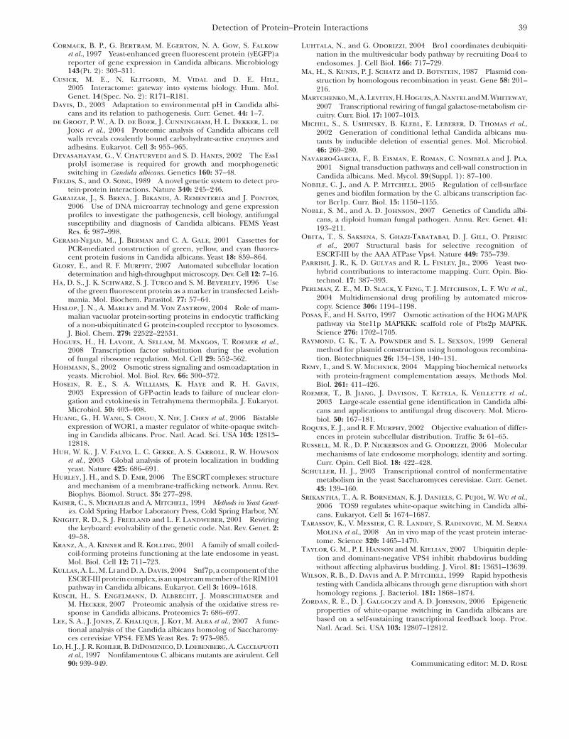
Figure S2.—S. cerevisiae PBS2GFP & VPS32 vps4-. Sets of 3-4 fields of GFP fluorescence images for S. cerevisiae interacting protein pairs and negative controls. All fields are shown at the same magnification.

J. Boysen et al. 4 SI
A
B
C
D
FIGURE S3.—S. cerevisiae SNF1GFP & SNF4VPS32 vps4-. Sets of 3-4 fields of GFP fluorescence images for S. cerevisiae
interacting protein pairs and negative controls. All fields are shown at the same magnification.
J. Boysen et al. 5 SI
A
B
C
D
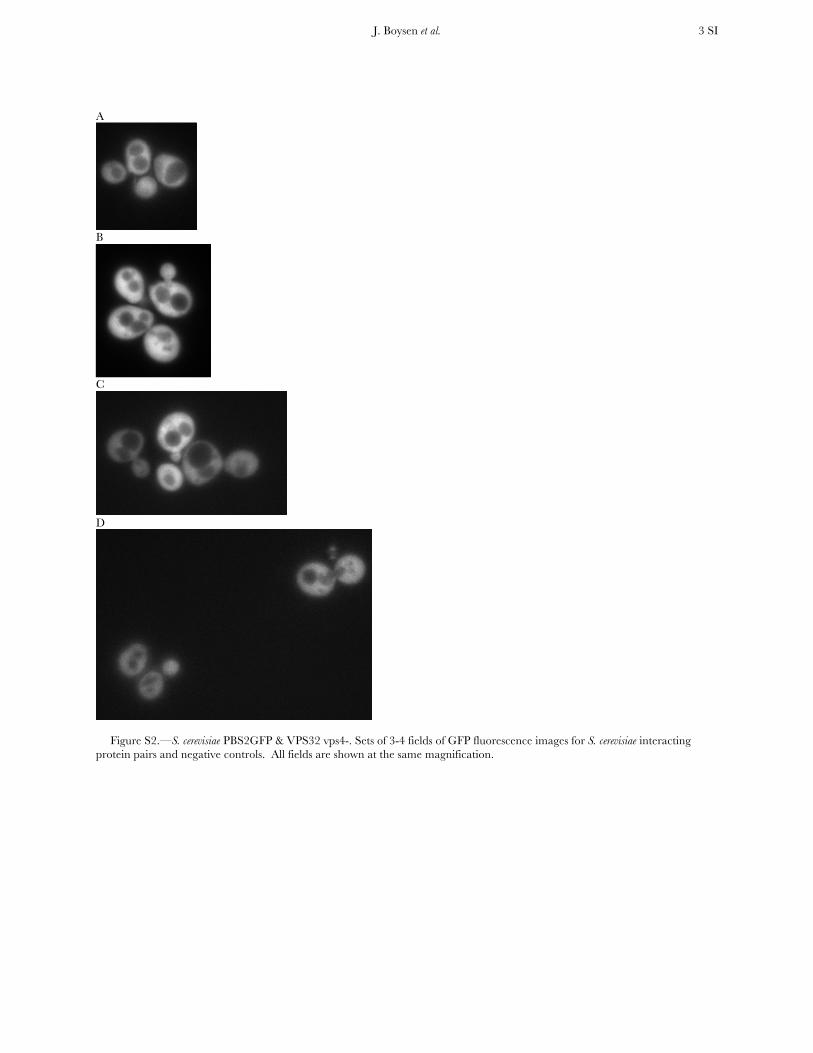
FIGURE S4.— S. cerevisiae SNF1GFP & VPS32 vps4-. Sets of 3-4 fields of GFP fluorescence images for S. cerevisiae interacting protein pairs and negative controls. All fields are shown at the same magnification.

J. Boysen et al. 6 SI
A
B
C
D
FIGURE S5.—S. cerevisiae PBS2GFP & HOG1VPS32 WT. Sets of 3-4 fields of GFP fluorescence images for S. cerevisiae interacting protein pairs and negative controls. All fields are shown at the same magnification.

J. Boysen et al. 7 SI
A
B
C
D
FIGURE S6.— S. cerevisiae PBS2GFP & VPS32 WT. Sets of 3-4 fields of GFP fluorescence images for S. cerevisiae interacting protein pairs and negative controls. All fields are shown at the same magnification.
J. Boysen et al. 8 SI
A
B
C
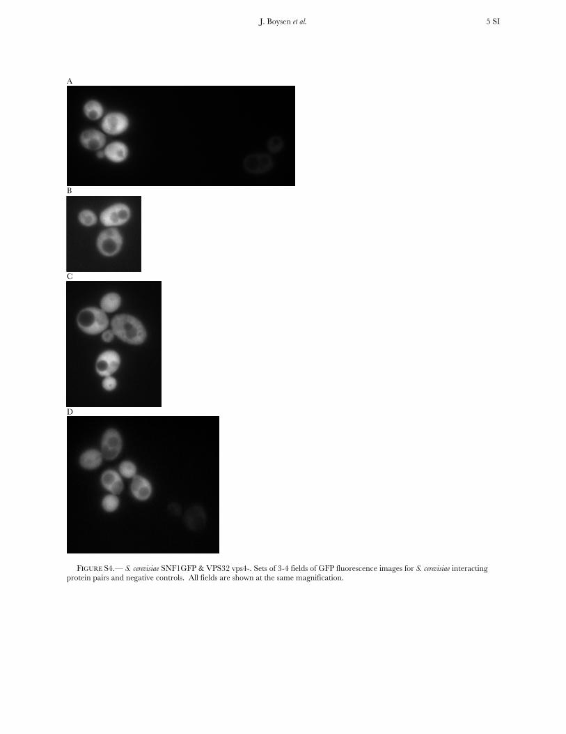
FIGURE S7.— S. cerevisiae SNF1GFP & SNF4VPS32 WT. Sets of 3-4 fields of GFP fluorescence images for S. cerevisiae interacting protein pairs and negative controls. All fields are shown at the same magnification.

J. Boysen et al. 9 SI
A
B
C
FIGURE S8.— S. cerevisiae SNF1GFP & VPS32 WT. Sets of 3-4 fields of GFP fluorescence images for S. cerevisiae interacting protein pairs and negative controls. All fields are shown at the same magnification.
J. Boysen et al. 10 SI
A
B
C
D
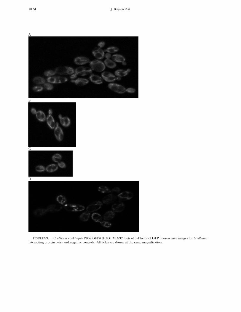
FIGURE S9.— C. albicans vps4/vps4 PBS2.GFP&HOG1.VPS32. Sets of 3-4 fields of GFP fluorescence images for C. albicans interacting protein pairs and negative controls. All fields are shown at the same magnification.

J. Boysen et al. 11 SI
A
B
C
FIGURE S10.— C. albicans vps4/vps4 PBS2.GFP&VPS32 EMPTY VECTOR. Sets of 3-4 fields of GFP fluorescence images for C. albicans interacting protein pairs and negative controls. All fields are shown at the same magnification.
J. Boysen et al. 12 SI
A
B
C
D
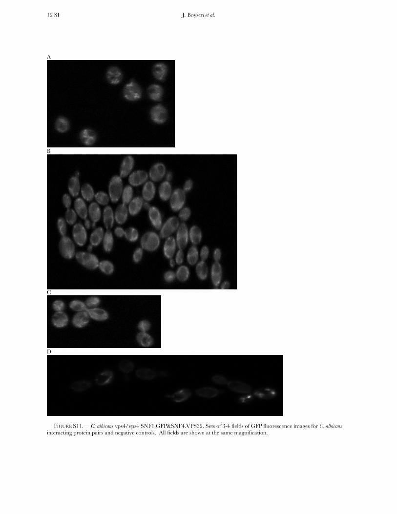
FIGURE S11.— C. albicans vps4/vps4 SNF1.GFP&SNF4.VPS32. Sets of 3-4 fields of GFP fluorescence images for C. albicans interacting protein pairs and negative controls. All fields are shown at the same magnification.

J. Boysen et al. 13 SI
A
B
C
D
FIGURE S12.— C. albicans vps4/vps4 SNF1.GFP&VPS32 EMPTY VECTOR. Sets of 3-4 fields of GFP fluorescence images for C. albicans interacting protein pairs and negative controls. All fields are shown at the same magnification.

J. Boysen et al. 14 SI
A
B
C
D
FIGURE S13.— C. albicans vps4/vps4 Non Interacting Pairs Control PBS2GFP&SNF4VPS32. Sets of 3-4 fields of GFP fluorescence images for C. albicans interacting protein pairs and negative controls. All fields are shown at the same magnification.