DESIGN, SYNTHESIS AND BIOLOGICAL EVALUATION OF NON ...
169
DESIGN, SYNTHESIS AND BIOLOGICAL EVALUATION OF NON-PEPTIDIC SMALL MOLECULAR SMAC MIMETICS AS POTENT IAP INHIBITORS by Yuefeng Peng A dissertation submitted in partial fulfillment of the requirements for the degree of Doctor of Philosophy (Medicinal Chemistry) in the University of Michigan 2008 Doctoral Committee: Professor Shaomeng Wang, Chair Professor Masato Koreeda Professor Anna K. Mapp Professor David H. Sherman Assistant Professor Jason E. Gestwicki
Transcript of DESIGN, SYNTHESIS AND BIOLOGICAL EVALUATION OF NON ...

DESIGN, SYNTHESIS AND BIOLOGICAL EVALUATION OF NON-PEPTIDIC SMALL MOLECULAR SMAC
MIMETICS AS POTENT IAP INHIBITORS
by
Yuefeng Peng
A dissertation submitted in partial fulfillment of the requirements for the degree of
Doctor of Philosophy (Medicinal Chemistry)
in the University of Michigan 2008
Doctoral Committee:
Professor Shaomeng Wang, Chair Professor Masato Koreeda Professor Anna K. Mapp Professor David H. Sherman Assistant Professor Jason E. Gestwicki

© Yuefeng Peng ————————————
All Rights Reserved
2008

ii
To my family

iii
Acknowledgements
I genuinely appreciate my advisor, Professor Shaomeng Wang for his expert
guidance and support. It is a really great honor for me to perform my PhD research under
the guidance of Professor Wang, a leading scientist in the area of Medicinal Chemistry.
I would like to thank my committee members, Professor Anna K. Mapp, Professor
David H. Sherman, Professor Masato Koreeda, and Professor Jason E. Gestwicki for their
time and efforts.
I would like to thank the Chair of Medical Chemistry Department, Professor
Ronald W. Woodard for providing me the opportunity to study in one of the top
Medicinal Chemistry Programs.
I would like to thank Dr. Haiying Sun for his advice and assistance in the synthesis
of Smac mimetics, Dr. Jianfeng Lu for his advice and assistance in the biological studies
of Smac mimetics, and Dr. Qian Cai for providing CQ-406 (SM-406) in the biological
evaluation experiments.
I would like to thank Dr. Chao-Yie Yang for computational studies, Dr. Zaneta
Nikolovska-Coleska for the FP assay and functional assay results. Besides, I would like
to thank everyone in the lab for their assistance in my research.
I need to extend a special thank to Dr. George W. A. Milne for his critical reading
and editorial assistance of and this dissertation
I appreciate my wife and my parents for their full support.

iv
Table of Contents
Dedication ................................................................................................................................. ii Acknowledgements ................................................................................................................. iii List of Figures ......................................................................................................................... vii List of Tables .......................................................................................................................... xii List of Abbreviations ............................................................................................................. xiii Chapter 1
INTRODUCTION ............................................................................................................ 1 1.1 Apoptosis and Cancer ........................................................................................ 1 1.2 IAP ...................................................................................................................... 3 1.3 Smac / DIABLO ................................................................................................. 4 1.4 Structure-Activity Relationships ....................................................................... 7
Chapter 2
DESIGN AND SYNTHESIS OF NON-PEPTIDIC SMALL MOLECULAR SMAC MIMETICS ..................................................................................................................... 22
2.1 Design Rational ................................................................................................ 22
2.1.1 Design of monovalent Smac mimetics ................................................. 22 2.1.2 Design of bivalent Smac mimetics ....................................................... 24
2.2 Retrosynthetic Analysis ................................................................................... 25 2.3 Results and Discussion .................................................................................... 28

v
2.4 Conclusion ........................................................................................................ 32 2.5 Synthesis of Smac Mimetics ............................................................................ 34 2.6 Methods and Materials ..................................................................................... 42
Chapter 3
BIOLOGICAL EVALUATION OF MONOVALENT SMAC MIMETICS .............. 68 3.1 Binding Potency of Monovalent Smac Mimetics ........................................... 68 3.2 Tumor Cell Growth Inhibition Activity of Monovalent Smac Mimetics ....... 70 3.3 Tumor Cell Death Induction Activity of Monovalent Smac Mimetics .......... 72 3.4 Apoptosis Induction Activity of Monovalent Smac Mimetics ....................... 73 3.5 Caspase Activation of Monovalent Smac Mimetics ....................................... 77 3.6 Drug Synergy Effect of Monovalent Smac Mimetics with TRAIL ............... 79 3.7 Cellular Molecular Effects of Monovalent Smac Mimetics ........................... 81 3.8 Conclusion ........................................................................................................ 83
Chapter 4
BIOLOGICAL EVALUATION OF BIVALENT SMAC MIMETICS ...................... 85 4.1 Binding Potency of Bivalent Smac Mimetics ................................................. 85 4.2 Tumor Cell Growth Inhibition Activity of Bivalent Smac Mimetics ............. 86 4.3 Tumor Cell Death Induction Activity of Bivalent Smac Mimetics ................ 88 4.4 Apoptosis Induction Activity of Bivalent Smac Mimetics ............................. 90 4.5 Cellular Molecular Effects of Bivalent Smac Mimetics ................................. 92 4.6 Conclusion ........................................................................................................ 94

vi
Chapter 5
CELLULAR MECHANISM STUDIES BASED ON SM-406 ................................... 96 5.1 SM-406 ............................................................................................................. 96 5.2 Further Biological Studies Based on SM-406 ................................................. 98
5.2.1 Apoptosis induction activity of SM-406 .............................................. 98 5.2.2 Tumor cell death induction activity of SM-406 ................................. 103 5.2.3 Cellular molecular effects of SM-406 ................................................ 105 5.2.4 Co-immunoprecipitation assays confirm c-IAP1 and XIAP as the cellular targets of SM-406 ........................................................................... 108 5.2.5 SM-406 can compensate for Smac knockdown in tumor cells ......... 110 5.2.6 Study of caspase dependence in the cellular activity of SM-406 in tumor cells .................................................................................................... 112 5.2.7 SM-406 can induce fast degradation of c-IAP1 but not XIAP .......... 116
5.3 Conclusion ...................................................................................................... 118 5.4 Methods and Materials ................................................................................... 119
Chapter 6
CONCLUSION ............................................................................................................ 126 BIBLIOGRAPHY ................................................................................................................ 131

vii
List of Figures
Figure 1.1 Biological pathways of apoptosis ....................................................................................... 2 1.2 Domain structure of XIAP, c-IAP1 and c-IAP2 ................................................................ 4 1.3 IAPs-Binding Motif (IBM) of Smac/DIABLO and caspase-9 ......................................... 5 1.4 X-ray structure of Smac IBM binding with the XIAP BIR3 domain ............................... 6 1.5 Design of the conformationally constrained Smac mimetics .......................................... 10 1.6 Design of the conformationally constrained Smac mimetics with higher potency ....... 13 1.7 Inhibition of cell growth by Smac mimetics in human breast cancer MDA-MB-231 cell
lines. Cells were treated for 4 days, and cell growth inhibition was determined using the WST-based assay .............................................................................................................. 16
1.8 Chemical structures of the monovalent and dimeric Smac mimetics ............................. 17 1.9 Inhibition of cell growth by Smac mimetics in the HL-60 leukemia cancer cell line. HL-
60 cells were treated with the Smac mimetics for 4 days and cell growth was analyzed by WST-based cell growth assay ..................................................................................... 20
1.10 Probing the interaction of Smac mimetics to cellular XIAP in the HL-60 leukemia cell
line by a competitive, co-immunoprecipitation pull-down assay using biotinylated Smac mimetic ................................................................................................................. 21
2.1 Chemical structures of SM-122 and designed new monomeric Smac mimetics ........... 22 2.2 Computational modeling structure of Smac mimetic compound YP-245P3 binding with
the XIAP BIR3 domain .................................................................................................... 23 2.3 Chemical structure of designed bivalent Smac mimetics ................................................ 24 2.4 Retro-synthetic analysis of designed Smac mimetics ..................................................... 26 2.5 Retro-synthetic analysis of the new route ........................................................................ 27

viii
2.6 Synthetic route to key intermediateYP-248P ................................................................... 34 2.7 New synthetic route to the key intermediate YP-248P ................................................... 36 2.8 Synthesis of key components YP-245 and YP-373 ......................................................... 37 2.9 Synthesis of monovalent Smac mimetics ........................................................................ 39 2.10 Synthesis of monovalent Smac mietics SM-376 and SM-377 ...................................... 40 2.11 Synthesis of bivalent Smac mietics ................................................................................ 41 3.1 Predicted binding models of SM-227 (2), SM-245 (3), SM-246 (4), and SM-330 (5) to
XIAP BIR3 domain, in superposition with Smac AVPI peptide. ................................... 69 3.2 Principles of WST-based cell proliferation assay ............................................................ 70 3.3 Inhibition of tumor cell growth by monovalent Smac mimetics in human breast cancer
MDA-MB-231 cells and human ovarian cancer SK-OV-3 cells .................................... 71 3.4 Chemical structure of Trypan blue ................................................................................... 72 3.5 Cell viabilities of human ovarian cancer SK-OV-3 cells and human breast cancer
MDA-MB-231 cells treated with different concentrations of monovalent Smac mimetics for 24 or 48 hours, as determined by Trypan blue cell death assays ............. 73
3.6 Annexin V and P.I. double staining flow cytometry of untreated MDA-MB-231 cells 74 3.7 Annexin V and P.I. double staining flow cytometry of human breast cancer MDA-MB-
231 cells treated with different concentrations of monovalent Smac mimetic SM-245, SM-337, or SM-376 for 24 hours ..................................................................................... 74
3.8 Annexin V and P.I. double staining flow cytometry of human ovarian cancer SK-OV-3
cells treated with different concentrations of monovalent Smac mimetic SM-245, SM-337, or SM-376 for 24 hours ............................................................................................ 75
3.9 Annexin V and P.I. double staining flow cytometry of human breast cancer MDA-MB-
231 cells treated with different concentrations of monovalent Smac mimetic SM-245, SM-337, or SM-376 for 48 hours ..................................................................................... 76
3.10 Inhibition of caspase-3/7 activity by XIAP and antagonism of Smac mimetics to XIAP
to recover the activity of caspase-3/7 in a cell-free functional assay ............................ 78

ix
3.11 Left: Inhibition of caspase-3/7 activity by XIAP and antagonism of Smac mimetic SM-246 to XIAP to recover the activity of caspase-3/7 in a cell-free functional assay. Right: Dose-dependent recovery of caspase-3/7 activity by SM-122, SM-246, and SM-337 to the maximum activation. Caspase-3/7 activity at 30 minute point was used79
3.12 Inhibition of cell growth by Smac mimetics SM-337, SM-376, and SM-377 in
combination with TRAIL in human breast cancer MDA-MB-231 cell lines. Cells were treated with TRAIL only or TRAIL in combination with Smac mimetics for 4 days and cell growth was analyzed by WST-based cell growth assay ........................ 80
3.13 Inhibition of cell growth by Smac mimetics SM-337, SM-376, and SM-377 in
combination with TRAIL in human breast cancer 2LMP cell lines ............................ 81 3.14 Western blot assays of human breast cancer MDA-MB-231 cells and human ovarian
cancer SK-OV-3 cells treated with different concentrations of Smac mimetic SM-122, SM-227, SM-245, or SM-337 for 24 hours ................................................................... 82
4.1 WST cell growth assays of human breast cancer MDA-MB-231 cells and human
ovarian cancer SK-OV-3 cells treated with bivalent Smac mimetics for 96 hours ....... 87 4.2 WST cell growth assay of human melanoma MALME-3M cells treated with bivalent
Smac mimetics for 96 hours. ........................................................................................... 88 4.3 Trypan blue assays of human breast cancer MDA-MB-231 cells and human ovarian
cancer SK-OV-3 cells treated with different concentrations of bivalent Smac mimetics for designated lengths of time ......................................................................................... 89
4.4 Human breast cancer MDA-MB-231cells were treated with different concentrations of
bivalent Smac mimetic SM-381 (8C) and SM-383 (10C) or 1 µM of inactive control SM-122 for 24 hours ........................................................................................................ 90
4.5 Human ovarian cancer SK-OV-3 cells were treated with different concentrations of
bivalent Smac mimetic SM-381 (8C) and SM-383 (10C) or 1 µM of inactive control SM-122 for 24 hours ....................................................................................................... 91
4.6 Western blotting assays of human breast cancer MDA-MB-231 cells treated with
different concentrations of bivalent Smac mimetic SM-164, SM-381 and SM-383 for 24 hours ............................................................................................................................ 92
4.7 Western blotting assays of human ovarian cancer SK-OV-3 cells were treated with
different concentrations of bivalent Smac mimetic SM-164, SM-381 and SM-383 for 24 hours ............................................................................................................................ 93
5.1 Chemical structure of SM-406 ......................................................................................... 97

x
5.2 Top: Chemical structure of SM-428, inactive control of Smac mimetics. Bottom: Annexin V and P.I. double staining flow cytometry of human ovarian cancer SK-OV-3 cells treated Smac mimetic SM-406 and inactive control SM-428 for 24 hours .......... 98
5.3 Annexin V and P.I. double staining flow cytometry of human ovarian cancer SK-OV-3
cells treated with 3 µM of Smac mimetic SM-406 for designated lengths of time ........ 99 5.4 Annexin V and P.I. double staining flow cytometry of human breast cancer MDA-MB-
231 cells treated with different doses of Smac mimetic SM-406 or inactive control SM-428 for 24 hours .............................................................................................................. 100
5.5 Annexin V and P.I. double staining flow cytometry of human breast cancer MDA-MB-
231 cells treated with 3 µM of Smac mimetic SM-406 or 3 µM of inactive control SM-428 for designated lengths of time ................................................................................. 101
5.6 Annexin V and P.I. double staining flow cytometry of human melanoma MALME-3M
cells treated with different doses of Smac mimetic SM-406 or inactive control SM-428 for 24 hours ..................................................................................................................... 102
5.7 Annexin V and P.I. double staining flow cytometry of human melanoma MALME-3M
cells treated with 3 µM of Smac mimetic SM-406 or 3 µM of inactive control SM-428 for designated lengths of time ........................................................................................ 103
5.8 Cell viabilities of human breast cancer MDA-MB-231 cells and human ovarian cancer
SK-OV-3 cells treated with different concentrations of Smac mimetic SM-406 or inactive control SM-428 for 24 hours, as analyzed by Trypan-blue-based cell death assay ................................................................................................................................ 103
5.9 Cell viabilities of human breast cancer MDA-MB-231 cells and human ovarian cancer
SK-OV-3 cells treated with 3 µM of Smac mimetic SM-406 or 3 µM of inactive control SM-428 for designated lengths of time, as analyzed by Trypan-blue-based cell death assay ................................................................................................................................ 104
5.10 Western blotting assays of human breast cancer MDA-MB-231 cells and human
ovarian cancer SK-OV-3 cells treated with different concentrations of Smac mimetic SM-406 or 3 µM of inactive control SM-428 for 24 hours ........................................ 105
5.11 Western blotting assays of human breast cancer MDA-MB-231 cells and human
ovarian cancer SK-OV-3 cells treated with 3 µM of Smac mimetic SM-406 or 3 µM of inactive control SM-428 for designated lengths of time ........................................ 107
5.12 Chemical structures of Smac mimetic SM-406 and biotinylated Smac mimetic SM-
222 ................................................................................................................................ 108

xi
5.13 Probing the interaction of Smac mimetics to cellular XIAP and c-IAP1 in human breast cancer MDA-MB-231 cells and human ovarian cancer SK-OV-3 cells by competitive, co-immunoprecipitation pull-down assays using biotinylated Smac mimetic SM-222 .......................................................................................................... 109
5.14 Western blotting assay of human ovarian cancer SK-OV-3 cells transfected with
control siRNA oligonucleotides targeting GFP, or siRNA oligonucleotides targeting Smac for 48 hours, then treated with 3 µM of Smac mimetic SM-406 for 24 hours 110
5.15 Western blotting assay of human breast cancer MDA-MB-231 cells transfected with
control siRNA oligonucleotides targeting GFP, or siRNA oligonucleotides targeting Smac for 48 hours, then treated with 3 µM of Smac mimetic SM-406 for 24 hours 111
5.16 Human ovarian cancer SK-OV-3 cells and human breast cancer MDA-MB-231 cells
were first transfected with control siRNA oligonucleotides targeting GFP, or siRNA oligonucleotides targeting Smac for 48 hours, then treated with 3 µM of Smac mimetic SM-406 for 24 hours. Cell viabilities were analyzed by using Trypan-blue-based cell death assay ................................................................................................... 112
5.17 Cell viabilities of human breast cancer MDA-MB-231 cells and human ovarian cancer
SK-OV-3 cells treated with designated concentrations of Smac mimetic SM-406 alone or in combination with 25 µM of caspase-9, -8, and -3 inhibitors for 24 hours, as analyzed by Trypan-blue-based cell death assay ......................................................... 113
5.18 Western blotting assays of human breast cancer MDA-MB-231 cells and human
ovarian cancer SK-OV-3 cells treated with 3 µM of Smac mimetic SM-406 alone or in combination with 25 µM of caspase-9, -8, and -3 inhibitors for 24 hours ............ 114
5.19 Top: Western blotting assay of human ovarian cancer SK-OV-3 cells transfected with
control siRNA oligonucleotides targeting GFP, or siRNA oligonucleotides targeting caspase-9, -8 and -3 for 48 hours, then treated with 3 µM of Smac mimetic SM-406 for 24 hours. Bottom: Cell viability of SK-OV-3 cells, as analyzed by Trypan-blue based cell death assay ................................................................................................... 115
5.20 Western blotting assays of human ovarian cancer SK-OV-3 cells and human breast
cancer MDA-MB-231 cells treated with 3 µM of Smac mimetic SM-406 for designated lengths of time ........................................................................................... 117

xii
List of Tables
Table 1.1 Chemical structures of Smac peptide-mimetics and their binding affinities to the XIAP
BIR3 protein determined using a fluorescence-polarization-based binding assay .......... 8 1.2 Chemical structures of 6,5-bicyclic Smac mimetics and their binding affinities to the
XIAP BIR3 protein determined with a fluorescence-polarization-based binding assay 12 1.3 Chemical structures of 7,5-bicyclic Smac mimetics and their binding affinities to the
XIAP BIR3 protein as determined using a fluorescence-polarization-based binding assay .................................................................................................................................. 14
1.4 Design of cell-permeable Smac mimetics and their binding affinities to the XIAP BIR3
protein as determined using a fluorescence-polarization-based binding assay .............. 15 1.5 Chemical structures of monovalent and bivalent 8,5-bicyclic Smac mimetic compounds
and their binding affinities with the XIAP BIR3 domain (residue 240-356) and XIAP BIR2-BIR3 domains (residue 120-356) determined in a competitive fluorescence-polarization-based assay ................................................................................................... 18
2.1 Chemical structures of synthesized monovalent and bivalent Smac mimetics and their
binding affinities to the XIAP BIR3 or XIAP linker-BIR2-BIR3 as determined using a fluorescence-polarization-based binding assay ............................................................... 29
3.1 Binding affinities of Smac mimetics to XIAP, c-IAP1/2, as determined by competitive,
fluorescence-polarization based assays ........................................................................... 68 4.1 Binding affinities of bivalent Smac mimetics against XIAP BIR3 and XIAP linker-
BIR2-BIR3 domains, as measured by fluorescence-polarization based assays ............. 86

xiii
List of Abbreviations
A or Ala Alanine Ac Acetyl Admin. Administration Apaf-1 Apoptotic Protease Activating Factor-1 ATP Adenosine Tri-Phosphate AUC Area Under Curve Bax Bcl-2 Associated X protein Bid BH3 Interacting Domain death agonist BIR Baculovirus Inhibitors of Apoptosis Protein Repeat Bn Benzyl Boc tert-Butyloxycarbonyl BRUCE Baculoviral IAP Repeat containing Ubquitin Conjugating
Enzyme Bu Butyl C3 Caspase-3 C7 Caspase-7 C8 Caspase-8 C9 Caspase-9 calcd. Calculated Cbz Carbobenzyloxy Cl. Clearance CL C3 Cleaved Caspase-3 CL C7 Cleaved Caspase-7 CARD Caspase Recruitment Domain Caspase Cysteine-dependent Aspartate Protease Comp. Compound Conc. Concentration co-IP co-Immunoprecipitation c-IAP1 cellular Inhibitors of Apoptosis Protein 1 c-IAP2 cellular Inhibitors of Apoptosis Protein 2 Cyt-c Cytochrome c DCM Dichloromethane DIABLO Direct IAP Binding protein with Low pI DIEA N,N-Diisopropylethylamine DISC Death-Inducing Signaling Complex DLB Double Lysis Buffer DTT Dithiothreitol E or Glu Glutamic acid

xiv
EDC 1-ethyl-3-(3-dimethylaminopropyl) carbodiimide hydrochloride
EDTA Ethylenediaminetetraacetic Acid 2 equiv. equivalent Et Ethyl EtOAc Ethyl estate F Bioavailability F or Ph Phenylalanine FADD Fas Associated Death Domain FasL Fas Ligand FP Fluorescence Polarization G or Gly Glycine GFP Green Fluorescent Protein h. hour(s) H or His Histidine HOBt Hydroxybenzotriazole HPLC High-performance liquid chromatography HRMS High Resolution Mass Spectrometry I or Ile Isoleucine IAC IAPs Antagonist Compound IAP Inhibitors of Apoptosis Protein IBM Inhibitors of Apoptosis Protein-Binding Motif K or Lys Lysine Kd Constant of Dissociation L or Leu Leucine Me Methyl min. minute(s) ML-IAP Melanoma Linked Inhibitors of Apoptosis Protein MRB Mitochondrial Resuspension Buffer MRT Mean Residence Time MS Mass Spectrometry NAIP Neuronal Apoptosis Inhibitory Protein NF-κB Nuclear Factor-κB NMR Nuclear magnetic resonance NIK1 Nim1-like Protein Kinase NP-40 Nonidet P 40 P or Pro Proline PAGE Polyacrylamide Gel Electrophoresis PARP Poly ADP Ribose Polymerase PBS Phosphate Buffer Saline PCD Programmed Cell Death Ph Phenyl pI Isoelectric Point P.I. Propidium Iodide PK Phamacokinetic PMSF Phenylmethylsulphonyl Fluoride

xv
Pr Propyl PS Phosphatidylserine PVDF Polyvinylidene Difluoride Q or Gln Glutamine R or Arg Arginine RING Really Interesting New Gene RIP Receptor-Interacting Protein R.T. Room Temperature S or Ser Serine SAR Structure Activity Relationship SD Standard Deviation SDS Sodium Dodecyl Sulfate siRNA small interfering RNA or silencing RNA or short interfering RNA Smac Second Mitochondria-derived Activator of Caspase STR Succinate-tetrazolium Reductase t1/2 Half life T or Thr Theronine TBS tert-butyldimethylsilyl TBS Tris-Buffered Saline t-Bu tert-butyl TFA Trifluoroacetic acid THF Tetrahydrofuran TMS Tetramethylsilane TNFα Tumor Necrosis Factor α TRAIL Tumor Necrosis Factor-Related Apoptosis Inducing Ligand Ts-IAP Tesis-specific Inhibitors of Apoptosis Protein V or Val Valine W or Trp Tryptophan WST 2-(2-methoxy-4-nitrophenyl)-3-(4-nitrophenyl)-5-(2, 4-
isulfophenyl)-2H-tetrazolium, monosodium salt XIAP X-chromosome linked Inhibitor of Apoptosis Protein Y or Tyr Tyrosine

1
CHAPTER 1
INTRODUCTION
1.1 Apoptosis and Cancer
Apoptosis, from the Greek, meaning “falling off” petals or leaves from plants or
trees,1-15 is a term used in cellular biology for programmed cell death (PCD). This process,
is distinct from necrosis, which is a form of traumatic cell death caused by cellular injury,
and follows a series of cellularly controlled steps, resulting ultimately in cell death. Most
of the current cancer therapies, such as radiation, chemotherapeutic agents and
immunotherapy work by directly or indirectly inducing apoptosis in cancer cells.16-21
Resistance to apoptosis leaves cancer cells unable to execute apoptosis,22-24 and is a major
problem in current cancer therapy.25-30 Successful anticancer therapies must include
strategies specifically targeting the resistance of cancer cells to apoptosis.31,32 Hence,
targeting the crucial negative regulators which play a role in inhibition of apoptosis of
cancer cells can be a promising therapeutic strategy for new anticancer drug design.33-43
Apoptosis can be induced by a variety of stimuli, including death ligands such as
Tumor Necrosis Factor α (TNFα), TNF-Related Apoptosis Inducing Ligand (TRAIL), or
Fas Ligand (FasL) as shown in the extrinsic pathway in Figure 1.1; heat, radiation,
hypoxia, viral infection, and nutrient deprivation, in the intrinsic pathway, are also

2
effective. In the extrinsic pathway, ligation of death ligand with the death receptor leads
to the formation of FADD (Fas-Associated Death Domain), which in turn recruits
procaspase-8 or procaspase-10, forming a Death-Inducing Signaling Complex (DISC),
and inducing auto-activation of the initiator caspase, caspase-8, or caspase-10.44,45
FADD
TRAIL/TNF Ligand
Death Receptors
Caspase‐8/10
tBid
Radio and chemotherapy
Bcl‐2Bcl‐xL
cytochrome c
Caspase‐3/7
Apoptosis
XIAP
Smac/
DIABLO
Caspase‐9
BaxChannel
Apoptosome
Apaf‐1Procaspase‐9
Mitochondria
PARP
Smac/
DIABLO
Extrinsic pathway
Intrinsic pathway
Figure 1.1 Biological pathways of apoptosis.
Initiator caspases cleave procaspase-3 and procaspase-7 to yield the executioners of
apoptosis, caspase-3 and caspase-7.46-67 In the intrinsic pathway, cellular stress, for
example radiation, induces the translocation of a Bcl-2 family protein such as Bax (Bcl-2
Associated X protein), causing the release of cytochrome c from the mitochondria into
the cytosol.68 Cytochrome c binds Apaf-1 (Apoptotic Protease Activating Factor-1),
procaspase-9 and ATP (Adenosine Triphosphate) to form the apoptosome complex,

3
which recruits the auto-activation of caspase-9.69-74 Caspase-9 in turn induces the
activation of the effectors, caspase-3 and caspase-7 to effect apoptosis.75-77
In some cases, the extrinsic pathway can induce the intrinsic pathway, in which a
pro-apoptotic member of Bcl-2 family, Bid (BH3 Interacting Domain death agonist) is
cleaved by caspase-8, then interacts with Bax, leading to the release of cytochrome c into
cytosol from mitochondria.78-85
1.2 IAP
IAPs (Inhibitors of Apoptosis Protein) are a class of proteins which can negatively
regulate the apoptosis process in cancer cells.86-93 Eight distinct IAPs are known:
• NAIP (Neuronal Apoptosis Inhibitory Protein)
• XIAP (X-chromosome linked Inhibitor of Apoptosis Protein)
• c-IAP1 (cellular IAP 1)
• c-IAP2 (cellular IAP 2)
• Ts-IAP (Tesis-specific IAP)
• ML-IAP (Melanoma Linked IAP) or Livivn
• BRUCE (Baculoviral IAP Repeat containing Ubiquitin Conjugating Enzyme)
or Apollon
• Survivin
The domain structures of XIAP, c-IAP1 and c-IAP2 are shown in Figure 1.2. Most
of the IAPs, except NAIP, BRUCE/Apollon, and Survivin, have a carboxy-terminal
RING (Really Interesting New Gene) domain which directs self-ubiquitination and

4
protein degradation.94,95 Of all the IAPs, only c-IAP1 and c-IAP2 have a CARD
(CAspase Recruitment Domain) domain, which mediates other CARD-containing
proteins.96,97 The BIR (Baculovirus IAP Repeat) domain, is the functional domain of
apoptosis inhibition and is the important characteristic of each IAP member.98-103 While
XIAP, c-IAP1, c-IAP2, ML-IAP, and NAIP can bind caspase-9, caspase-3 and caspase-7
directly to their BIR domains, thus inhibiting caspase activity, Survivin and BRUCE
regulate the cytokines and mitotic spindle formation in order to inhibit the apoptosis
process.104-111
XIAP: BIR1 BIR2 BIR3 RING
c-IAP1: BIR1 BIR2 BIR3 RINGCARD
c-IAP2: BIR1 BIR2 BIR3 RINGCARD
Figure 1.2 Domain structures of XIAP, c-IAP1 and c-IAP2.
XIAP binds both the initiator caspase, caspase-9, with its BIR3 domain and the
effector caspase-3 or caspase-7, with BIR2 and the linker before the BIR2 domain.112,113
By blocking the activity of caspase-3/7, XIAP inhibits apoptosis at the down-stream
effector phase, where multiple signal pathways converge. Hence, strategies targeting
XIAP can be an effective method to overcome the resistance of cancer cells to the
apoptosis.114-118
1.3 Smac/DIABLO

5
Smac (Second Mitochondria-derived Activator of Caspase), also known as
DIABLO (Direct IAP Binding protein with Low pI), was recently identified as a protein
released from mitochondria in response to apoptotic stimuli.119,120 Smac is a 239 amino-
acid protein. Its amino-terminal 55 residues are removed during translocation to yield
active Smac.119,120 As shown in Figure 1.3, the amino-terminal tetrapeptide in Smac, Ala-
Val-Pro-Ile (residue 56 to 59) is homologous with the exposed amino-terminal
tetrapeptide of caspase-9 (Ala-Thr-Pro-Phe). The amino-terminal tetrapeptides of Smac
and caspase-9, also known as IBM (IAP-Binding Motif), bind to a well-defined surface
groove in the BIR3 domain of XIAP. By binding with the BIR3 domain of XIAP, Smac
inhibits the interaction of XIAP BIR3 domain and caspase-9. This interaction releases
caspase-9 and promotes apoptosis.121
Smac / DIABLO: A-V-P-I-A-Q-K-S-E-P-H
Caspase-9: A-T-P-F-Q-E-G-L-R-T-F
Figure 1.3 IAPs-Binding Motif (IBM, in red) of Smac/DIABLO and caspase-9.
The structures of the XIAP BIR3 domain complexed with either Smac protein or
Smac amino-terminal peptide have been determined by X-ray crystallography and NMR
spectroscopy.122,123 The amino-terminal tetrapeptide Ala-Val-Pro-Ile binds the XIAP
BIR3 domain and is equipotent (Kd = 0.4 µM) with the mature Smac protein (Kd = 0.4
µM). Therefore, it is possible to use small molecule non-peptide Smac mimetics to mimic
the interaction between the mature Smac protein and the XIAP BIR3 domain, releasing
initiator caspase-9 to promote the apoptosis process.

6
Figure 1.4 X-ray structure of Smac IBM binding with the XIAP BIR3 domain. (Hydrogen bonds are shown in light-blue dashed lines.)
Figure 1.4 shows the interaction of the IAP-binding motif (IBM) of Smac with the
XIAP BIR3 domain.123 The amino group of the amino-terminal alanine (A1 in Figure 1.4)
of mature Smac is positively charged and its hydrogen atoms form a total of four
hydrogen bonds with the two oxygen atoms in the carboxyl group of XIAP glutamic acid
314 side chain, the carbonyl oxygen of the side chain of glutamine 319, and the backbone
carbonyl oxygen of aspartic acid 309. These hydrogen bonds are crucial because
mutagenesis experiments show that in the Smac mutant A1M (alanine is replaced with an
methionine) the interaction of Smac and the XIAP BIR3 domain is completely
disrupted.124 The methyl side chain of A1 fits tightly in a small hydrophobic pocket
formed by the side chains of leucine 307, tryptophan 310, and glutamine 319 of XIAP.
The amino hydrogen in the indole ring of tryptophan 323 forms a hydrogen bond with the

7
backbone carbonyl oxygen of A1 and the indole ring participates in a hydrophobic
interaction with the five-member ring of proline (P3 in Figure 1.4). The amino hydrogen
and the carbonyl oxygen of the valine 2 (V2 in Figure 1.4) form two hydrogen bonds
with the backbone carbonyl oxygen and amino hydrogen of threonine 308 respectively,
while the methyl side chain of threonine 308 enjoys a hydrophobic interaction with the
isopropyl side chain of valine 2. A further hydrogen bond interaction is present between
the amino hydrogen of isoleucine 4 (I4 in Figure 1.4) and the backbone carbonyl oxygen
of glycine 306. The backbone carbonyl of isoleucine 4 is directed towards the solvent and
fails to interact with the XIAP BIR3 domain, while the isobutyl side chain of I4 is
inserted into the large hydrophobic pocket formed by lysine 297 and lysine 299 of the
XIAP BIR3 domain. V2 and P3 form a reverse turn structure forcing the amino terminal
tetrapeptide Ala-Val-Pro-Ile of the mature Smac protein to bind in the Smac binding
groove of the XIAP BIR3 domain.122
1.4 Structure-Activity Relationships
As discussed in Section 1.3, above, the amino-terminal tetrapeptide Ala-Val-Pro-
Ile of Smac binds the XIAP BIR3 domain with potency (Kd = 0.4 µM) similar to that of
the mature Smac protein (Kd = 0.4 µM), allowing the design of a small molecule non-
peptidic Smac mimetic which can mimic the interaction of the mature Smac protein with
the XIAP BIR3 domain. Several groups have been investigating small molecular non-
peptidic Smac mimetics as potent inhibitors of XIAP which can overcome apoptosis

8
resistance in cancer therapy. The Structure Activity Relationships (SAR) of small
molecular Smac mimetics have been extensively explored.125-130
H2NNH
N
R1
O
O R4
Comp R1 R4 Ki/µM ± SD Comp R1 R4 Ki/µM ± SD 1 Me -CONHCH2 0.29 ± 0.07 2 Me -CONHCH2 13.40 ± 1.6 3 Me -CONHCH2 2.45 ± 0.7 4 Me -CONHCH2 4.41 ± 1.5 5 Me -CONHCH2 1.27 ± 0.2 6 Me -CONHCH2
O
0.22 ± 0.07 7 Me -CONHCH2
S
0.18 ± 0.07 8 Me -CONH 4.9 ± 2.1 9
Me
-CONHCH2CH2
0.15 ± 0.09
10
Me -CONHCH2
0.028 ± 0.020
11
Et -CONHCH2
0.024 ± 0.020
12
Me
-CH2CH2CH2
1.2 ± 0.4
13 H -CONHCH2CH2
68 ± 7 14 Et -CONHCH2 0.081 ± 0.06
15 i-Pr -CONHCH2
4.15 ± 1.2 16 Pr -CONHCH2
54 ± 7
Table 1.1 Chemical structures of Smac peptide-mimetics and their binding affinities to the XIAP BIR3 protein determined using a fluorescence-polarization-based binding assay.125
The SAR of variations in the side chain of alanine 1 and of isoleucine 4 were
studied and the results are shown in Table 1.1.125 The backbone carbonyl of isoleucine 4
is directed towards the solvent and does not interact with the XIAP BIR3 domain while
the isobutyl side chain of I4 is in the large hydrophobic pocket formed by lysine 297 and
lysine 299 of the XIAP BIR3 domain. Replacement of the backbone carbonyl of
isoleucine 4 with a benzyl as in the caspase-9 phenylalanine residue yields compound 1
in Table 1.1. Compound 1, with Ki = 0.29 µM, was as twice as potent as the original

9
amino terminal tetrapeptide of mature Smac protein (Ala-Val-Pro-Ile; Ki = 0.58 µM),
determined using the fluorescence-polarization-based assay.126
To further explore the SAR concerning R4, a series of compounds (2-10) with R1 =
CH3 were synthesized. When the phenyl group in compound 1 was replaced by isopropyl,
2’-ethylbutyl, cyclopropyl or cyclohexyl, the potency of the compounds as Smac
mimetics dropped dramatically. When the phenyl group in compound 1 was substituted
with another aromatic group however, as in compounds 6 and 7, the potency of Smac
mimetics persisted. Hence, an aromatic group in R4 appears to bind preferentially in the
hydrophobic pocket formed by lysine 297 and lysine 299 in the XIAP BIR3 domain. In
order to determine the optimum length of the R4 side chain, compounds 8 and 9, with one
more and one less carbon in the R4 side chain respectively, were tested. As shown in
Table 1.1, increase in the chain length was correlated with a slight increase in the potency
of the compound but a decrease of the chain length decreased the compound’s potency
dramatically. Although the backbone carbonyl group of isoleucine 4 is oriented towards
the solvent and has no specific interaction with the XIAP BIR3 domain, it plays a role in
orientating the isoleucine 4 side chain toward the relative hydrophobic pocket in the
XIAP BIR3 domain. Hence, in compound 10, another phenyl ring was added to orientate
the other phenyl ring and reduce the conformational flexibility and, as expected,
compound 10, a highly potent Smac mimetic, resulted.
The methyl group of R1 was substituted with an ethyl group to yield compound 11,
R4 remaining unchanged. This change resulted in a slight increase in the Smac mimetic
potency as a result of the increased hydrophobic interaction between the R1 side chain

10
and hydrophobic pocket formed by leucine 307, tryptophan 310, and glutamine 319 in the
XIAP BIR3 domain.
The amino hydrogen of isoleucine 4 and the backbone carbonyl oxygen of glycine
306 interact through a hydrogen bond. In order to test its importance in the binding of
Smac mimetics with the XIAP BIR3 domain, the amide bond between these two residues
was removed to yield compound 12. The compound potency was dramatically reduced by
the removal of this hydrogen bond interaction.
To further explore the SAR of R1, a series of groups in R1 (-H, -C2H5, -CH(CH3)2, -
CH2CH2CH3) were tested as shown in compounds 13-16 in Table 1.1. Evidently, the
small hydrophobic pocket formed by leucine 307, tryptophan 310, and glutamine 319 in
the XIAP BIR3 domain can accommodate only a small hydrophobic group such as
methyl or ethyl, the latter being slightly better than methyl due to enhancement of the
hydrophobic interaction. A functional group with more than two carbon atoms appears to
be too large for this hydrophobic pocket, witness the dramatic decrease in potency of
compound 15 and 16 in Table 1.1.
H2NNH
NO
ONH
O
CONH2
AVPI-NH2
H2NNH
NO
ONH
O
xy
H2NNH
NO
O
y
NH
O1 17 - 25
Figure 1.5 Design of conformationally constrained Smac mimetics.

11
Although the Smac mimetic compound 11 is as twenty times as potent as the Smac
tetrapeptide Ala-Val-Pro-Ile, it may have limited in vivo stability due to its peptide nature.
Hence, the small molecule non-peptidic Smac mimetics shown in Figure 1.5 were
explored.127 Compound 1, developed from the original Smac Ala-Val-Pro-Ile tetrapeptide,
had double its potency. Computational studies showed that the distance between carbon x
in the valine side chain and carbon y in the five-membered proline ring is close to the
length of a carbon-carbon single bond. Hence, a strategy to reduce the peptide character
of our Smac mimetics was to link the two carbon atoms to form a new fused six-
membered ring. The other carbon atom in the isopropyl side chain in valine 2 was
removed for ease of synthesis. In this way, Smac mimetic compounds 17 to 25 were
prepared, in which the fused 6,5-bicyclic ring mimics the reverse turn structure formed
by valine 2 and proline 3 of mature Smac mimetics.
H2NNH
N
R1
O
O
y
NH
R4O
Compound Stereochemistry of Cy R1 R4 Ki/µM ± SD
17 R Me CH2 4.47 ± 0.65
18 S Me CH2 >100
19 R Et CH2 1.41 ± 0.16
20 R n-Pr CH2 >100
21 R i-Pr CH2 43.11 ± 1.51

12
22 R Me >100
23 R Me CH2CH2 22.4 ± 1.87
24
R
Me
CH
2.33 ± 0.68
25
R
Et
CH
0.35 ±0.01
Table 1.2 Chemical structures of 6,5-bicyclic Smac mimetics and their binding affinities to the XIAP BIR3 protein determined with a fluorescence-polarization-based binding assay.127
Table 1.2 shows the chemical structures of 6,5-bicyclic Smac mimetics and their
binding affinities to the XIAP BIR3 domain, determined using a fluorescence-
polarization-based binding assay.127 When carbon x of the isoleucine was linked to
carbon y of the proline by a carbon-carbon single bond, a chiral center (carbon y) was
created (Figure 1.5). Both of the resulting stereoisomers were tested, and the R isomer
was found to be preferred over the S isomer. This was supported by computational
modeling study which confirmed that the S isomer of 6,5-bicyclic Smac mimetics was
unable to mimic the hydrogen bonding and hydrophobic interactions between Smac
amino-terminal Ala-Val-Pro-Ile tetrapeptide and the XIAP BIR3 domain.127 The R
isomer of 6,5-bicyclic Smac mimetic (compound 17) was much more potent than the S
isomer but was 10 fold less potent then original Ala-Val-Pro-Ile tetrapeptide. In an effort
to increase the potency of these 6,5-bicyclic Smac mimetics, we tested a series of
different hydrophobic groups for R1 (compounds 19-21). An ethyl group was found to be
preferred over methyl group for the R1 substituent due to its increase in the hydrophobic
interaction with the hydrophobic pocket formed by leucine 307, tryptophan 310, and
glutamine 319 in the XIAP BIR3 domain. However, propyl and isopropyl may seem too
large for this small hydrophobic binding pocket, as can be seen the decrease in binding

13
potency of compound 20 and 21. While the R1 substituent remained as methyl, a series of
different aromatic groups were tested for the R4 substituent. Diphenylmethyl was found
to be preferred for the R4 substituent, as a consequence of the increase of both the
interaction with the hydrophobic pocket formed by leucine 307, tryptophan 310, and
glutamine 319 in the XIAP BIR3 domain and the reduction in the conformational
flexibility of the compound. When R1 substituent was substituted with an ethyl and R4
substituent was substituted with a diphenylmethyl, as in compound 25, the most potent
6,5-bicyclic Smac mimetic was produced. This compound was slightly more potent than
the original Smac amino-terminal Ala-Val-Pro-Ile tetrapeptide.
Figure 1.6 Design of conformationally constrained Smac mimetics with higher potency.
Our computational modeling studies showed that the 6,5-bicyclic system was more
constrained than the reverse turn formed by valine 2 and proline 3 in the amino-terminus
of mature Smac protein. Relaxation of the 6-membered ring in the 6,5-bicyclic system
may generate a conformation better able to mimic the conformation of the original N-
terminal Ala-Val-Pro-Ile tetrapeptide of mature Smac. To probe this, one more carbon
atom was inserted in the 6-membered ring to increase the conformational flexibility of
the bicyclic Smac mimetics.128

14
H2NNH
N
R1
O
OO N
HR4
Compound R1 R4 Ki ± SD (µM)
26 Me CH2 0.15 ± 0.02
27 Me CH
0.060 ± 0.02
28 Et CH
0.025 ± 0.004
Table 1.3 Chemical structures of 7,5-bicyclic Smac mimetics and their binding affinities to the XIAP BIR3 protein as determined using a fluorescence-polarization-based binding assay.128
While both R1 and R4 remained the same, increasing the size of the 6-membered
ring by insertion of a single carbon atom improves the potency of bicyclic Smac mimetics
binding with the XIAP BIR3 domain by a factor of 30, as can be seen from a comparison
of the binding potency of compound 26 with that of 17. As expected, when R4 was
substituted with a diphenylmethyl group or R1 was substituted with an ethyl group, the
potency of these 7,5-bicyclic Smac mimetics binding with the XIAP BIR3 domain was
improved. In this way, a 7,5-bicyclic Smac mimetic, compound 28, with an excellent
potency in binding with the XIAP BIR3 domain (Ki = 25 nM), was obtained.
R1 NH
NO
OO NHCHPh2
R0

15
Compound R0 R1 Ki ± SD (µM)
28 NH2 C2H5 0.025 ± 0.004
29 NHCH3 C2H5 0.061 ± 0.006
30 N(CH3)2 C2H5 14.4 ± 0.6
31 OH CH3 29.0 ± 1.4
Table 1.4 Design of cell-permeable Smac mimetics and their binding affinities to the XIAP BIR3 protein as determined using a fluorescence-polarization-based binding assay.129
The Smac mimetic compound 28 has however very weak activity in cell-based
assays. It was recently reported that methylation of amino-terminal nitrogen atom could
increase the cellular potency of Smac mimetics130 and so the Smac mimetic compounds
29 and 30 were developed, with single and double methylations of the amino-terminal
nitrogen atoms respectively. As expected, Smac mimetic compound 29 was as 150 times
as potent as the unmethylated Smac mimetic compound 28 in tumor cell growth
inhibition in human breast cancer MDA-MB-231 cell lines (Figure 1.7). The doubly
methylated Smac mimetic compound 30 was 60 times less potent in binding with the
XIAP BIR3 domain compound 28 due to the disruption of the hydrogen bonding
interaction between the amino terminal of Smac mimetic and aspartic acid 309, glycine
314, and glutamine 319 in the XIAP BIR3 domain. Compound 30 was however 4 times
more potent than compound 28 in a cell growth inhibition activity in human breast cancer
MDA-MB-231 cells, and this was attributed to its superior cell-permeability. Compound
31, in which the amino-terminal group of is substituted by a hydroxyl group, was weaker
than compound 28 both in binding with the XIAP BIR3 domain and in a cell growth
inhibition activity measured in human breast cancer MDA-MB-231 cells and this was

16
assumed to be a result of the complete disruption of its amino-terminal hydrogen bonding
interaction with the XIAP BIR3 domain. In this way, a potent, cell-permeable, small-
molecular and non-peptidic Smac mimetic, compound 29 (IC50 = 0.1 µM), was
obtained.129
0.01 0.1 1 10 1000
25
50
75
100
2930
28 (IC50=18µM)
31(IC50=4µM)(IC50=0.1µM)
(IC50=22µM)
Drug conc. (µM)
Cel
l Gro
wth
(% o
f con
trol
)
Figure 1.7 Inhibition of cell growth by Smac mimetics in human breast cancer MDA-MB-231 cell lines. Cells were treated for 4 days, and cell growth inhibition was determined using the WST-based assay129.
Mature wild-type Smac can form an elongated dimer which binds to both the BIR2
and BIR3 domains of XIAP.131-133 Hence, it is possible to develop a bivalent small-
molecular non-peptidic Smac mimetic to mimic this Smac dimer in binding with XIAP.
Such a bivalent, bidentate Smac mimetic should be a more effective promoter of
apoptosis in cancer cells because it can release both the initiator caspase-9 and the
effector caspase-3 and caspase-7, by binding to both the BIR2 and BIR3 domains of
XIAP.

17
O
O N
HN OO
NH
NNN N
SN
NHOO
HN
N NNN
S
O N
HN OO
NH
NNN N
S
33 K i=0.12 M32 K i=0.51 M
Figure 1.8 Chemical structures of the monovalent and bivalent Smac mimetics.134
Recently, a bivalent Smac mimetic was developed (compound 33 in Figure 1.8)
from a monovalent Smac mimetic (compound 32).134 As expected, bivalent Smac
mimetic compound 33 was slightly more potent than its relative monovalent Smac
mimetic compound 32. However, compound 33 was dramatically more potent than
compound 32 in inducing apoptosis in combination with TRAIL or TNF-α in human
glioblastoma T98G cell lines.134 The potency of the bivalent Smac mimetic could be a
consequence of its ability to bind both the BIR2 and BIR3 domains of XIAP, releasing
both initiator and effector caspases.

18
NHHN
O N
O O NHCHPh2
NHHN
O N
O O NH
N
Ph
N N(CH2)4 (CH2)4 N
N N
NH NH
ON
OONH
Ph
34
35
NHHN
O N
O O NH
N
Ph
N N(CH2)4 (CH2)4 N
N N
NH NH
ON
OONH
Ph
36
NHNHAc
O N
O O NH
N
Ph
N N(CH2)4 (CH2)4 N
N N
NH NHAc
ON
OONH
Ph
37
NHHN
Compound Ki ± SD (nM) (BIR3) Ki ± SD (nM) (BIR2-BIR3)
34 26 ± 5.0 438 ± 137
35 N/A 1.39 ± 0.17
36 N/A 71.5 ± 34.9
37 N/A > 100,000
Table 1.5 Chemical structures of monovalent and bivalent 8,5-bicyclic Smac mimetic compounds and their binding affinities with the XIAP BIR3 domain (residue 240-356) and XIAP BIR2-BIR3 domains (residue 120-356) determined in a competitive fluorescence-polarization-based assay.135
A potent, non-peptidic, cell-permeable, bivalent Smac mimetic, compound 35, was
developed recently.135 Computational modeling studies showed that the 7-membered
fused ring of the 7,5-bicyclic Smac mimetic compounds 26, 27 and 28 could be further
relaxed by increasing the ring size. The 8,5-bicyclic Smac mimetic compound 34 was
developed and found to be slightly more potent (Table 1.5) than the related 7,5-bicyclic

19
Smac mimetic compound 27 (Table 1.3). Modeling studies also showed that the phenyl
ring of the Smac mimetic compound 34 in the pro-(S) configuration is directed towards
the solvent and has no interaction with the XIAP protein, making it a suitable anchoring
site at which to tether another monovalent Smac mimetic unit. The pro-(S) phenyl ring
was substituted with a [1,2,3]-triazole ring using “click chemistry”,136-142 yielding the
bivalent Smac mimetic compound 35. Another bivalent Smac mimetic, compound 37, in
which pro-(R) phenyl ring was used to tether another monovalent Smac mimetic unit, was
also developed. As expected, both these bivalent Smac mimetic compounds (35 and 36)
were dramatically more potent than the monovalent Smac mimetic compound 34 in
binding with the XIAP protein containing both BIR2 and BIR3 domains (residues 120-
356). The bivalent Smac mimetic compound 35 was 50 times more potent than the
bivalent Smac mimetic compound 36 in binding to the XIAP BIR2-BIR3 protein, and is
the most potent Smac mimetic reported to date. Compound 37 in which both the
important hydrogen bonding interaction between the Smac mimetic amino terminal and
glutamic acid 314, glutamine 319 and aspartic acid 309, and the hydrophobic interaction
of Smac amino-terminal methyl group with the hydrophobic pocket formed by leucine
307, tryptophan 310, and glutamine 319 in the XIAP are disrupted, was designed as an
inactive control for the bivalent Smac mimetics,. As shown in Table 1.5, the bivalent
Smac mimetic inactive control, compound 37, essentially fails to bind with the XIAP
BIR2-BIR3 protein.135
20
0.01 0.1 1 10 100
1000
1000
00
25
50
75
100
34 (1400) 35 (1.0)
37 (>3000)
Compounds (IC50, nM)
36 (90)
[Compound] (nM)
Cel
l Gro
wth
( % o
f Con
trol )
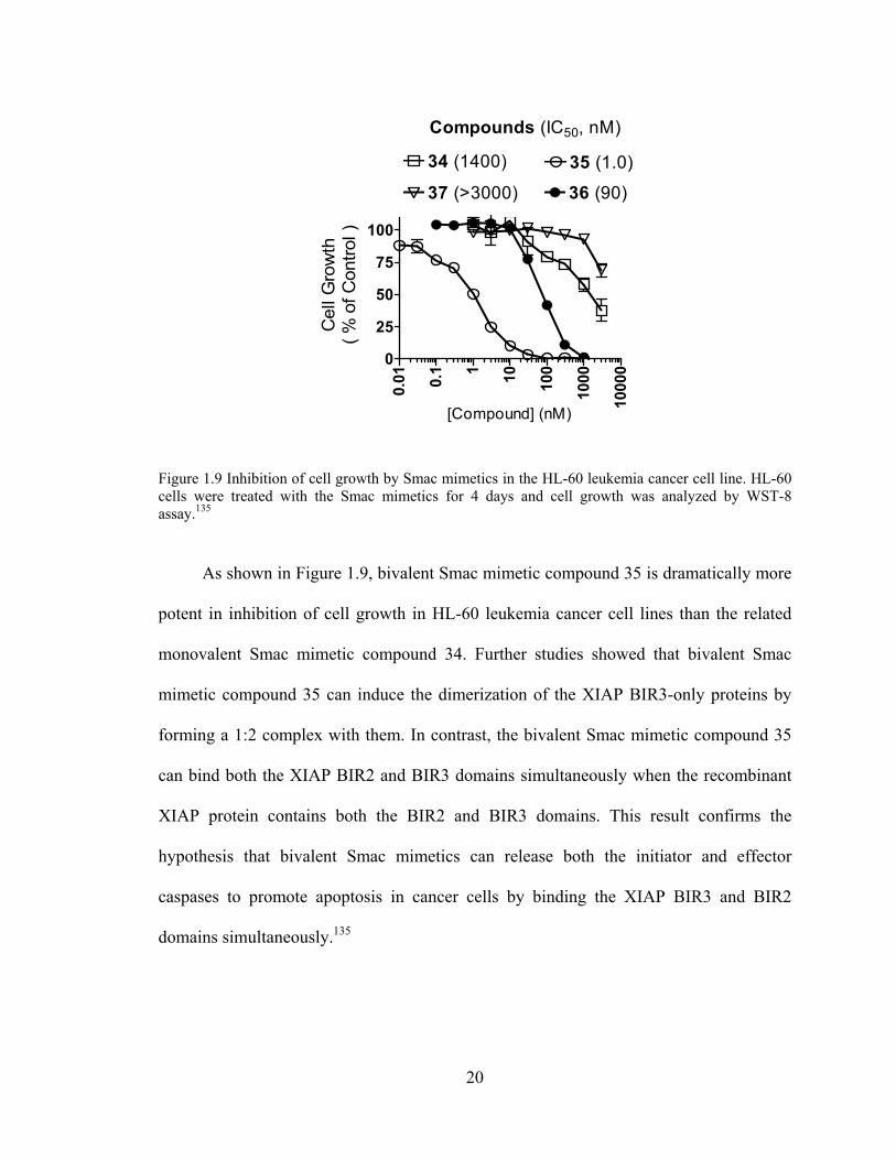
Figure 1.9 Inhibition of cell growth by Smac mimetics in the HL-60 leukemia cancer cell line. HL-60 cells were treated with the Smac mimetics for 4 days and cell growth was analyzed by WST-8 assay.135
As shown in Figure 1.9, bivalent Smac mimetic compound 35 is dramatically more
potent in inhibition of cell growth in HL-60 leukemia cancer cell lines than the related
monovalent Smac mimetic compound 34. Further studies showed that bivalent Smac
mimetic compound 35 can induce the dimerization of the XIAP BIR3-only proteins by
forming a 1:2 complex with them. In contrast, the bivalent Smac mimetic compound 35
can bind both the XIAP BIR2 and BIR3 domains simultaneously when the recombinant
XIAP protein contains both the BIR2 and BIR3 domains. This result confirms the
hypothesis that bivalent Smac mimetics can release both the initiator and effector
caspases to promote apoptosis in cancer cells by binding the XIAP BIR3 and BIR2
domains simultaneously.135

C
C
Fiby(cwcoXm
b
st
te
sp
d
m
to
sh
X
m
co
ce
HN
O
Compound 38
Compound 35
igure 1.10 Proy a competicompound 38)
with compounompound 38 a
XIAP protein amonoclonal XIA
In orde
iotinylated 7
trategy as in
ethered to th
pecific inter
esigned biot
mimetic com
o verify that
hown in Fig
XIAP protein
mimetic com
ompound 38
ellular target
NH
O N
O O
8 (µM)
5 (nM)
obing the intertive, co-imm). HL-60 who
nd 38, followand its targetedassociated witAP antibody.
er to further
7,5- and 8,5
n the develo
he pro-(S) ri
action with
tinylated 8,5
mpound 34 w
the XIAP p
gure 1.10, t
n in the HL-
mpound 35 co
8 and XIAP
t of designed
O NH
Ph
- 1
- -
raction of Smamunoprecipitatole cell lysateswed by coincu
d proteins werth beads was
probe the ce
-bicyclic Sm
pment of th
ing, which l
the XIAP p
5-bicyclic Sm
were equipote
protein is the
the biotinyla
60 leukemia
ompeted off
P protein in
d Smac mim
21
HN
38: Ki =
3 10
- -
ac mimetics toion pull-dows were incubaubation with re recovered beluted by hea
ellular molec
mac mimetic
he bivalent S
ies outside t
protein. In bi
mac mimetic
ent. A co-im
e intracellula
ated Smac m
a cell lines d
f the binding
n a dose-dep
metics.135
O
= 34±5.0 nM (X
10 10
1 10
o cellular XIAPwn assay usinated with com
compound 3by incubation wating and dete
cular targets
cs were deve
Smac mimet
the XIAP B
inding with
c compound
mmunoprecip
ar target of d
mimetic com
dose-depend
g between th
pendent man
NH
O
XIAP BIR3)
10 10
100 1000
P in the HL-6ng biotinylat
mpound 38 alon35. Complexewith Streptavi
ected by weste
of designed
eloped.125,135
tics, the biot
BIR3 binding
the XIAP B
d 38 and the
pitation assa
designed Sm
mpound 38
dently, and th
he biotinylate
nner, verifyi
S
NHN
O
0 leukemia ceted Smac mine, or preincu
es formed beidin-argarose bern blotting u
d Smac mim
5 Using the
tin molecule
g site and ha
BIR3 domain
e unlabeled S
ay was perfo
mac mimetic
pulled down
he bivalent S
ed Smac mim
ing XIAP a
H
O
ell line imetic ubated etween beads. sing a
metics,
same
e was
as no
n, the
Smac
ormed
cs. As
n the
Smac
metic
as the

22
CHAPTER 2
DESIGN AND SYNTHESIS OF NON-PEPTIDIC SMALL MOLECULE SMAC MIMETICS
2.1 Design Rationale
2.1.1 Design of monovalent Smac mimetics
NHHN
O N
O O NHCHPh2
SM-122
x
NHHN
O NN
O O NHR4
Rx
NHHN
O NN
O O NHR4
O
Ry
NHHN
O NN
O O NHCHPh2
YP-245P3
39
40
Figure 2.1 Chemical structures of SM-122 and designed new monovalent Smac mimetics.
The 7,5-bicyclic system in Smac mimetic compound 28 (Table 1.4) and the 8,5-
bicylic system in compound 34 (Table 1.5) are the core structures of previously
published Smac mimetics.125,135 The bicyclic core structure mimics the original

co
h
p
d
S
p
st
FiX
onformation
ead part and
enetrate dee
omain.122,123
mac mimeti
otency bind
tructure as th
igure 2.2 ComXIAP BIR3 dom
n formed by
d the termina
eply into th
3 The 8,5-b
ics, and our
ding with th
he optimal b
mputational momain.
y Smac AVP
al R1 group o
he relatively
icyclic Sma
recently de
he XIAP BI
bicyclic syste
odeling structu
23
PI tetrapepti
of the compo
y hydrophob
ac mimetics
eveloped 9,5
IR3 domain
em in the dev
ure of Smac m
ide, ensuring
ounds have t
bic binding
are more p
-bicyclic Sm
. Therefore,
velopment o
mimetic compo
g the methy
the appropri
pockets of
potent than
mac mimetic
, we regard
of future Sma
ound YP-245P
yl or ethyl in
iate orientati
the XIAP B
the 7,5-bic
cs have the
the 8,5-bic
ac mimetics
P3 binding wit
n the
ion to
BIR3
cyclic
same
cyclic
.
th the

24
Our modeling studies showed that while the 8-membered ring in compound 34
(SM-122) has van der Waals contacts with Trp323 of the XIAP BIR3 domain, the middle
portion (carbon X) is largely exposed to solvent. Based upon this model, we predicted
that replacement of carbon X by a nitrogen atom will not be detrimental to the binding of
XIAP. Two types of functional groups can then be tethered to this newly introduced
nitrogen atom, as shown in compounds 39 and 40 respectively in Figure 2.1. Compounds
39 and 40 will have two slightly different conformations compared to compound 34, and
it is hoped that one of them may bind more effectively than compound 34. Meanwhile,
modification of the Rx or Ry group in the new Smac mimetics (Figure 2.1) having no
detrimental effect on the binding potency with XIAP, it is hoped that through this
modification new Smac mimetics can be developed with improved pharmacokinetic (PK)
properties compared to the previously studied Smac mimetics.
2.1.2 Design of Bivalent Smac mimetics
NHHN
O NN
O O NHR4NH NH
ONN
OOR4HN
linker
Figure 2.3 Chemical structure of designed bivalent Smac mimetics.

25
As shown in Figure 2.2, the methyl group tethered to the nitrogen atom in the 8-
membered ring is oriented towards the solvent and has no specific interaction with the
XIAP BIR3 domain. This nitrogen atom is therefore a suitable site at which to chemically
tether a second monovalent Smac mimetic moiety, forming a bidentate Smac mimetic.
Similarly, both the carbon-nitrogen single bond and the amide bond were tested as sites at
which to link the two monovalent Smac mimetic moieties. As can be seen in Figure 2.3,
the newly developed bivalent Smac mimetics are structurally different from those
previously published, in which monovalent Smac mimetic moieties were linked via their
aromatic tails (R4).135
2.2 Retrosynthetic Analysis

26
NHHN
O NN
O O NHR4
R
NHHN
O NN
O O NHR4NH NH
ONN
OOR4HN
linker
bivalent Smac mimetics
monomeric Smac mimetics
NHHN
O NN
O O NHR4
R
monomeric Smac moiety
NN
O COOMe
Cbz
BocHN
YP-248P
NHN
O COOt-BuBocHN
YP-239P
N COOt-Bu
O OCbzHN NHBoc
N COOt-Bu
HO
COOH
CbzHN NHBoc
YP-7 commercially available
NH
COOHO
YP-1Bn
YP-239
Figure 2.4 Retro-synthetic analysis of designed Smac mimetics.
A retrosynthetic analysis of designed Smac mimetics is shown in Figure 2.4. Both
bivalent Smac mimetics and monovalent Smac mimetics can be prepared from the related
monovalent moieties which bear the common core structure, a nitrogen-containing 8,5-
bicyclic fused ring system. Compound YP-248P which, with the common core structure,
is the most important intermediate in the synthetic route can be prepared from YP-239P
by protection of the secondary amine as its carbobenzyloxy derivative followed by
transesterification. The cyclic amine YP-239P can be obtained from the aldehyde YP-239
by catalytic hydrogenation and the aldehyde YP-239 can be prepared by the condensation

27
of the commercially available N-α-t-butyloxycarbonyl-N-β-benzyloxycarbonyl-L-2,3-
diaminopropionic acid with the deprotected free secondary amine of compound YP-7.
Compound YP-7 is a known compound which can be obtained in six steps from (S)-(-)-2-
pyrrolidone-5-carboxylic acid (compound YP-1 in Figure 2.4).143,144
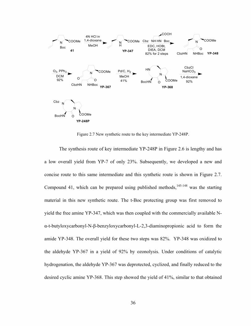
Figure 2.5 Retro-synthetic analysis of the new route.
Subsequently, a new concise synthetic route for the core structure YP-248P was
developed based on the retrosynthetic analysis of compound YP-248P shown in Figure
2.5. In the newly developed route, the transesterification reaction following the
cyclization of YP-367 was avoided, increasing the overall yield. The key intermediate
YP-248P can be directly prepared from its relative cyclic amine YP-369, which can be
obtained using the same catalytic hydrogenation condition as in synthesis of YP-239P.
The aldehyde YP-367 can be prepared from the terminal alkene YP-348 by ozonolysis.

28
The amide YP-348 is prepared by condensation of the commercially available N-α-t-
butyloxycarbonyl-N-β-benzyloxycarbonyl-L-2,3-diaminopropionic acid and the
deprotected secondary amine derived from compound 41. Compound 41 is a known
compound and can be synthesized by published methods.145-148
2.3 Results and Discussion
Comp. Structure BIR3 Ki±SD (nM)
YP-245P3 (SM-245)
340 ± 65.9
YP-246P (SM-246)
NN
O CONHCHPh2NH
HN
O
Bn 91.8 ± 30.4
YP-330 (SM-330)
5.4 ± 3.0
YP-337 (SM-337)
8.4 ± 1.6
YP-350 (SM-350)
6.0 ± 1.5
YP-356 (SM-356)
6.8 ± 0.8

29
YP-376 (SM-376)
2.0 ± 1.0
YP-377 (SM-377)
1.4 ± 0.7
Comp. Structure BIR3 Ki±SD (nM)
L-BIR2-BIR3 Ki±SD (nM)
YP-317 (SM-317)
185 ± 5.7 43 ± 0.8
YP-343 (SM-343)
21 ± 7.0 2.7 ± 0.5
YP-381 (SM-381)
27.5 ± 6.2 1.8 ± 0.6
YP-383 (SM-383)
43.1 ± 3.6 1.8 ± 0.5
YP-385 (SM-385)
12.7 ± 3.9 1.0 ± 0.5
Table 2.1 Chemical structures of synthesized monovalent and bivalent Smac mimetics and their binding affinities to the XIAP BIR3 or XIAP linker-BIR2-BIR3 determined using a fluorescence-polarization-based binding assay.126 XIAP BIR3 binding was tested for both monovalent and bivalent Smac mimetics, while additionally, XIAP linker-BIR2-BIR3 binding was tested for bivalent Smac mimetics.

30
A series of monovalent and bivalent Smac mimetics was designed and
synthesized, and their binding affinities were determined using the fluorescence-
polarization(FP)-based binding assay.126 The results are shown in Table 2.1. In the
monovalent Smac mimetics YP-245P3 (SM-245) and YP-246P (SM-246), a methyl
group and a benzyl group are directly tethered to the newly introduced nitrogen atom in
the 8-membered ring respectively; these two Smac mimetics, with binding affinities of
340 nM and 92 nM respectively, have less potency than the original lead compound SM-
122 (compound 34 in Chapter 1) in binding with the XIAP BIR3 domain.
Our computational modeling studies showed that both the tethered phenyl group
in SM-246 and the newly introduced methyl group in SM-245 have no specific
interactions with the XIAP BIR3 domain. However, SM-246 is 5 times more potent than
SM-245 in binding with the XIAP BIR3 domain. This increase in potency could be
caused by the increased rigidity of the fused 8-membered ring, while the decrease in
potency of both SM-245 and SM-246 compared with lead compound SM-122 could be
caused by the decreased rigidity of that ring.
The monovalent Smac mimetic compounds SM-330 and SM-337 have a carbonyl
group inserted between the alkyl group and the nitrogen atom in the 8-membered ring of
SM-245 and SM-246, respectively. This carbonyl group forms an amide bond with the
adjacent nitrogen atom and increases the rigidity of the ring. Both new Smac mimetics
are much more potent than the related SM-245 and SM-246 in binding with the XIAP

31
BIR3 domain as shown in Table 2.1 and they are 3-4 times more potent than the lead
compound SM-122 in binding affinity with the XIAP BIR3 domain.
Consequently, SM-337 was used as a new lead compound in the development of
nitrogen-containing 8,5-bicyclic Smac mimetics. The para and ortho sites of the benzyl
group tethered to the nitrogen atom in the 8-membered ring can be metabolized by
cytochrome P450 in vivo, and with this in mind, we designed SM-350 and SM-356 with a
view to improving the pharmacokinetic properties. In SM-350, the para site of the phenyl
ring was blocked with a fluorine atom while in SM-356 the para site and one of the ortho
sites of the phenyl ring were both blocked with fluorine atoms. Both SM-350 and SM-
356 were found to have similar potency compared with the lead compound SM-337 in
binding with the XIAP BIR3 domain.
Previous studies showed that an (R)-tetrahydronaphthyl group can be well
accommodated in the large hydrophobic pocket formed by lysine 297 and lysine 299 of
the XIAP BIR3 domain. These compounds might be expected to have superior potency
than the corresponding diphenylmethyl compounds, for example, 245P3, in binding to the
XIAP BIR3 domain.130,149 Compound SM-376 in which the terminal diphenylmethyl
group was replaced by a (R)-tetrahydronaphthyl group was designed and synthesized.
This compound was found to be the most potent monovalent Smac mimetic to date both
in binding with the XIAP BIR3 domain and in inhibition of tumor cell growth. The Smac
mimetic SM-377, in which the para site of the phenyl ring is blocked with a fluorine

32
atom, was also designed and synthesized in the hope that it would demonstrate improved
pharmacokinetic properties.
Using Smac mimetic SM-246 as the monovalent moiety and 1,4-
dimethylenephenyl as the linker, the bivalent Smac mimetic SM-317 was synthesized and
another bivalent Smac mimetic, SM-343, was also developed using the more potent Smac
mimetic SM-337 as the monovalent moiety. As expected, SM-343 is much more potent
than SM-317 in binding with the XIAP BIR3 domain and it is dramatically more potent
than SM-317 in binding with the XIAP linked-BIR2-BIR3 protein.
Using the monovalent Smac mimetic compound SM-330 as the template, we
designed and synthesized SM-381, SM-383 and SM-385, bivalent Smac mimetic
compounds with dicarboxylic acid linkers of different lengths as shown in Table 2.1. The
linkers in SM-381, SM-383 and SM-385 have 8, 10, and 6 methylene groups respectively.
The binding affinities of these bivalent Smac mimetics against XIAP BIR3 domain
increase as the lengths of the linker decrease, but they have similar potency in binding
with the XIAP linker-BIR2-BIR3 protein, as shown in Table 2.1.
The activities in tumor cells of these synthetic monovalent and bivalent Smac
mimetics were determined and analyzed in detail in Chapters 3-5, below.
2.4 Conclusion

33
Various monovalent and bivalent small-molecular non-peptidic Smac mimetics
were successfully designed and synthesized. SM-337 has a dramatically improved
potency in binding to XIAP BIR 3 compared with our original lead compound SM-122.
Using this compound, SM-337, as our new design template, several potent monovalent
and bivalent Smac mimetics were developed.
SM-376 and SM-377 are the most potent monovalent Smac mimetics reported to
date, with a binding affinity (Ki) to XIAP BIR3 domain as 2.0 and 1.4 nM respectively.
The cell-permeable Smac mimetics, SM-376 and SM-377, are as potent as the bivalent
Smac mimetic lead compound SM-164 in tumor cell growth inhibition in human breast
cancer MDA-MB-231 cells. SM-406 (Chapter 5), a derivative of SM-376 and SM-377,
shows excellent pharmacokinetic properties and is a promising drug candidate.
The bivalent Smac mimetics SM-381, SM-383 and SM-385 are as potent as the
bivalent Smac mimetic lead compound SM-164 in both binding the XIAP BIR3 protein
and the XIAP linker-BIR2-BIR3 protein. These three newly developed bivalent Smac
mimetics have a linker that differs from the triazole linker in SM-164. This has
advantages in terms of ease of synthesis and further optimization of the aromatic binding
in the hydrophobic pocket formed by lysine 297 and lysine 299 of the XIAP BIR3
domain. Further optimization of this aromatic fragment and the length of the linker may
yield bivalent Smac mimetics that are even better than the original lead compound SM-
164.

34
2.5 Synthesis of Smac Mimetics
NH
COOHO6 steps
N COOt-Bu
HO BnYP-7
NH
TBSO COOt-Bu
YP-237P
TBSClDIEA, DCM NTBSO COOt-Bu
YP-237Bn
Pd/C, H2
EtOAc
N COOt-Bu
TBSO OCbzHN NHBoc YP-238
COOH
NH HNCbz Boc
EDC, HOBt,DIEA, DCM88% for 2 steps
N COOt-Bu
HO OCbzHN NHBoc YP-238P
TBAF
THF87% for 2 steps 96%
Dess-MartinDCM
N COOt-Bu
O OCbzHN NHBoc YP-239
Pd/C, H2
MeOH42%
HNN
O COOt-BuBocHN
YP-239P
NN
O COOt-BuBocHN
YP-246P2
CbzCbzClNaHCO3
1,4-dioxane
SOCl2MeOH
NN
O COOMeH2N
YP-248
Cbz Boc2ONaHCO3
1,4-dioxane74% for 3 steps
NN
O COOMeBocHN
Cbz
YP-248P Figure 2.6 Synthetic route to key intermediate YP-248P.
Figure 2.6 shows the synthetic route to compound YP-248P, the key intermediate
for the synthesis of both monovalent and bivalent Smac mimetics. Commercially
available (S)-(-)-2-pyrrolidone-5-carboxylic acid was used as the starting material and
known compound YP-7 was obtained after 6 published steps.143,144 The free hydroxyl
group in compound YP-7 was first protected with a tert-butyldimethylsilyl (TBS) group

35
to yield compound YP-237, whose benzyl protecting group was then removed by
catalytic hydrogenation. The free secondary amine YP-237P was obtained after these two
steps of reactions with an overall yield from YP-7 of 88%.
The pyrrolidine YP-237P was coupled with commercially available N-α-t-
butyloxycarbonyl-N-β-benzyloxycarbonyl-L-2,3-diaminopropionic acid to yield the
amide YP-238. The TBS protective group in compound YP-238 was then removed with
tetra-n-butylammonium fluoride (TBAF) in tetrahydrofuran to give the free alcohol YP-
238 with an 87% overall yield for the two reactions. The alcohol YP-238 was oxidized
with Dess-Martin reagent to yield the aldehyde YP-239 in a yield of 96%. Upon catalytic
hydrogenation, the desired nitrogen-containing 8,5-bicyclic structure was formed. Three
chemical reactions occur in this single step: first, the carbobenzyloxy (Cbz) protective
group is removed to yield a free terminal amine; then the terminal amine reacts
intramolecularly with the aldehyde group forming the cyclic imine and finally, this cyclic
imine is reduced to the desired cyclic amine. This step was performed at low
concentrations so as to encourage the intramolecular reaction, and gave a yield of 42%.
The free cyclic amine was then protected with a carbobenzyloxy group to yield
compound YP-246P2. The tert-butyl ester YP-246P2 was allowed to react with SOCl2,
added dropwise in methanol at 0°C to yield the methyl ester YP-248. In this step, the tert-
butyloxycarbonyl (t-Boc) group was also removed and so in a final step, the free amino
group was protected with a t-Boc protective group to yield the key intermediate YP-248P.
These three reaction steps gave an overall yield of 74%.150
36
N COOMe
Boc
HNN
O COOMeBocHN
YP-368
NN
O COOMeBocHN
YP-248P
Cbz
NH
COOMe
41
4N HCl in 1,4-dioxane
MeOH
YP-347
COOH
NH HNCbz BocEDC, HOBt, DIEA, DCM
N COOMe
OCbzHN NHBoc YP-34882% for 2 steps
O3, PPh3
DCM92%
N COOMe
O OCbzHN NHBoc
YP-367
Pd/C, H2
MeOH41%
CbzClNaHCO3
1,4-dioxane92%
Figure 2.7 New synthetic route to the key intermediate YP-248P.
The synthesis route of key intermediate YP-248P in Figure 2.6 is lengthy and has
a low overall yield from YP-7 of only 23%. Subsequently, we developed a new and
concise route to this same intermediate and this synthetic route is shown in Figure 2.7.
Compound 41, which can be prepared using published methods,145-148 was the starting
material in this new synthetic route. The t-Boc protecting group was first removed to
yield the free amine YP-347, which was then coupled with the commercially available N-
α-t-butyloxycarbonyl-N-β-benzyloxycarbonyl-L-2,3-diaminopropionic acid to form the
amide YP-348. The overall yield for these two steps was 82%. YP-348 was oxidized to
the aldehyde YP-367 in a yield of 92% by ozonolysis. Under conditions of catalytic
hydrogenation, the aldehyde YP-367 was deprotected, cyclized, and finally reduced to the
desired cyclic amine YP-368. This step showed the yield of 41%, similar to that obtained

37
in the previous route. Finally, the key intermediate YP-248P was obtained by protection
of the secondary amino group in compound YP-368. The yield for this step was 92%.
NN
O COOMeBocHN
Cbz
YP-248P
2N LiOH in H2O
1,4-dioxane
NN
O COOHBocHN
Cbz
YP-248P2
EDC, HOBtDCM, DIEA
82% for 2 steps
NN
O CONHCHPh2BocHNYP-244
Cbz 4N HCl in 1,4-dioxane
MeOH
NN
O CONHCHPh2H2N
Cbz
YP-244P
N COOHBoc
EDC, HOBtDIEA, CH2Cl2
83% over 2 steps
NN
O CONHCHPh2NH
Cbz
YP-244P2
NOBoc
10% Pd/C, H2
MeOH
HNN
O CONHCHPh2NH
YP-245
NOBoc
92%
NN
O COOMeBocHN
Cbz
YP-248P
2N LiOH in H2O
1,4-dioxane
NN
O COOHBocHN
Cbz
YP-248P2
EDC, HOBtDCM, DIEA
80% for 2 steps
NH2
NN
O CONHBocHN
Cbz
YP-370
4N HCl in 1,4-dioxane
MeOH
NOBoc
NN
O CONHNH
Cbz
YP-372
NN
O CONHH2N
Cbz
YP-371
N COOHBoc
EDC, HOBtDIEA, CH2Cl2
80% over 2 steps
10% Pd/C, H2
MeOH91%
NOBocHN
N
O CONHNH
YP-373
NH2CHPh2
Figure 2.8 Synthesis of key components YP-245 and YP-373.

38
Figure 2.8 shows the synthetic route to compounds YP-245 and YP-373, which
are the key intermediates in the syntheses of the monovalent and bivalent Smac mimetic
compounds. The methyl ester YP-248P was first converted to the free carboxylic acid
YP-248P2, which was then coupled with the commercially available diphenylmethyl
amine to form the amide YP-244. The overall yield for these two steps was 82%.
Removal of the t-Boc protective group from compound YP-244 gave the free
amine YP-244P, which was coupled with the commercially t-Boc-N-methylalanine to
form the amide YP-244P with an overall yield of 83% for these two steps. The
carbobenzyloxy protecting group was removed by catalytic hydrogenation to yield the
free cyclic amine YP-245 with a yield of 92%.
Similarly, the methyl ester YP-248P was first converted to the free carboxylic
acid YP-248P2, which was then coupled with the commercially available (R)-1,2,3,4-
tetrahydro-1-naphthylamine to form the amide YP-370 with an overall yield of 80% for
these two steps. Then the t-Boc protecting group in compound YP-370 was removed to
yield the free amine YP-371, which was coupled with the commercially available t-Boc-
N-methylalanine to form the amide YP-372 with an overall yield of 80% for the two
steps. The carbobenzyloxy protecting group was finally removed by catalytic
hydrogenation to yield the free cyclic amine YP-373 with a yield of 91%.

39
HNN
O CONHCHPh2NH
YP-245
NOBoc
4N HCl in 1,4-dioxane
MeOH
NN
O CONHCHPh2NH
YP-245P2
NO
37% wt. HCHO in H2O BocNaCNBH3
HOAc
NN
O CONHCHPh2NH
YP-245P3HN
O
71% over 2 steps
HNN
O CONHCHPh2NH
YP-245
NOBoc BnBr, NaHCO3
1,4-dioxane
NN
O CONHCHPh2NH
YP-246
NOBoc
Bn
4N HCl in 1,4-dioxane
MeOH77% over 2 steps
NN
O CONHCHPh2NH
YP-246PHN
O
Bn
(SM-245)
(SM-246)
HNN
O CONHCHPh2NH
YP-245
NOBoc
EDC, HOBtDIEA, CH2Cl2
RCOOHN
N
O CONHCHPh2NH
NOBoc
R
O
4N HCl in 1,4-dioxane
MeOH
NN
O CONHCHPh2NH
HN
OR
OYP-330 (SM-330): R=Me, 85% over 2 stepsYP-337 (SM-337): R=Bn, 87% over 2 stepsYP-350 (SM-350): R=4-fluorine-benzyl, 84% over 2 stepsYP-356 (SM-356): R=2,4-difluorine-benzyl, 83% over 2 steps
Figure 2.9 Synthesis of monovalent Smac mimetics.
The synthesis of monovalent Smac mimetics is shown in Figure 2.9. A methyl
group was attached to the nitrogen atom in the 8-membered ring via a reductive
amination reaction forming compound YP-245P2, whose t-Boc protecting group was
subsequently removed to yield the monovalent Smac mimetic compound YP-245P3 (SM-
245) with an overall yield of 71% for the two steps. A benzyl group was attached to the

40
nitrogen atom in the 8-membered ring via an SN2 reaction, in which the cyclic amine YP-
245 was treated with benzyl bromide to form YP-246, whose t-Boc protecting group was
removed to yield the monovalent Smac mimetic compound YP-246P (SM-246) with an
overall yield of 77% for the two steps. Condensation reactions between the cyclic amine
YP-245 and the appropriate carboxylic acid, followed by the removal of the t-Boc groups,
were performed to obtain the relative monovalent Smac mimetic compounds YP-330
(SM-330), YP-337 (SM-337), YP-350 (SM-350), and YP-356 (SM-356). The yield,
reagents and conditions for these two reactions are shown in Figure 2.9.
NOBocHN
N
O CONHNH
YP-373
EDC, HOBtDIEA, CH2Cl2
RCOOH
NO
NN
O CONHNH
R
O
4N HCl in 1,4-dioxane
MeOH
Boc
HN
ON
N
O CONHNH
R
O
YP-376 (SM-376): R=Bn, 85% over 2 stepsYP-377 (SM-377): R=4-fluorine-benzyl, 84% over 2 steps
Figure 2.10 Synthesis of monovalent Smac mimetics SM-376 and SM-377.
Syntheses of the monovalent Smac mimetic compounds SM-376 and SM-377 are
shown in Figure 2.10. Condensation of the cyclic amine YP-373 with phenylacetic acid
or 4-fluorophenylacetic acid, followed by the deprotection of the t-Boc protecting groups,

41
was performed to yield the monovalent Smac mimetic compounds YP-376 (SM-376) and
YP-377 (SM-377) with overall yields of 85% and 84% respectively.
Figure 2.11 Synthesis of bivalent Smac mimetics.

42
The syntheses of bivalent Smac mimetic compounds are shown in Figure 2.17.
Cyclic amine YP-245 underwent an SN2 reaction with a half equivalent of p-xylylene
dibromide to form the bivalent compound YP-316, whose t-Boc protective groups were
then removed to obtain the bivalent Smac mimetic compound YP-317 (SM-317) with an
overall yield of 54% for the two steps. Condensation of the cyclic amine YP-245 and the
appropriate dicarboxylic acid, followed by removal of the t-Boc groups, was performed
to obtain the bivalent Smac mimetic compounds YP-343 (SM-343), YP-381 (SM-381),
YP-383 (SM-383), and YP-385 (SM-385) respectively. The yields, reagents and
conditions for these steps are shown in Figure 2.11.
2.6 Methods and Materials
General methods: NMR spectra were measured at a proton frequency of 300 MHz. 1H
chemical shifts are reported relative to tetramethylsilane (0.00 ppm) or DHO (4.70 ppm)
as internal standards. 13C chemical shifts are reported relative to CDCl3 (77.00 ppm) or
CD3OD (49.00 ppm) as internal standards. The final products were purified by C18
reverse phase semi-preparative HPLC column with solvent A (0.1% of TFA in water) and
solvent B (0.1% of TFA in CH3CN) as eluents.
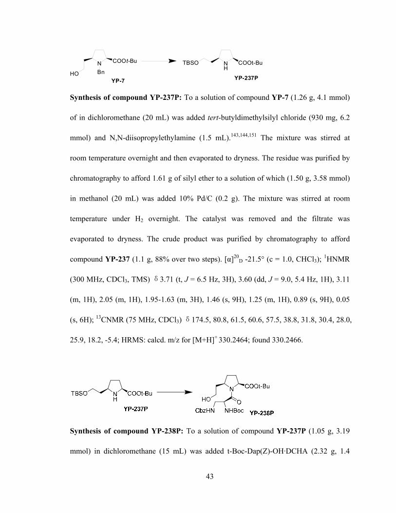
43
N COOt-Bu
HO BnYP-7
NH
TBSO COOt-Bu
YP-237P
Synthesis of compound YP-237P: To a solution of compound YP-7 (1.26 g, 4.1 mmol)
of in dichloromethane (20 mL) was added tert-butyldimethylsilyl chloride (930 mg, 6.2
mmol) and N,N-diisopropylethylamine (1.5 mL).143,144,151 The mixture was stirred at
room temperature overnight and then evaporated to dryness. The residue was purified by
chromatography to afford 1.61 g of silyl ether to a solution of which (1.50 g, 3.58 mmol)
in methanol (20 mL) was added 10% Pd/C (0.2 g). The mixture was stirred at room
temperature under H2 overnight. The catalyst was removed and the filtrate was
evaporated to dryness. The crude product was purified by chromatography to afford
compound YP-237 (1.1 g, 88% over two steps). [α]20D -21.5° (c = 1.0, CHCl3); 1HNMR
(300 MHz, CDCl3, TMS) δ3.71 (t, J = 6.5 Hz, 3H), 3.60 (dd, J = 9.0, 5.4 Hz, 1H), 3.11
(m, 1H), 2.05 (m, 1H), 1.95-1.63 (m, 3H), 1.46 (s, 9H), 1.25 (m, 1H), 0.89 (s, 9H), 0.05
(s, 6H); 13CNMR (75 MHz, CDCl3) δ174.5, 80.8, 61.5, 60.6, 57.5, 38.8, 31.8, 30.4, 28.0,
25.9, 18.2, -5.4; HRMS: calcd. m/z for [M+H]+ 330.2464; found 330.2466.
Synthesis of compound YP-238P: To a solution of compound YP-237P (1.05 g, 3.19
mmol) in dichloromethane (15 mL) was added t-Boc-Dap(Z)-OH·DCHA (2.32 g, 1.4

44
equiv.), EDC (1-ethyl-3-(3-dimethylaminopropyl) carbodiimide hydrochloride) (978 mg,
1.6 equiv.), HOBt (Hydroxybenzotriazole) (690 mg, 1.6 equiv.), and N,N-
diisopropylethylamine (3 mL). The mixture was stirred at room temperature overnight
and then concentrated to give a residue which was purified by chromatography to afford
an amide (1.86 g). To a solution of the above amide (860 mg, 1.32 mmol) in THF (10 mL)
was added tetrabutylammonium fluoride solution (1M, 1.6 mL, 1.2 equiv.) in THF and
the solution was stirred at room temperature for 3 h.152-154 After adding dichloromethane
(50 mL), the solution was washed with brine, dried over Na2SO4 and then condensed.
The crude product was purified by chromatography to yield compound YP-238P (690 mg,
87% over two steps). [α] 20D -90.0° (c = 1.67, CHCl3); 1H NMR shows this compound is a
1:1 mixture of two rotamers. 1HNMR (300 MHz, CDCl3, TMS) δ7.28 (m, 5H), 5.59
(m,1H), 5.35 (m, 1H), 5.20-5.05 (m, 2H), 4.85 (m, ½ H), 4.65 (m, ½ H), 4.46 (m, 1H),
4.35 (m, 1H), 3.80 (m, ½ H), 3.70-3.50 (m, 2H), 3.40 (m, 1H), 3.25 (m, ½ H), 2.32 (m,
1H), 2.20-1.50 (m, 4H), 1.46 (s, 4.5H), 1.44 (s, 4.5H), 1.43 (s, 4.5H), 1.41 (s, 4.5H);
HRMS: calcd m/z 558.2791 for [M+Na]+; found 558.2794.
Synthesis of compound YP-239: To a solution of compound YP-238P (667 mg, 1.25
mmol) in dichloromethane (10 mL) was added Dess-Martin periodinane (636 mg, 1.5

45
mmol, 1.2 equiv.) at room temperature. The mixture was stirred for 3 h, filtered through
celite and then evaporated. The residue was purified by chromatography to give YP-239
(639 mg, 96%). [α] 20D -51.6° (c = 1.67, CHCl3); 1HNMR shows that this compound has
two rotamers with a ratio of 2:1. 1HNMR (300 MHz, CDCl3, TMS) δ 9.76 (s, 2/3 H), 9.71
(s, 1/3 H), 7.40-7.28 (m, 5H), 5.72-5.30 (m, 2H), 5.20-4.95 (m, 2H), 4.90-4.25 (m, 3H),
3.52-3.05 (m, 3H), 2.90-1.60 (m, 4H), 1.50-1.35 (m, 18H); HRMS: calcd m/z 556.2635
for [M+Na]+; found 556.2629.
Synthesis of compound YP-239P: To a solution of compound YP-239 (380 mg, 0.713
mmol) in methanol (20 mL) was added 10% Pd/C (0.2 g). The mixture was stirred at
room temperature under H2 overnight, filtered through celite and then condensed. The
residue was purified by chromatography to give compound YP-239P (114 mg, 42%).150
[α] 20D -8.4° (c = 0.65, CHCl3); 1HNMR (300 MHz, CDCl3, TMS) δ 5.49 (brd, J = 8.1 Hz,
1H), 4.70 (m, 1H), 4.41 (t, J = 9.3 Hz, 1H), 4.30 (m, 1H), 3.25-3.18 (m, 2H), 2.89 (m,
1H), 2.75 (dd, J = 13.5, 11.1 Hz, 1H), 2.34 (m, 1H), 2.18-1.60 (m, 6H), 1.49 (s, 9H), 1.44
(s, 9H); 13CNMR(75 MHz, CDCl3) δ171.8, 170.4, 155.2, 81.7, 79.5, 60.6, 58.5, 54.9,
52.3, 46.9, 37.5, 32.1, 28.3, 28.0, 27.0; HRMS: calcd m/z 406.2318 for [M+Na]+; found
406.2317.

46
HNN
O COOt-BuBocHNYP-239P
NN
O COOMeBocHN
Cbz
YP-248P
Synthesis of compound YP-248P: To a solution of compound YP-239P (1.5 g, 3.9
mmol) in 1,4-dioxane (15 ml) were added CbzCl (0.66 mL, 1.2 equiv.) and NaHCO3 (820
mg, 2.5 equiv.).155 The mixture was stirred overnight at room temperature, filtered and
concentrated under vacuum. The residue was purified by chromatography to give 1.9 g of
Cbz-protected compound (Rf = 0.5 when EtOAc/Hexane = 1/1). To a well-stirred solution
of this Cbz-protected compound (0.9 g, 1.7 mmol) in MeOH (5 mL) at 0°C, SOCl2 (1 mL,
14 mmol) was added dropwise. The mixture was stirred at room temperature for 1 h, then
was quenched with saturated NaHCO3 solution and the pH adjusted to 8. The aqueous
layer was extracted with CH2Cl2, and combined organic phases were dried over Na2SO4,
filtered, and concentrated under vacuum. The crude amine was dissolved in 1,4-dioxane
(10 mL), then t-Boc2O (0.44 g, 1.2 equiv.) and NaHCO3 (0.35 g, 2.5 equiv.) were added.
The mixture was stirred overnight at room temperature and then filtered and concentrated
under vacuum. The residue was purified by chromatography to give 0.6 g of compound
YP-248P (Rf = 0.4 when EtOAc/Hexane= 2/1) (74% over 3 steps). 1H NMR shows that
this compound has two rotamers with a ratio of 2:1. 1H NMR (300 MHz, CDCl3, TMS) δ
7.47-7.25 (m, 5H), 5.63 (brd, J=8Hz, 1H), 5.31-5.16 (m, 2H), 4.64-4.60 (m, 1H), 4.51-

47
4.46 (t, J=8Hz, 1H), 4.30-3.98 (m, 2H), 3.74 (brs, 3H), 3.63 (m, 1H), 3.50 (m, 1H), 3.30
(m, 1H), 2.38 (m, 1H), 2.28-1.76 (m, 5H), 1.44-1.45 (s, 9H). ESI MS: m/z 476.2 [M+H]+.
N COOMe
Boc41
N COOMe
OCbzHN NHBoc YP-348
Synthesis of compound YP-348: To a solution of compound 41 (3.2 g, 12 mmol) in
MeOH (20 mL) was added a solution of 4N HCl in 1,4-dioxane (6 mL, 2 equiv.).145-148,156
The mixture was stirred overnight at room temperature, and then concentrated to yield
2.2 g of crude amine to a solution of which in DCM (20 mL) was added t-Boc-Dap(Z)-
OH·DCHA (6.6 g, 1.1 equiv.), EDC (2.9 g, 1.2 equiv.), HOBt (2.0 g, 1.2 equiv.), and
DIEA (5.5 mL, 2.5 equiv.). The mixture was stirred overnight at room temperature, and
then concentrated and purified by chromatography to give compound YP-348 (Rf = 0.3
EtOAc/Hexane= 1/1) (5.8 g, 82% over two steps). 1HNMR shows that this compound has
two rotamers with a ratio of 2:1. 1HNMR (300 MHz, CDCl3, TMS) δ 7.34-7.28 (m, 5H),
5.80-5.77 (m, 1H), 5.59 (m, 1H), 5.36-5.33 (d, J=10 Hz, 2H), 5.19-5.01 (m, 4H), 4.67-
4.62 (m, 1H), 4.47-4.44 (m, 1H), 3.76-3.74 (s, 1H), 3.74-3.71 (s, 2H), 2.32-2.30 (m, 1H),
2.16-2.12 (m, 1H), 1.99-1.95 (m, 2H), 1.42 (s, 9H). 13CNMR (75 MHz, CDCl3) δ 172.4,
170.5, 156.5, 155.2, 136.4, 134.6, 133.8, 128.3, 127.9, 118.5, 117.1, 80.0, 66.6, 59.7, 58.2,
52.6, 43.4, 29.2, 28.1, 26.6.

48
N COOMe
OCbzHN NHBoc YP-348
N COOMe
O OCbzHN NHBoc YP-367
Synthesis of compound YP-367: A solution of compound YP-348 (0.75 g, 1.5 mmol) in
DCM (10 mL) was cooled to -78 oC. Ozone was passed through the solution until a blue
color persisted, then air was passed through until the blue color disappeared. PPh3 (0.8 g,
2 equiv.) was added and the mixture was stirred for 30 min at room temperature. The
mixture was condensed and purified by chromatography to give compound YP-367 (Rf =
0.3 EtOAc/Hexane= 1/1) (0.68 g, 92%). 1HNMR shows that this compound has two
rotamers with a ratio of 1:1. 1HNMR (300 MHz, CDCl3, TMS) δ 9.78-9.67 (m, 1H), 7.53-
7.32 (m, 5H), 5.44 (s, 1/2 H), 5.32 (s, 1/2 H), 5.15-5.06 (m, 2H), 4.64 (m, 1H), 4.40-4.39
(m, 1H), 3.78-3.76 (s, 3/2 H), 3.76-3.74 (s, 3/2H), 3.48-3.42 (m, 3H), 2.78-2.52 (m, 1H),
2.40-2.20 (m, 1H), 2.16 (m, 2H), 2.06-1.89 (m, 1H), 1.44-1.43 (m, 9H). 13CNMR (75
MHz, CDCl3) δ 200.3, 199.5, 172.6, 172.2, 170.3, 156.5, 136.4, 128.4, 128.0, 66.7, 59.7,
59.1, 54.3, 52.4, 52.3, 48.4, 43.3, 29.6, 28.2, 21.0.
N COOMe
O OCbzHN NHBoc YP-367
HNN
O COOMeBocHNYP-368
Synthesis of compound YP-368: To a solution of compound YP-367 (0.6 g, 1.2 mmol)
in MeOH (40 mL) was added 10% Pd/C (0.4 g). The mixture was stirred at room
temperature under H2 overnight, filtered through celite and then evaporated. The residue

49
was purified by chromatography to give compound YP-368 (0.25 g, 41%). 1HNMR (300
MHz, CDCl3, TMS) δ 5.45-5.42 (d, J=8.0 Hz, 1H), 4.72-4.71 (m, 1H), 4.57-4.51 (t, J=9.1
Hz, 1H), 4.38-4.28 (m, 1H), 3.77 (s, 3H), 3.25-3.18 (m, 1H), 2.80-2.70 (m, 1H), 2.35 (m,
1H), 2.18-1.60 (m, 6H), 1.46 (s, 9H). 13CNMR (75 MHz, CDCl3) δ 173.4, 170.6, 155.1,
131.9, 79.6, 59.4, 58.3, 54.9, 52.4, 46.7, 37.4, 32.1, 29.6, 28.4, 26.9, 22.6.
HNN
O COOMeBocHN
YP-368
NN
O COOMeBocHNYP-248P
Cbz
Synthesis of compound YP-248P: To a solution of compound YP-368 (1.6 g, 4.5 mmol)
in 1,4-dioxane (20 ml) were added CbzCl (0.8 mL, 1.2 equiv.) and NaHCO3 (0.9 g, 2.5
equiv.). The mixture was stirred overnight at room temperature and then filtered and
concentrated under vacuum.157,158 The residue was purified by chromatography to give
compound YP-248P (Rf = 0.5 when EtOAc/Hexane= 1/1) (2.0 g, 92%). 1H NMR shows
that this compound has two rotamers with a ratio of 2:1. 1H NMR (300 MHz, CDCl3,
TMS) δ 7.47-7.25 (m, 5H), 5.63 (brd, J=8Hz, 1H), 5.31-5.16 (m, 2H), 4.64-4.60 (m, 1H),
4.51-4.46 (t, J=8Hz, 1H), 4.30-3.98 (m, 2H), 3.74 (brs, 3H), 3.63 (m, 1H), 3.50 (m, 1H),
3.30 (m, 1H), 2.38 (m, 1H), 2.28-1.76 (m, 5H), 1.44-1.45 (s, 9H). ESI MS: m/z 476.2
[M+H]+.

50
NN
O CONHCHPh2BocHNYP-244
CbzNN
O COOMeBocHN
Cbz
YP-248P
Synthesis of compound YP-244: To a solution of compound YP-248P (0.3 g, 0.6 mmol)
in 1,4-dioxane (5 mL) was added 2N LiOH/H2O (0.8 mL, 2.5 equiv.). The mixture was
stirred 1h at room temperature, then 1N HCl was added to adjust the pH to 3. The
aqueous layer was extracted with CH2Cl2, and combined organic phases were dried
(Na2SO4), filtered, and concentrated under vacuum. The crude acid was dissolved in
CH2Cl2 (15 mL), and then NH2CHPh2 (0.1 mL, 1.1 equiv.), EDC (0.14 g, 1.2 equiv.),
HOBt (0.1 g, 1.2 equiv.) and DIEA (diisopropylethylamine, 0.26 mL, 2.5 equiv.) were
added. The mixture was stirred at overnight room temperature and then concentrated. The
residue was purified by chromatography to give compound YP-244 (Rf = 0.3 when
EtOAc/Hexane= 1/1) (0.3 g, 82% over 2 steps). 1HNMR (300 MHz, CDCl3, TMS) δ
7.92-7.75 (m, 1H), 7.48-7.24 (m, 15H), 6.20 (m, 1H), 5.72 (m, 1H), 5.15 (brs, 2H), 4.68
(m, 1H), 4.56 (m, 1H), 4.35-4.05 (m, 2H), 3.55 (m, 1H), 2.73-2.58 (m, 2H), 2.34 (m, 1H),
2.20-1.65 (m, 5H), 1.48 (brs, 9H). ESI MS: m/z 627.3 [M+H]+.
NN
O CONHCHPh2BocHN
YP-244
Cbz NN
O CONHCHPh2NH
Cbz
YP-244P2
NOBoc
Synthesis of compound YP-244P2: To a solution of compound YP-244 (0.3 g, 0.5
mmol) in MeOH (5 mL), was added 0.5 mL of a solution of 4N HCl in 1,4-dioxane.159,160

51
The mixture was stirred overnight at room temperature and then condensed and quenched
with saturated NaHCO3 solution and adjusted to pH = 8. The aqueous layer was extracted
with CH2Cl2, and combined organic phases were dried over Na2SO4, filtered, and
concentrated under vacuum. The crude amine was dissolved in CH2Cl2 (15 mL), and then
Boc-N-methyl-alanine (0.1 g, 1.1 equiv.), EDC (0.11 g, 1.2 equiv.), HOBt (0.08 g, 1.2
equiv.) and DIEA (0.2 mL, 2.5 equiv.) were added. The mixture was stirred overnight at
room temperature and then condensed and purified by chromatography to give compound
YP-244P2 (Rf = 0.3 when EtOAc/Hexane= 3/1) (0.28 g, 83% over two steps). 1H NMR
shows that this compound has two rotamers with a ratio of 1:1. 1H NMR (300 MHz,
CDCl3, TMS) δ 7.83-4.69 (m, 1H), 7.47-7.45 (m, 1H), 7.36-7.26 (m, 15H), 6.24-6.18 (t,
J= 8.2 Hz, 1H), 5.15 (s, 2H), 4.89-4.79 (m, 1H), 4.70-4.62 (q, J=6.9 Hz, 1H), 4.23-4.07
(m, 2H), 3.60-3.47 (1H), 2.82 (s, 3/2 H), 2.79 (s, 3/2 H), 2.62-2.59 (m, 2H), 2.48-2.30 (m,
1H), 2.13-2.03 (m, 2H), 1.86-1.79 (m, 2H), 1.51 (s, 9/2 H), 1.49 (s, 9/2 H), 1.38-1.35 (d,
J= 7.0 Hz, 3H).
NN
O CONHCHPh2NH
Cbz
YP-244P2
NOBoc
HNN
O CONHCHPh2NH
YP-245
NOBoc
Synthesis of compound YP-245: To a solution of compound YP-244P2 (0.28 g, 0.4
mmol) in MeOH (20 mL) was added 10% Pd/C (0.1 g). The mixture was stirred at room
temperature under H2 overnight.161 The catalyst was removed and the filtrate was
evaporated. The crude product was purified by chromatography to afford compound YP-
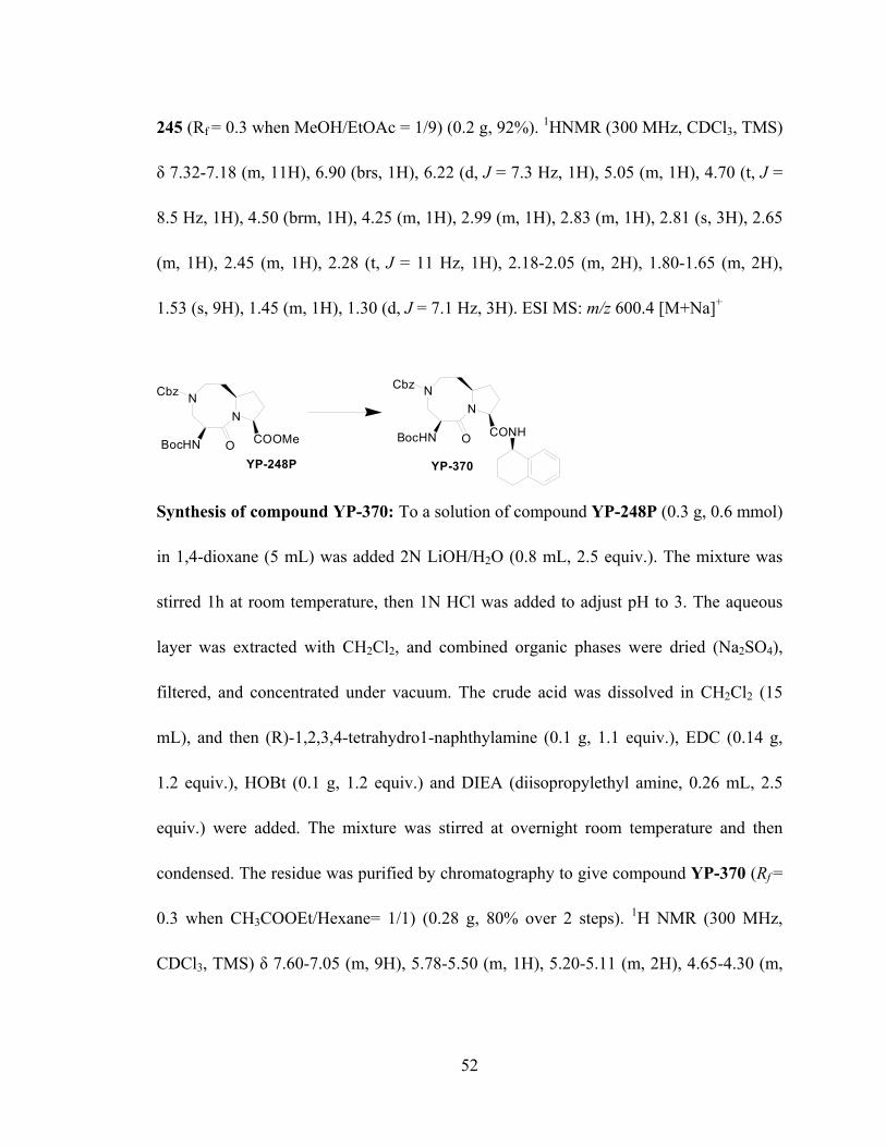
52
245 (Rf = 0.3 when MeOH/EtOAc = 1/9) (0.2 g, 92%). 1HNMR (300 MHz, CDCl3, TMS)
δ 7.32-7.18 (m, 11H), 6.90 (brs, 1H), 6.22 (d, J = 7.3 Hz, 1H), 5.05 (m, 1H), 4.70 (t, J =
8.5 Hz, 1H), 4.50 (brm, 1H), 4.25 (m, 1H), 2.99 (m, 1H), 2.83 (m, 1H), 2.81 (s, 3H), 2.65
(m, 1H), 2.45 (m, 1H), 2.28 (t, J = 11 Hz, 1H), 2.18-2.05 (m, 2H), 1.80-1.65 (m, 2H),
1.53 (s, 9H), 1.45 (m, 1H), 1.30 (d, J = 7.1 Hz, 3H). ESI MS: m/z 600.4 [M+Na]+
NN
O COOMeBocHN
Cbz
YP-248P
NN
O CONHBocHN
Cbz
YP-370
Synthesis of compound YP-370: To a solution of compound YP-248P (0.3 g, 0.6 mmol)
in 1,4-dioxane (5 mL) was added 2N LiOH/H2O (0.8 mL, 2.5 equiv.). The mixture was
stirred 1h at room temperature, then 1N HCl was added to adjust pH to 3. The aqueous
layer was extracted with CH2Cl2, and combined organic phases were dried (Na2SO4),
filtered, and concentrated under vacuum. The crude acid was dissolved in CH2Cl2 (15
mL), and then (R)-1,2,3,4-tetrahydro1-naphthylamine (0.1 g, 1.1 equiv.), EDC (0.14 g,
1.2 equiv.), HOBt (0.1 g, 1.2 equiv.) and DIEA (diisopropylethyl amine, 0.26 mL, 2.5
equiv.) were added. The mixture was stirred at overnight room temperature and then
condensed. The residue was purified by chromatography to give compound YP-370 (Rf =
0.3 when CH3COOEt/Hexane= 1/1) (0.28 g, 80% over 2 steps). 1H NMR (300 MHz,
CDCl3, TMS) δ 7.60-7.05 (m, 9H), 5.78-5.50 (m, 1H), 5.20-5.11 (m, 2H), 4.65-4.30 (m,

53
2H), 4.28-4.22 (m, 1H), 3.60-3.48 (m, 1H), 3.42-3.38 (m, 1H), 2.93-2.68 (m, 2H), 2.58-
2.40 (m, 1H), 2.28-1.98 (m, 4H), 1.98-1.70 (m, 6H), 1.44 (s, 9H).
NN
O CONHBocHN
Cbz
YP-370
NOBoc
NN
O CONHNH
Cbz
YP-372
Synthesis of compound YP-372: To a solution of compound YP-370 (0.28 g, 0.48 mmol)
in MeOH (5 mL), was added a solution of 4N HCl in 1,4-dioxane (0.5 mL). The mixture
was stirred overnight at room temperature and then condensed and quenched with
saturated NaHCO3 solution and adjusted to pH = 8. The aqueous layer was extracted with
CH2Cl2, and combined organic phases were dried over Na2SO4, filtered, and concentrated
under vacuum. The crude amine was dissolved in CH2Cl2 (15 mL), and then t-Boc-N-
methylalanine (0.1 g, 1.1 equiv.), EDC (0.11 g, 1.2 equiv.), HOBt (0.08 g, 1.2 equiv.) and
DIEA (0.2 mL, 2.5 equiv.) were added. The mixture was stirred overnight at room
temperature and then condensed and purified by chromatography to give compound YP-
372 (Rf = 0.3 when EtOAc/Hexane= 3/1) (0.25 g, 80% over two steps). 1H NMR shows
that this compound has two rotamers with a ratio of 1:1. 1H NMR (300 MHz, CDCl3,
TMS) δ 7.45 (m, 1H), 7.36-7.10 (m, 9H), 6.72-6.58 (m, 1H), 5.21-5.14 (t, J= 9.9 Hz, 2H),
4.82 (m, 1H), 4.48-4.41 (m, 1H), 4.14-4.09 (m, 2H), 3.82-3.60 (m, 1H), 3.20-2.90 (m,
2H), 2.79 (s, 3/2 H), 2.77 (s, 3/2 H), 2.50-2.36 (m, 1H), 2.18-2.01 (m, 4H), 1.89-1.82 (m,

54
6H), 1.49 (s, 9/2 H), 1.46 (s, 9/2 H), 1.37-1.33 (m, 3H). HRMS: calcd m/z 698.3530 for
[M+Na]+; found 698.3541.
NOBoc
NN
O CONHNH
Cbz
YP-372
NOBocHN
N
O CONHNH
YP-373
Synthesis of compound YP-373: To a solution of compound YP-372 (0.25 g, 0.37 mmol)
in MeOH (20 mL) was added 10% Pd/C (0.1 g).162,163 The mixture was stirred at room
temperature under H2 overnight, then the catalyst was removed and the filtrate was
evaporated to dryness. The crude product was purified by chromatography to afford
compound YP-373 (Rf = 0.3 when MeOH/EtOAc = 1/4) (0.18 g, 91%). 1H NMR (300
MHz, CDCl3, TMS) δ 8.77-8.75 (d, J= 7.1 Hz, 1H), 7.21-7.05 (m, 4H), 6.90-6.73 (br, 1H),
5.06-4.98 (m, 2H), 4.65-4.59 (t, J= 8.1 Hz, 1H), 4.23-4.17 (m, 1H), 3.02-3.01 (m, 1H),
2.76 (s, 3H), 2.70-2.68 (m, 2H), 2.60-2.48 (m, 2H), 2.38 (m, 1H), 2.12-2.03 (m, 4H),
1.82-1.72 (m, 6H), 1.47 (s, 9H), 1.32-1.29 (d, J= 7.1 Hz, 3H). 13C NMR (75 MHz, CDCl3)
δ 170.6, 168.4, 137.6, 136.4, 129.3, 128.9, 127.2, 125.8, 60.3, 58.2 54.4, 49.3, 47.4, 46.8,
34.9, 31.8, 29.0, 28.2, 27.6, 18.7, 14.1; HRMS: calcd m/z 564.3162 for [M+Na]+; found
564.3163.

55
HNN
O CONHCHPh2NH
YP-245
NOBoc
NN
O CONHCHPh2NH
YP-245P3
HN
O
Synthesis of compound YP-245P3: To a solution of compound YP-245 (40 mg, 0.07
mmol) in MeOH (5 mL) was added a solution of HCHO 37% w.t. in H2O (0.025 mL, 5
equiv.). The mixture was stirred for 30 min at room temperature, and then NaCNBH3 (26
mg, 6 equiv.) and a catalytic amount of acetic acid were added. The mixture was stirred
for 1 h at room temperature, and then quenched with 10 mL of water. The aqueous layer
was extracted with CH2Cl2, and combined organic phases were dried over Na2SO4,
filtered, and concentrated under vacuum. The crude amine was dissolved in MeOH (5
mL), and then a solution of 4N HCl in 1,4-dioxane (0.2 mL) was added. The mixture was
stirred overnight at room temperature, concentrated and purified by chromatography on a
C18 reverse phase semi-preparative HPLC column and then lyophilized to give
compound YP-245P3 (24 mg, 71% over 2 steps). The gradient ran from 90% of solvent
A and 10% solvent B to 60% solvent A and 40% solvent B in 30 min. The purity of YP-
245P3 was confirmed by analytical HPLC to be over 98%. 1HNMR (300 MHz, D2O) δ
7.42-7.25 (m, 10H), 6.07 (d, J = 6.8 Hz, 1H), 5.35 (m, 1H), 4.70 (m, 1H), 4.61 (m, 1H),
3.99 (dd, J = 14.0, 7.0 Hz, 1H), 3.86 (m, 1H), 3.65 (m, 1H), 3.56 (m, 1H), 3.30 (m, 1H),
3.00 (s, 3H), 2.68 (s, 3H), 2.49 (m, 1H), 2.32-1.82 (m, 5H), 1.51 (d, J = 7.1 Hz, 3H).
HRMS: calcd. m/z for [M+H]+ 492.2975; found 492.2971. Anal. (C28H37N5O3⋅3.1CF3COOH):
C, H, N.

56
HNN
O CONHCHPh2NH
YP-245
NOBoc
NN
O CONHCHPh2NH
YP-246P
HN
O
Bn
Synthesis of compound YP-246P: To a solution of compound YP-245 (40 mg, 0.069
mmol) in 1,4-dioxane (10 mL) were added BnBr (0.01 mL, 1.2 equiv.) and NaHCO3 (15
mg, 2.5 equiv.).164,165 The mixture was stirred overnight at room temperature, and then
filtered, concentrated, and purified by chromatography to afford the desired amine (Rf =
0.3 when EtOAc/Hexane = 3/1). This amine was dissolved in MeOH (5 mL) and then a
solution of 4N HCl in 1,4-dioxane (0.2 mL) was added. The mixture was stirred
overnight at room temperature, evaporated and purified by chromatography on a C18
reverse phase semi-preparative HPLC column then lyophilized to give compound YP-
246P (30 mg, 77% over 2 steps). The gradient ran from 90% of solvent A and 10%
solvent B to 60% solvent A and 40% solvent B in 30 min. The purity was confirmed by
analytical HPLC to be over 98%. 1HNMR (300 MHz, D2O) δ 7.34 (m, 2H), 7.28-7.02 (m,
13H), 6.05 (d, J = 6.9 Hz, 1H), 5.38 (m, 1H), 4.72 (m, 1H), 4.52 (m, 1H), 4.25 (ABq, J =
8.4 Hz, 2H), 3.97 (dd, J = 13.5, 6.8 Hz, 1H), 3.82-3.56 (m, 2H), 3.49 (m, 1H), 3.18 (m,
1H), 2.67 (s, 3H), 2.35 (m, 1H), 2.08 (m, 1H), 1.75-1.52 (m, 4H), 1.47 (d, J = 7.0 Hz, 3H).
HRMS: calcd. m/z for [M+H]+ 568.3288; found 568.3284. Anal. (C34H41N5O3⋅
2.5CF3COOH): C, H, N.

57
HNN
O CONHCHPh2NH
YP-245
NOBoc
NN
O CONHCHPh2NH
YP-330
HN
O
Ac
Synthesis of compound YP-330: To a solution of compound YP-245 (20 mg, 0.035
mmol) in CH2Cl2 (10 mL) were added Ac2O (0.004 mL, 1.2 equiv.) and DIEA (0.015 mL,
2.5 equiv.). The mixture was stirred 4h at room temperature, and then concentrated and
purified by chromatography to give the desired amide (Rf = 0.3 in EtOAc). The desired
amide was dissolved in MeOH (5 mL), and then a solution of 4N HCl in 1,4-dioxane (0.2
mL) was added. The mixture was stirred overnight at room temperature, condensed and
purified by chromatography on a C18 reverse phase semi-preparative HPLC column and
then lyophilized to give compound YP-330 (15 mg, 85% over 2 steps). The gradient ran
from 80% of solvent A and 20% solvent B to 50% solvent A and 50% solvent B in 30
min. The purity was confirmed by analytical HPLC to be over 98%. 1HNMR (300 MHz,
D2O) δ 7.38-7.19 (m, 10H), 5.95 (brs, 1H), 4.96 (m, 1H), 4.40 (m, 1H), 4.25 (m, 1H),
3.94 (m, 1H), 3.66 (m, 1H), 3.60-3.35 (m, 3H), 2.63 (s, 3H), 2.25 (m, 1H), 2.15-1.65 (m,
8H), 1.47 (d, J = 7.1Hz, 3H). 13C NMR (75 MHz, D2O) δ 175.3, 173.3, 172.9, 170.2,
141.2, 129.2, 128.1, 127.7, 62.3, 62.1 58.5, 57.8, 57.3, 52.6, 51.8, 32.2, 31.3, 27.5 21.5,
20.7, 15.5. HRMS: calcd. m/z for [M+H]+ 520.2924; found 520.2924. Anal. (C29H37N5O4⋅
1.0HCl⋅1.5CF3COOH): C, H, N.

58
HNN
O CONHCHPh2NH
YP-245
NOBoc
NN
O CONHCHPh2NH
YP-337
HN
O
O
Bn
Synthesis of compound YP-337: To a solution of compound YP-245 (20 mg, 0.035
mmol) in CH2Cl2 (10 mL) was added BnCOOH (6 mg, 1.2 equiv.), EDC (9 mg, 1.4
equiv.), HOBt (6.5 mg, 1.4 equiv.) and DIEA (0.015 mL, 2.5 equiv.). The mixture was
stirred overnight at room temperature, and then condensed and purified by
chromatography to give the desired amide (Rf = 0.3 when EtOAc/Hexane= 4/1). The
desired amide was dissolved in MeOH (5 mL), and a solution of 4N HCl in 1,4-dioxane
(0.2 mL) was added. The mixture was stirred overnight at room temperature, condensed
and purified by chromatography on a C18 reverse phase semi-preparative HPLC column
and then lyophilized to give compound YP-337 (18 mg, 87% over 2 steps). The gradient
ran from 80% of solvent A and 20% solvent B to 50% solvent A and 50% solvent B in 30
min. The purity was confirmed by analytical HPLC to be over 98%. 1HNMR (300 MHz,
D2O) δ 7.27-6.90 (m, 15H), 5.95 (brs, 1H), 4.65 (m, 1H), 4.38 (m, 1H), 4.06 (m, 1H),
3.85 (m, 1H), 3.78-3.30 (m, 6H), 2.55 (brs, 3H), 2.08 (m, 1H), 1.98-1.30 (m, 8H). 13C
NMR (75 MHz, CD3OD) δ 174.6, 174.0, 169.6, 169.4, 143.1, 136.4, 130.3, 129.5, 128.6,
128.2, 127.8, 62.7, 58.3, 54.2, 41.6, 33.3, 31.9, 28.2, 16.3. HRMS: calcd. m/z for [M+H]+
596.3237, found 596.3250. Anal. (C35H41N5O4⋅1.0HCl⋅1.2CF3COOH): C, H, N.

59
HNN
O CONHCHPh2NH
YP-245
NOBoc
YP-350
NN
O CONHCHPh2NH
HN
O
OF
Synthesis of compound YP-350: To a solution of compound YP-245 (20 mg, 0.035
mmol) in CH2Cl2 (10 mL) were added 4-fluorophenylacetic acid (6.5 mg, 1.2 equiv.),
EDC (9 mg, 1.4 equiv.), HOBt (6.5 mg, 1.4 equiv.) and DIEA (0.015 mL, 2.5 equiv.).
The mixture was stirred overnight at room temperature, and then condensed and purified
by chromatography to give the desired amide (Rf = 0.3 when EtOAc/Hexane= 3/1). This
product was dissolved in MeOH (5 mL), and then a solution of 4N HCl in 1,4-dioxane
(0.2 mL) was added. The mixture was stirred at overnight room temperature, condensed
and purified by chromatography on a C18 reverse phase semi-preparative HPLC column
and then lyophilized to give compound YP-350 (18 mg, 84% over 2 steps). The gradient
ran from 80% of solvent A and 20% solvent B to 50% solvent A and 50% solvent B in 30
min. The purity was confirmed by analytical HPLC to be over 98%. 1H NMR (300 MHz,
CD3OD, TMS) δ 8.94-8.92 (d, J = 7.9 Hz, 1H), 7.34-7.26 (m, 12H), 7.04-6.98 (m, 2H),
6.18-6.15 (d, J = 7.9 Hz, 1H), 4.60-4.57 (m, 1H), 4.31 (br, 1H), 4.02-3.76 (m, 4H), 3.50
(m, 1H), 2.68 (s, 3H), 2.34 (m, 1H), 2.11-1.82 (m, 5H), 1.55-1.53 (d, J = 7.0 Hz, 3H).
13CNMR (75 MHz, CD3OD) δ 173.2, 170.2, 169.7, 164.8, 161.5, 143.1, 132.5, 131.9,
129.6, 128.6, 128.1, 116.2, 62.7, 58.4, 53.9, 40.5, 33.3, 32.3, 31.8, 28.3, 16.3. HRMS:
calcd. m/z for [M+Na]+ 636.2962; found 636.2974.

60
HNN
O CONHCHPh2NH
YP-245
NOBoc
YP-356
NN
O CONHCHPh2NH
HN
O
OF F
Synthesis of compound YP-356: To a solution of compound YP-245 (20 mg, 0.035
mmol) in CH2Cl2 (10 mL) was added 2,4-difluorophenylacetic acid (7.2 mg, 1.2 equiv.),
EDC (9 mg, 1.4 equiv.), HOBt (6.5 mg, 1.4 equiv.) and DIEA (0.015 mL, 2.5 equiv.).
The mixture was stirred overnight at room temperature, and then evaporated and purified
by chromatography to give the desired amide (Rf = 0.3 when EtOAc/Hexane= 2/1). The
desired amide was dissolved in MeOH (5 mL), and a solution of 4N HCl in 1,4-dioxane
(0.2 mL) was added. The mixture was stirred overnight at room temperature, condensed
and purified by chromatography on a C18 reverse phase semi-preparative HPLC column
and then lyophilized to give compound YP-356 (18.4 mg, 83% over 2 steps). The
gradient ran from 80% of solvent A and 20% solvent B to 50% solvent A and 50%
solvent B in 30 min. The purity was confirmed by analytical HPLC to be over 98%. 1H
NMR (300 MHz, CD3OD, TMS) δ 8.93-8.91 (d, J = 8.0 Hz, 1H), 7.38-7.26 (m, 11H),
6.90-6.86 (m, 2H), 6.18-6.16 (d, J = 7.9 Hz, 1H), 5.10-5.00 (m, 1H), 4.61-4.58 (m, 1H),
4.40-4.28 (m, 1H), 4.15-3.91 (m, 4H), 3.36 (m, 2H), 2.69 (s, 2H), 2.67 (s, 1H), 2.42-2.28
(m, 1H), 2.02-1.95 (m, 5H), 1.55-1.53 (d, J = 7.0 Hz, 3H). 13CNMR (75 MHz, CD3OD) δ
173.2, 172.4, 170.2, 169.7, 169.4, 143.2, 134.1, 129.6, 128.4, 128.1, 120.1, 112.0, 104.1,
62.7, 58.4, 53.9, 34.2, 33.3, 32.3, 31.7, 28.3, 16.2. HRMS: calcd. m/z for [M+Na]+
654.2868; found 654.2866.

61
NOBocHN
N
O CONHNH
YP-373
HN
ON
N
O CONHNH
Bn
O
YP-376
Synthesis of compound YP-376: To a solution of compound YP-373 (80 mg, 0.15
mmol) in CH2Cl2 (20 mL) were added phenylacetic acid (24 mg, 1.2 equiv.), EDC (40
mg, 1.4 equiv.), HOBt (28 mg, 1.4 equiv.) and DIEA (0.06 mL, 2.5 equiv.). The mixture
was stirred overnight at room temperature, and then condensed and purified by
chromatography to give the desired amide (Rf = 0.3 in EtOAc). The desired amide was
dissolved in MeOH (10 mL), and then a solution of 4N HCl in 1,4-dioxane (0.3 mL) was
added. The mixture was stirred overnight at room temperature, concentrated and purified
by chromatography on a C18 reverse phase semi-preparative HPLC column and then
lyophilized to give compound YP-376 (70 mg, 85% over 2 steps). The gradient ran from
80% of solvent A and 20% solvent B to 50% solvent A and 50% solvent B in 30 min.
The purity was confirmed by analytical HPLC to be over 98%. 1H NMR (300 MHz,
CD3OD, TMS) δ 8.50-8.43 (m, 1H), 7.46-7.44 (m, 1H), 7.32-7.31 (m, 4H), 7.26-7.24 (m,
1H), 7.15-7.11 (m, 3H), 5.09 (m, 2H), 4.43 (m, 1H), 4.20(m, 1H), 4.01-3.92 (m, 4H),
3.58-3.42 (m, 1H), 2.83-2.81 (m, 2H), 2.70 (s, 1H), 2.68 (s, 2H), 2.38-2.25 (m, 1H), 2.09-
1.82 (m, 8H), 1.27-1.53 (d, J = 7.0 Hz, 3H). 13C NMR (75 MHz, CD3OD) δ 174.1, 173.6,
169.6, 169.3, 138.5, 137.9, 136.5, 130.2, 129.8, 128.0, 127.0, 62.9, 58.3, 54.4, 41.6, 32.1,

62
31.9, 31.4, 30.2, 28.4, 21.7, 16.4. HRMS: calcd. m/z for [M+Na]+ 582.3056; found
582.3080.
NOBocHN
N
O CONHNH
YP-373
HN
ON
N
O CONHNH
O
YP-377
F
Synthesis of compound YP-377: To a solution of compound YP-373 (80 mg, 0.15
mmol) in CH2Cl2 (20 mL) were added 4-fluorophenylacetic acid (28 mg, 1.2 equiv.),
EDC (40 mg, 1.4 equiv.), HOBt (28 mg, 1.4 equiv.) and DIEA (0.06 mL, 2.5 equiv.). The
mixture was stirred overnight at room temperature, and then concentrated and purified by
chromatography to give the desired amide (Rf = 0.3 in EtOAc). The desired amide was
dissolved in MeOH (10 mL), and a solution of 4N HCl in 1,4-dioxane (0.3 mL) was
added. The mixture was stirred overnight at room temperature, concentrated and purified
by chromatography on a C18 reverse phase semi-preparative HPLC column and then
lyophilized to give compound YP-377 (73 mg, 84% over 2 steps). Gradient ran from 80%
of solvent A and 20% solvent B to 50% solvent A and 50% solvent B in 30 min. The
purity was confirmed by analytical HPLC to be over 98%. 1H NMR (300 MHz, CD3OD,
TMS) δ 8.50-8.40 (m, 1H), 7.47-7.44 (m, 1H), 7.36-7.31 (m, 2H), 7.15-7.01 (m, 5H),
5.09 (m, 2H), 4.43 (m, 1H), 4.38-4.28 (m, 1H), 4.12 (m, 1H), 4.02-3.93 (m, 4H), 2.83-
2.81 (m, 2H), 2.70 (s, 3H), 2.38-2.28 (m, 1H), 2.12-1.82 (m, 8H), 1.56-1.53 (d, J = 7.0
Hz, 3H). 13CNMR (75 MHz, CD3OD) δ 173.8, 173.5, 169.7, 169.3, 138.5, 132.5, 131.8,

63
129.9, 126.9, 116.3, 62.9, 58.3, 57.8, 54.1, 40.6, 33.4, 32.2, 31.7, 30.2, 28.4, 21.7, 16.3.
HRMS: calcd. m/z for [M+H]+ 578.3143; found 578.3147.
Synthesis of compound YP-317: To a solution of compound YP-245P (20 mg, 0.035
mmol) in 1,4-dioxane (10 mL) were added p-xylylene dibromide (10 mg, 1.1 equiv.) and
NaHCO3 (7 mg, 1.2 equiv.). The mixture was stirred overnight at room temperature, and
then . YP-245P (20 mg, 0.035 mmol, 1 equiv.) and NaHCO3 (7 mg, 1.2 equiv.) were
added. The mixture was stirred overnight at room temperature, and then filtered,
condensed, and purified by chromatography to afford the desired amine (Rf = 0.3 when
EtOAc/MeOH= 9/1). This amine was dissolved in MeOH (5 mL) and a solution of 4N
HCl in 1,4-dioxane (0.2 mL) was added. The mixture was stirred overnight at room
temperature, concentrated and purified by chromatography on a C18 reverse phase semi-
preparative HPLC column and then lyophilized to give compound YP-317 (24 mg, 54%
over 2 steps). The gradient ran from 90% of solvent A and 10% solvent B to 60% solvent
A and 40% solvent B in 30 min. The purity was confirmed by analytical HPLC to be over
98%. 1H NMR (300 MHz, D2O, TMS) δ 9.26-9.23 (d, J = 7.6 Hz, 1H), 7.22-6.09 (m,
12H), 6.05-6.02 (d, J = 7.2 Hz, 1H), 5.36-5.30 (m, 1H), 4.52 (m, 1H), 3.86-3.78 (m, 2H),
3.58-3.53 (m, 2H), 2.99 (t, J = 12 Hz, 1H), 2.56 (s, 3H), 2.32 (m, 1H), 2.11-2.00 (m, 1H),

64
1.75-1.67 (m, 4H), 1.37-1.35 (d, J = 7.1 Hz, 3H). 13CNMR (75 MHz, D2O) δ 173.9, 170.1,
167.9, 140.7, 132.3, 130.5, 128.4, 114.7, 62.9, 61.0, 59.0, 58.2, 57.1, 56.4, 55.2, 54.1,
47.9, 31.6, 31.2, 27.0, 15.4. HRMS: calcd. m/z for [M+H]+ 1057.6028; found 1057.6057.
HNN
O CONHCHPh2NH
YP-245
NOBoc
NN
OPh2HCHNOC NH
HN
O
(CH2)8O
NN
O CONHCHPh2HN
NH
O
O
YP-381
Synthesis of compound YP-381: To a solution of compound YP-245 (20 mg, 0.035
mmol) in CH2Cl2 (10 mL) was added 1,8-octanedicarboxylic acid (4 mg, 0.55 equiv.),
EDC (9 mg, 1.4 equiv.), HOBt (6.5 mg, 1.4 equiv.) and DIEA (0.015 mL, 2.5 equiv.).
The mixture was stirred overnight at room temperature, and then condensed and purified
by chromatography to give the desired amide (Rf = 0.3 with EtOAc). The desired amide
was dissolved in MeOH (5 mL), and then a solution of 4N HCl in 1,4-dioxane (0.2 mL)
was added. The mixture was stirred overnight at room temperature, concentrated and
purified by chromatography on a C18 reverse phase semi-preparative HPLC column and
then lyophilized to give compound YP-381 (14 mg, 72% over 2 steps). The gradient ran
from 80% of solvent A and 20% solvent B to 50% solvent A and 50% solvent B in 30
min. The purity was confirmed by analytical HPLC to be over 98%. 1H NMR (300 MHz,
CD3OD, TMS) δ 8.94-8.91 (d, J = 7.9 Hz, 1H), 7.37- 7.24 (m, 10H), 6.17-6.14 (d, J = 8.2
Hz, 1H), 4.58-4.55 (m, 1H), 4.24 (br, 1H), 3.99-3.90 (m, 2H), 3.50-3.38 (m, 1H), 2.70 (s,
3H), 2.65-2.40 (m, 2H), 2.31 (m, 1H), 2.09-1.78 (m, 6H), 1.61 (m, 2H), 1.56-1.53 (d, J =
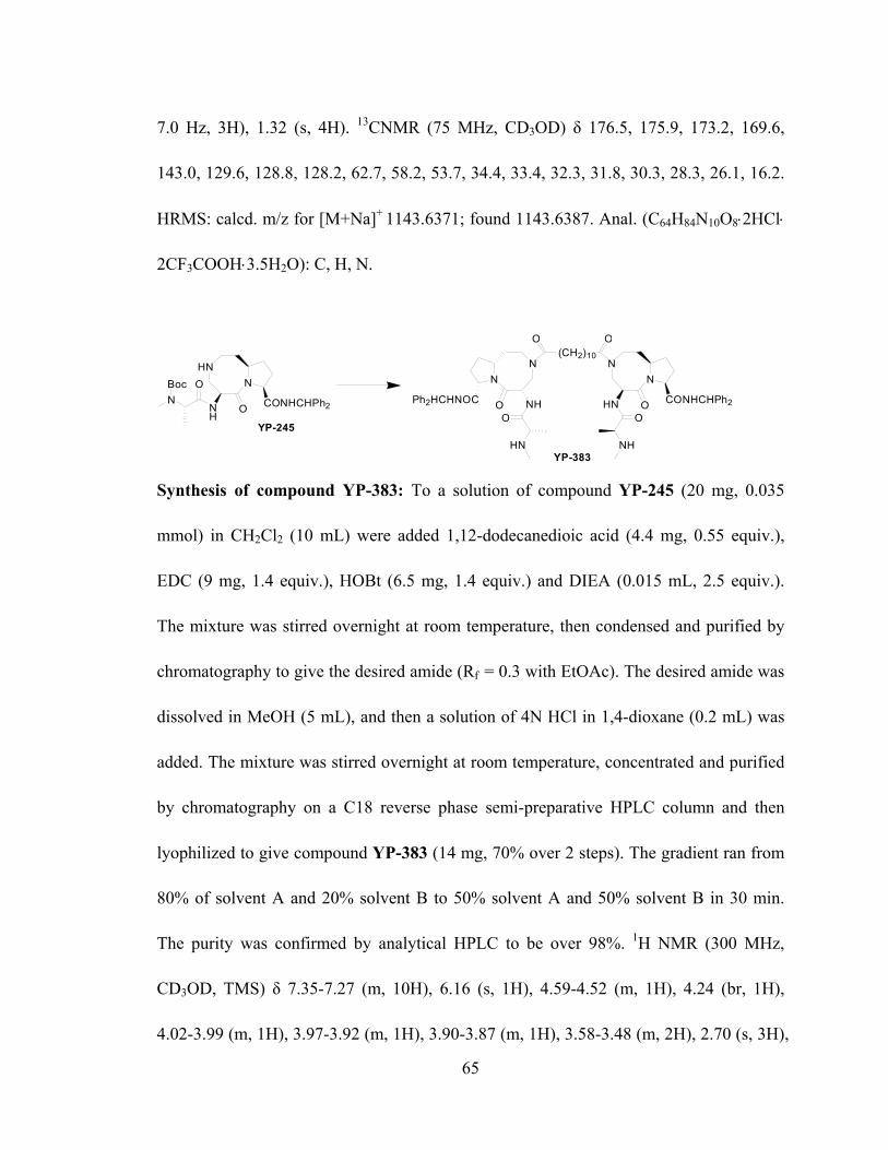
65
7.0 Hz, 3H), 1.32 (s, 4H). 13CNMR (75 MHz, CD3OD) δ 176.5, 175.9, 173.2, 169.6,
143.0, 129.6, 128.8, 128.2, 62.7, 58.2, 53.7, 34.4, 33.4, 32.3, 31.8, 30.3, 28.3, 26.1, 16.2.
HRMS: calcd. m/z for [M+Na]+ 1143.6371; found 1143.6387. Anal. (C64H84N10O8⋅2HCl⋅
2CF3COOH⋅3.5H2O): C, H, N.
HNN
O CONHCHPh2NH
YP-245
NOBoc
YP-383
NN
OPh2HCHNOC NH
HN
O
(CH2)10
O
NN
O CONHCHPh2HN
NH
O
O
Synthesis of compound YP-383: To a solution of compound YP-245 (20 mg, 0.035
mmol) in CH2Cl2 (10 mL) were added 1,12-dodecanedioic acid (4.4 mg, 0.55 equiv.),
EDC (9 mg, 1.4 equiv.), HOBt (6.5 mg, 1.4 equiv.) and DIEA (0.015 mL, 2.5 equiv.).
The mixture was stirred overnight at room temperature, then condensed and purified by
chromatography to give the desired amide (Rf = 0.3 with EtOAc). The desired amide was
dissolved in MeOH (5 mL), and then a solution of 4N HCl in 1,4-dioxane (0.2 mL) was
added. The mixture was stirred overnight at room temperature, concentrated and purified
by chromatography on a C18 reverse phase semi-preparative HPLC column and then
lyophilized to give compound YP-383 (14 mg, 70% over 2 steps). The gradient ran from
80% of solvent A and 20% solvent B to 50% solvent A and 50% solvent B in 30 min.
The purity was confirmed by analytical HPLC to be over 98%. 1H NMR (300 MHz,
CD3OD, TMS) δ 7.35-7.27 (m, 10H), 6.16 (s, 1H), 4.59-4.52 (m, 1H), 4.24 (br, 1H),
4.02-3.99 (m, 1H), 3.97-3.92 (m, 1H), 3.90-3.87 (m, 1H), 3.58-3.48 (m, 2H), 2.70 (s, 3H),

66
2.56-2.34 (m, 2H), 2.34-2.32 (m, 1H), 2.06-1.19 (m, 6H), 1.56-1.53 (d, J = 7.0 Hz, 3H),
1.30 (s, 6H). 13CNMR (75 MHz, CD3OD) δ 176.5, 175.9, 173.2, 169.6, 143.0, 129.6,
128.8, 128.2, 62.7, 58.2, 53.7, 34.4, 33.4, 31.7, 30.6, 30.4, 28.3, 26.3, 16.1. HRMS: calcd.
m/z for [M+Na]+ 1171.6684; found 1171.6680.
HNN
O CONHCHPh2NH
YP-245
NOBoc
NN
OPh2HCHNOC NH
HN
O
(CH2)6O
NN
O CONHCHPh2HN
NH
O
O
YP-385
Synthesis of compound YP-385: To a solution of compound YP-245 (20 mg, 0.035
mmol) in CH2Cl2 (10 mL) was added 1,6-hexanedicarboxylic acid (3.4 mg, 0.55 equiv.),
EDC (9 mg, 1.4 equiv.), HOBt (6.5 mg, 1.4 equiv.) and DIEA (0.015 mL, 2.5 equiv.).
The mixture was stirred overnight at room temperature, and then condensed and purified
by chromatography to give the desired amide (Rf = 0.3 EtOAc/MeOH= 99/1). The
desired amide was dissolved in MeOH (5 mL), and a solution of 4N HCl in 1,4-dioxane
(0.2 mL) was added. The mixture was stirred overnight at room temperature,
concentrated and purified by chromatography on a C18 reverse phase semi-preparative
HPLC column and then lyophilized to give compound YP-385 (14 mg, 76% over 2 steps).
The gradient ran from 80% of solvent A and 20% solvent B to 50% solvent A and 50%
solvent B in 30 min. The purity was confirmed by analytical HPLC to be over 98%. 1H
NMR (300 MHz, CD3OD, TMS) δ 8.95-8.93 (d, J = 8.1 Hz, 1H), 7.37-7.24 (m, 10H),
6.17-6.14 (d, J = 8.0 Hz, 1H), 4.58-4.56 (m, 1H), 4.24 (br, 1H), 3.99-3.92 (m, 2H), 3.82-

67
3.70 (m, 1H), 2.70 (s, 3H), 2.62-2.40 (m, 2H), 2.33 (m, 1H), 2.04-1.75 (m, 6H), 1.60-1.56
(m, 2H), 1.55-1.52 (d, J = 7.0 Hz, 3H), 1.34-1.30 (m, 2H). 13CNMR (75 MHz, CD3OD) δ
176.5, 175.8, 173.3, 169.7, 143.0, 129.6, 128.8, 128.2, 62.7, 58.2, 53.7, 34.5, 33.4, 31.7,
30.2, 28.3, 26.2, 16.1. HRMS: calcd. m/z for [M+H]+ 1093.6239; found 1093.6252. Anal.
(C62H80N10O8⋅2HCl⋅2CF3COOH⋅2H2O): C, H, N.

68
CHAPTER 3
BIOLOGICAL EVALUATION OF MONOVALENT SMAC MIMETICS
3.1 Binding Potency of Monovalent Smac Mimetics
Compound X R2 XIAP (nM) c-IAP1 (nM) c-IAP2 (nM)
SM-122 CH2 N/A 26 ± 4 1.0 ± 0.3 1.8 ± 0.6
SM-227 N H- 20.0 ±14.5 1.2 ± 0.1 4.6 ± 1.5
SM-245 N CH3- 341 ± 65.9 3.3 ± 1.3 17.5 ± 4.3
SM-246 N PhCH2- 91.8 ±30.4 3.7 ± 1.4 9.8 ± 4.1
SM-330 N CH3CO- 5.4 ± 3.0 1.3 ± 0.3 1.9 ± 0.8
SM-337 N PhCH2CO- 8.4 ± 1.6 1.5 ± 0.5 4.2 ± 0.6
Table 3.1 Binding affinities of Smac mimetics to XIAP, c-IAP1/2, as determined by competitive, fluorescence-polarization based assays. Data are shown in the form of Ki ± SD (nM).
To further explore the SAR of monovalent Smac mimetics, SM-227 was recently
developed in our laboratory. In SM-227, no additional functional group is tethered to the
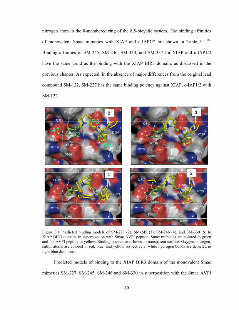
69
nitrogen atom in the 8-membered ring of the 8,5-bicyclic system. The binding affinities
of monovalent Smac mimetics with XIAP and c-IAP1/2 are shown in Table 3.1.166
Binding affinities of SM-245, SM-246, SM-330, and SM-337 for XIAP and c-IAP1/2
have the same trend as the binding with the XIAP BIR3 domain, as discussed in the
previous chapter. As expected, in the absence of major differences from the original lead
compound SM-122, SM-227 has the same binding potency against XIAP, c-IAP1/2 with
SM-122.
Figure 3.1 Predicted binding models of SM-227 (2), SM-245 (3), SM-246 (4), and SM-330 (5) to XIAP BIR3 domain, in superposition with Smac AVPI peptide. Smac mimetics are colored in green and the AVPI peptide in yellow. Binding pockets are shown in transparent surface. Oxygen, nitrogen, sulfur atoms are colored in red, blue, and yellow respectively, while hydrogen bonds are depicted in light blue dash lines.
Predicted models of binding to the XIAP BIR3 domain of the monovalent Smac
mimetics SM-227, SM-245, SM-246 and SM-330 in superposition with the Smac AVPI

70
peptide are shown in Figure 3.1. As expected, SM-227, SM-245, SM-246 and SM-330
can mimic the reverse turn conformation of Smac AVPI tetrapeptide, while the functional
groups tethered to the nitrogen atom in the 8,5-bicyclic system in SM-245, SM-246, and
SM-330 are directed towards the solvent, and have no specific interaction with the XIAP
BIR3 domain.
3.2 Tumor Cell Growth Inhibition Activity of Monovalent Smac Mimetics
Figure 3.2 Principles of WST-based cell proliferation assay.
WST (Water Soluble Tetrazolium)-based cell proliferation assays were preformed
to test the capability of Smac mimetics in tumor cell growth inhibition.167 The principles
of WST assays are shown in Figure 3.2. STR (Succinate Tetrazolium Reductase) is active
in the mitochondrial respiratory chain, transforming slightly red WST-1 to dark red
formazan only in viable cells. Therefore, the formation of formazan is in proportion to the
number of viable cells.168,169 By measuring the relative light absorbance at 450 nm, the

71
ratio of viable cells in the measured group to the untreated group (cell viability
percentage) can be obtained, as shown in Figure 3.3.
0.1 1 10 100 1000 100000
25
50
75
100
SM-122SM-227SM-245SM-246
IC50=Aver.+SD (nM)
890.0±383.3 1326.0±4.31011.6±206.3
SM-330 68.5±36.5SM-337 31.4±7.6SM-376 23.1±15.5
259.2±66.8
Human breast cancer MDA-MB-231 cells
Smac mimetic (nM)
Cel
l Via
bilit
y%
0.1 1 10 100 1000 100000
25
50
75
100
SM-122SM-227SM-245SM-246
325.6±76.6 484.6±111.7 892.6±19.5 720.3±94.3
SM-330 59.6±25.5SM-337 27.3±3.0SM-376 6 .7±3.8
IC50=Aver.+SD (nM)
Human ovarian cancer SK-OV-3 cells
Smac mimetic (nM)
Cel
l Via
bilit
y%
Figure 3.3 Inhibition of tumor cell growth by monovalent Smac mimetics in human breast cancer MDA-MB-231 cells and human ovarian cancer SK-OV-3 cells. Tumor cells were treated with Smac mimetics for 96 hours and cell growth was determined using a WST-based cell growth assay.
After human breast cancer MDA-MB-231 cells and human ovarian cancer
SK-OV-3 cells were treated with Smac mimetics for 96 hours, tumor cell viabilities were
analyzed by using WST-based cell growth assay. As shown in Figure 3.3, the potency of
monovalent Smac mimetics in inhibiting tumor cell growth is consistent with the binding
potency with XIAP, c-IAP1/2. In both tumor cell lines, Smac mimetics SM-227, SM-245,
and SM-246 have less potency than the lead compound SM-122 in tumor cell growth
inhibition. The Smac mimetics SM-330 and SM-337, with improved binding potency
with XIAP, are 4 to 10 times more potent than SM-122 in both tumor cell lines. SM-376,

72
with an (R)-tetrahydronaphthyl group which serves to increase the hydrophobic
interaction with the large hydrophobic pocket formed by lysine 297 and lysine 299 of the
XIAP BIR3 domain, has the best potency in inhibition of cell growth in both tumor cell
lines, consistent with its significant binding potency with the XIAP BIR 3 domain.
3.3 Tumor Cell Death Induction Activity of Monovalent Smac Mimetics
HO3S
NH2
SO3H
OH
N NSO3H
H2N
HO3S
HO
NN
Figure 3.4 Chemical structure of Trypan blue.
Trypan blue cell death assays were performed to evaluate the activity of
monovalent Smac mimetics in tumor cell death induction. Trypan blue (Figure 3.4) can
pass through the cell membrane of dead cells to color the cells in dark blue, while live
cells remain unaffected. Therefore, cell viabilities can be obtained after counting both
live and non-live cells distinguished by Trypan blue.
After human breast cancer MDA-MB-231 cells and human ovarian cancer
SK-OV-3 cells were treated with 0.1, 1, and 3 µM of the monovalent Smac mimetics
SM-122, SM-245, SM-246, SM-337, SM-350, or SM-376 for 24 and 48 hours, tumor cell
viabilities were analyzed by using Trypan-blue-based cell death assay. As shown in
Figure 3.5, every tested monovalent Smac mimetic can dose- and time-dependently
induce tumor cell death in both SK-OV-3 and MDA-MB-231 cells. Smac mimetics

73
SM-245 and SM-246 have same potency compared with lead compound SM-122 in both
tumor cell lines, while SM-337 and SM-350 are more potent than SM-122, consistent
with their better binding potency against XIAP BIR 3 domain. As expected, SM-376,
with the best binding potency among all the monovalent Smac mimetics, shows the best
potency in tumor cell death induction in both MDA-MB-231 and SK-OV-3 cells.
0
20
40
60
80
100
Time/d 1 2 1 2 1 2 1 2 1 2 1 2 1 2 1 2 1 2 1 2 1 2 1 2 1 2 1 2 1 2 1 2 1 2 1 2 1 2c (µM) 0 0.1 1 3 0.1 1 3 0.1 1 3 0.1 1 3 0.1 1 3 0.1 1 3
SK-O
V-3
Viab
ility
%
0
20
40
60
80
100
Untreated SM-122 SM-245 SM-246SM-337 SM-350 SM-376
Time/d 1 2 1 2 1 2 1 2 1 2 1 2 1 2 1 2 1 2 1 2 1 2 1 2 1 2 1 2 1 2 1 2 1 2 1 2 1 2c (µM) 0 0.1 1 3 0.1 1 3 0.1 1 3 0.1 1 3 0.1 1 3 0.1 1 3
MD
A-M
B-2
31 V
iabi
lity
%
Figure 3.5 Cell viabilities of human ovarian cancer SK-OV-3 cells and human breast cancer MDA-MB-231 cells treated with different concentrations of monovalent Smac mimetics for 24 or 48 hours, as determined by Trypan blue cell death assays.
3.4 Apoptosis Induction Activity of Monovalent Smac Mimetics
Our hypothesis is that Smac mimetics induce tumor cell death and inhibit tumor
cell growth by inhibiting IAPs and activating apoptosis in tumor cells. To further test this

74
hypothesis, the abilities of monovalent Smac mimetics in tumor apoptosis induction were
analyzed by using Annexin V and Propidium Iodide (P.I.) double staining flow
cytometry.
Figure 3.6 Annexin V and P.I. double staining flow cytometry of untreated MDA-MB-231 cells.
2%
3%1%
3%
4%1%
3%
4%1%
6%
6%2%
6%
5%1%
14%
13%4%
14%
16%8%
8%
7%2%
14%
14%7%
14%
16%7%UT SM245
10 nM100 nM1 µM
SM33710 nM100 nM1 µM
SM37610 nM100 nM1 µM
Figure 3.7 Annexin V and P.I. double staining flow cytometry of human breast cancer MDA-MB-231 cells treated with different concentrations of monovalent Smac mimetic SM-245, SM-337, or SM-376 for 24 hours.
1%
4% 95%

75
1%
2%1%
2%
3%1%
2%
5%2%
3%
5%2%
3%
4%1%
6%
6%3%
9%
9%5%
3%
4%2%
6%
8%4%
7%
7%5%UT SM245
10 nM100 nM1 µM
SM33710 nM100 nM1 µM
SM37610 nM100 nM1 µM
Figure 3.8 Annexin V and P.I. double staining flow cytometry of human ovarian cancer SK-OV-3 cells treated with different concentrations of monovalent Smac mimetic SM-245, SM-337, or SM-376 for 24 hours.
Figure 3.6 shows the results of Annexin V and P.I. double staining flow cytometry
of untreated MDA-MB-231 cells. Annexin V competes for the binding site of
phosphatidylserine, which is normally confined to the inner leaflet of the cell membrane
and is externalized in the early stages of apoptosis, while P.I. can intercalate DNA which
is released after damage of the cell nucleus in the late stage of apoptosis.170-179 Hence,
cells in the lower right quadrant are in the early phase of apoptosis, binding Annexin V
but not taking up P.I. Cells in the upper right quadrant are in the late phase of apoptosis,
both binding Annexin V and taking up P.I.. Cells in the lower left quadrant are live cells,
neither binding Annexin V nor taking up P.I., while cells in the upper left quadrant are
dead cells, only taking up P.I.. Therefore, flow cytometry with Annexin V and P.I. double

76
staining can be used to evaluate the ability of Smac mimetics in inducing apoptosis, since
it can successfully distinguish the different apoptosis phases in which cells are
present.180-191
1%
2%3%
6%
4%1%
10%
7%2%
8%
5%1%
10%
8%1%
31%
13%3%
39%
18%5%
16%
10%2%
32%
14%3%
39%
14%5%UT SM24510 nM100 nM1 µM
SM33710 nM100 nM1 µM
SM37610 nM100 nM1 µM
Figure 3.9 Annexin V and P.I. double staining flow cytometry of human breast cancer MDA-MB-231 cells treated with different concentrations of monovalent Smac mimetic SM-245, SM-337, or SM-376 for 48 hours.
After human breast cancer MDA-MB-231 cells and human ovarian cancer
SK-OV-3 cells were treated with 10, 100, and 1000 nM of the Smac mimetic SM-245,
SM-337, or SM-376 for 24 hours, apoptosis in tumor cells was analyzed using Annexin V
and P.I. double staining flow cytometry. As shown in Figure 3.7 and 3.8, every Smac
mimetic can induce apoptosis in both tumor cell lines in a dose-dependent manner.
SM-337 and SM-376 show similar potency in inducing apoptosis in both MDA-MB-231

77
and SK-OV-3 cells, while both Smac mimetics are more potent than SM-245, consistent
with their superior potency in binding against XIAP BIR 3 domain.
After human breast cancer MDA-MB-231 cells were treated with 10, 100, and
1000 nM of Smac mimetic SM-245, SM-337, or SM-376 for 48 hours, apoptosis in tumor
cells was analyzed by Annexin V and P.I. double staining flow cytometry, as shown in
Figure 3.9. As expected, each Smac mimetic can dose-dependently induce apoptosis in
MDA-MB-231 cells. Meanwhile, each Smac mimetic time-dependently induces
apoptosis in MDA-MB-231 cells, when compared with the results in Figure 3.7.
Similarly, SM-337 and SM-376 have better potency than SM-245, which bears a
relatively lower binding affinity against XIAP BIR 3 domain.
3.5 Caspase Activation of Monovalent Smac Mimetics
A cell-free functional assay was performed to test further whether Smac mimetics
can function as antagonists of XIAP. While the XIAP protein effectively inhibits the
activity of caspase-3/7, the Smac mimetics SM-122, SM-227, SM-330, and SM-337 can
all dose-dependently antagonize XIAP and restore the activity of casapase-3/7. As shown
in Figure 3.10, newly developed Smac mimetic SM-227, SM-330, and SM-337 all have
better potency than original lead compound SM-122 in caspase activity recovery.

78
0 10 20 30 40 50 600
10000
20000
+ XIAP + SM-122 312.5 nM
+ XIAP + SM-122 2,500 nM+ XIAP + SM-122 5,000 nM
Control (Maximum activation)+ XIAP 500 nM
+ XIAP + SM-122 1,250 nM+ XIAP + SM-122 625 nM
+ XIAP + SM-122 156.25 nM
Time (minutes)0 10 20 30 40 50 60
0
10000
20000
+ XIAP + SM-227 312.5 nM
+ XIAP + SM-227 2,500 nM+ XIAP + SM-227 5,000 nM
Control (Maximum activation)+ XIAP 500 nM
+ XIAP + SM-227 1,250 nM+ XIAP + SM-227 625 nM
+ XIAP + SM-227 156.25 nM
Time (minutes)
RFU
(Cas
pase
-3/7
act
ivity
)
0 10 20 30 40 50 600
10000
20000
+ XIAP + SM-330 312.5 nM
+ XIAP + SM-330 2,500 nM+ XIAP + SM-330 5,000 nM
Control (Maximum activation)+ XIAP 500 nM
+ XIAP + SM-330 1,250 nM+ XIAP + SM-330 625 nM
+ XIAP + SM-330 156.25 nM
Time (minutes)
RFU
(Cas
pase
-3/7
act
ivity
)
0 10 20 30 40 50 600
10000
20000
+ XIAP + SM-337 312.5 nM
+ XIAP + SM-337 2,500 nM+ XIAP + SM-337 5,000 nM
Control (Maximum activation)+ XIAP 500 nM
+ XIAP + SM-337 1,250 nM+ XIAP + SM-337 625 nM
+ XIAP + SM-337 156.25 nM
Time (minutes)
Figure 3.10 Inhibition of caspase-3/7 activity by XIAP and antagonism of Smac mimetics to XIAP to recover the activity of caspase-3/7 in a cell-free functional assay.

79
0 10 20 30 40 50 600
10000
20000
+ XIAP + SM-246 10,000 nM+ XIAP + SM-246 100,000 nM
Control (Maximum activation)+ XIAP 500 nM
+ XIAP + SM-246 1,000 nM+ XIAP + SM-246 100 nM
Time (minutes)
RFU
(Cas
pase
-3/7
act
ivity
)
-1 0 10
25
50
75
100
SM-122 (1.03)
SM-246 (4.86)
SM-337 (0.50)
Compounds (EC50, µM)
[Compounds] (log [µM])% R
ecov
ery
of C
aspa
se-3
/7 A
ctiv
ity
Figure 3.11 Left: Inhibition of caspase-3/7 activity by XIAP and antagonism of Smac mimetic SM-246 to XIAP to recover the activity of caspase-3/7 in a cell-free functional assay. Right: Dose-dependent recovery of caspase-3/7 activity by SM-122, SM-246, and SM-337 to the maximum activation. Caspase-3/7 activity at 30 minute point was used.
The ability of Smac mimetic SM-246 in caspase-3/7 activity recovery was also
analyzed in a cell-free functional assay. The result shows that SM-246 can
dose-dependently recover the activity of caspase-3/7 by antagonizing XIAP. However, as
shown in Figure 3.11, SM-246 has less potency in caspase activity recovery than SM-122
and SM-330, consistent with its lower binding potency with the XIAP BIR 3 domain.
3.6 Drug Synergy Effect of Monovalent Smac Mimetics with TRAIL

80
Figure 3.12 Inhibition of cell growth by Smac mimetics SM-337, SM-376, and SM-377 in combination with TRAIL in human breast cancer MDA-MB-231 cell lines. Cells were treated with TRAIL only or TRAIL in combination with Smac mimetics for 4 days and cell growth was analyzed by WST-based cell growth assay.
We hypothesize that our Smac mimetics can inhibit tumor cell growth by inhibiting
XIAP thus promoting apoptosis in tumor cells. Hence, our Smac mimetics may synergize
with Tumor necrosis factor-Related Apoptosis Inducing Ligand (TRAIL) which can
induce the extrinsic apoptotic pathway. To verify this, WST-based cell growth assays of
human breast cancer MDA-MB-435 cells treated with TRAIL alone or in combination
with different concentrations of Smac mimetic SM-337, SM-376, or SM-377, were
performed. As shown in Figure 3.12, each monovalent Smac mimetic is
dose-dependently synergistic with TRAIL in inhibiting MDA-MB-435 cell growth.
SM-376 and SM-377 have similar potency in tumor cell growth inhibition, while both are
more potent than SM-337, consistent with their better binding potency to XIAP protein.
TRAIL + SM-337 wst assay in MDA-MB-435 cells
1 10 1000
25
50
75
100
TRAILT + 0.1nM of SM-337T + 1nM of SM-337T + 10nM of SM-337
(IC50=280)
(IC50=180)(IC50=80)
(IC50=10)
Drug conc. (ng/ml)
Cel
l Gro
wth
( % o
f con
trol
)
TRAIL + SM-376 wst assay in MDA-MB-435 cells
1 10 1000
25
50
75
100
TRAILT + 0.1nM of SM-376T + 1nM of SM-376T + 10nM of SM-376
(IC50=220)
(IC50=95)(IC50=12)
(IC50=2.8)
Drug conc. (ng/ml)
Cel
l Gro
wth
( % o
f con
trol
)
TRAIL + SM-377 wst assay in MDA-MB-435 cells
1 10 1000
25
50
75
100
TRAILT + 0.1nM of SM-377T + 1nM of SM-377T + 10nM of SM-377
(IC50=210)
(IC50=190)(IC50=18)
(IC50=3)
Drug conc. (ng/ml)
Cel
l Gro
wth
( % o
f con
trol
)

81
Figure 3.13 Inhibition of cell growth by Smac mimetics SM-337, SM-376, and SM-377 in combination with TRAIL in human breast cancer 2LMP cell lines. Cells were treated with TRAIL only or TRAIL in combination with Smac mimetics for 4 days and cell growth was analyzed by WST-based cell growth assay.
The synergistic effect of Smac mimetics with TRAIL in human breast cancer
2LMP cells was also analyzed by using WST-based cell growth assay. As shown in
Figure 3.13, SM-337, SM-376, and SM-377 can all dose-dependently synergize with
TRAIL in inhibiting 2LMP cell growth. Similarly, in MDA-MB-435 cells, SM-376 and
SM-377 are slightly more potent than SM-337 in inhibiting 2LMP cell growth, consistent
with their better binding potency to XIAP protein.
3. 7 Cellular Molecular Effects of Monovalent Smac Mimetics
TRAIL + SM-337 wst assay in 2LMP cells
0.01 0.1 1 1000
25
50
75
100
TRAILT + 0.1nM of SM-337T + 1nM of SM-337T + 10nM of SM-337
(IC50=2)
(IC50=1.8)(IC50=1.5)
(IC50=0.35)
Drug conc. (ng/ml)
Cel
l Gro
wth
( % o
f con
trol
)
TRAIL + SM-376 wst assay in 2LMP cells
0.01 0.1 1 1000
25
50
75
100
TRAILT + 0.1nM of SM-376T + 1nM of SM-376T + 10nM of SM-376
(IC50=2.1)
(IC50=1.5)(IC50=0.5)
(IC50=0.2)
Drug conc. (ng/ml)
Cel
l Gro
wth
( % o
f con
trol
)
TRAIL + SM-377 wst assay in 2LMP cells
0.01 0.1 1 1000
25
50
75
100
TRAILT + 0.1nM of SM-377T + 1nM of SM-377T + 10nM of SM-377
(IC50=2.8)
(IC50=2.5)(IC50=1)
(IC50=0.25)
Drug conc. (ng/ml)
Cel
l Gro
wth
( % o
f con
trol
)

82
CL PARP 85kd
CL C3 21kd
CL C3 19kd
CL C3 17kd
cIAP1 72kd
β‐actin 42kd
UT 0.01 0.1 1 0.01 0.1 1 0.01 0.1 1 0.01 0.1 1 Conc. (µM)
SM-122 SM-227 SM-245 SM-337(MDA-MB-231 cells)
(SK-OV-3 cells)
CL PARP 85kd
CL C3 21kd
CL C3 19kd
CL C3 17kd
cIAP1 72kd
β‐actin 42kd
Figure 3.14 Western blot assays of human breast cancer MDA-MB-231 cells and human ovarian cancer SK-OV-3 cells treated with different concentrations of Smac mimetic SM-122, SM-227, SM-245, or SM-337 for 24 hours.
To further explore the cellular molecular effects induced by our monovalent Smac
mimetics, human breast cancer MDA-MB-231 cells and human ovarian cancer SK-OV-3
cells were treated with 10, 100, and 1000 nM of Smac mimetic SM-122, SM-227,
SM-245, or SM-337 for 24 hours, and then Poly ADP Ribose Polymerase (PARP)
cleavage. Degradation of c-IAP1, and caspase activation were analyzed with Western
blotting assays. As shown in Figure 3.14, each Smac mimetic can dose-dependently
induce the cleavage of PARP, a substrate of active caspase-3, and also a marker of cells
undergoing apoptosis in both tumor cell lines. Our designed Smac mimetics bind to
cIAP-1 with very high affinities (Table 3.1) and recent studies have shown that Smac

83
mimetics induce cIAP-1 degradation in cancer cells, mediating apoptosis induction.192-195
Hence, the ability of Smac mimetic in inducing cIAP-1 degradation was also analyzed.
As expected, each Smac mimetic can effectively induce c-IAP1 degradations in both
tumor cell lines. Consistent with the ability of Smac mimetics in inducing PARP cleavage,
each Smac mimetic dose-dependently induces the activation of caspase-3 in both tumor
cell lines.
3.8 Conclusion
A series of potent non-peptidic small molecular Smac mimetics has been
successfully developed. SM-330, SM-337, SM-376, and SM-377 with improved binding
potency with XIAP and c-IAP1/2, compared with our original lead compound SM-122,
are cell-permeable and have excellent activity in inhibiting tumor cell growth, inducing
tumor cell death, and inducing apoptosis in tumor cells, as tested in both human breast
cancer MDA-MB-231 cells and human ovarian cancer SK-OV-3 cells. As expected, our
Smac mimetics can synergize with TRAIL in inhibiting tumor cell growth, as tested in
both human breast cancer MDA-MB-435 cells and 2LMP cells. Cell-free functional
assays verify that our Smac mimetics can recover the activity of caspase-3,7 previously
inhibited by XIAP protein. The cellular molecular events induced by our Smac mimetics
were also analyzed with Western blotting assays, which show that each Smac mimetic
can dose-dependently induce PARP cleavage, c-IAP1 degradation and caspase activation
in tumor cells. Overall, these potent Smac mimetics with excellent cellular activity are

84
currently being used as our new lead template to develop future drug candidates with
excellent PK (pharmacokinetic) properties.

85
CHAPTER 4
BIOLOGICAL EVALUATION OF BIVALENT SMAC MIMETICS
4.1 Binding Potency of Bivalent Smac Mimetics
To further explore the optimal length of the linker for the bivalent Smac mimetics,
bivalent Smac mimetics SM-1252 and SM-1253 were recently developed. A series of
bivalent Smac mimetics with linkers of different lengths were tested for their binding
potency against both XIAP BIR3 protein and XIAP linker-BIR2-BIR3 protein, using our
fluorescence-polarization based assays. As shown in Table 4.1, as the length of the
bivalent linker increases, the binding potency against XIAP BIR3 domain decreases.
However, all the five bivalent Smac mimetics show similar excellent potency in binding
against XIAP linker-BIR2-BIR3 domains. All the bivalent Smac mimetics show excellent
binding potency in binding with linker-BIR2-BIR3 protein compared with BIR3 protein
only. Their cellular activities were explored in detail in the subsequent parts of this
chapter.

86
Comp. (linker)
Structure BIR3 Ki±SD (nM)
L-BIR2-BIR3Ki±SD (nM)
SM-1252 (2C)
9.7 ± 3.8 1.0 ± 0.7
SM-1253 (4C)
7.6 ± 2.3 0.3 ± 0.2
SM-385 (6C)
NN
O CONHCHPh2HN
NN
OPh2HCHNOC NH
NH
O
HN
O
O O
HClHCl
12.7 ± 3.9 1.0 ± 0.5
SM-381 (8C)
NN
O CONHCHPh2HN
NN
OPh2HCHNOC NH
NH
O
HN
O
O O
HClHCl
27.5 ± 6.2 1.8 ± 0.6
SM-383 (10C)
NN
O CONHCHPh2HN
NN
OPh2HCHNOC NH
NH
O
HN
O
O O
HClHCl
43.1 ± 3.6 1.8 ± 0.5
Table 4.1 Binding affinities of bivalent Smac mimetics against XIAP BIR3 and XIAP linker-BIR2-BIR3 domains, as measured by fluorescence-polarization based assays
4.2 Tumor Cell Growth Inhibition Activity of Bivalent Smac Mimetics
SM-1252, SM-1253, SM-385, SM-381, and SM-383 have 2, 4, 6, 8, and 10
methylene groups respectively in the bivalent linker. To explore the ability of these

87
bivalent Smac mimetics in inhibition of tumor cell growth, WST-based cell growth
assays were performed. Human breast cancer MDA-MB-231 cells and human ovarian
cancer SK-OV-3 cells were treated with bivalent Smac mimetics for 96 hours, and then
cell viabilities were analyzed by using WST-based cell growth assay.
0.1 1 10 100 1000 100001000000
25
50
75
100 2C (10.5± 3.1 nM)4C (11.4± 3.2 nM)6C (4.7± 1.5 nM)8C (2.3± 0.7 nM)10C (2.6± 1.2 nM)SM123 (6.5± 1.3 µM)
Drug conc. (nM)
MD
A-M
B-3
21 V
iabi
lity
%
0.01 0.1 1 10 100 10000
25
50
75
100 2C (28± 10 nM)4C (14.6± 5.7 nM)6C (10.9± 3.1 nM)8C (3.9± 1.0 nM)10C (4.6± 1.9 nM)
0[Drug] (nM)
SK-O
V-3
Viab
ility
%
Figure 4.1 WST cell growth assays of human breast cancer MDA-MB-231 cells and human ovarian cancer SK-OV-3 cells treated with bivalent Smac mimetics for 96 hours. Experiments were performed three times. Data are reported in the form of IC50 ± SD. 2C (SM-1252), 4C (SM-1253), 6C (SM-385), 8C (SM-381), 10C (SM-383).
As shown in Figure 4.1, all the bivalent Smac mimetics show excellent potency in
tumor cell growth induction with IC50 values (the concentration in which Smac mimetic
inhibits 50% tumor cell growth compared with untreated group) in the neighborhood of
30 nM in both MDA-MB-231 and SK-OV-3 cells. In comparison, the inactive control
SM-123 is over 600 times less potent than the weakest bivalent Smac mimetic SM-1252.
In both tumor models, SM-1252, with the shortest linker, has the weakest potency among

88
all the bivalent Smac mimetics. As the linker increases in length, the cellular potency
increases. However, SM-381 and SM-383, with 8 and 10 methylenes in the bivalent
linker respectively, have equivalent cellular potency in tumor cell growth inhibition.
0.01 0.1 1 10 100 10000
25
50
75
1002C (2.5± 1.7 nM)4C (3.1± 2.2 nM)6C (1.7± 1.2 nM)8C (0.8± 0.2 nM)10C (1.6± 0.9 nM)
0Drug Conc. (nM)
MAL
ME-
3M V
iabi
lity%
Figure 4.2 WST cell growth assay of human melanoma MALME-3M cells treated with bivalent Smac mimetics for 96 hours. Experiments were performed three times. Data are reported in the form of IC50 ± SD. 2C (SM-1252), 4C (SM-1253), 6C (SM-385), 8C (SM-381), 10C (SM-383).
Human melanoma MALME-3M cells were also treated with bivalent Smac
mimetics for 96 hours, and then cell viabilities were analyzed using WST cell growth
assays. As shown in Figure 4.2, all the bivalent Smac mimetics show excellent cellular
potency in tumor cell growth inhibition, with an IC50 of approximately 5 nM. As expected,
the cellular potency of bivalent Smac mimetics increases as the linker increases in its
length. Just as in MDA-MB-231 and SK-OV-3 cells, SM-381 and SM-383, with the
longest linkers, have the best cellular potency in MALME-3M cells.
4.3 Tumor Cell Death Induction Activity of Bivalent Smac Mimetics
To further explore the ability of bivalent Smac mimetics in inducing tumor cell
death, Trypan blue cell death assays were performed. Human breast cancer

89
MDA-MB-231 cells and human ovarian cancer SK-OV-3 cells were treated with 0.1, 1,
10, and 100 nM of bivalent Smac mimetic SM-1252 (2C), SM-381 (8C), and SM-383
(10C) for 2, 6, 12, and 24 hours respectively, and cell viabilities were analyzed by using
Trypan blue assays.
0
20
40
60
80
100
Time/h 2h 6h 12h 24hUT
viab
ility
% (M
DA
-MB
-231
)
0
20
40
60
80
100
0.1 nM of Smac mimetic1 nM of Smac mimetic
10 nM of Smac mimetic100 nM of Smac mimetic
Time/h 2h 6h 12h 24hUT
viab
ility
% (S
K-O
V-3)
Figure 4.3 Trypan blue assays of human breast cancer MDA-MB-231 cells and human ovarian cancer SK-OV-3 cells treated with different concentrations of bivalent Smac mimetics for designated lengths of time. In each group of three, left is SM-1252 (2C), middle is SM-381 (8C), and right is SM-383 (10C).
As shown in Figure 4.3, each bivalent Smac mimetic can dose- and
time-dependently induce tumor cell death in both MDA-MB-231 and SK-OV-3 cells. All
the three bivalent Smac mimetics show similar potency within 12 hours of treatment in
both tumor cell lines. However, at higher concentrations (10 nM and 100 nM) of bivalent
Smac mimetics for 24 hours, SM-381 and SM-383 are dramatically more potent than

90
SM-1252 in inducing tumor cell death in both tumor cell lines, consistent with the
potency trend shown in the results in WST-based cell growth assays.
4.4 Tumor Apoptosis Induction Activity of Bivalent Smac Mimetics
MDA‐MB‐231 cells
UT SM-123: 1000 nM8C: 0.1, 1, 10, 100 nM10C: 0.1, 1, 10, 100 nM
3%
4%0%
4%
9%1%
4%
11%1%
9%
8%20%
6%
7%31%
4%
5%35%
15%
12%15%
4%
4%32%
14%
11%24%
11%
15%26%
Figure 4.4 Human breast cancer MDA-MB-231cells were treated with different concentrations of bivalent Smac mimetic SM-381 (8C) and SM-383 (10C) or 1 µM of inactive control SM-122 for 24 hours. Apoptosis in tumor cells was analyzed by Annexin V and P.I. double staining flow cytometry.
To further explore the ability of bivalent Smac mimetics in inducing apoptosis in
tumor cells, Annexin V and P.I. double staining flow cytometry was performed. Human
breast cancer MDA-MB-231 cells were treated with 0.1, 1, 10, and 100 nM of the
bivalent Smac mimetics SM-381 (8C) and SM-383 (10C) or 1000 nM of inactive control

91
SM-123 for 24 hours, and apoptosis was analyzed by flow cytometry. As shown in Figure
4.4, both bivalent these Smac mimetics can dose-dependently induce apoptosis in
MDA-MB-231 cells. In contrast, the inactive control SM-123 can only induce a small
percentage of apoptosis compared with the untreated group, although a high
concentration of 1 µM was used.
5%
7%0%
2%
4%0%
17%
20%2%
9%
45%27%
12%
38%21%
4%
5%1%
15%
18%3%
15%
44%14%
10%
41%27%
5%
7%0% SK‐OV‐3 cells
UT SM-123: 1000 nM8C: 0.1, 1, 10, 100 nM10C: 0.1, 1, 10, 100 nM
Figure 4.5 Human ovarian cancer SK-OV-3 cells were treated with different concentrations of bivalent Smac mimetic SM-381 (8C) and SM-383 (10C) or 1 µM of inactive control SM-122 for 24 hours. Apoptosis in tumor cells was analyzed by Annexin V and P.I. double staining flow cytometry.
The ability of bivalent Smac mimetics to induce apoptosis in human ovarian cancer
SK-OV-3 cells was also analyzed. SK-OV-3 cells were treated with 0.1, 1, 10, and 100
nM of bivalent Smac mimetics SM-381 (8C) and SM-383 (10C) or 1000 nM of inactive
control SM-123 for 24 hours, and apoptosis was analyzed by flow cytometry. As in

92
MDA-MB-231 cells, both bivalent Smac mimetics were found to dose-dependently
induce apoptosis in SK-OV-3 cells, while the inactive control SM-123 fails to induce
more apoptosis than the untreated group, as shown in Figure 4.5.
4.5 Cellular Molecular Effects of Bivalent Smac Mimetics
UT 1 10 100 1 10 100 1 10 100 1000 Conc. (µM)SM-164 SM-383 SM-385 SM-428
CL PARP 85kd
CL C3 21kdCL C3 19kdCL C3 17kd
Pro-C9 46kd
CL C7 20kd
β-actin 42kd
cIAP1 72kd
XIAP 57kd
Pro-C8 57kdPro-C8 55kd
Figure 4.6 Western blotting assays of human breast cancer MDA-MB-231 cells treated with different concentrations of bivalent Smac mimetic SM-164, SM-381 and SM-383 for 24 hours.
To further probe the cellular molecular events induced by bivalent Smac mimetics,
Western blotting assays were performed. Human breast cancer MDA-MB-231 cells were
treated with 1, 10, and 100 nM of bivalent Smac mimetic SM-164, SM-381, and SM-383
for 24 hours, and then PARP cleavage, IAP degradations, and caspase activations were

93
analyzed using Western blotting assays. As shown in Figure 4.6, each bivalent Smac
mimetic can dose-dependently induce PARP cleavage, c-IAP1 degradation and
caspase-3/7 activations. SM-381 is as potent as the previously developed bivalent Smac
mimetic SM-164,135 while SM-383 is slightly less potent than SM-164. However, both
SM-381 and SM-383 are dramatically more potent than the related monovalent Smac
mimetics SM-330 and SM-337.
CL PARP 85kd
CL C3 21kdCL C3 19kdCL C3 17kd
Pro-C9 46kd
CL C7 20kd
β-actin 42kd
cIAP1 72kd
XIAP 57kd
Pro-C7 35kd
Pro-C8 57kdPro-C8 55kd
UT 1 10 100 1 10 100 1 10 100 Conc. (µM)SM-164 SM-383 SM-385
Figure 4.7 Western blotting assays of human ovarian cancer SK-OV-3 cells were treated with different concentrations of bivalent Smac mimetic SM-164, SM-381 and SM-383 for 24 hours.
The cellular activity of bivalent Smac mimetics in human ovarian cancer SK-OV-3
cells was also analyzed. SK-OV-3 cells were treated with 1, 10, and 100 nM of the
bivalent Smac mimetics SM-164, SM-381, and SM-383 for 24 hours, and then PARP

94
cleavage, IAP degradations, and caspase activations were analyzed using Western
blotting assays. Consistent with the results in MDA-MB-231 cells, SM-383 is as potent
as SM-164 in PARP cleavage induction, c-IAP1 degradation, and caspase-3/7 activations,
while SM-383 is slightly less potent than SM-164. However, both bivalent Smac
mimetics SM-381 and SM-383 are 10 to 100 times more potent than monovalent Smac
mimetics SM-227 and SM-337, whose performance is presented in a previous chapter.
4.6 Conclusion
Compared with monovalent Smac mimetics, all the bivalent Smac mimetics show
excellent cellular activity as inhibitors of tumor cell growth of human breast cancer
MDA-MB-231 cells, human ovarian cancer SK-OV-3 cells and human melanoma
MALME-3M. As the length of bivalent linker increases, the cellular activity potency of
bivalent Smac mimetics decreases. However, SM-381 and SM-383, with 8 and 10
methylene groups in the bivalent linker, have similar potency in all the tested tumor cell
lines.
Bivalent Smac mimetics can dose- and time- dependently induce tumor cell death
in both MDA-MB-231 and SK-OV-3 cells. In treatment of 12 hours, SM-1252, SM-381,
and SM-383 show similar potency in tumor cell death induction. However, SM-381 and
SM-383 are dramatically more potent than SM-1252 after 24 hours’ treatment at higher
concentrations, consistent with the potency trend shown in the WST-based cell growth
assays.

95
The capability of bivalent Smac mimetic SM-381 and SM-383 in induction of
apoptosis in MDA-MB-231 cells and SK-OV-3 cells was further analyzed. As expected,
SM-381 and SM-383 were found to dose-dependently induce apoptosis in both tumor cell
lines.
The cellular molecular effects of bivalent Smac mimetics were analyzed in
MDA-MB-231 and SK-OV-3 cells. SM-381 shows excellent activity in PARP cleavage
induction, c-IAP1 degradation induction, and caspase activations, and is as potent as our
previously developed bivalent Smac mimetic SM-164.135 Although SM-383 is slightly
less potent than SM-381, both SM-381 and SM-383 are 10 to 100 times more potent than
monomeric Smac mimetics SM-227 and SM-337 in cellular activities in tumor cells.
Further modification of these bivalent Smac mimetics is in process, and we hope that our
potent bivalent Smac mimetics can lead to excellent drug candidates in the future.

96
CHAPTER 5
CELLULAR MECHANISM STUDIES BASED ON SM-406
5.1 SM-406
The Smac mimetics SM-337, SM-350, SM-376 and SM-377 can potently inhibit
tumor cell growth of human breast cancer MDA-MB-231 cells, MDA-MB-453 cells,
2LMP cells and human ovarian cancer SK-OV-3 cells, both alone and in combination
with TRAIL, as shown in the results of our WST-based cell growth assays in Chapter 3.
Pharmacokinetic studies, completed by Medicilon Company (Shanghai), showed that all
of these potent Smac mimetics, in rats, have relatively low oral bioavailability (F%).
With the lead compound SM-337 as template, a series of derived Smac mimetics were
further designed and synthesized, with the aim of developing novel Smac mimetics with
improved PK properties. The only difference between the newly developed Smac
mimetics and SM-337 is the carboxylic acid tethering the cyclic amine of the
8-membered fused ring in the bicyclic structure. This modification has no detrimental
effect on the potency of Smac mimetics, because the side chain tethered to the nitrogen
atom in the bicyclic system is directed towards the solvent, and as predicted by our
modeling studies (Figure 2.2), has no specific interaction with the XIAP protein.

97
NN
O CONHCHPh2NH
HN
O
O
(SM-406)CQ-406
Figure 5.1 Chemical structure of SM-406.
Among these newly developed Smac mimetics, compound CQ-406 (or SM-406),
in which a 3-methylbutanoic acid is attached to the cyclic amine in the left fused ring,
made by Dr. Cai Qian in our lab, has an excellent oral bioavailability (45%) in rats
compared with the previously synthesized Smac mimetics SM-337 (24%), SM-350
(22%), SM-376 (26%), and SM-377 (23%), although it has a slightly weaker potency in
binding with the XIAP BIR3 domain (Ki = 13.80 ± 2.93 nM). Further pharmacokinetic
studies of the Smac mimetic SM-406 in dogs and monkeys are under investigation, and
the molecular cellular mechanism studies of our Smac mimetics detailed here are based
on this promising drug candidate SM-406.
5.2 Further Biological Studies Based on SM-406
5.2.1 Apoptosis induction activity of SM-406
To test the ability of SM-406 in inducing apoptosis in tumor cells, Annexin V and
P.I. (Propidium Iodide) double staining flow cytometry was performed to monitor the
apoptosis process in tumor cells treated with SM-406.

98
NN
O CONHCHPh2NH
HN
O
O
SM-428NH
Figure 5.2 Top: Chemical structure of SM-428, inactive control of Smac mimetics. Bottom: Annexin V and P.I. double staining flow cytometry of human ovarian cancer SK-OV-3 cells treated Smac mimetic SM-406 and inactive control SM-428 for 24 hours.
SM-428, used as an inactive control of Smac mimetics, is a derivative of the Smac
mimetic SM-406, in which the methyl side chain of the amino terminal alanine moiety is
substituted with an indole ring, yielding compound SM-428. The methyl side chain in the
amino terminal alanine residue of mature Smac protein penetrates the small hydrophobic
pocket formed by the side chains of leucine 307, tryptophan 310, and glutamine 319 in
XIAP.122 Further study showed that this small hydrophobic pocket can also accommodate
an ethyl group, however, larger hydrophobic groups can eliminate this hydrophobic
interaction.127 Predictably, the newly introduced large indole ring completely abolishes
SM-406 (0.1 µM) SM-406 (0.3 µM) SM-406 (1 µM) SM-406 (3 µM)
Untreated SM-428 (1 µM) SM-428 (3 µM)
0% 0% 1% 3% 1% 3%
6% 4% 4%
2% 6% 2% 13% 4% 22% 4% 24%
8% 13% 15% 18%

99
this interaction and dramatically reduces the binding affinity of SM-428 with the XIAP
BIR3 domain, as verified by our fluorescence-polarization-based binding assay (Ki = 45.0
± 8.7 µM). Consequently, compound SM-428 was used as an inactive control in our
molecular biological studies of Smac mimetics.
Figure 5.3 Annexin V and P.I. double staining flow cytometry of human ovarian cancer SK-OV-3 cells treated with 3 µM of Smac mimetic SM-406 for designated lengths of time.
Human ovarian cancer SK-OV-3 cells were first treated with different doses of
Smac mimetic SM-406 and 3 µM of inactive control SM-428 for 24 hours, and then the
apoptosis of SK-OV-3 cells was analyzed by Annexin V and P.I. double staining flow
cytometry, as shown in Figure 5.2. As expected, SM-406 can induce apoptosis of
SK-OV-3 cells in a dose-dependent manner.
The apoptosis induction behavior of SM-406 in SK-OV-3 cells was also studied
in a time course. SK-OV-3 cells were treated with 3 µM of Smac mimetic SM-406 for
Untreated
SM-406 (3 hours) SM-406 (6 hours) SM-406 (12 hours) SM-406(24 hours)
Human ovarian cancer SK-OV-3 cells were treated with SM-406 (3 µM) for designated lengths of time.
0% 3%
4%
2% 8% 2% 15% 3% 25% 5% 35%
11% 13% 12% 14%

100
different lengths of time, and the apoptosis of SK-OV-3 cells was analyzed by using flow
cytometry, as shown in Figure 5.3. The Smac mimetic SM-406 was shown to induce
apoptosis of human ovarian cancer SK-OV-3 cells in a time-dependent manner.
Untreated SM-428 (3 µM)
0% 2%
2%
0% 2%
2%
0% 4%
7%
1% 6%
14%
2% 7%
28%
1% 6%
1%
SM-406 (0.1 µM) SM-406 (0.3 µM) SM-406 (1 µM) SM-406 (3 µM)
Cells were treated with Smacmimetic SM-406 or inactive control SM-428 for 24 h.
Figure 5.4 Annexin V and P.I. double staining flow cytometry of human breast cancer MDA-MB-231 cells treated with different doses of Smac mimetic SM-406 or inactive control SM-428 for 24 hours.
Human breast cancer MDA-MB-231 cells were also treated with different doses
of Smac mimetic SM-406 or 3 µM of inactive control SM-428 for 24 hours. Apoptosis
was analyzed by using flow cytometry as shown in Figure 5.4. The result shows that
SM-406 can also induce apoptosis of MDA-MB-231 cells in a dose-dependent manner,
while the inactive control SM-428 fails to induce apoptosis of tumor cells, compared with
an untreated group. The time dependency of apoptosis induction of SM-406 in
MDA-MB-231 cells was also studied. After MDA-MB-231 cells were treated with 3 µM

101
of Smac mimetic SM-406 or 3 µM of inactive control SM-428 for different lengths of
time, the apoptosis of MDA-MB-231 cells was analyzed by Annexin V and P.I. double
staining flow cytometry, as shown in Figure 5.5. Consistent with the results in human
ovarian cancer SK-OV-3 cells, our Smac mimetic SM-406 was found to induce apoptosis
in human breast cancer MDA-MB-231 cells in a time-dependent manner.
Figure 5.5 Annexin V and P.I. double staining flow cytometry of human breast cancer MDA-MB-231 cells treated with 3 µM of Smac mimetic SM-406 or 3 µM of inactive control SM-428 for designated lengths of time.
The apoptosis induction behavior of the Smac mimetic SM-406 in human
melanoma MALME-3M cells were also analyzed by using Annexin V and P.I. double
staining flow cytometry. As shown in Figure 5.6, the Smac mimetic SM-406 can
dose-dependently induce apoptosis in MALME-3M cells, while the inactive control
SM-428 cannot induce apoptosis of tumor cells compared with the untreated group.
Untreated (24 hours) SM-428 (24 hours)
SM-406 (3 hours) SM-406 (6 hours) SM-406 (12 hours) SM-406 (24 hours)
Cells were treated with Smac mimetic SM-406 (3 µM)or inactive control SM-428 (3 µM) for designated time.
0% 3%
2%
0% 6%
3%
1% 7% 2% 12% 2% 24%1% 2%
5% 35% 28%2%

102
Similarly, Smac mimetic SM-406 can also time-dependently induce apoptosis in
MALME-3M cells, while the group treated with the same concentration of the inactive
control SM-428 has almost no further induced apoptosis compared with the untreated
group of MALME-3M cells, as shown in Figure 5.7.
Untreated SM-428 (3 µM)
SM-406 (0.1 µM) SM-406 (0.3 µM) SM-406 (1 µM) SM-406 (3 µM)
Cells were treated with Smacmimetic SM-406 or inactive control SM-428 for 24 h.
0% 2%
2%
0% 2%
2%
1% 4%
7%
2% 6%
14%
2% 7%
27%
1% 6%
1%
Figure 5.6 Annexin V and P.I. double staining flow cytometry of human melanoma MALME-3M cells treated with different doses of Smac mimetic SM-406 or inactive control SM-428 for 24 hours.

103
Untreated SM-428 (3 µM) (24h)
SM-406 (3 µM) 1h 3h 12h 24h
0% 2%
2%
0% 2%
2%
0% 4%
8%
1% 6%
16%
1% 6%
30%
0% 6%
2%
Cells were treated with 3 µM of Smacmimetic SM-406 or inactive control SM-428 for designated lengths of time.
Figure 5.7 Annexin V and P.I. double staining flow cytometry of human melanoma MALME-3M cells treated with 3 µM of Smac mimetic SM-406 or 3 µM of inactive control SM-428 for designated lengths of time.
5.2.2 Tumor cell death induction activity of SM-406.
MDA-MB-231 cells treated with Smac mimeticSM-406 and inactive control SM-428 for 24 hours.
0
20
40
60
80
100
Drug(µM) 0 0.1 0.3 1 3 3
UT SM-406 SM-428
Cell
Viab
ility
%
0
20
40
60
80
100
Drug(µM) 0 0.1 0.3 1 3 3
UT SM-406 SM-428
SK-OV-3 cells treated with Smac mimeticSM-406 and inactive control SM-428 for 24 hours.
Cell
Viab
ility
%
Figure 5.8 Cell viabilities of human breast cancer MDA-MB-231 cells and human ovarian cancer SK-OV-3 cells treated with different concentrations of Smac mimetic SM-406 or inactive control SM-428 for 24 hours, as analyzed by Trypan-blue-based cell death assay.

104
MDA-MB-231 cells treated by Smac mimetic SM-406 orinactive control SM-428 for designated lengths of time.
0
20
40
60
80
100
Time/h UT 1 3 6 12 24 12 24
SM-406 (3µM) SM-428 (3µM)
Cell
Viab
ility
%
SK-OV-3 cells treated by Smac mimetic
SM-406 or inactive control SM-428
0
20
40
60
80
100
Time/h UT 1 3 6 12 24 12 24
SM-406 (3µM) SM-428 (3µM)
viab
lity
%
Figure 5.9 Cell viabilities of human breast cancer MDA-MB-231 cells and human ovarian cancer SK-OV-3 cells treated with 3 µM of Smac mimetic SM-406 or 3 µM of inactive control SM-428 for designated lengths of time, as analyzed by Trypan-blue-based cell death assay.
The cell death induction activity of Smac mimetic SM-406 in human breast
cancer MDA-MB-231 cells and human ovarian cancer SK-OV-3 cells was investigated.
Tumor cell lines were treated with different doses of Smac mimetic SM-406 or 3 µM of
inactive control SM-428 for 24 hours. Then cell viabilities in these tumor cell lines were
analyzed by using Trypan-blue based assay, in which Trypan blue can color dead cells
blue but fails to color live cells with intact cell membranes. As shown in Figure 5.8,
SM-406 can induce cell death in both human breast cancer MDA-MB-231 cells and

105
human ovarian cancer SK-OV-3 cells in a dose-dependent manner, while the inactive
control SM-428 exhibits no tumor cell death induction activity.
The tumor cell death induction activity of SM-406 was also studied in a time
course. MDA-MB-231 cells and SK-OV-3 cells were treated with 3 µM of Smac mimetic
SM-406 or 3 µM of inactive control SM-428 for different lengths of time as shown in
Figure 5.9, and tumor cell viabilities were analyzed by using Trypan-blue-based cell
death assay. As expected, it was found that the Smac mimetic SM-406 can induce tumor
cell death in these two different types of tumor cell lines in a time-dependent manner.
5.2.3 Cellular molecular effects of SM-406.
CL PARP 85kd
Pro-C7 35kd
CL C3 21kdCL C3 19kdCL C3 17kd
Pro-C8 57kd
β-actin 42kd
cIAP1 72kd
CL C7 20kd
(nM) 0 100 300 1000 3000 1000 3000
UT SM-406 SM-428
MDA-MB-231 cells treated with Smac mimeticSM-406 or inactive control SM-428 for 24 hours.
CL PARP 85kd
Pro-C9 46kd
CL C3 21kdCL C3 19kdCL C3 17kd
Pro-C8 57/55kd
β-actin 42kd
XIAP 57kd
CL C7 20kd
Pro-C7 35kd
Pro-C3 36kd
(nM) 0 100 300 1000 3000 1000 3000
UT SM-406 SM-428
SK-OV-3 cells treated with Smac mimeticSM-406 or inactive control SM-428 for 24 hours.
Smac mimetic SM-406 induces PARP cleavage, cIAP-1 and XIAP degradation, and caspase-8,9,3 and 7 activation in both MDA-MB-231 and SK-OV-3 cells in a dose-dependent manner.
Figure 5.10 Western blotting assays of human breast cancer MDA-MB-231 cells and human ovarian cancer SK-OV-3 cells treated with different concentrations of Smac mimetic SM-406 or 3 µM of inactive control SM-428 for 24 hours.

106
To further explore the cellular activity of Smac mimetic SM-406 in tumor cells,
PARP cleavage, IAP degradations, and caspase activations were monitored with Western
blotting assays. Human breast cancer MDA-MB-231 cells and human ovarian cancer
SK-OV-3 cells were treated with different doses of Smac mimetic SM-406 or 3 µM of
inactive control SM-428. Then the cells were harvested, lysed and analyzed by Western
blotting assays. As shown in Figure 5.10, Smac mimetic SM-406 can induce c-IAP1 and
XIAP degradation, caspase-8, -3 and -7 activations in a dose-dependent manner in both
MDA-MB-231 cells and SK-OV-3 cells. As expected, SM-406 can dose-dependently
induce the cleavage of PARP (Poly ADP Ribose Polymerase), a substrate of active
caspase-3 and also a marker of cells undergoing apoptosis in both tumor cell lines,
consistent with the results of apoptosis analysis by using Annexin V and P.I. double
staining flow cytometry. In contrast, inactive control SM-428 fails to induce PARP
cleavage, IAP degradation, or caspase activation in either MDA-MB-231 cells or
SK-OV-3 cells.

107
CL PARP 85kd
CL C7 30kd
CL C3 21kdCL C3 19kdCL C3 17kd
Pro-C8 57kd
β-actin 42kd
cIAP1 72kd
CL C7 20kd
(Time/h) 0 1 3 6 12 24 48 24 48
UT SM-406 SM-428
MDA-MB-231 cells treated with Smac mimetic SM-406 or inactive control SM-428
XIAP 57kd
Pro-C9 46kd
β-actin 42kd
Smac 25kd
Cytosol:
CL PARP 85kd
CL C7 30kd
CL C3 21kd
CL C3 19kd
CL C3 17kd
Pro-C8 57kd
β-actin 42kd
CL C7 20kd
(Time/h) 0 1 3 6 12 24 12 24
UT SM-406 SM-428
SK-OV-3 cells treated with Smac mimetic SM-406 or inactive control SM-428
XIAP 57kd
Pro-C9 46kd
β-actin 42kd
Smac 25kd
Cytosol:
Pro-C8 55kd
Pro-C7 35kd
Pro-C3 36kd
Smac mimetic SM-406 can induce PARP cleavage, cIAP-1 and XIAP degradation, caspase-8,9,3 and 7 activation and Smac release in both MDA-MB-231 and SK-OV-3
cells in a time-dependent manner.
Figure 5.11 Western blotting assays of human breast cancer MDA-MB-231 cells and human ovarian cancer SK-OV-3 cells treated with 3 µM of Smac mimetic SM-406 or 3 µM of inactive control SM-428 for designated lengths of time.
To explore the cellular molecular events induced by the Smac mimetic SM-406,
MDA-MB-231 cells and SK-OV-3 cells treated with 3 µM of Smac mimetic SM-406 or 3
µM of the inactive control SM-428 for different lengths of time, and then PARP cleavage,
IAP degradation and caspase activation were analyzed using Western blotting assays. As
shown in Figure 5.11, SM-406 can induce PARP cleavage, c-IAP1 and XIAP degradation,
and caspase-8, -9, -3 and -7 activations in a time-dependent manner in both
MDA-MB-231 cells and SK-OV-3 cells. Smac proteins in cell cytosol were also probed,
and the results show that SM-406 can also induce the release of Smac proteins in a
time-dependent manner in both MDA-MB-231 cells and SK-OV-3 cells. In contrast, the

108
inactive control SM-428 cannot induce PARP cleavage, IAP degradation, caspase
activation, or Smac release in either MDA-MB-231 cells or SK-OV-3 cells.
5.2.4 Co-immunoprecipitation assays confirm c-IAP1 and XIAP as the cellular targets of SM-406.
NN
NH O
O
NH ONH
O
SM-222N N
NHN
NH
O
O
S
NHHN
O
NN
NH O
O
NH ONHCHPh2
O
SM-406
Figure 5.12 Chemical structures of Smac mimetic SM-406 and biotinylated Smac mimetic SM-222.
To provide direct evidence that SM-406 targets XIAP and c-IAP1 in cells, a
biotinylated Smac mimetic SM-222 (Figure 5.12) was developed in our lab, and
co-immunoprecipitation (co-IP) pull-down assays were performed using this biotinylated
Smac mimetic SM-222 in both human breast cancer MDA-MB-231 cell lysates and
human ovarian cancer SK-OV-3 cell lysates.
Our computational modeling studies show that the pro-(S) phenyl group in the tail
region of SM-406 is oriented toward the solvent and has no specific interaction with the
XIAP BIR3 domain. Hence, a linker tethering a biotin moiety can be linked to this pro-(S)
phenyl ring to yield the biotinylated Smac mimetic SM-222. For ease of synthesis, this
phenyl ring was replaced with a triazole ring, which can be easily linked by “click
chemistry”.136-142 As shown in Figure 5.12, biotinylated Smac mimetic SM-222 shares the

109
same core structure with Smac mimetic SM-406. Our Fluorescence Polarization-based
assay showed that biotinylated Smac mimetic SM-222 (Ki = 6.19 ± 3.0 nM) can bind with
the XIAP BIR 3 domain as effectively as SM-406.
Figure 5.13 Probing the interaction of Smac mimetics to cellular XIAP and c-IAP1 in human breast cancer MDA-MB-231 cells and human ovarian cancer SK-OV-3 cells by competitive, co-immunoprecipitation pull-down assays using biotinylated Smac mimetic SM-222. MDA-MB-231 and SK-OV-3 cell lysates were incubated with SM-222 alone, or followed by co- incubation with Smac mimetic SM-406. Complexes formed between SM-222 and its targeted proteins were recovered by incubation with Streptavidin-argarose beads. XIAP and c-IAP1 proteins associated with beads were eluted by heating and detected by western blotting using monoclonal XIAP and c-IAP1 antibodies.
In Figure 5.13, the results of co-immunoprecipitation pull-down assays showed
that biotinylated Smac mimetic SM-222 can dose-dependently pull down XIAP and
c-IAP1 proteins in both human breast cancer MDA-MB-231 cell lysates and human
ovarian cancer SK-OV-3 cell lysates. Meanwhile, Smac mimetic SM-406 can effectively
compete off the binding between the biotinylated Smac mimetic SM-222 and c-IAP1 or
XIAP in a dose-dependent manner in lysates of both MDA-MB-231 cells and SK-OV-3
cells. In contrast, the inactive control SM-428 cannot compete off the binding between
SM-222 and c-IAP1 or XIAP in either MDA-MB-231 cell lysates or SK-OV-3 cell
lysates. Thus, the results of co-immunoprecipitation assays verify XIAP and c-IAP1 as
the intracellular targets of our Smac mimetics.
cIAP1 72kd
XIAP 57kd
cIAP1 72kd
XIAP 57kd
MDA-MB-231 cells
SK-OV-3 cells
SM-222 (uM) - 0.3 1 3 3 3 3 3 3 SM-406 (uM) - - - - 0.3 1 3 - -SM-428 (uM) - - - - - - - 3 10
cIAP1 72kd
XIAP 57kd
cIAP1 72kd
XIAP 57kd
MDA-MB-231 cells
SK-OV-3 cells
SM-222 (uM) - 0.3 1 3 3 3 3 3 3 SM-406 (uM) - - - - 0.3 1 3 - -SM-428 (uM) - - - - - - - 3 10

110
5.2.5 SM-406 can compensate for Smac knockdown in tumor cells.
Pro-C8 57/55kd
CL PARP 85kd
Pro-C7 35kd
β‐actin 42kd
CL C3 21kdCL C3 19kdCL C3 17kd
cIAP-1 72kd
Smac 25kd
CL C7 20kd
Control
SM406 (3 µM) ‐ + ‐ + ‐ +
siGFP siSmac
Figure 5.14 Western blotting assay of human ovarian cancer SK-OV-3 cells transfected with control siRNA oligonucleotides targeting GFP, or siRNA oligonucleotides targeting Smac for 48 hours, then treated with 3 µM of Smac mimetic SM-406 for 24 hours.
Cellular activities of SM-406 in Smac knockdown cells were tested to further
verify the hypothesis that our small molecular Smac mimetics acquire cellular activity in
tumor cells by mimicking Smac proteins,. Human ovarian cancer SK-OV-3 cells were
first transfected with control Small Interfering RNA or Silencing RNA (siRNA)
oligonucleotides targeting Green Fluorescent Protein (GFP) or Smac proteins for 48
hours, then treated with 3 µM of the Smac mimetic SM-406 for 24 hours. PARP cleavage,
c-IAP1 and XIAP degradations, and caspase activations were analyzed by a Western
blotting assay. As shown in Figure 5.14, SM-406 shows the same cellular activities in
inducing PARP cleavage, IAP degradation and caspase activation in Smac protein
knockdown SK-OV-3 cells, compared with untreated group and GFP knockdown group,
verifying that Smac mimetic SM-406 can fully compensate for the reduction of Smac

111
proteins in SK-OV-3 cells.
Similarly, human breast cancer MDA-MB-231 cells were also transfected with
siRNA oligonucleotides targeting GFP or Smac proteins for 48 hours, and then treated
with 3 µM of Smac mimetic SM-406 for 24 hours. As shown in Figure 5.15, Smac
mimetic SM-406 can fully compensate the reduction of Smac proteins in MDA-MB-231
cells in the activity of inducing PARP cleavage, IAP degradation and caspase activation
in tumor cells.
XIAP 57kd
CL PARP 85kd
Pro-C7 35kd
β‐actin 42kd
CL C3 21kdCL C3 19kdCL C3 17kd
cIAP-1 72kd
Smac 25kd
Pro-C9 46kd
Pro-C3 36kd
Control
SM406 (3 µM) ‐ + ‐ + ‐ +
siGFP siSmac
CL C7 20kd
Figure 5.15 Western blotting assay of human breast cancer MDA-MB-231 cells transfected with control siRNA oligonucleotides targeting GFP, or siRNA oligonucleotides targeting Smac for 48 hours, then treated with 3 µM of Smac mimetic SM-406 for 24 hours.
The ability of Smac mimetic SM-406 to induce tumor cell death in MDA-MB-231
cells and SK-OV-3 cells which were transfected with siRNA against Smac protein, was
analyzed by using Trypan blue cell death assay. As shown in Figure 5.16, Smac
knockdown has no influence on the ability of the Smac mimetic SM-406 in inducing

112
tumor cell death in both MDA-MB-231 and SK-OV-3 cells, further verifying that Smac
mimetic SM-406 can fully compensate the reduction of Smac proteins in tumor cells,
consistent with the results of Western blotting assays.
0
20
40
60
80
100
1 2 3 4 5 6
1: Untreated 2: SM406 (3 µM)3: siG 4: siG + SM4065: siSmac 6: siSmac + SM406
SK-O
V-3
Viab
ility
%
0
20
40
60
80
100
1 2 3 4 5 61: Untreated 2: SM406 (3 µM)3: siG 4: siG + SM4065: siSmac 6: siSmac + SM406
MD
A-M
B-2
31 V
iabi
lity
%
Figure 5.16 Human ovarian cancer SK-OV-3 cells and human breast cancer MDA-MB-231 cells were first transfected with control siRNA oligonucleotides targeting GFP, or siRNA oligonucleotides targeting Smac for 48 hours, then treated with 3 µM of Smac mimetic SM-406 for 24 hours. Cell viabilities were analyzed by using Trypan-blue-based cell death assay.
5.2.6 Study of caspase dependence in the cellular activity of SM-406 in tumor cells
To further investigate caspase dependence in the cellular activity of the Smac
mimetic SM-406, caspase inhibitors were used to inhibit caspase activity in tumor cells.
After human breast cancer MDA-MB-231 cells and human ovarian cancer SK-OV-3 cells
were treated with Smac mimetic SM-406 alone or in combination of 25 µM of caspase-9,
-8, -3 inhibitors respectively, cell viabilities were analyzed by using Trypan blue cell
death assays. As shown in Figure 5.17, the Smac mimetic SM-406 can induce cell death
in both tumor cell lines in a dose-dependent manner, consistent with the previous study.
Inhibition of active caspase-3 can rescue cells from death in both tumor cell lines,
suggesting that the ability of SM-406 in dose-dependently inducing cell death in these
two tumor cell lines is dependent on the activation of caspase-3. However, inhibition of

113
active caspase-9 cannot rescue tumor cells from death in either MDA-MB-231 cells or
SK-OV-3 cells; in contrast, inhibition of active caspase-8 can rescue cells from death in
both tumor cell lines.
0
20
40
60
80
100
1 2 3 4 5 1 2 3 4 5 1 2 3 4 5 1 2 3 4 5
Viab
ility
% (M
DA
-MB
-231
)
0
20
40
60
80
100
SM-406 onlySM-406 + Caspase-9 inhibitor (25µM)SM-406 + Caspase-8 inhibitor (25µM)SM-406 + Caspase-3 inhibitor (25µM)
1 2 3 4 5 1 2 3 4 5 1 2 3 4 5 1 2 3 4 5
1: no SM-406 added2: SM-406 (0.3µM)3: SM-406 (1 µM)4: SM-406 (3 µM)5: SM-406 (10 µM)
Viab
ility
% (S
K-O
V-3)
Figure 5.17 Cell viabilities of human breast cancer MDA-MB-231 cells and human ovarian cancer SK-OV-3 cells treated with designated concentrations of Smac mimetic SM-406 alone or in combination with 25 µM of caspase-9, -8, and -3 inhibitors for 24 hours, as analyzed by Trypan-blue-based cell death assay.
Therefore, Trypan-blue assay results show that the ability of Smac mimetic
SM-406 in dose-dependently inducing cell death in MDA-MB-231 cells and SK-OV-3
cells is dependent on the activities of caspase-8 and caspase-3, but only partially on the
activity of caspase-9. This interesting result contradicts our hypothesis that the Smac
mimetic SM-406 induces tumor cell death by binding with XIAP and antagonizing its
inhibition of caspase-9 to promote apoptosis in tumor cells. In order to further investigate
the importance of caspase-9, -8 and -3, Western blotting assays were performed to

114
monitor caspase activation and cleavage of PARP by using caspase inhibitors and
siRNAs against caspase-9, -8, and -3 (Figure 5.18 and 5.19).
CL PARP 85kd
CL C3 21kdCL C3 19kdCL C3 17kd
Pro-C8 57kd
β-actin 42kd
Pro-C9 46kd
Pro-C8 55kd
Pro-C7 35kd
CL PARP 85kd
CL C7 30kd
CL C3 21kdCL C3 19kdCL C3 17kd
Pro-C8 57kd
β-actin 42kd
CL C7 20kd
XIAP 57kd
Pro-C9 46kd
Pro-C8 55kd
Pro-C7 35kd
Pro-C3 36kd
cIAP-1 72kdcIAP-1 72kd
SM-406 - + - + - + - +
ControlCaspase-3inhibitor
Caspase-8inhibitor
Caspase-9inhibitor Control
Caspase-9inhibitor
Caspase-3inhibitor
Caspase-8inhibitor
MDA-MB-231 cells treated with caspaseinhibitors and 3 uM of SM-406 for 24 hours.
SK-OV-3 cells treated with caspaseinhibitors and 3 uM of SM-406 for 24 hours.
SM-406 - + - + - + - +
Figure 5.18 Western blotting assays of human breast cancer MDA-MB-231 cells and human ovarian cancer SK-OV-3 cells treated with 3 µM of Smac mimetic SM-406 alone or in combination with 25 µM of caspase-9, -8, and -3 inhibitors for 24 hours.
Consistent with the results of Trypan-blue-based cell death assays, Western
blotting assays of MDA-MB-231 cells and SK-OV-3 cells treated with 3 µM of Smac
mimetic SM-406 alone or in combination with 25 µM of caspase-9, -8, and -3 inhibitors
respectively, show that SM-406 with the caspase-9 activity blocked can still induce
PARP cleavage and caspase-3 and -7 activations, but has less potency compared with the
treatment of SM-406 alone in both tumor cell lines. In contrast, SM-406 loses its ability
in inducing PARP cleavage and caspase-3 and -7 activations, when the activity of
caspase-8 is blocked by caspase-8 inhibitor. Interestingly, our Smac mimetic SM-406 can

115
induce the degradation of c-IAP1 in both tumor cell lines, whether caspase-9, -8, or -3 is
blocked or not. However, there was no dramatic degradation of XIAP compared with
c-IAP1, when cells were with SM-406.
control
SM-406 (3 µM) - + - + - + - + - +
CL PARP 85kd
CL C3 21kdCL C3 19kdCL C3 17kd
Pro-C8 57kd
β-actin 42kd
CL C7 20kd
Pro-C8 55kd
Pro-C7 35kd
Pro-C3 36kd
siGFP siC3 siC8 siC9
0
20
40
60
80
100
SM406 (3µM) - + - + - + - + - +
UTsiGFPsiC3siC8siC9SK
-OV-
3C
ell V
iabi
lity
%SK-OV-3 cells
Figure 5.19 Top: Western blotting assay of human ovarian cancer SK-OV-3 cells transfected with control siRNA oligonucleotides targeting GFP, or siRNA oligonucleotides targeting caspase-9, -8 and -3 for 48 hours, then treated with 3 µM of Smac mimetic SM-406 for 24 hours. Bottom: Cell viability of SK-OV-3 cells, as analyzed by Trypan-blue based cell death assay.
SiRNAs against caspase-9, -8, and -3 were used to further investigate caspase
dependence in the activity of SM-406 in tumor cells,. Human ovarian cancer SK-OV-3
cells were first transfected for 48 hours with control siRNA oligonucleotides targeting
either GFP or caspase-9, -8, and -3, then treated with 3 µM of the Smac mimetic SM-406
for 24 hours. SK-OV-3 cell viability was analyzed by Trypan-blue based cell death assay,
giving results consistent with the previous studies. SM-406 induced tumor cell death even
116
though caspase-9 was reduced; however, tumor cells were almost completely rescued
from the treatment with SM-406 when caspase-8 or caspase-3 was reduced. Western
blotting assays show the same result: SM-406 still induces PARP cleavage and caspase-3
and -7 activations when caspase-9 is reduced; however, SM-406 loses its activity when
either caspase-8 or caspase-3 was reduced by siRNAs.
Hence, from the above investigations by using both caspase inhibitors and
siRNAs, it appears that the activity of SM-406 in inducing cell death, PARP cleavage,
and caspase-3 and -7 activations in MDA-MB-231 cells and SK-OV-3 cells is mainly
dependent on caspase-8 and caspase-3 rather than caspase-9.
5.2.7 SM-406 can induce fast degradation of c-IAP1 but not XIAP.
Recently, it was found that Smac mimetics can induce fast degradation of c-IAP1
but not XIAP,192-194 and this may assist in elucidation of the molecular mechanism of
Smac mimetics in inducing tumor cell apoptosis.195 The ability of our Smac mimetic
SM-406 in inducing c-IAP1 and XIAP degradations was further probed. As shown in
Figure 5.20, SM-406 can induce fast degradation of c-IAP1 but not XIAP in both human
breast cancer MDA-MB-231 cells and human ovarian cancer SK-OV-3 cells. Complete
degradation of c-IAP1 was complete within five minutes of treatment; however, the
degradation of XIAP was not observed until 12 hours of treatment in SK-OV-3 cells or 6
hours in MDA-MB-231 cells, when tumor cells were treated with 3 µM of SM-406.

117
Human ovarian cancer SK-OV-3 cells treated with 3 µM of Smac mimetic SM 406 for designated lengths of time.
β-actin 42kd
UT 5min 15min 30min 1h 3h 6h 12h 24h
c-IAP1 72kd
XIAP 57kd
Human breast cancer MDA-MB-231 cells treated with 3 µM of Smac mimetic SM 406 for designated lengths of time.
β-actin 42kd
UT 5min 15min 30min 1h 3h 6h 12h 24h
c-IAP1 72kd
XIAP 57kd
Figure 5.20 Western blotting assays of human ovarian cancer SK-OV-3 cells and human breast cancer MDA-MB-231 cells treated with 3 µM of Smac mimetic SM-406 for designated lengths of time.
The results that Smac mimetic SM-406 can induce fast c-IAP1 degradation but
not XIAP are consistent with the recently published work,192-195 in which a plausible
cellular mechanism of IAC (IAPs Antagonist Compound)-induced tumor cell death in
IAC sensitive cells was advanced. Fast degradation of c-IAP1 caused by the Smac
mimetic SM-406 recruits Receptor-Interacting Protein (RIP) and activates canonical
NF-κB (Nuclear Factor-κB) pathway. The non-canonical pathway is also activated by the
inhibition of c-AIP1 with an increase in levels of Nim1-like Protein Kinase (NIK1) and
processing of p100. In IAC-sensitive cells such as MDA-MB-231 and SK-OV-3 cells, the
activation of NF-κB causes an increase in levels of TNFα, killing these tumor cells
through enhanced apoptotic TNF-R1 signaling.192-194 A recent study also shows that the

118
degradation of c-IAP1/2 caused by Smac mimetics leads to the release of receptor
interacting protein kinase (RIPK1) from the activated TNF receptor complex to form a
caspase-8-activating complex consisting of RIPK1, FADD, and caspase-8. The activated
initiator caspase-8 then activates effector caspases to promote apoptosis in tumor cells.195
5.3 Conclusion
The investigations of cellular molecular activity based on the potent Smac
mimetic SM-406 show that SM-406 can induce apoptosis and cell death in
MDA-MB-231 cells and SK-OV-3 cells in a dose- and time-dependent manner, as
analyzed by Annexin V, P.I. double staining flow cytometry and Trypan-blue based cell
death assays respectively. SM-406 can also dose- and time-dependently induce PARP
cleavage, c-IAP1 and XIAP degradation, and caspase activation in MDA-MB-231 cells
and SK-OV-3 cells, as tested by Western blotting assays. Co-immunoprecipitation assays
using both the biotinylated Smac mimetic SM-222 and potent Smac mimetic SM-406
verify c-IAP1 and XIAP as the cellular targets of our Smac mimetics.
Caspase dependence in the cellular activity of SM-406 was studied by blocking
caspase-9, -8, and -3 activities with either caspase inhibitors or siRNAs against each
caspase. Both Trypan-blue based cell death assays and Western blotting assays show that
the ability of SM-406 in inducing tumor cell death, PARP cleavage, and caspase
activations is mainly dependent on caspase-8 and caspase-3, but only partially dependent
on caspase-9.

119
The Smac mimetic SM-406 can induce a complete and fast degradation of c-IAP1
but not XIAP. The complete degradation of c-IAP1 was finished within 5 minutes, while
the degradation of XIAP did not show up until 6 hours of treatment in either
MDA-MB-231 or SK-OV-3 cells. As the half life of SM-406 determined
bypharmacokinetic studies (Table 5.1) is 1.78 hours, the main cellular target of SM-406
can be c-IAP1 instead of XIAP. Recently, a possible cellular mechanism of SM-406 in
inducing tumor cell apoptosis was proposed: fast degradation of c-IAP1 caused by the
Smac mimetic SM-406 recruits RIP (Receptor-Interacting Protein) and activates the
canonical NF-κB pathway. The non-canonical pathway is also activated by the inhibition
of c-IAP1 with an increase in levels of NIK1 and processing of p100. In IAC-sensitive
cells such as MDA-MB-231 and SK-OV-3 cells, the activation of NF-κB causes an
increase in levels of TNFα, killing these tumor cells through enhanced apoptotic TNF-R1
signaling.192-194 Recent study also showed that the degradations of c-IAP1/2 caused by
Smac mimetics lead to the release of RIPK1 from the activated TNF receptor complex to
form a caspase-8-activating complex consisting of RIPK1, FADD, and caspase-8. The
activated initiator caspase-8 then activates effector caspases to promote apoptosis in
tumor cells.195 Further studies aimed at elucidating the cellular mechanism of Smac
mimetics are continuing.196
5.4 Methods and Materials

120
Cell Culture: Human breast cancer cells (ATCC®) were maintained at 37ºC, 95% air, 5%
carbon dioxide in modified IMEM (with L-glutamine, without gentamicin sulfate,
GIBCO™, Invitrogen Corp.) supplemented with 10% Fetal Bovine Serum (FBS,
Hyclone®, QB Perbio) and passaged twice weekly. Human ovarian cancer cells (ATCC®)
were maintained at 37ºC, 95% air, 5% carbon dioxide in HyQ® RPMI-1640 medium
(with 2.05 mM L-glutamine, 0.1 µM sterile filtered, Hyclone®, QB Perbio) supplemented
with 10% FBS and passaged twice weekly. Human melanoma MALME-3M cells were
maintained at 37ºC, 95% air, 7% carbon dioxide in HyQ® RPMI-1640 medium
supplemented with 15% FBS and passaged twice weekly.
Cell Transient Transfection by siRNA: To 1 mL of reduced serum medium (Opti-
MEM®, GIBCO®, Invitrogen™ Inc.) in a 50 mm Petri Dish were added 100 pmol of
siRNA and 10 µL of Lipofectamine RNAiMAX reagent (Lipofectamine™, Invitrogen™
Inc.) The mixture was incubated at room temperature for 15 minutes, followed by adding
5 mL of diluted cells in medium without antibiotics, and mixed gently and were allowed
to grow for 48 hours in the incubator. Western Blotting assay was performed to
determine the efficiency when downregulation of targeting genes and transfected cells
were subjected to further experiments. For studying the effect of siRNA knockdown on
cell growth inhibition, after mixing with reduced serum medium containing siRNA and
Lipofectamine RNAiMAX, cells were transferred into 96-well plates, and subjected to
WST-based cell growth assay. Small interfering RNA oligonucleotides (siRNAs): against
Smac (targets 156-176 aaccctgtgtgcggttcctat, QIAGEN Inc.); against caspase-3 (Singal
Silence™ pool caspase-3 siRNA, Cell Signaling Technology®); against caspase-8

121
(caspase-8 siRNA pool, Eurogentec Corp.); against caspase-9 (Smart Pool™ caspase-9
siRNA, Dharmacon Inc.); against negative control nonsilencing GFP (Green
Fluorescence Protein) (targets 322-342 aagacccgcgccgaggtgaag, QIAGEN Inc.).
Antibodies and Caspase Inhibitors: Primary antibodies against cleaved PARP (85 kDa,
source: rabbit, Eptiomics® Inc.),197-200 caspase-9 (46 kDa, source: mouse, Stressgen
Biotechnologies),201-207 caspase-8 (57 kDa, 55 kDa, source: mouse, Stressgen
Biotechnologies),208-210 caspase-3 (35 kDa, 19 kDa, 17 kDa, source: rabbit, Stressgen
Biotechnologies),211-219 caspase-7 (35 kDa, 30 kDa, 20 kDa, source: mouse, Cell
Signaling Technology®),220-226 c-IAP1 (72 kDa, source: rabbit, R&D Systems Inc.),227-230
XIAP (57 kDa, source: mouse, BD Biosciences Pharmingen™),231-233 Smac (25 kDa,
source: rabbit, Calbiochem®),234 Cyt C (14 kDa, source: rabbit, Cell Signaling
Technology®),235-237 β-actin (42 kDa, source: mouse, Sigma®).238,239 New primary
antibody against c-IAP1 was kindly provided by Dr. John Silke.193 Second antibodies
against mouse IgG (H+L) (host: goat, Immuno Pure®, Thermo Scientific), rabbit IgG
(H+L) (host: goat, Immuno Pure®, Thermo Scientific). Caspase-9 inhibitor I
(C32H43FN6O10, 668.7 g/mol, Calbiochem®), Caspase-8 inhibitor I (C21H34FN4O10, 502.5
g/mol, Calbiochem®), Capase-3 inhibitor II (C30H41FN4O12, 690.7 g/mol, Calbiochem®).
Apoptosis Flow Cytometry: Apoptosis flow cytometry was performed with an
Annexin-V and Propidium Iodide (PI) apoptosis detection kit (Annexin-V- FLUOS
staining kit, Roche® Diagnostics) according to the manufacturer’s instructions. After
treated with Smac mimetics for the designated time, cells were harvested, washed with

122
ice-cold PBS (Phosphate Buffer Saline pH 7.4, 1X, GIBCO®, Invitrogen Corp.), and then
stained with Annexin-V-FITC and PI for 15 minutes at room temperature in the dark.
Stained cells were analyzed in a FACS caliber flow cytometer. Annexin-V positive cells
were measured as apoptotic cells while Annexin-V negative and PI positive cells were
analyzed as death cells.240-246
Trypan-blue-based Cell Death Assay: Cell viability was quantitated by a microscopic
examination (model: CKX41, 10X, Olympus®) in a Trypan blue exclusion assay. After
treated with Smac mimetics for a designated time, cells were harvested, and stained with
an equal volume of 0.4% Trypan Blue (membrane filtered prepared in 0.85% saline,
GIBCO®, Invitrogen Corp.). For the combination treatment of Smac mimetic and caspase
inhibitor, cells were treated with Smac mimetic after 1 hour treatment with the designated
caspase inhibitor. Both blue cells and morphological shrunk cells were scored as
nonviable cells. At least 100 cells from each treatment, performed in triplicate, were
counted.
WST-based Cell Growth Assay: Cells were seeded in 96-well flat bottom cell culture
plates at a density of 3-4×10³ cells/well with compounds and incubated for 4 days. The
rate of cell growth inhibition after treatment with different concentrations of the
inhibitors was determined by WST (2-(2-methoxy-4-nitrophenyl)-3-(4-nitrophenyl)-5-
(2,4-disulfophenyl)-2H-tetrazolium monosodium salt (Dojindo Molecular Technologies
Inc., Gaithersburg, Maryland). WST was added at a final concentration of 10% to each
well, and then the plates were incubated at 37°C for 2-3 hrs. The absorbance of the

123
samples was measured at 450 nm using a TECAN ULTRA Reader. The concentration of
the compounds that inhibited cell growth by 50% (IC50) was calculated by comparing
absorbance in the untreated and treated cells.
Western Blotting Assay: After treatment with Smac mimetics for a designated time,
cells were harvested and washed with ice-cold PBS. Cell pellets were lysed in DLB
(Double Lysis Buffer: 50 mmol/L Tris, 150 mmol/L Sodium Chloride, 1 mmol/EDTA
[Ethylenediaminetetraacetic Acid], 0.1% SDS [Sodium Dodecyl Sulfate] and 1% NP-40
[Nonidet P 40, Igepal CA 630, BioChemika™, Fluka®, Sigma-Aldrich]) in the presence
of 1 mmol/L PMSF (Phenylmethylsulphonyl Fluoride) and 2 mmol/L protease inhibitor
cocktail (Complete™, Roche® Diagnostics) for 10 minutes on ice and then centrifuged at
13,000 rpm at 4ºC for 10 minutes. Protein concentrations were determined with a
Bio-Rad Protein assay kit (Bio-Rad protein assay, dye reagent concentrate, Bio-Rad
Laboratories® Inc.) according to manufacturer’s instructions. Proteins were
electrophoresed onto 4% SDS-PAGE (Polyacrylamide Gel Electrophoresis, Invitrogen™
Corp.) and then transferred to PVDF (Polyvinylidene Difluoride, Immun-Blot® PVDF
membrane for protein blotting, Bio-Rad® Laboratories) membranes. Following blocking
with 5% milk (blotting grade blocker non-fat dry milk, Bio-Rad® Laboratories), 0.1%
Tween 20 (Fisher BioReagents®, Fisher Scientific) in 10X TBS (10X Tris-Buffered
Saline, Bio-Rad® Laboratories); membranes were incubated with specific primary
antibodies, washed with 10X TBS in triplicate, and incubated with linked second
antibodies. The signals were visualized with the Lumi-Light Western Blotting substrate

124
detection kit (Lumi-Light Luminol/Enhancer solution and Sable Peroxide solution,
Roche® Diagnostics).
Cell Fractionation: After treated with Smac mimetics for a designated time, cells were
harvested and washed with ice-cold PBS. Cells were suspended at 3 × 10⁷ cells/ml in
MRB (Mitochondrial Resuspension Buffer: 250 mmol/L Sucrose, 10 mmol/L Potassium
Chloride, 1.5 mmol/L Magnesium Dichloride, 1 mmol/L EDTA, 1 mol/L DTT
[Dithiothreitol], 1 mmol/L PMSF, and 700 µg/mL of Digitonin), and 2 mmol/L protease
inhibitor cocktail (Complete™, Roche® Diagnostics) for 10 minutes on ice and then
centrifuged at 13,000 rpm at 4ºC for 10 minutes. The supernatants (cytosol fraction) were
collected and the membrane pellets were lysed in DLB as described above, and the
supernatants (mitochondrial fraction) were collected. The two fractions were subjected to
Western Blotting assay.
Co-immunoprecipitation Assay: Cellular interactions of Smac mimetic SM-406,
biotinylated Smac mimetic SM-222, inactive control SM-428 and c-IAP1 and XIAP were
investigated by using a Biotin-Streptavidin pull-down assay. Cells were lysed in lysis
buffer (20 mmol/L Tris, 150 mmol/L Sodium Chloride, and 1% NP-40) for 20 minutes.
Cell lysates were precleared with Streptadvidin-Agarose beads (Upstate®, Milipore
Corp.), incubated with biotinylated Smac mimetic SM-222, alone for pull down assay, or
preincubated with SM-406 or SM-428 followed by co-incubation with SM-222 for
competitive experiments. Complexes formed by Smac mimetics and the associated
proteins were recovered by incubation with 100 µL of Streptadvidin-Agarose beads on a

125
shaker at 4ºC for 2 hours and then centrifuged at 10,000 rpm for 1 minute. The
complexes were then washed three times with lysis buffer at 4ºC and eluted from the
beads by boiling in 100 µL of SDS loading buffer. The eluted proteins were detected by
Western Blotting assay by using the procedure described above.

126
CHAPTER 6
CONCLUSION
A series of monovalent and bivalent small-molecular, non-peptidic and
cell-permeable Smac mimetics have been successfully developed.
For monovalent Smac mimetics, SM-245 and SM-246, in which an alkyl group is
attached to the nitrogen atom in the 8-membered ring in the bicyclic system, have less
potency in binding with XIAP BIR3 domain than the original lead compound SM-122.
However, with the insertion of a carbonyl between the alkyl group and the nitrogen atom,
SM-330 and SM-337 have improved binding potency compared with SM-245 and
SM-246 against the XIAP BIR3 domain. Both SM-330 and SM-337 are 5 and 7 folds
more potent than the original lead compound SM-122 in binding to the XIAP BIR3
domain respectively. These potent Smac mimetics can also bind c-IAP1/2 with high
affinities as was shown in Chapter 3, with high potency in antagonizing c-IAP1/2.
Cellular studies of these monovalent Smac mimetics show that each Smac
mimetic can dose- and time-dependently induce apoptosis and cell death in human breast
cancer MDA-MB-231 and SK-OV-3 cells. Monovalent Smac mimetics can also
efficiently inhibit tumor cell growth of MDA-MB-231, SK-OV-3 and human melanoma
MALME-3M cells, as tested by using WST-based cell growth assays. The cellular

127
potency of these monovalent Smac mimetics is consistent with their binding potency
against the XIAP BIR3 domain.
Our cell-free functional assays show that these monovalent Smac mimetics can
compete off the inhibition of XIAP to recover the activity of caspase-3/7 in a
dose-dependent manner, further showing their excellent potency in antagonizing XIAP.
Monovalent Smac mimetics show a drug synergy effect with TRAIL in inhibiting tumor
cell growth of human breast cancer MDA-MB-231 and 2LMP cells in a dose-dependent
manner, as tested by using WST-based cell growth assays.
The cellular molecular events caused by our monovalent Smac mimetics were
further probed by using Western blotting assays. The results show that monovalent Smac
mimetics can dose-dependently induce PARP cleavage, c-IAP1 degradation, and caspase
activations in MDA-MB-231 and SK-OV-3 cells.
For bivalent Smac mimetics, SAR for the bivalent linker was explored. In the five
tested bivalent Smac mimetics, the linker has 2 to 10 methylene groups. The results show
that the binding potency against XIAP BIR3 domain decreases as the length of the
bivalent linker increases. However, all the bivalent Smac mimetics have similar binding
potency against linker-BIR2-BIR3 protein as measured by our fluorescence-polarization
based assay. Each bivalent Smac mimetic is 10 to 20 times more potent in binding with
linker-BIR2-BIR3 protein than BIR3 only. Meanwhile, the cellular potency of bivalent
Smac mimetics increases as the length of the bivalent linker increases, suggesting that
bivalent Smac mimetics do not interact with XIAP BIR3 domain only.
Each bivalent Smac mimetic can efficiently inhibit tumor cell growth of
MDA-MB-231, SK-OV-3 and MALME-3M cells, as tested in WST-based cell growth

128
assays. This tumor cell growth inhibition activity increases as the bivalent linker
increases in length. However, SM-381 and SM-383, with 8 and 10 methylene groups in
the bivalent linker, show the same cellular potency, suggesting that SM-381 may have
already reached the optimal length for the bivalent linker.
As tested by Trypan blue cell death assays, each bivalent Smac mimetic can dose-
and time-dependently induce tumor cell death in MDA-MB-231 and SK-OV-3 cells.
Bivalent Smac mimetics can also induce apoptosis in MDA-MB-231 and SK-OV-3 cells
in a dose-dependent manner, as tested by using Annexin V and P.I. double staining flow
cytometry. Consistent with the potency trend in tumor cell growth inhibition activity as
tested by WST-based cell growth assays, SM-381 and SM-383 have same potency in
inducing tumor cell death and apoptosis in MDA-MB-231 cells and SK-OV3 cells, with a
potency superior to that of SM-1252.
The cellular molecular events caused by our bivalent Smac mimetics were further
probed by using Western blotting assays. The results show that new bivalent Smac
mimetics SM-381 and SM-383 can dose-dependently induce PARP cleavage, c-IAP1
degradation, and caspase activations in MDA-MB-231 and SK-OV-3 cells, as potently as
previously developed bivalent Smac mimetic SM-164. However, SM-381 and SM-383
are more synthetically accessible than SM-164.
Using potent monovalent Smac mimetic SM-337 as the template, a series of
different carboxylic acids attached to the nitrogen atom of the 8-membered ring in the
bicyclic system were tested. Among these new monovalent Smac mimetics, SM-406,
with 3-methylbutanoic acid attached to the amine in the bicyclic system, shows much

129
improved pharmacokinetic properties compared with SM-337. Further cellular
mechanism studies were performed by using this excellent drug candidate.
Same as the previously developed monovalent Smac mimetics, SM-406 can dose-
and time-dependently induce apoptosis in MDA-MB-231, SK-OV-3 and MALME-3M
cells. SM-406 can also dose- and time-dependently induce tumor cell death, PARP
cleavage, c-IAP1 degradation and caspase activations in tumor cells.
Co-immunoprecipitation assays further confirm that c-IAP1 and XIAP are the
cellular targets of SM-406. The biotinylated Smac mimetic SM-222 can pull down
c-IAP1 and XIAP in a dose-dependent manner in both MDA-MB-231 and SK-OV-3 cell
lysates, while SM-406 can dose-dependently compete off their interactions. In contrast,
inactive control SM-428 cannot compete off the interaction between biotinylated Smac
mimetic and IAPs.
The dependence on each caspase in the cellular activity of SM-406 was further
studied by using either caspase inhibitor or siRNA. The results are interesting, because
the cellular activity of SM-406 is mainly dependent on caspase-8 and caspase-3, but only
partially on caspase-9. Recent studies proposed a plausible cellular molecular mechanism
of Smac mimetics: the degradations of c-IAP1/2 caused by Smac mimetics lead to the
release of receptor interacting protein kinase (RIPK1) from the activated TNF receptor
complex to form a caspase-8-activating complex consisting of RIPK1, FADD
(Fas-Associated protein with Death Domain), and caspase-8. The activated initiator
caspase-8 then activates effector caspases to promote apoptosis in tumor cells.195 Further
studies of the cellular mechanism of Smac mimetics are still under investigation, and are
expected to assist in elucidating the mechanism.196

130
In conclusion, a series of monovalent and bivalent small-molecular, non-peptidic
and cell-permeable Smac mimetics were successfully developed. SM-406, with improved
pharmacokinetic properties, is a promising drug candidate for further clinical
development. We hope that our further efforts may lead to a new treatment for cancer
diseases.

131
BIBLIOGRAPHY

132
1. Lowe, S.W. and Lin, A.W. (2000). Apoptosis in cancer. Carcinogenesis 21, 485-495. 2. Fesus, L., Davies, P.J., and Piacentini, M. (1991). Apoptosis: molecular mechanisms in programmed cell death. Eur. J. Cell Biol. 56, 170-177. 3. Williams, G.T. (1991). Programmed cell death: apoptosis and oncogenesis. Cell 65, 1097-1098. 4. Waring, P., Kos, F.J., and Mullbacher, A. (1991). Apoptosis or programmed cell death. Med. Res. Rev. 11, 219-236. 5. Vaux, D.L., and Strasser, A. (1996). The molecular biology of apoptosis. Proc. Nat. Acad. Sci. USA 93, 2239-2244. 6. Hale, A.J., Smith, C.A., Sutherland, L.C., Stoneman, V.E., Longthorne, V.L., Culhane, A.C., and Williams, G.T. (1996). Apoptosis: molecular regulation of cell death. Eur. J. Biochem. 236, 1-26. 7. Green, D.R., and Reed, J.C. (1998). Mitochondria and apoptosis. Science 281, 1309-1312. 8. King, K.L., and Cidlowski, J.A. (1998). Cell cycle regulation and apoptosis. Annu. Rev. Physiol. 60, 309-325. 9. O’Connor, L., Huang, D.C., O’Reilly, L.A., and Strasser, A. (2000). Apoptosis and cell division. Curr. Opin. Cell Biol. 12, 257-263. 10. Kroemer, G. (2003). Mitochondrial control of apoptosis: an introduction. Biochem Biophys Res Commun. 304, 433-435. 11. Martin, D.A., and Elkon, K.B. (2004). Mechanisms of apoptosis. Rheum. Dis. Clin. North Am. 30, 441-454. 12. Modjtahedi, N., Giordanetto, F., Madeo, F., and Kroemer, G. (2006). Apoptosis-inducing factor: vital and lethal. Trends Cell Biol. 16, 264-272. 13. Perl, M., Chung, C.S., and Ayala, A. (2005) Apoptosis. Crit. Care Med. 33, 526-529. 14. Yan, N., and Shi, Y. (2005). Mechanisms of apoptosis through structural biology. Annu. Rev. Cell Dev. Biol. 21, 35-56.
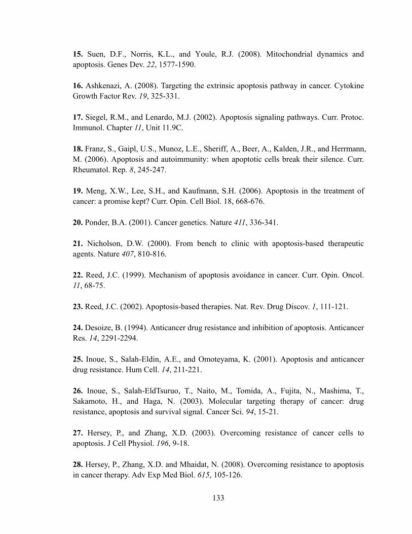
133
15. Suen, D.F., Norris, K.L., and Youle, R.J. (2008). Mitochondrial dynamics and apoptosis. Genes Dev. 22, 1577-1590. 16. Ashkenazi, A. (2008). Targeting the extrinsic apoptosis pathway in cancer. Cytokine Growth Factor Rev. 19, 325-331. 17. Siegel, R.M., and Lenardo, M.J. (2002). Apoptosis signaling pathways. Curr. Protoc. Immunol. Chapter 11, Unit 11.9C. 18. Franz, S., Gaipl, U.S., Munoz, L.E., Sheriff, A., Beer, A., Kalden, J.R., and Herrmann, M. (2006). Apoptosis and autoimmunity: when apoptotic cells break their silence. Curr. Rheumatol. Rep. 8, 245-247. 19. Meng, X.W., Lee, S.H., and Kaufmann, S.H. (2006). Apoptosis in the treatment of cancer: a promise kept? Curr. Opin. Cell Biol. 18, 668-676. 20. Ponder, B.A. (2001). Cancer genetics. Nature 411, 336-341. 21. Nicholson, D.W. (2000). From bench to clinic with apoptosis-based therapeutic agents. Nature 407, 810-816. 22. Reed, J.C. (1999). Mechanism of apoptosis avoidance in cancer. Curr. Opin. Oncol. 11, 68-75. 23. Reed, J.C. (2002). Apoptosis-based therapies. Nat. Rev. Drug Discov. 1, 111-121. 24. Desoize, B. (1994). Anticancer drug resistance and inhibition of apoptosis. Anticancer Res. 14, 2291-2294. 25. Inoue, S., Salah-Eldin, A.E., and Omoteyama, K. (2001). Apoptosis and anticancer drug resistance. Hum Cell. 14, 211-221. 26. Inoue, S., Salah-EldTsuruo, T., Naito, M., Tomida, A., Fujita, N., Mashima, T., Sakamoto, H., and Haga, N. (2003). Molecular targeting therapy of cancer: drug resistance, apoptosis and survival signal. Cancer Sci. 94, 15-21. 27. Hersey, P., and Zhang, X.D. (2003). Overcoming resistance of cancer cells to apoptosis. J Cell Physiol. 196, 9-18. 28. Hersey, P., Zhang, X.D. and Mhaidat, N. (2008). Overcoming resistance to apoptosis in cancer therapy. Adv Exp Med Biol. 615, 105-126.

134
29. Hickman J.A. and Dive C. (1999). Apoptosis and cancer chemotherapy. Humana Press Inc. 30. Studziniski, G..P. (1999). Apoptosis, a practical approach. Oxford University Press. 31. Green, D.R. (1998). Apoptotic pathways: the roads to ruin. Cell 94, 695-698. 32. Cameron R.G. and Feuer G.. (2000). Apoptosis and its modulation by drugs. Springer. 33. Evan, G.I. and Vousden, K.H. (2001). Proliferation, cell cycle and apoptosis in cancer. Nature 411, 342-348. 34. Green, D.G. and Evan G..I. (2002). A matter of life and death. Cancer Cell 1, 19-30. 35. Hunter, A.M., LaCasse E.C., and Korneluk R.G. (2007). The inhibitors of apoptosis (IAPs) as cancer targets. Apoptosis 12, 1543-1568. 36. Fulda, S. (2008). Targeting inhibitor of apoptosis proteins (IAPs) for cancer therapy. Anticancer Agents Med Chem. 8, 533-539. 37. Srinivasula, S.M., and Ashwell, J.D. (2008). IAPs: what's in a name? Mol. Cell 30, 123-135. 38. Vucic, D. (2008). Targeting IAP (inhibitor of apoptosis) proteins for therapeutic intervention in tumors. Curr. Cancer Drug Targets 8, 110-117. 39. Danson, S., Dean, E., Dive, C., and Ranson, M. (2007). IAPs as a target for anticancer therapy. Curr. Cancer Drug Targets 7, 785-794. 40. Mor, G., Montagna, M.K., and Alvero, A.B. (2008). Modulation of apoptosis to reverse chemoresistance. Methods Mol. Biol. 414, 1-12. 41. Dean, E.J., Ranson, M., Blackhall, F., and Dive, C. (2007). X-linked inhibitor of apoptosis protein as a therapeutic target. Expert Opin. Ther. Targets 11, 1459-1471. 42. Vucic, D., and Fairbrother, W.J. (2007). The inhibitor of apoptosis proteins as therapeutic targets in cancer. Clin. Cancer Res. 13, 5995-6000. 43. Rajapakse, H.A. (2007). Small molecule inhibitors of the XIAP protein-protein interaction. Curr. Top. Med. Chem. 7, 966-971.

135
44. Kischkel F.C., Helbardt, S., Behrmann I. Germer, M., Pawlita, M., Krammer, P.H., and Peter, M.E. (1995). Cytotoxicity-dependent APO-1(Fas/CD95)-associated proteins form a death-inducing signalling complex (DISC) with the receptor. EMBO J. 14, 5579-5588. 45. Fulda, S., and Debatin, K.M. (2006). Extrinsic versus intrinsic apoptosis pathways in anticancer chemotherapy. Oncogene 25, 4798-4811. 46. Kang, J.J., Schaber, M.D., Srinivasula, S.M., Alnemri, E.S., Litwack, G., Hall, D.J., and Bjornsti, M.A. (1999). Cascades of Mammalian Caspase Activation in the Yeast Saccharomyces cerevisiae. J. Biol. Chem. 274, 3189-3198. 47. Stennicke, H.R., Jurgensmeier, J.M., Shin, H., Deveraux, Q., Wolf, B.B., Yang, X., Zhou, Q., Ellerby, H.M., Bredesen, D., Green, D.R., Reed, J.C., Froelich, C.J., and Salvasen, G.S. (1998) Pro-casepase-3 is a major physiologic target of caspase-8. J. Biol. Chem. 273, 27084-27090. 48. Salvesen, G.S., and Dixit, V.M. (1999). Caspase activation: the induced-proximity model. Proc. Nat. Acad. Sci. USA. 96, 10964-10967. 49. Frisch, S.M. (2008). Caspase-8: fly or die. Cancer Res. 68, 4491-4493. 50. Salvesen, G.S., and Riedl, S.J. (2008). Caspase mechanisms. Adv. Exp. Med. Biol. 615, 13-23. 51. Denault, J.B., and Salvesen, G.S. (2008). Apoptotic caspase activation and activity. Methods Mol. Biol. 414, 191-220. 52. Timmer, J.C., and Salvesen, G.S. (2007). Caspase substrates. Cell Death Differ. 14, 66-72. 53. Kumar, S. (2007). Caspase function in programmed cell death. Cell Death Differ. 14, 32-43. 54. Callus, B.A., and Vaux, D.L. (2007). Caspase inhibitors: viral, cellular and chemical. Cell Death Differ. 14, 73-78. 55. Linton, S.D. (2005). Caspase inhibitors: a pharmaceutical industry perspective. Curr. Top. Med. Chem. 5, 1697-1717.

136
56. Wang, Z.B., Liu, Y.Q., and Cui, Y.F. (2005). Pathways to caspase activation. Cell Biol. Int. 29, 489-496. 57. Riedl, S.J., and Shi, Y. (2005). Molecular mechanisms of caspase regulation during apoptosis. Nat. Rev. Mol. Cell Biol. 5, 897-907. 58. Shi, Y. (2004). Caspase activation, inhibition, and reactivation: a mechanistic view. Protein Sci. 13, 1979-1987. 59. Shi, Y. (2004). Caspase activation: revisiting the induced proximity model. Cell 117, 855-858. 60. Boatright, K.M., and Salvesen, G.S. (2003). Mechanisms of caspase activation. Curr. Opin. Cell Biol. 15, 725-731. 61. Creagh, E.M., Conroy. H., and Martin, S.J. (2003). Caspase-activation pathways in apoptosis and immunity. Immunol Rev. 193, 10-21. 62. Shi, Y. (2002). Mechanisms of caspase activation and inhibition during apoptosis. Mol. Cell 9, 459-470. 63. Kuida, K. (2000). Caspase-9. Int. J. Biochem. Cell Biol. 32, 121-124. 64. Takahashi, A. (1999). Caspase: executioner and undertaker of apoptosis. Int. J. Hematol. 70, 226-232. 65. Ekert, P.G., Silke, J., and Vaux, D.L. (1999). Caspase inhibitors. Cell Death Differ. 6, 1081-1086. 66. Kumar, S. (1999). Mechanisms mediating caspase activation in cell death. Cell Death Differ. 6, 1060-1066. 67. Villa P, Kaufmann SH, Earnshaw WC. (1997). Caspases and caspase inhibitors. Trends Biochem. Sci. 22, 388-393. 68. Kluck, R.M., Bossey-Wetzel E., Green, D.R., and Newmeyer D.D. (1997). The release of cytochrome c from mitochondria: a primary site for bcl-2 regulation of apoptosis. Science 275, 1132-1136. 69. Ow, Y.L., Green, D.R., Hao, Z., and Mak, T.W. (2008). Cytochrome c: functions beyond respiration. Nat. Rev. Mol. Cell Biol. 9, 532-542.

137
70. Hacker, G., and Weber, A. (2007). BH3-only proteins trigger cytochrome c release, but how? Arch. Biochem. Biophys. 15, 150-155. 71. Garrido, C., Galluzzi, L., Brunet, M., Puig, P.E., Didelot, C., and Kroemer, G. (2006). Mechanisms of cytochrome c release from mitochondria. Cell Death Differ. 136, 1423-1433. 72. Bertini I, Cavallaro G, Rosato A. (2006). Cytochrome c: occurrence and functions. Chem. Rev. 106, 90-115. 73. Jiang, X., and Wang, X. (2004). Cytochrome C-mediated apoptosis. Annu. Rev. Biochem. 73, 87-106. 74. Scorrano, L., and Korsmeyer, S.J. (2003). Mechanisms of cytochrome c release by proapoptotic BCL-2 family members. Biochem. Biophys. Res. Commun. 304, 437-444. 75. Saleh, A., Srinivasula, S. Acharya, S., Fishel, R., Alnemri, E.S. (1999). Cytochrome c and dATP-mediated oligomerization of Apaf-1 is a prerequisite for procaspase-9 activation. J. Biol. Chem. 274, 17941-17945. 76. Zou, H., Henzel, W.J., Liu, X., Lutschg, A., Wang, X. (1997). Apaf-1, a human protein homologous to C. elegans CED-4, participates in cytochrome c–dependent activation of caspase-3. Cell 90, 405-413. 77. Zou, H., Li, Y., Liu, X., Wang, X. (1999). An Apaf-1 cytochrome c multimeric complex is a functional apoptosome that activates procaspase-9. J. Biol. Chem. 274, 11549-11556. 78. Li, H., Zhu, H., Xu, C., amd Yuan, J. (1998). Cleavage of Bid by caspase-8 mediates the mitochondrial damage in the Fas pathway of apoptosis. Cell 94, 491-501. 79. Luo X., Budihardjo, I., Zou, H., Slaughter, C., and Wang, X. (1998). Bid, a Bcl-2 interacting protein, mediates cytochrome c release from mitochondria in response to activation of cell surface death receptors. Cell 94, 481-490. 80. Chipuk, J.E., and Green, D.R. (2008). How do BCL-2 proteins induce mitochondrial outer membrane permeabilization? Trends Cell Biol. 18, 157-164. 81. Danial, N.N. (2007). BCL-2 family proteins: critical checkpoints of apoptotic cell death. Clin. Cancer Res. 13, 7254-7263.

138
82. Brooks, C., and Dong, Z. (2007). Regulation of mitochondrial morphological dynamics during apoptosis by Bcl-2 family proteins: a key in Bak? Cell Cycle 6, 3043-3047. 83. Adams, J.M., and Cory, S. (2007). Bcl-2-regulated apoptosis: mechanism and therapeutic potential. Curr. Opin. Immunol. 19, 488-496. 84. Adams, J.M., and Cory, S. (2007). The Bcl-2 apoptotic switch in cancer development and therapy.Oncogene 26, 1324-1337. 85. Reed, J.C. (2006). Proapoptotic multidomain Bcl-2/Bax-family proteins: mechanisms, physiological roles, and therapeutic opportunities. Cell Death Differ. 13, 1378-1386. 86. Devraux, Q.L., and Reed, J.C. (1999). IAP family proteins suppressors of apoptosis. Genes Dev. 13, 239-252. 87. Salvesen, G.S., and Duckett, C.S. (2002). Apoptosis: IAP proteins: blocking the road to death's door. Nat. Rev. Mol. Cell Biol. 3, 401-410. 88. Holick, M., Gibson, H., and Korneluk, R.G. (2001). XIAP: apoptotic brake and promising therapeutic target. Apoptosis 6, 253-261. 89. Vucic, D. (2008). Targeting IAP (inhibitor of apoptosis) proteins for therapeutic intervention in tumors.Curr. Cancer Drug Target 8, 110-117. 90. Martin, S.J. (2002). Destabilizing influences in apoptosis: sowing the seeds of IAP destruction. Cell 109, 793-796. 91. Verhagen, A.M., and Vaux, D.L. (2002). Cell death regulation by the mammalian IAP antagonist Diablo/Smac. Apoptosis 7, 163-166. 92. Yang, Y.L., and Li, X.M. (2000). The IAP family: endogenous caspase inhibitors with multiple biological activities. Cell Res. 10, 169-177. 93. Eiben, L.J., and Duckett, C.S. (1998). The IAP family of apoptotic regulators. Results Probl Cell Differ. 24, 91-104. 94. Yang Y., Fang, S., Jensen, J.P., Weissman, A.M., and Ashwell, J.D. (2000). Ubiquitin protein ligase activity of IAPs and their degradation in proteasomes in response to apoptotic stimuli. Science 288, 874-877.

139
95. Silke, J., Kratina, T., Chu, D., Ekert, P.G., Day, C.L., Pakusch, M., Huang, D.C., and Vaux, D.L. (2005). Determination of cell survival by RING-mediated regulation of inhibitor of apoptosis (IAP) protein abundance. Proc. Nat. Acad. Sci. USA 102, 16182-16187. 96. Martin, S. J. (2001). Dealing with CARDs between life and death. Trends Cell. Biol. 11, 188-189. 97. Bouchier-Hayes, L., and Martin, S.J. (2002). CARD games in apoptosis and immunity. EMBO 3, 616-621. 98. Verhagen, A.M., Coulson, E.J., and Vaux, D.L. (2001). IAPs and their relatives: IAPs and other BIRPs. Genome Biology 2, 3009.1-3009.10. 99. Eckelman, B.P., Drag, M., Snipas, S.J., and Salvesen, G.S. (2008). The mechanism of peptide-binding specificity of IAP BIR domains. Cell Death Differ. 15, 920-928. 100. Samuel, T., Welsh, K., Lober, T., Togo, S.H., Zapata, J.M., and Reed, J.C. (2006). Distinct BIR domains of cIAP1 mediate binding to and ubiquitination of tumor necrosis factor receptor-associated factor 2 and second mitochondrial activator of caspases. J. Biol. Chem. 281, 1080-1090. 101. Shin, H., Renatus, M., Eckelman, B.P., Nunes, V.A., Sampaio, C.A., and Salvesen, G.S. (2005). The BIR domain of IAP-like protein 2 is conformationally unstable: implications for caspase inhibition.Biochem. J.385, 1-10. 102. Huang,Y., Rich, R.L., Myszka, D.G., and Wu, H. (2003). Requirement of both the second and third BIR domains for the relief of X-linked inhibitor of apoptosis protein (XIAP)-mediated caspase inhibition by Smac.J. Biol. Chem. 278, 49517-49522 103. Miller, L.K. (1999). An exegesis of IAPs: salvation and surprises from BIR motifs.Trends Cell Biol. 9, 323-328. 104. Lacasse, E.C., Barid, S., Korneluk, R.G., MacKenzie, A.E., (1998). The inhibitors of apoptosis (IAPs) and their emerging role in cancer. Oncogene 17, 3247-3259. 105. Altieri, D.C. (2008). Survivin, cancer networks and pathway-directed drug discovery.Nat. Rev. Cancer. 8, 61-70. 106. Duffy, M.J., O'Donovan, N., Brennan, D.J., Gallagher, W.M., and Ryan, B.M. (2007). Survivin: a promising tumor biomarker.Cancer Lett. 249, 49-60.

140
107. Lens, S.M., Vader, G., and Medema, R.H. (2006). The case for Survivin as mitotic regulator.Curr. Opin. Cell Biol. 18, 616-622. 108. Li, F., and Ling, X. (2006). Survivin study: an update of "what is the next wave"? J. Cell Physiol. 208, 476-486. 109. Li, F. (2003). Survivin study: what is the next wave? J. Cell Physiol. 197, 8-29. 110. Altieri, D.C. (2003). Survivin in apoptosis control and cell cycle regulation in cancer. Prog. Cell Cycle Res. 5, 447-452. 111. Pohl, C., and Jentsch, S. (2008). Final stages of cytokinesis and midbody ring formation are controlled by BRUCE.Cell 132, 832-845. 112. Takahashi, R., Deveraux, Q.L., Tamm, I., Welsh, K., Assa-Munt, N., Salvesen, G.S., and Reed, J.C. (1998). A single BIR domain of XIAP sufficient for inhibiting caspases. J. Biol. Chem. 273, 7787-7790. 113. Asselin, E., Mills, G.B., and Tsang, B.K. (2001). XIAP regulates Akt activity and caspase-3-dependent cleavage during cisplatin-induced apoptosis in human ovarian epithelial cancer cells. Cancer Res. 61, 1862-1868. 114. Deveraux, Q.L., Tahashi, R., Salvesen G.S., and Reed, J.C. (1997) X-linked IAP is a direct inhibitor of cell-death proteases. Nature 388, 300-304. 115. Deveraux, Q.L., Leo, E., Stennicke, H.R., Welsh, K., Salvesen, G.S. and Reed, J.C. (1999). Cleavage of human inhibitor of apoptosis protein XIAP results in fragments with distinct specificities for caspases. EMBO J. 18, 5242-5251. 116. Rajapakse, H.A. (2007). Small molecule inhibitors of the XIAP protein-protein interaction.Curr Top Med Chem. 7, 966-971. 117. Eckelman, B.P., Salvesen, G.S., and Scott, F.L. (2007). Human inhibitor of apoptosis proteins: why XIAP is the black sheep of the family.EMBO Rep.7, 988-994. 118. Schimmer, A.D., Dalili, S., Batey, R.A., and Riedl, S.J. (2006). Targeting XIAP for the treatment of malignancy.Cell Death Differ. 13, 179-188. 119. Du, C., Fang, M., Li, Y., Li, L., and Wang, X. (2000). Smac, a mitochondrial protein that promotes cytochrome c–dependent caspase activation by eliminating IAP inhibition. Cell 102, 33-42.

141
120. Verhagen, A.M., Kkert, P.G., Pakusch, M., Silke, J., Connolly, L.M., Reid, G.E., Moritz, R.L., Simpson, R.J., and Vaux, D.L. (2000). Identification of DIABLO, a mammalian protein that promotes apoptosis by binding to and antagonizing IAP proteins. Cell 102, 43-53. 121. Ekert, P.G., Silke, J., Hawkins, C.J., Verhagen, A.M., and Vaux, D.L. (2001). DIABLO promotes apoptosis by removing MIHA/XIAP from processed caspase-9. J. Cell Biol. 152, 483-490. 122. Liu, Z., Sun, C., Olejniczak, E.T., Meadows, R.P., Betz, S.F., Oost, T., Herrmann, J., Wu, J.C., and Feisk, S.K. (2000). Structural basis for binding of Smac/DIABLO to the XIAP BIR3 domain. Nature 408, 1004-1008. 123. Wu, G., Chai, J. Suber, T.L., Wu, J.W., Du, C., Wang, X., and Shi, Y. (2000). Structural basis of IAP recognition by Smac/DIABLO. Nature 408, 1008-1012. 124. Sun, C., Cai, M., Meadows, R.P., Xu, N., Gunasekera, A.H., Hermann, J., Wu, J.C., and Fesik, S.W. (2000). NMR structure and mutagenesis of the third BIR domain of the inhibitor of apoptosis protein XIAP. J. Biol. Chem. 275, 33777-33781. 125. Sun, H., Nikolovska-Coleska, Z., Chen, J., Yang, C.-Y., Tomita, Y., Pan H., Yoshioka, Y., Krajewski, K., Roller, P.P., and Wang, S. (2005). Bioorg. Med. Chem. Lett. 15, 793-797. 126. Nikolovska-Coleska, Z., Wang, R., Fang, X., Pan, H., Tomita, Y., Li, P., Roller, P.P., Krajewski, K., Saito, N.G., Stuckey, J.A., and Wang, S. (2004). Development and optimization of a binding assay for the XIAP BIR3 domain using fluorescence polarization. Anal. Biochem. 332, 261-273. 127. Sun, H., Nikolovska-Coleska, Z., Yang, C.-Y., Xu, L., Liu, M., Tomita, Y., Pan, H., Yoshioka, Y., Krajewski, K., Roller, P.P., and Wang, S. (2004). Structure-based design of potent, conformationally constrained Smac mimetics. J. Am. Chem. Soc. 126, 16686-16687. 128. Sun, H., Nikolovska-Coleska, Z., Yang, C.-Y., Xu, L., Tomita, Y., Krajewski, K., Roller, P.P., and Wang, S. (2004). Structure-based design, synthesis, and evaluation of conformationally constrained mimetics of the second mitochondria-derived activator of caspase that target the X-linked inhibitor of apoptosis protein / caspase-9 interaction site. J. Med. Chem. 47, 4147-4150.

142
129. Sun, H. Nikolovska-Coleska, Z., Lu, J., Qiu, S., Yang, C.-Y., Gao, W., Meagher, J., Stuckey, J., and Wang, S. (2006). Design, synthesis, and evaluation of a potent, cell-permeable, conformationally constrained second mitochondria derived activator of caspase (Smac) mimetic. J. Med. Chem. 49, 7916-7920. 130. Oost, T.K., Sun, C., Armstrong, R.C., Al-Assaad, A.S., Betz, S.F., Deckwerth, T.L., Ding, H., Elmore, S.W., Meadows, R.P., Olejniczak, E.T., Oleksijew, A., Oltersdorf, T., Roseberg, S.H., Shoemaker, A.R., Tomaselli, K.J., Zou, H., and Fesik, S.W. (2004). Discovery of potent antagonists of the antipoptotic protein XIAP for the treatment of cancer. J. Med. Chem. 47, 4417-4426. 131. Shi, Y. (2001). The Mechanisms of caspase activation and inhibition during apoptosis. Mol. Cell 9, 459-470. 132. Huang Y., Park, Y.C., Rich, R.L., Segal, D., Myszka, D.G., and Wu, H. (2001). Structure basis of caspase inhibition by XIAP: differential roles of the linker versus the BIR domain. Cell 104, 781-790. 133. Shiozaki, E.N., and Shi, Y. (2004). Caspases, IAPs and Smac/DIABLO: mechanism from structural biology. Trends Biochem. Sci. 29, 486-494. 134. Li, L., Thomas, R.M., Suzuki, H., Brabander, J.K.D., Wang, X., and Harran, P.G. (2004). A small molecule Smac mimic potentiates TRAIL- and TNFα-mediated cell death. Science 305, 1471-1474. 135. Sun, H. Nikolovska-Coleska, Z., Lu, J., Meagher, J., Yang, C.-Y., Qiu, Su., Tomita, Y., Jiang, S., Krajewski, K., Roller, P.P., Stuckey, J.A., and Wang, S. (2007). Design, synthesis, and characterization of a potent, nonpeptide, cell-permeable, bivalent Smac mimetic that concurrently targets both the BIR2 and BIR3 domains in XIAP. J. Am. Chem. Soc. 129, 15279-15294. 136. Kolb, H.C., Sharpless, K.B. (2003). The growing impact of click chemistry on drug discovery. Drug Discov Today 8, 1128-1137. 137. Rostovstev, V.V., Green, L.G., Fokin, V.V., and Sharpless, K.B. (2002). A stepwise Huisgen cycloaddition process: copper(I)-catalyzed regioselective ligation of azides and terminal alkynes. Angew. Chem. Int. Ed. 41, 2596-2599. 138. Moses, J.E., and Moorhouse, A.D. (2007). The growing applications of click chemistry. Chem. Soc. Rev. 36, 1249-1262.

143
139. Tron, G.C., Pirali, T., Billington, R.A., Canonico, P.L., Sorba, G., and Genazzani, A.A. (2008). Click chemistry reactions in medicinal chemistry: applications of the 1,3-dipolar cycloaddition between azides and alkynes. Med. Res. Rev. 28, 278-308. 140. Moorhouse, A.D., and Moses, J.E. (2008). Click chemistry and medicinal chemistry: a case of "cyclo-addiction". ChemMedChem 3, 715-723. 141. Hein, C.D., Liu, X.M., and Wang, D. (2008). Click Chemistry, A Powerful Tool for Pharmaceutical Sciences. Pharm. Res. 25, 2216-2230. 142. Service, R.F. (2008). Chemistry. Click chemistry clicks along. Science 320, 868-869. 143. Petersen, J.S., Fels, G., and Rapoport, H. (1984). Chirospecific syntheses of (+)- and (-)-Anatoxin α. J. Am. Chem. Soc. 106, 4539-4547. 144. Campcell, J.A. and Rapoport, H. (1996). Chirospecific syntheses of conformationally constrained 7-azabicycloheptane amino acids by transannular alkylation. J. Org. Chem. 61, 6313-6325. 145. Aggarwal, V.K., Astle, C.J., Christopher, J.I., Iding, H., Wirz, B., and Rogers-Evans, M. (2005). Separation of pyrrolidine allylation products by diastereoselective enzymatic ester hydrolysis. Tetrahedron Lett. 46, 945-947. 146. Shinada, T., Hamada, M., Kawasaki, M., and Ohfune, Y. (2005). Stereoselective synthesis of 2,5-Di- and 2,2,5-trisubstituted pyrrolidines by allylation reaction of acyliminium ion. Heterocycles 66, 511-525. 147. Colombo, L., Di Giacomo, M., Vinci, V., Colombo, M., Manzoni, L., and Scolastico, C. (2003). Synthesis of new bicyclic lactam peptidomimetics by ring-closing metathesis reactions. Tetrahedron 59, 4501-4513. 148. Grossmith, C.E., Senia, F., and Wagner, J. (1999). Synthesis of novel unsaturated bicyclic lactams by ring-closing metathesis. Synlett 10, 1660-1662. 149. Park, C.-M., Sun, C., Olejniczak, E.T., Wilson, A.E., Meadows, R.P., Betz, S.F., Elmore, S.W., and Fesik, S.W. (2005). Non-peptidic small molecular inhibitors of XIAP. Bioorg. Med. Chem. Lett. 15, 771-775.

144
150. Peng, Y., Sun, H., and Wang, S. (2006). Design and synthesis of a 1,5- diazabicyclo[6,3,0] dodecane amino acid derivative as a novel dipeptide reverse-turn mimetic. Tetrahedron Lett. 47, 4769-4770. 151. Chaudhary, S.K., and Hernandez, O. (1979). 4-dimethylaminopyridine: an efficient and selective catalyst for the silylation of alcohols. Tetrahedron Lett. 20, 99-102 152. Debenham, J.S., Rodebaugh, R., and Fraser-Reid, B. (1997). TCP- and phthalimide- protected n-pentenyl glucosaminide precursors for the synthesis of nodulation factors as illustrated by the total synthesis of NodRf-III (C18:1, MeFuc). J. Org. Chem. 62, 4591-4600. 153. Boger, D.L., Borzilleri, R.M., Nukui, S., Beresis, R.T. (1997). Synthesis of the Vancomycin CD and DE ring systems. J. Org. Chem. 62, 4721-4736. 154. Scheidt, K.A., Chen, H., Follows, B.C., Chemler, S.R., Coffey, D.S., Roush, W.R. (1998). Tris(dimethylamino)sulfonium difluorotrimethylsilicate, a mild reagent for the removal of silicon protecting groups. J. Org. Chem. 63, 6436-6437. 155. Bodanszky, M., and Bodanszky, A. (1994). The practice of peptide Synthesis 2nd edition. Springer-Verlag. Chapter 2. 156. Gibson, F.S., Bergmeier, S.C., and Rapoport, H. (1994). Selective removal of an N-Boc protecting group in the presence of a tert-butyl ester and other acid-sensitive groups. J. Org. Chem. 59, 3216-3218. 157. Atwell, G.J., and Denny, W.A. (1984). Monoprotection of α,ω-alkanediamines with the N-benzyloxycarbonyl group. Synthesis 12, 1032-1033. 158. Berkowitz, D.B., and Pedersen, M.L. (1994). Simultaneous amino and carboxyl group protection for .alpha.-branched amino acids. J. Org. Chem. 59, 5476-5478. 159. Stahl, G.L., Walter, R., and Smith, C.W. (1978). General procedure for the synthesis of mono-N-acylated 1,6-diaminohexanes. J. Org. Chem. 43, 2286-2286. 160. Cavelier, F. and Enjalbal, C. (1996). Studies of selective Boc removal in the presence of silyl ethers. Tetrahedron Lett. 37, 5131-5134. 161. Evans, D.A., and Ellman, J.A. (1989). The total syntheses of the isodityrosine-derived cyclic tripeptides OF4949-III and K-13. Determination of the absolute configuration of K-13. J. Am. Chem. Soc. 111, 1063-1072.

145
162. Sakaitani, M., Hori, K., and Ohfune Y. (1988). One-pot conversion of N-benzyloxycarbonyl group into N-tert-butoxycarbonyl group. Tetrahedron Lett. 29, 2983-2984. 163. Mazaleyrat, J.-P., Xie, J., and Wakselman, M. (1992). Selective hydrogenolysis of the benzyloxycarbonyl protecting group of Nε-lysine in cyclopeptides containing a benzylic phenyl ether function. Evidence for Nε-methylated lysine side products. Tetrahedron Lett. 33, 4301-4302. 164. Yamazaki, N., and Kibayashi, C. (1989). Asymmetric synthesis with α,β.-bis [(methoxymethyl)oxy] ketones. Enantioselective total synthesis of natural (+)- indolizidine 195B (bicyclic gephyrotoxin 195B) and (-)-pinidine and their enantiomers from a common chiral synthon. J. Am. Chem. Soc. 111, 1396-1408. 165. Gary, B.D., and Jeffs, P.W. (1987). Alkylation and condensation reactions of N,N-dibenzylglycine esters: synthesis of α-amino acid derivatives. J. Chem. Soc. Chem. Commun. 18, 1329-1330. 166. Peng, Y., Sun, H., Nikolovska-Coleska, Z., Yang, C.-Y., Qiu, S., Lu, J., Cai, Q., Yi, H., and Wang, S. (2008). Design, synthesis and evaluation of potent and orally bioavailable diazabicyclic Smac mimetics. Submitted to J. Med. Chem. 167. Peskin, A.V. and Winterbourn, C.C. (2000). A microtiter plate assay for superoxide dismutase using a water-soluble tetrazolium salt (WST-1). Clinnica Chimica Acta 293, 157-166. 168. Berridge, M.V., Herst, P.M., and Tan A.S. (2005). Tetrazolium dyes as tools in cell biology: new insights into their cellular reduction. Biotechnol. Annu. Rev. 11, 127-152. 169. Sulic, S., Panic, L., Dikic, I., and Volarevic, S. (2005). Deregulation of cell growth and malignant transformation. Croat. Med. J. 46, 622-638. 170. Herzenberg, L.A., and DeRosa, S.C. (2000). Monoclonal antibodies and the FACS: complementary tools for immunobiology and medicine. Immunol. Today 21, 383-390. 171. Tung, J.W., Parks, D.R., Moore, W.A., and Herzenberg, L.A. (2004). New approaches to fluorescence compensation and visualization of FACS data. Clin. Immunol. 110, 277-283.

146
172. Diaz, D., Prieto, A., Reyes, E., Barcenilla, H., Monserrat, J., and Alvarez-Mon, M. (2008). Flow cytometry enumeration of apoptotic cancer cells by apoptotic rate. Methods Mol. Biol. 414, 23-33. 173. Ibrahim, S.F., and Van Den Engh, G. (2007). Flow cytometry and cell sorting. Adv. Biochem. Eng. Biotechnol. 106, 19-39. 174. Tung, J.W., Heydari, K., Tirouvanziam, R., Sahaf, B., Parks, D.R., and Herzenberg, L.A. (2007). Modern flow cytometry: a practical approach. Clin. Lab. Med. 27, 453-468. 175. Sklar, L.A., Carter, M.B., and Edwards, B.S. (2007). Flow cytometry for drug discovery, receptor pharmacology and high-throughput screening. Curr. Opin. Pharmacol. 7, 527-534. 176. Delude, R.L. (2005). Flow cytometry. Crit. Care Med. 33, 426-428. 177. Carroll, S., and Al-Rubeai, M. (2004). The selection of high-producing cell lines using flow cytometry and cell sorting. Expert Opin. Biol. Ther. 4, 1821-1829. 178. Givan, A.L. (2004). Flow cytometry: an introduction. Methods Mol. Biol. 263, 1-32. 179. Cram, L.S. (2002). Flow cytometry, an overview. Methods Cell Sci. 24, 1-9. 180. McCoy, J.P. Jr. (2002). Basic principles of flow cytometry. Hematol. Oncol. Clin. North Am. 16, 229-243. 181. Ormerod, M.G. (2001). Using flow cytometry to follow the apoptotic cascade. Redox. Rep. 6, 275-287. 182. Darzynkiewicz, Z., Smolewski, P., and Bedner, E. (2001). Use of flow and laser scanning cytometry to study mechanisms regulating cell cycle and controlling cell death. Clin. Lab Med. 21, 857-873. 183. Schwartz, A., and Fernandez-Repollet, E. (2001). Quantitative flow cytometry. Clin. Lab Med. 21, 743-761. 184. O'Connor, J.E., Callaghan, R.C., Escudero, M., Herrera, G., Martinez, A., Monteiro, M.D., and Montoliu, H. (2001). IUBMB Life 51, 231-239. 185. Rieseberg, M., Kasper, C., Reardon, K.F., and Scheper, T. (2001). Flow cytometry in biotechnology. Appl. Microbiol. Biotechnol. 56, 350-360.

147
186. Boddy, L., Wilkins, M.F., and Morris, C.W. (2001). Pattern recognition in flow cytometry. Cytometry 44, 195-209. 187. Darzynkiewicz, Z., Bedner, E., and Smolewski, P. (2001). Flow cytometry in analysis of cell cycle and apoptosis. Semin. Hematol. 38, 179-193. 188. Marti, G.E., Stetler-Stevenson, M., Bleesing, J.J., Fleisher, T.A. (2001). Introduction to flow cytometry. 38, 93-99. 189. Scheffold, A., and Kern, F. (2001). Recent developments in flow cytometry. J. Clin. Immunol. 20, 400-407. 190. Givan AL. (2001). Principles of flow cytometry: an overview. Methods Cell Biol. 63, 19-50. 191. Vermes, I., Haanen, C., and Reutelingsperger, C. (2000). Flow cytometry of apoptotic cell death. J. Immunol. Methods 21, 167-190. 192. Varfolomeev, E., Blankenship, J.W., Wayson, S.M., Fedorava, A.V., Kayagaki, N., Garg, P., Zobel, K., Dynek, J.N., Elliott, L.O., Wallweber, H.J.A., Flygare, J.A., Fairbrother, W.J., Deshayes, K., Dixit, V.M., and Vucic, D. (2007). IAP antagonists induced autoubiquitination of c-IAPs, NF-κB activation, and TNFα-dependent apoptosis. Cell 131, 669-681. 193. Vince, J.E., Wong, W.W.-L., Khan, N., Feltham, R., Chau, D., Ahmed, A.U., Benetatos, C.A., Chunduru, S.K., Condon, S.M., McKinlay, M., Brink, R., Leverkus, M., Tergaokar, V., Schneider, P., Callus, B.A., Koentgen, F., Vaux, D.L., and Silke J. (2007). IAP antagonists target cIAP1 to induce TNFα-dependent apoptosis. Cell 131, 682-693. 194. Petersen, S.L., Wang, L., Yalcin-Chin, A., Li, L., Peyton, M., Minna, J., Harran, P., and Wang, X. (2007). Autocrine TNFα signaling renders human cancer cells susceptible to Smac-mimetic-induced apoptosis. Cancer Cell 12, 445-456. 195. Wang, L., Du, F., and Wang, X. (2008). TNF-α induces two distinct caspase-8 activation pathways. Cell 133, 693-703. 196. Varfolomeev, E., Goncharov, T., Fedorova, A.V., Dynek, J.N., Zobel, K., Deshayes, K., Fairbrother, W.J., and Vucic, D. (2008). c-IAP1 and c-IAP2 are critical mediators of tumor necrosis factor alpha (TNFalpha)-induced NF-kappaB activation. J. Biol. Chem. 283, 24295-24299.

148
197. Ikejima, M., Noguchi, S., Yamashita, R., Ogura, T., Sugimura, T., Gill, D.M., and Miwa, M. (1990). The zinc fingers of human poly(ADP-ribose) polymerase are differentially required for the recognition of DNA breaks and nicks and the consequent enzyme activation. Other structures recognize intact DNA. J. Biol. Chem. 265, 21907-21913. 198. Lazebnik, Y.A., Kaufmann, S.H., Desnoyers, S., Poirier, G.G., and Earnshaw, W.C. (1994). Cleavage of poly(ADP-ribose) polymerase by a proteinase with properties like ICE. Nature 371, 346-347. 199. Tewari, M., Quan, L.T., O'Rourke, K., Desnoyers, S., Zeng, Z., Beidler, D.R., Poirier, G.G., Salvesen, G.S., and Dixit, V.M. (1995). Yama/CPP32 beta, a mammalian homolog of CED-3, is a CrmA-inhibitable protease that cleaves the death substrate poly(ADP-ribose) polymerase. Cell 81, 801-809. 200. Kaufmann, S.H., Desnoyers, S., Ottaviano, Y., Davidson, N.E., and Poirier, G.G. (1993). Specific proteolytic cleavage of poly(ADP-ribose) polymerase: an early marker of chemotherapy-induced apoptosis. Cancer Res. 53, 3976-3985. 201. Duan, H., Chinnaiyan, A.M., Hudson, P.L., Wing, J.P., He, W.-W. and Dixit, V.M. (1996). ICE-LAP3, a novel mammalian homologue of the Caenorhabditis elegans cell death protein Ced-3 is activated during Fas- and tumor necrosis factor-induced apoptosis. J. Biol. Chem. 271, 1621-1625. 202. Srinivasula, S.M., Fernandes-Alnemri, T., Zangrilli, J., Robertson, N., Armstrong, R.C., Wang, L., Trapani, J.A., Tomaselli, K.J., Litwack, G. and Alnemri, E.S. (1996). The ced-3/interleukin 1 converting enzyme-like homolog Mch6 and the lamin-cleaving enzyme Mch2 are substrates for the apoptotic mediator CPP32. J. Biol. Chem. 271, 27099-27106. 203. Li, P., Nijhawan, D., Budihardjo, I., Srinivasula, S.M., Ahmad, M., Alnemri, E.S., and Wang, X. (2001). Cytochrome c and dATP-dependent formation of Apaf-1/caspase-9 complex initiates an apoptotic protease cascade. Cell 91, 479-489. 204. Liu, J.R., Opipari, A.W., Tan, L., Jiang, Y., Zhang, Y., Tang, H., and Nuñez, G. (2002). Dysfunctional apoptosome activation in ovarian cancer: implications for chemoresistance. Cancer Res. 62, 924-931.

149
205. Greene, B.T., Thorburn, J., Willingham, M.C., Thorburn, A., Planalp, R.P., Brechbiel, M.W., Jennings-Gee, J., Wilkinson, J., Torti, F.M., and Torti, S.V. (2002). Activation of caspase pathways during iron chelator-mediated apoptosis. J. Biol. Chem. 277, 25568-25575. 206. Degenhardt, K., Sundararajan, R., Lindsten, T., Thompson, C., and White, E. (2002). Bax and Bak independently promote cytochrome c release from mitochondria. J. Biol. Chem. 277, 14127-14134. 207. Liu, F.T., Kelsey, S.M., Newland, A.C., and Jia, L. (2002). Liposomal encapsulation diminishes daunorubicin-induced generation of reactive oxygen species, depletion of ATP and necrotic cell death in human leukaemic cells. Br. J. Haematol. 117, 333-342. 208. Muzio, M., Chinnaiyan, A.M., Kischkel, F.C., O'Rourke, K., Shevchenko, A., Ni, J., Scaffidi, C., Bretz, J.D., Zhang, M., Gentz, R., Mann, M., Krammer, P.H., Peter M.E., and Dixit, V.M. (1996). FLICE, a Novel FADD-homologous ICE/CED-3–like protease, is recruited to the CD95 (Fas/APO-1) death-inducing signaling complex. Cell 85, 817-827. 209. Boldin, M.P., Goncharov, T.M., Goltseve, Y.V., and Wallach D. (1996). Involvement of MACH, a novel MORT1/FADD-interacting protease, in Fas/APO-1- and TNF receptor–induced cell death. Cell 85, 803-815. 210. Fernandes-Alnemri, T., Armstrong, R.C., Krebs, J., Srinivasula, S.M., Wang, L., Bullrich, F., Fritz, L.C., Trapani, J.A., Tomaselli, K.J., Litwack, G., and Alnemri, E.S. (1996). In vitro activation of CPP32 and Mch3 by Mch4, a novel human apoptotic cysteine protease containing two FADD-like domains. Proc. Natl. Acad. Sci. USA 93, 7464-7469. 211. Mancini, M., Nicholson, D.W., Roy, S., Thornberry, N.A., Peterson, E.P., Casciola-Rosen, L.A., and Rosen, A. (1998). The caspase-3 precursor has a cytosolic and mitochondrial distribution: implications for apoptotic signaling. J. Cell Biol. 140, 1485-1495. 212. Samali, A., Cai, J., Zhivotovsky, B., Jones, D.P., and Orrenius, S. (1999). Presence of a pre-apoptotic complex of pro-caspase-3, Hsp60 and Hsp10 in the mitochondrial fraction of Jurkat cells. EMBO J. 18, 2040-2048. 213. Kirsch, D.G., Doseff, A., Chau, B.N., Lim, D.-S., Souza-Pinto, N.C., Hansford, R., Kastan, M.B., Lazebnik, Y.A., and Hardwick, J.M. (1999). Caspase-3-dependent cleavage of Bcl-2 promotes release of cytochrome c. J. Biol. Che,. 274, 21155-21161

150
214. Suria, H., Chau, L.A., Negrou, E., Kelvin D.J., and Madrenas, J. (1999). Cytoskeletal disruption induces T cell apoptosis by a caspase-3 mediated mechanism. Life Sci. 65, 2697-2707. 215. Faleiro, L., Kobayashi, R., Fearnhead H., and Lazebnik, Y. (1997). Multiple species of CPP32 and Mch2 are the major active caspases present in apoptotic cells. EMBO J. 16, 2271-2281. 216. Schafer, T., Scheuer, C., Roemer, K., Menger, M.D., and Vollmar, B. (2003). Inhibition of p53 protects liver tissue against endotoxin-induced apoptotic and necrotic cell death. FASEB J. 17, 660-667. 217. Jia, L., Patwari, Y., Kelsey, S.M., Srinivasula, S.M., Agrawal, S.G., Alnemri E.S., and Newland, A.C. (2003) Role of Smac in human leukaemic cell apoptosis and proliferation. Oncogene 22, 1589-1599. 218. Xiao, C., Yang, B.F., Asadi, N., Beguinot, F., and Hao, C. (2002). Tumor necrosis factor-related apoptosis-inducing ligand-induced death-inducing signaling complex and its modulation by c-FLIP and PED/PEA-15 in glioma cells. J. Biol. Che,. 277, 25020-25025. 219. Hoshi, T., Sasano, H., Kato, K., Yabuki, N., Ohara, S., Konno, R., Asaki, S., Toyota, T., Tateno, H., and Nagura, H. (1998). Immunohistochemistry of Caspase3/CPP32 in human stomach and its correlation with cell proliferation and apoptosis. Anticancer Res. 18, 4347-4353. 220. Fernandes-Alnemri, T., Takahashi, A., Armstrong, R., Krebs, J., Fritz, L., Tomaselli, K.J., Wang, L., Yu, Z., Croce, C.M., Salveson, G., Earnshaw, W.C., Litwack, G., and Alnemri, E.S. (1995). Mch3, a novel human apoptotic cysteine protease highly related to CPP32. Cancer Res. 55, 6045-6052. 221. Lippke, J.A., Gu, Y., Sarnecki, C., Caron, P.R., and Su, M.S.-S. (1996) Identification and characterization of CPP32/Mch2 homolog 1, a novel cysteine protease similar to CPP32. J. Biol. Chem. 271, 1825-1828. 222. Thornberry, N.A., Rano, T.A., Peterson, E.P., Rasper, D.M., Timkey, T., Garcia-Calvo, M., Houtzager, V.M., Nordstrom, P.A., Roy, S., Vaillancourt, J.P., Chapman, K.T., and Nicholson, D.W. (1997). A combinatorial approach defines specificities of members of the caspase family and granzyme B. J. Biol. Chem. 272, 17907-17911.

151
223. Chandler, J.M., Cohen, G.M., and MacFarlane, M. (1998). Different subcellular distribution of caspase-3 and caspase-7 following Fas-induced apoptosis in mouse liver. J. Biol. Chem. 273, 10815-10818. 224. MacFarlane, M., Cain, K., Sun, X.-M., Alnemri, E.S., and Cohen, G.M. (1997). Processing/activation of at least four interleukin-1 converting enzyme-like proteases occurs during the execution phase of apoptosis in human monocytic tumor cells. J. Cell Biol. 137, 469-479. 225. Cohen, G.M. (1997). Caspases: the executioners of apoptosis. Biochem J. 326, 1-16. 226. Nunez, G., Benedict, M.A., Hu, Y., and Inohara, N. (1998). Caspases: the proteases of the apoptotic pathway. Oncogene 17, 3237-3245. 227. Goyal, L. (2001). Cell death inhibition: keeping caspases in check. Cell 104, 805-808. 228. Roy, N., Deveraux, Q.L., Takahashi, R., Salvesen, G.S., and Reed, J.C. (1997). The c-IAP-1 and c-IAP-2 proteins are direct inhibitors of specific caspases. EMBO J. 16, 6914-6925. 229. Roy, N., Mahadevan, M.S., McLean, M., Shutler, G., Yaraghi, Z. Farahani, R., Baird, S., besner-Johnston, A., Lefebvre, C., and Kang, C. (1995). The gene for neuronal apoptosis inhibitory protein is partially deleted in individuals with spial muscular atrophy. Cell 80, 167-178. 230. Rothe, M., Pan, M.G., Henzel, W.J., Ayres, T.M., and Goeddel, D.V. (1995). The TNFR2-TRAF signaling complex contains two novel proteins related to baculoviral inhibitor of apoptosis proteins. Cell 83, 1243-1252. 231. Duckett, C.S., Nava, V.E., Gedrich, R.W., Clem, R.J., Van Dongen, J.L., Gilfillan, M.C., Shiels, H., Hardwick, J.M., and Thompson, C.B. (1996). A conserved family of cellular genes related to the baculovirus iap gene and encoding apoptosis inhibitors. EMBO J. 15, 2685-2694. 232. Lee, Y.-J., and Shacter, E. (2001). Fas aggregation does not correlate with Fas-mediated apoptosis. J. Immunol. 167, 82-89. 233. Reffey, S.B., Wurthner, J.U., Parks, W.T., Roberts, A.B., and Duckett, C.S. (2001). X-linked inhibitor of apoptosis protein functions as a cofactor in transforming growth factor-β signaling. J. Biol. Chem. 276, 26589-26596.

152
234. Wang, G.-Q., Gastman, B.R., Wieckowski, E., Goldstein, L.A., Rabinovitz, A., Yin, X.-M., and Rabinowich, H. (2001). Apoptosis-resistant mitochondria in T cells selected for resistance to Fas signaling. J. Biol. Chem. 276, 3650-3659. 235. Li, P., Nijhawan, D., Budihardjo, I., Srinivasula, S.M., Ahmad, M., Alnemri, E.S., and Wang, X. (1997). Cytochrome c and dATP-dependent formation of Apaf-1/caspase-9 complex initiates an apoptotic protease cascade. Cell 91, 479-489. 236. Schagger, H. (2002). Respiratory chain supercomplexes of mitochondria and bacteria. Biochem. Biophys. Acta. 1555, 154-159. 237. Liu, X., Kim, C.N., Yang, J., Jemmerson, R., and Wang, X. (1996). Induction of apoptotic program in cell-free extracts: requirement for dATP and cytochrome c. Cell 86, 147-157. 238. Sawyer, C., Sturge, J., Bennett, D.C., O'Hare, M.J., Allen, W.E., Bain, J., Jones, G.E., and Vanhaesebroeck, B. (2003). Regulation of breast cancer cell chemotaxis by the phosphoinositide 3-kinase p110delta. Cancer Res. 63, 1667-1675. 239. Lacor, P.N., Grayson, D.R., Auta, J., Sugaya, I., Costa, E., and Guidotti, A. (2000). Reelin secretion from glutamatergic neurons in culture is independent from neurotransmitter regulation. Proc. Natl. Acad. Sci. USA 97, 3556-3561. 240. Andree, H.A., Reutelingsperger, C.P., Hauptmann, R., Hemker, H.C., Hermens, W.T., and Willems, G.M. (1990). Binding of vascular anticoagulant alpha (VAC alpha) to planar phospholipid bilayers. J. Biol. Chem. 265, 4923-4928. 241. Fadok, V.A., Savill, J.S., Haslett, C., Bratton, D.L., Doherty, D.E., Campbell, P.A., and Henson, P.M. (1992). Different populations of macrophages use either the vitronectin receptor or the phosphatidylserine receptor to recognize and remove apoptotic cells. J. Immunol. 149, 4029-4035. 242. Creutz, C.E. (1992). The annexins and exocytosis. Science 258, 924-931. 243. Vermes, I., Haanen, C., Steffens-Nakken, H., and Reutelingsperger, C. (1995). A novel assay for apoptosis. Flow cytometric detection of phosphatidylserine expression on early apoptotic cells using fluorescein labelled Annexin V. J. Immunol. Methods 184, 39-51.

153
244. Koopman, G., Reutelingsperger, C.P., Kuijten, G.A., Keehnen, R.M., Pals, S.T., van Oers, M.H. (1994). Annexin V for flow cytometric detection of phosphatidylserine expression on B cells undergoing apoptosis. Blood 84, 1415-1420. 245. Homburg, C.H., de Haas, M., von dem Borne, A.E., Verhoeven, A.J., Reutelingsperger, C.P., and Roos, D. (1995). Human neutrophils lose their surface Fc gamma RIII and acquire Annexin V binding sites during apoptosis in vitro. Blood 85, 532-540. 246. Verhoven, B., Schlegel, R.A., and Williamson, P. (1995). Mechanisms of phosphatidylserine exposure, a phagocyte recognition signal, on apoptotic T lymphocytes. J. Exp. Med. 182, 1597-1601.