DESIGN AND SYNTHESIS OF NEW EGFR - TYROSINE...
16
Bull. Pharm. Sci., Assiut University, Vol. 35, Part 1, 2012, pp. 27-42. ــــــــــــــــــــــــــــــــــــــــــــــــــــــــــــــــــــــــــReceived in 24/1/2012 & Accepted in 29/8/2012 * Corresponding author: Eman K. A.Abdelall, E-mail: [email protected] DESIGN AND SYNTHESIS OF NEW EGFR- TYROSINE KINASE INHIBITORS CONTAINING PYRAZOLO[3,4-d]PYRIMIDINE CORES AS ANTICANCER AGENTS Rania B. Bakr 1 , Eman K. A.Abdelall* 1 , Mohamed K. Abdel-Hamid 2 and Manal M. Kandeel 2 1 Department of Pharmacutical Organic Chemistry, Faculty of Pharmacy, Beni-Suef University, Beni-Suef, Egypt 2 Department of Organic Chemistry, Faculty of Pharmacy, Cairo University Cairo, Egypt 14 - . 4 - 7 - 11 . . EGFR ﻟﺨﻼﻴﺎ17 ﻟﻪIC 50 (14.86μM) = New designed EGFR inhibitors (7-11) were prepared from the pyrazolo[3,4-d]pyrimidine intermediates 4a-d including different moieties. All newly synthesized compounds were confirmed by elemental analyses and spectral data. The molecular simulation docking to protein tyrosine kinase (EGFR), using erlotinib (Tarceva TM ) as a lead compound was also studied. Some of the prepared compounds were screened for in-vitro cytotoxic activity. The docking results were in coincidence with the biological results that indicated compound 7a showed an inhibitory activity against human breast carcinoma cell line (MCF-7) [IC 50 (14.86μM)]. INTRODUCTION Protein kinases have become one of the most intensively pursued classes of drug target with approximately 30 distinct kinase targets being developed to the level of a phase I clinical trial 1 . Receptor protein tyrosine kinases play a role in signal transduction pathways that regulate cell division and differentiation. Among the growth factors receptor kinases that have been identified as being important in cancer is epidermal growth factor (EGFR) kinase (also known as erb-1 or Her-1 and the related human epidermal growth factor receptor (also known as erb-2 or HER-2) 2 . EGFR- dependent aberrant signaling is associated with cancer cell proliferation, apoptosis, angiogenesis and metastasis 3 . A number of small molecule EGFR kinase inhibitors have been evaluated in cancer clinical trials. Anilinoquinazoline-containing compounds, erlotinib I (Tarceva TM ) 3-5 , gefitinib II (Iressa TM ) 6 (Fig. 1) and lapatinib (Tykerb TM ) 7-9 have been approved for the chemotherapeutic treatment of patients with advanced non small lung cancer. They are considered as 4- substituted amino pyrimidine pharmacophoric core derivatives that bind to the hinge region of the kinase enzyme. On the basis of the bioisosterism between benzene and pyrazole which is well known and widely documented in the biologically active drugs, pyrazolo- pyrimidine cores were evaluated as isosteres for quinazoline cores. Several analogues
Transcript of DESIGN AND SYNTHESIS OF NEW EGFR - TYROSINE...

Bull. Pharm. Sci., Assiut University, Vol. 35, Part 1, 2012, pp. 27-42.
ــــــــــــــــــــــــــــــــــــــــــــــــــــــــــــــــــــــــــReceived in 24/1/2012 & Accepted in 29/8/2012
* Corresponding author: Eman K. A.Abdelall, E-mail: [email protected]
DESIGN AND SYNTHESIS OF NEW EGFR- TYROSINE KINASEINHIBITORS CONTAINING PYRAZOLO[3,4-d]PYRIMIDINE CORESAS ANTICANCER AGENTS
Rania B. Bakr1, Eman K. A.Abdelall*1, Mohamed K. Abdel-Hamid2 and Manal M. Kandeel2
1 Department of Pharmacutical Organic Chemistry, Faculty of Pharmacy, Beni-SuefUniversity, Beni-Suef, Egypt
2 Department of Organic Chemistry, Faculty of Pharmacy, Cairo University Cairo, Egypt
14- .4-
7-11 . .EGFR
17لخاليا=IC50 (14.86μM)له
New designed EGFR inhibitors (7-11) were prepared from the pyrazolo[3,4-d]pyrimidineintermediates 4a-d including different moieties. All newly synthesized compounds wereconfirmed by elemental analyses and spectral data. The molecular simulation docking toprotein tyrosine kinase (EGFR), using erlotinib (Tarceva TM) as a lead compound was alsostudied. Some of the prepared compounds were screened for in-vitro cytotoxic activity. Thedocking results were in coincidence with the biological results that indicated compound 7ashowed an inhibitory activity against human breast carcinoma cell line (MCF-7) [IC50
(14.86μM)].
INTRODUCTION
Protein kinases have become one of themost intensively pursued classes of drug targetwith approximately 30 distinct kinase targetsbeing developed to the level of a phase Iclinical trial1. Receptor protein tyrosine kinasesplay a role in signal transduction pathways thatregulate cell division and differentiation.Among the growth factors receptor kinases thathave been identified as being important incancer is epidermal growth factor (EGFR)kinase (also known as erb-1 or Her-1 and therelated human epidermal growth factor receptor(also known as erb-2 or HER-2)2. EGFR-dependent aberrant signaling is associated withcancer cell proliferation, apoptosis,
angiogenesis and metastasis3. A number ofsmall molecule EGFR kinase inhibitors havebeen evaluated in cancer clinical trials.Anilinoquinazoline-containing compounds,erlotinib I (TarcevaTM)3-5, gefitinib II(IressaTM)6 (Fig. 1) and lapatinib (TykerbTM)7-9
have been approved for the chemotherapeutictreatment of patients with advanced non smalllung cancer. They are considered as 4-substituted amino pyrimidine pharmacophoriccore derivatives that bind to the hinge region ofthe kinase enzyme. On the basis of thebioisosterism between benzene and pyrazolewhich is well known and widely documented inthe biologically active drugs, pyrazolo-pyrimidine cores were evaluated as isosteresfor quinazoline cores. Several analogues

Rania B. Bakr, et al.
28
O
O
O
O
Erlotinib I
Gefitinib II
N
NO
O
N
O
N
NN
N
R
Ph
N
NN
N
Ph
ON
NHN
N-C2H5-
N,
III
O
Ar
NN
Ar
O
1
NR2
O
R1 = , ,..................
HN
HN
O
R
HN Cl
F
HN
HN
R1
N
N
R=
, ,
Fig. 1: Planned modification of certain kinase inhibitors.
containing 4-substituted phenylaminopyrazolo[3,4-d]pyrimidine III core have shown goodaffinity for enzyme binding site10-16 and led tonew models of kinase inhibitors.
In the present work, several pyrazolo-pyrimidines were designed and prepared thatincluded different moieties which reported tobe present in all active kinase inhibitors. Thesemoieties are represented by amidic, fusedquinazoline, chalcone, substituted pyrazoline,o-alkylamino to hydroxyl moiety as incompounds (7-11) respectively. Estimating theessentiality of phenylamino moiety, compounds5 and 8 were prepared where this phenylaminomoiety was omitted and replaced by C=O as incompound 5 and by a bicyclic structure fusedwith pyrazolopyrimidines as in 8.
RESULTS AND DISCUSSION
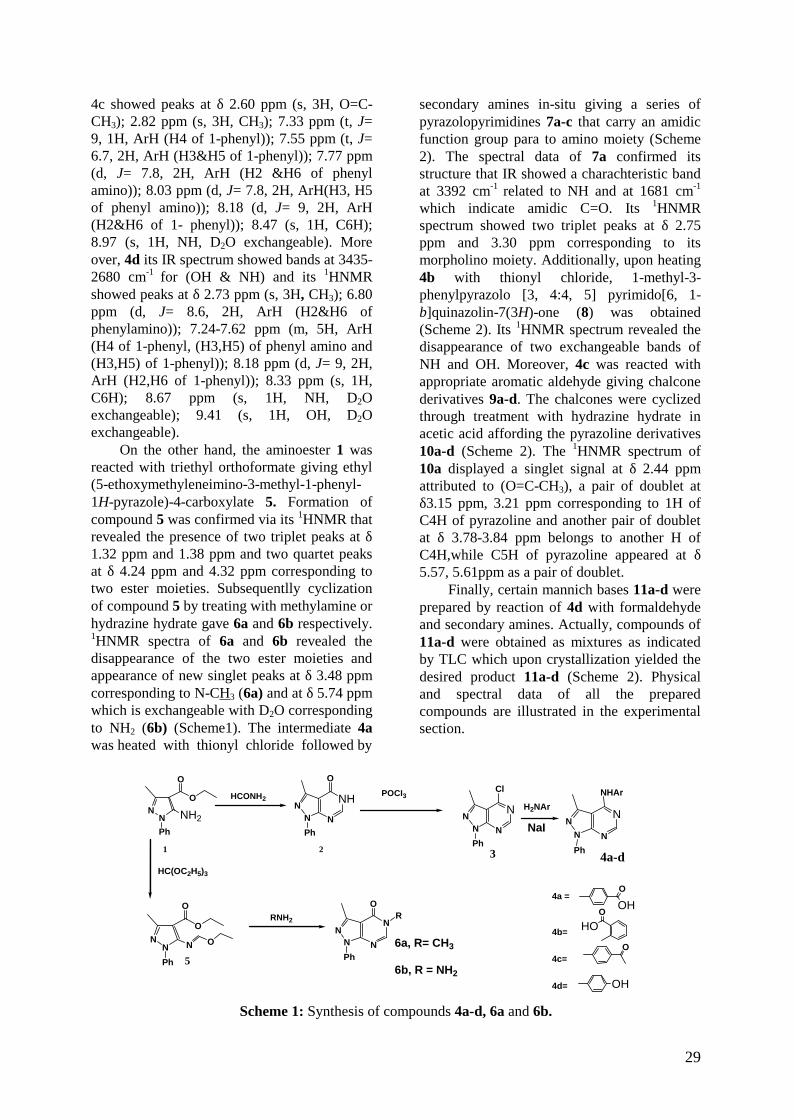
ChemistryReacting ethyl 5-amino-1-phenyl-3-
methyl-1H-pyrazole-4-carboxylate 117 withformamide resulted in formation ofpyrazolopyrimidinone 218 via a ring closure.The latter was chlorinated with phosphorousoxychloride to give 4-chloro-1-phenyl-3-methyl-1H-pyrazolo[3,4-d]pyrimidine 319
which was considered as the key intermediateused for alkylation of different aromatic aminesgiving 4a-d (Scheme1). The structures of
compounds 4a-d were elucidated by theirelemental analyses and their spectral data.Compound 4a showed IR absorption bands at3600-2787 cm-1 indicated for (OH & NH) and1684 cm-1 for (C=O). Also its 1HNMR showeda characteristic peaks at δ 2.81ppm (s, 3H,CH3); 7.33 ppm (dd J1= 9 Hz, J2= 3 Hz, 1H,ArH(H4 of 1-phenyl)); 7.54 ppm (t, 2H, ArH(H3&H5 of 1-phenyl)); 7.91 ppm (d, J= 9 Hz,2H, ArH (H2&H6 of phenyl amino)); 7.97 ppm(d, J= 9 Hz, 2H, ArH (H3&H5 of phenylamino)); 8.18 ppm (d, J= 9 Hz, 2H, ArH(H2,H6 of 1-phenyl)); 8.53 ppm (s, 1H, C6H);8.99 (s,1H, NH, D2O exchangeable. Alsocompound 4b showed IR absorption bands at3600-3063 cm-1 for (OH & NH) and at 1691cm-1 for (C=O). While its 1HNMR showedpeaks at δ 2.80 ppm (s, 3H, CH3); 7.14 ppm (t,J= 6 Hz, 1H, ArH (H4 of phenylamino)); 7.32ppm (t, J= 9 Hz, 1H, ArH (H4 of 1-phenyl));7.53 ppm (t, J= 6 Hz, 2H, ArH(H3&H5 of 1-phenyl)); 7.64 ppm (t, J= 9 Hz, 1H, ArH (H5 ofphenyl amino)); 8.03 ppm (d, J= 6 Hz, 1H, ArH(H6 of phenyl amino)); 8.15 ppm (d, J= 4.5 Hz,2H, ArH (H2&H6 of 1-phenyl)); 8.57 ppm (s,1H, C6H); 9.12 ppm (d, J= 9 Hz, 1H, ArH (H3of phenyl amino)); 9.28 ppm (brs, 1H, NH,D2O exchangeable); 11.56 ppm (s, 1H, OH,D2O exchangeable). In addition compound 4cits IR showed absorption bands at 3409 cm-1 for(NH) and at 1671 cm-1 for (C=O). 1HNMR of
29
4c showed peaks at δ 2.60 ppm (s, 3H, O=C-CH3); 2.82 ppm (s, 3H, CH3); 7.33 ppm (t, J=9, 1H, ArH (H4 of 1-phenyl)); 7.55 ppm (t, J=6.7, 2H, ArH (H3&H5 of 1-phenyl)); 7.77 ppm(d, J= 7.8, 2H, ArH (H2 &H6 of phenylamino)); 8.03 ppm (d, J= 7.8, 2H, ArH(H3, H5of phenyl amino)); 8.18 (d, J= 9, 2H, ArH(H2&H6 of 1- phenyl)); 8.47 (s, 1H, C6H);8.97 (s, 1H, NH, D2O exchangeable). Moreover, 4d its IR spectrum showed bands at 3435-2680 cm-1 for (OH & NH) and its 1HNMRshowed peaks at δ 2.73 ppm (s, 3H, CH3); 6.80ppm (d, J= 8.6, 2H, ArH (H2&H6 ofphenylamino)); 7.24-7.62 ppm (m, 5H, ArH(H4 of 1-phenyl, (H3,H5) of phenyl amino and(H3,H5) of 1-phenyl)); 8.18 ppm (d, J= 9, 2H,ArH (H2,H6 of 1-phenyl)); 8.33 ppm (s, 1H,C6H); 8.67 ppm (s, 1H, NH, D2Oexchangeable); 9.41 (s, 1H, OH, D2Oexchangeable).
On the other hand, the aminoester 1 wasreacted with triethyl orthoformate giving ethyl(5-ethoxymethyleneimino-3-methyl-1-phenyl-1H-pyrazole)-4-carboxylate 5. Formation ofcompound 5 was confirmed via its 1HNMR thatrevealed the presence of two triplet peaks at δ1.32 ppm and 1.38 ppm and two quartet peaksat δ 4.24 ppm and 4.32 ppm corresponding totwo ester moieties. Subsequentlly cyclizationof compound 5 by treating with methylamine orhydrazine hydrate gave 6a and 6b respectively.1HNMR spectra of 6a and 6b revealed thedisappearance of the two ester moieties andappearance of new singlet peaks at δ 3.48 ppmcorresponding to N-CH3 (6a) and at δ 5.74 ppmwhich is exchangeable with D2O correspondingto NH2 (6b) (Scheme1). The intermediate 4awas heated with thionyl chloride followed by
secondary amines in-situ giving a series ofpyrazolopyrimidines 7a-c that carry an amidicfunction group para to amino moiety (Scheme2). The spectral data of 7a confirmed itsstructure that IR showed a charachteristic bandat 3392 cm-1 related to NH and at 1681 cm-1
which indicate amidic C=O. Its 1HNMRspectrum showed two triplet peaks at δ 2.75ppm and 3.30 ppm corresponding to itsmorpholino moiety. Additionally, upon heating4b with thionyl chloride, 1-methyl-3-phenylpyrazolo [3, 4:4, 5] pyrimido[6, 1-b]quinazolin-7(3H)-one (8) was obtained(Scheme 2). Its 1HNMR spectrum revealed thedisappearance of two exchangeable bands ofNH and OH. Moreover, 4c was reacted withappropriate aromatic aldehyde giving chalconederivatives 9a-d. The chalcones were cyclizedthrough treatment with hydrazine hydrate inacetic acid affording the pyrazoline derivatives10a-d (Scheme 2). The 1HNMR spectrum of10a displayed a singlet signal at δ 2.44 ppmattributed to (O=C-CH3), a pair of doublet atδ3.15 ppm, 3.21 ppm corresponding to 1H ofC4H of pyrazoline and another pair of doubletat δ 3.78-3.84 ppm belongs to another H ofC4H,while C5H of pyrazoline appeared at δ5.57, 5.61ppm as a pair of doublet.
Finally, certain mannich bases 11a-d wereprepared by reaction of 4d with formaldehydeand secondary amines. Actually, compounds of11a-d were obtained as mixtures as indicatedby TLC which upon crystallization yielded thedesired product 11a-d (Scheme 2). Physicaland spectral data of all the preparedcompounds are illustrated in the experimentalsection.
NN
O
O
NH2Ph
NNN N
Cl
Ph
NHNN N
O
Ph
HC(OC2H5)3
NN
O
O
N O
Ph
N
NN
N
OR
Ph
NHAr
NN
N NPh
H2NAr
OHO
OHO
O
OH
1
5
4a-d2 3
4a =
4b=
4c=
4d=
6a, R= CH3
6b, R = NH2
HCONH2POCI3
RNH2
NaI
Scheme 1: Synthesis of compounds 4a-d, 6a and 6b.

Rania B. Bakr, et al.
30
HN
NN
N N
OH
R
Ph
NH2NH2/CH3COOH
NN
O
NN
N N
NH
Ph
HCHO/
O
NN
N N
NH
Ar1
Ph
NN N
N
N
O
Ph
R
O
NN
N N
HN
Ph
SOCl2NaOH
N
ON
N CH3
8
4a-d
Ar1CHO /
1- SOCl2
10 a Ar1= C6H5b, Ar1 = 2-Br-C6H4c,Ar1= 4-CI-C6H4d,Ar1= 4-NO2-C6H4
9a Ar1= C6H5b, Ar1 = 2-Br-C6H4c,Ar1= 4-CI-C6H4d,Ar1= 4-NO2-C6H4
11a, R = N(CH3)2b, R = N(C2H5)2c,R = morpholinod, R = piperidino
7a,R=
b,R=
c,R=
4a4d
4b4c
1
secondry amine
Ar
secondry amine
Scheme 2: Synthesis of compounds 7a-c, 8, 9a-d, 10a-d and 11a-d.
Molecular dockingProtein kinases (PKs) are essential
enzymes in cellular signaling processes thatregulate cell growth, differentiation, migrationand metabolism20. They transfer phosphatefrom ATP to tyrosine, serine and threonineresidues in protein substrates21. Aberrantcatalytic activity of many PKs via mutation oroverexpression plays an important role innumerous pathological conditions includingcancer21&22 Protein kinase inhibitors have beenwidely used to probe the role of proteinphosphorylation in cellular signaling andconstitute an important new class of potentialtherapeutic agents in the management ofcancer23-25. Literature review26&27 revealed thatthe kinase inhibitor binding site can bevisualized as five main pharmacophoricregions: hydrophilic region formed of adeninebinding site, sugar and phosphate regions inaddition to two hydrophobic areas I and II.Most kinase inhibitors share commonproperties: low molecular weight (smallmolecules) hydrophobic heterocycles which actby competing with ATP for binding in kinaseATP binding site26 Kinase inhibitors shouldcontain the following attributes to gainselectivity and potency28. A portion that closelymimics ATP molecule and one to threehydrogen bonds to the amino acids located in
the hinge region of the target kinases, as inerlotinib I26, gefitinib II29 and lapatinib26. Anadditional hydrophobic binding site which isdirectly adjacent to the ATP binding site(allosteric site), as in imatinib mesylate30 andsorafenib31. However, other mechanism couldbe achieved through binding outside the ATP-binding site at an allosteric site32 and byforming irreversible covalent bond to thekinase active site33&34 EGFR tyrosine kinase isa target for a remarkable variety of antitumordrugs, such as erlotinib I4. The 3D structure ofEGFR protein kinase complexed with erlotinibwas obtained from Protein Data Bank (PDBentry: 1M17) at Research Collaboration forStructural Bioinformatics (RCSB) proteindatabase35 (Fig. 2).
Fig. 2: 3D Structure of EGFR tyrosine kinase witherlotinib.

31
In order to qualify the molecular dockingresults in terms of accuracy of the predictedbinding conformations in comparison with theexperimental results, the internal ligand(erlotinib) was used as a testing molecule. Themolecular docking result indicated that thebinding conformation of the internal ligand,derived by MOE (Molecular OperatingEnvironment, 2008, 10) superposed well withthe crystallographic one. The vicinity whereerlotinib bound was considered as the activesite of EGFR tyrosine kinase and erlotinib astemplate. The interactions of erlotinib withEGFR (can be visualized using MOE sitefinder program), red dotted line representing H-bonding interaction between N3 and Thr 766through a water bridge (red sphere), and violetdotted line representing H-bonding interactionbetween N1 and Met 769 as in figure 3.
Fig. 3: Binding interaction of erlotinib with EGFRbinding site35.
The molecular docking study revealed thatsome of the designed compounds showedpromising activity to inhibit EGFR tyrosinekinase active site. The data obtained for theprepared compounds from the docking studywere explained in table 1.
Table 1: Results of molecular docking analyses, binding energy scores (Kcal/mol): energy of ligandsand erlotinb in the active site of EGFR tyrosine kinase.
Compound
number
Number of
H-bonds
Atoms of
compound forming
H-bonds
Amino acid Residues
forming-bonds (H-bond
length in Aо)
Binding
Energy Score
Kcal/mol
Erlotinib 2 N1,N4 Met769 (2.70),
Thr766 (2.78)
-21.35
6a 2 N7,
C=O
Thr766 (2.78),
Lys721 (2.43)
-11.80
6b 1 NH2 Met769 (1.93) -15.05
7a 2 N5,N7 Met769 (2.89),
Thr766 (2.71)
-17.23
7b 1 NH Met769 (1.64) -18.05
7c 2 N5,N7 Met769 (2.896),
Thr766 (2.94)
-16.75
8 0 - - -13.28
10a 1 N7 Thr766 (2.80) -16.02
10b 4 N5,
NH,
N7,
C=O
Met769 (1.92),
Met769 (2.807),
Thr766 (2.90),
Lys692 (2.64)
-17.38
10c 3 N5,
NH,
N7
Met769 (2.15),
Met769 (3.07),
Thr766 (2.97)
-18.70
11a 2 OH Asp831 (1.37)
Lys721 (2.64)
-14.49

Rania B. Bakr, et al.
32
Attachment of a morpholino carbonylmoiety para to amino phenyl function as incompound 7a resulted in the same mode ofligand erlotinib interaction with the aminoacids of ATP active site in EGFR tyrosinekinase (Thr 766, and Met 769). It seems thatcompound 7a has favorable binding to thekinase that led to high docking score (-17.23).Figures 4A & 4B show the 3D and 2Dinteraction of compound 7a with the active siteof EGFR tyrosine kinase respectively.
Fig. 4(A): 3D Interaction of 7a with the binding siteof EGFR tyrosine kinase.
Fig. (4B): 2D Interaction of 7a the binding site ofEGFR tyrosine kinase.
Biological studyFive compounds 6b,7a, 8, 10c, 11a were
tested for their anticancer activity using humanbreast carcinoma cell line (MCF-7)36: Thesurvival fraction ratio was calculated accordingto the following equation:Survival fraction=optical density (O.D.) (treated cells)/O.D.(control cells).
IC50 values (the concentration required toproduce 50% inhibition of cell growth) werecalculated using sigmodial dose response curvefitting models (GraphPad, Prizm software
incorporated). Human breast cancer cell line(MCF7) was challenged to the antiproliferativeeffect of the tested compounds at fourconcentrations and doxorubicin was used as areference in the biology experiment. From thesynthesized compounds, those compoundshaving the moderate binding energy score(Kcal/mol) were chosen for the in-vitrocytotoxic activity and the results are shown in(Table 2).
Table 2: Biological screening results ofDoxorubicin, 6b,7a, 8, 10c and 11aagainst MCF7.
Survival fraction ratio
Concentration (μM)Compound5 12.5 25 50
IC50
(μM)
Doxorubicin 0.19 0.17 0.18 0.20 5.476b 0.73 0.64 0.44 0.53 89.217a 0.53 0.32 0.18 0.30 14.868 0.82 0.69 0.72 0.90 -ve
10c 0.51 0.46 0.34 0.38 14.9411a 0.90 0.72 0.69 0.80 -ve
Each concentration was repeated 3 times.
The results of biological screeningindicated that MCF-7 cells appeared to besensitive to inhibitory activity of three of thetarget compounds. (6b, 7a, and 10c) Table 2.They showed more than 50% inhibition activitytowards cells, while the other testedcompounds (8 and 11a) were devoid ofactivity.
It was observed that the substitutedpyrazoline 10c resulted in an additionalhydrogen bonding with Met 769 besides thetwo interactions reported for the lead erlotiniband which may rationalize the cytotoxicactivity of 10c (IC50 = 14.94) which is lesspotant than doxorubicin.
In an attempts to rationalize therelationship between the cytotoxic activity andthe presence of a phenylamino moiety,compound 6 was prepared in which thephenylamino was replaced by oxo, as incompound 6b, that showed lower activityagainst MCF-7 less potent than doxorubicin.Docking results showed that in 6b only onehydrogen bond with EGFR tyrosine kinase sitewas detected and could explain its lowerpotency. On the other hand compound 8 and11a were prepared, such modification were not

33
successful hence giving two compounds withabolished inhibitory activity against MCF-7cells. Exploring the docking result ofcompound 8 no any hydrogen bondinginteraction was seen. Also 11a interact withreceptor active sire with different amino acidsAsp 831 and Lys 721 that seems not efficientfor activity.
Experimental
ChemistryMelting points were determined on Griffin
apparatus (U.K) and are uncorrected. IRspectra were recorded on Shimadzu 435Spectrometer (Japan), using KBr discs andvalues were represented in cm-1. 1H-NMRspectra were carried out on Varian Gemini 200or 300 MHz Spectrometer (Germany) at themicroanalytical center, Cairo University, Egyptusing TMS as an internal standard andchemical shifts were recorded in ppm on δscales. The electron Impact (EI)mass spectrarecorded on Hewlett Packard 5988Spectrometer (U.S.A), at the microanalyticalcenter, Cairo University, Egypt and NationalResearch Center, Cairo, Egypt. Elementalanalyses were carried out at the microanalyticalcenter, Cairo, Egypt. Progress of all reactionswas monitored by TLC using TLC sheetsprecoated with UV fluorescent silica gelMERCK 60 F 254 that was visualized by UVlamp, using CHCl3/ CH3OH (9.5/ 0.5 or 9/1) aseluent. Compounds 1, 2 and 3 were preparedaccording to reported17-19 procedures.
3-Methyl-1-phenyl-4-substituted amino-1H-pyrazolo[3,4-d]pyrimidines (4a-d)
A mixture of 4-chloro-3-methyl-1-phenyl-1H-pyrazolo [3, 4-d] pyrimidine (3) (2.44g,0.01mol), the appropriate aromatic amine (0.01mol) and sodium iodide (0.2 gm) in isopropylalcohol (20 mL) was heated under reflux for 2h as indicated by TLC. After cooling, thereaction mixture was neutralized to litmuspaper with sodium carbonate solution (20%).The formed precipitate was collected byfiltration, washed with water and crystallizedfrom ethanol to afford 4a-d, their physical andspectral data are given in table 3.
Ethyl (5-ethoxymethyleneimino-3-methyl-1-phenyl-1H-pyrazole)-4-carboxylate (5)
A mixture of ethyl (5-amino-3-methyl-1-phenyl-1H-pyrazole)-4-carboxylate (1) (2.45gm, 0.01 mol) and triethyl orthoformate (2.96gm, 0.02 mol) in acetic anhydride (25 mL) washeated under reflux for 5 h as indicated byTLC. After cooling, the solution was poured onice-cold water and the precipitate formed wasfiltered, dried and crystallized from ethanol togive white crystals of 5.Yield: (66%), m.p.: 52-4°C, IR cm-1: 1701(C=O); 1640 (C=N).1H-NMR(CDCl3): δ 1.32 (t, J=7.5Hz, 3H,OCH2CH3); 1.38 (t, J=7.5Hz,3H,COOCH2CH3); 2.49 (s, 3H, CH3); 4.24 (q,J=7.5 Hz, 2H, OCH2CH3); 4.32 (q, J=7.5 Hz,2H, COOCH2CH3); 7.27-7.64 (m, 5H, ArH of1-phenyl); 8.05 (s, 1H, N=CH).Analysis for C16H19N3O3 (301.35): Calcd.C%63.77, H%6.36, N%13.94 Found C%63.38,H%5.90, N%13.70
3-Methyl-1-phenyl-5-substituted-1H-pyrazolo[3,4-d] pyrimidin-4(5H)-ones (6a,b)
A mixture of 5 (3 gm, 0.01 mol) andmethyl amine or hydrazine hydrate (99.9%)(0.01 mol) in benzene (25 mL) was heatedunder reflux for 1h as indicated by TLC. Aftercooling, the precipitate formed was filtered,dried and crystallized from benzene to give 6a,b their physical and spectral data are given inTable 4.
3-Methyl-1-phenyl-4-[4-(substitutedaminocarbonyl)anilino]-1H-pyrazolo[3,4-d]pyrimidines (7a-c)
A mixture of 4a (3.4 gm, 0.01 mol) andexcess of thionyl chloride (15 mL) was heatedunder reflux for 4h as indicated by TLC.Excess thionyl chloride was distilled undervacuum; the obtained residue was washed withdiethyl ether and dried in vacuum oven. Theappropriate secondary amine (0.01 mol) wasadded to the resulting acid chloride, followedby dioxane (15 mL) and triethylamine (2.8 mL,0.02 mol) and the reaction mixture was stirredat room temperature for an overnight. Water(15 mL) was added and the stirring wascontinued for further 0.5 h. The separatedproduct was filtered, dried and crystallizedfrom ethanol to give 7a-c (Table 5).

Rania B. Bakr, et al.
34
Table 3: Physical and spectral data of compounds 4a-d.
HN X
NNN N
Y
4a-d
4\
3\
2\
5\
6\\
2\\
3\\4\\
5\\
6\
4a: X=COOH, Y=H4b: X=H, Y= COOH4c: X= COCH3,Y= H4d: X=OH, ,Y=H
Elemental Analyses %4
m.p.( оC)
Yield%,
Mol. formula
(M.Wt.) Calcd. FoundIR (cm-1)
1H-NMR(DMSO), δ(ppm),
J(HZ)
a > 300
(88)
C19H15N5O2
(345.36)
C
H
N
66.08
4.38
20.28
65.90
4.48
20.19
3600-2787 (OH &
NH); 1684 (C=O)
and 1608 (C=N).
2.81 (s, 3H, CH3); 7.33 (t, J= 5.6, 1H,
ArH(H4\); 7.54 (t, J= 5.8 , 2H,
ArH(H3\&H5\ ); 7.91 (d, J= 9 , 2H
,ArH(H2\\&H6\\ ); 7.97 (d, J= 9, 2H,
ArH ( H3\\&H5\\); 8.18 (d, J= 6, 2H,
ArH(H2\,H6\ ); 8.53 (s, 1H, C6H);
8.99 (s,1H, NH, D2O exchangeable).
b 235-7
(72)
C19H15N5O2
(345.36)
C
H
N
66.08
4.38
20.28
65.90
4.40
20.38
3600-3063 (OH &
NH); 1691 (C=O)
and 1578 (C=N).
2.80 (s, 3H, CH3); 7.14 (t, J= 6 , 1H,
ArH (H4\\); 7.32 (t, J= 9 , 1H, ArH (H4
); 7.53 (t, J= 6 , 2H, ArH( H3\&H5\ );
7.64 (t, J= 9, 1H, ArH (H5\\); 8.03 (d,
J= 6, 1H, ArH (H6\\); 8.15 (d, J= 4.5,
2H, ArH (H2\&H6\); 8.57 (s, 1H,
C6H); 9.12 (d, J= 9, 1H, ArH (H3\\);
9.28 (brs, 1H, NH, D2O exchangeable);
11.56 (s, 1H, OH, D2O exchangeable).
c 170-1
(76)
C20H17N5O
(343.39)
C
H
N
69.96
4.99
20.39
70.18
5.14
20.51
3409 (NH);
1671 (C=O) and
1614 (C=N).
2.60 (s, 3H, O=C-CH3 ); 2.82 (s,
3H, CH3); 7.33 (t, J= 9 , 1H, ArH
(H4\ ); 7.55 (t, J= 6.7 , 2H, ArH
(H3\&H5\); 7.77 (d, J= 7.8 , 2H,
ArH (H2\\ &H6\\); 8.03 (d, J= 7.8,
2H, ArH(H3\\, H5\\ ); 8.18 (d, J= 9 ,
2H, ArH (H2\&H6\ ); 8.47 (s, 1H,
C6H); 8.97 (s, 1H, NH, D2O
exchangeable).
d 274-5
(62)
C18H15N5O
(317.35)
C
H
N
68.13
4.76
22.07
68.09
4.75
21.91
3435-2680 (OH
& NH) and 1613
(C=N).
2.73 (s, 3H, CH3); 6.80 (d, J= 8.6 ,
2H, ArH (H2\\&H6\\); 7.24-7.62 (m,
5H, ArH (H4, (H3\\,H5\\) and
(H3\,H5\) ); 8.18 (d, J= 9 , 2H, ArH
(H2\,H6\); 8.33 (s, 1H, C6H); 8.67
(s, 1H, NH, D2O exchangeable);
9.41 (s, 1H, OH, D2O
exchangeable).

35
Table 4: Physical and spectral data of compounds 6a,b.
N
NN
N
OR
6a, R= CH3
6b, R = NH2
4\3\
2\6\
5\
Elemental Analyses %6
%, m.p.(оC)Yield
Mol.Formul(M.Wt.)
Calcd. FoundIR (cm-1)
1H-NMR(DMSO), δ(ppm),J(HZ)
a 150-2(60)
C13H12N4O(240.27)
CHN
64.995.03
23.32
64.924.60
23.60
1681 (C=O) and1587 (C=N).
2.54 (s, 3H, CH3); 3.48 (s, 3H, N-CH3); 7.37 (t, J= 6 , 1H, ArH (H4\);7.53 (t, J= 6 , 2H, ArH (H3\&H5\);8.02 (d, J= 9 , 2H, ArH (H2\&H6\);8.43 (s, 1H, C6H).
b 214-5(65)
C12H11N5O(241.25)
CHN
59.744.60
29.03
59.814.91
29.36
3319, 3272(NH2);1676 (C=O) and1576 (C=N).
2.91 (s, 3H, CH3); 5.74 (s, 2H,NH2, D2O exchangeable); 7.31-7.33(m, 1H, ArH (H4\); 7.45-7.48 (m,2H, ArH (H3\&H5\); 7.96 (d, J= 9,2H, ArH (H2\&H6\) ; 8.40 (s, 1H,C6H).
Table 5: Physical and spectral data of compounds 7a-c.
R
O
NNN N
HN
N
N
CH3
7a,R=
b,R=
c,R=2\
3\
4\5\
6\
3\\
5\\
2\\
6\\
Elemental Analysis %7
m.p. (оC)Yield %
Mol.Formula(M.Wt) Calcd Found
IR (cm-1)1H-NMR(DMSO), δ(ppm),
J(HZ)
a 205-7(60)
C23H22N6O2
(414.47)CHN
66.655.35
20.28
66.525.20
19.88
3392 (NH);1681 (C=O);and 1604(C=N).
2.46 (s, 3H, CH3); 2.75 (t, J= 6, 4H,CH(H3,H5 of morpholinyl)); 3.30 (t, J=6, 4H, CH(H2&H6 of morpholinyl));7.32-7.57 (m, 3H, ArH(H4\,H3\&H5\ );7.75 (d, J= 8.5 , 2H, ArH (H2\\&H6\\);7.79 (d, J= 8.5, 2H, ArH (H3\\&H5\\ );8.14 (d, J= 9 , 2H, ArH( H2\&H6 \));8.46 (s, 1H, C6H).
b 235-7(56)
C24H24N6O(412.50)
CHN
69.885.86
20.37
69.885.50
20.83
3412 (NH);1686 (C=O)and 1594(C=N).
1.42-1.65 (m, 6H, CH (H3,H4&H5 ofpiperidinyl)); 2.75 (s, 3H, CH3); 3.25-3.45 (m, 4H, CH (H2&H6 ofpiperidinyl)); 7.26-7.56 (m, 5H,ArH(H3,H5,H4,H2\\&H6\\); 8.09 (d, J=9, 2H, ArH(H3\\&H5\\); 8.19 (d, J= 6 ,2H, ArH(H2\&H6\ ); 8.59 (s, 1H, C6H);8.82 (s, 1H, NH, D2O exchangeable).
c 229-31(49)
C25H26N6O(426.53)
CHN
70.406.14
19.70
70.335.88
19.24
3335 (NH);1693 (C=O);and 1611(C=N).
0.92 (s, 3H, CH3,(4-methylpiperdinyl)); 1.45-1.62 (m, 5H,CH,H3,H4&H5 of 4-methylpiperidinyl)); 2.80 (s, 3H, CH3); 4.25-4.35 (m, 4H, CH(H2,H6 of 4-methylpiperidinyl)); 7.33-7.42 (m, 3H,ArH(H4\ and H2\\,H6\\); 7.50-7.55 (m,2H, ArH(H3\,H5\ ); 7.79 (d, J= 9 , 2H,ArH(H3\\&H5\\ ); 8.19 (d, J= 9 , 2H,ArH(H2\&H6\); 8.49 (s, 1H, C6H); 8.90(s,1H, NH, D2O exchangeable).
Rania B. Bakr, et al.
36
1-Methyl-3-phenylpyrazolo[3,4:4,5]pyrimido[6,1-b]quinazolin-7(3H)-one (8)
NN N
N
N
O
8
2\
3\
45\
6\
8
910
11
\
A mixture of 4b (0.34 gm, 0001 mol) andexcess thionyl chloride (4 mL) was heated underreflux for 4 h as indicated by TLC. Excess thionylchloride was distilled off and the residue wascrystallized from ethanol to give 8.Yield: (62.5%), m.p.: 170-171°C, IR cm-1: 1708(C=O); 1617 (C=N), 1H-NMR (CDCl3): δ 2.66 (s,3H, CH3);.738-7.51 (m, 2H, ArH(H4\ and H11);7.54-7.57 (m, 4H,ArH(H3\,H5\ and H9, H10);
7.73 (d J=8.1Hz, 2H, ArH(H2\,H6\); 8.23 (d, J=6.3 Hz,1H, ArH(H8); 8.26 (s, 1H, C5H ofpyrimidinyl)). Analysis for C19H13N5O (327.35):Calcd. C% 69.52, H% 4.00, N% 21.39 Found C%69.52, H% 4.21, N% 20.89.
(E) 3-Methyl-1-phenyl-4-[4-(2-arylvinyl-carbonyl)anilino]-1H-pyrazolo[3,4-d]pyrimidines (9a-d)
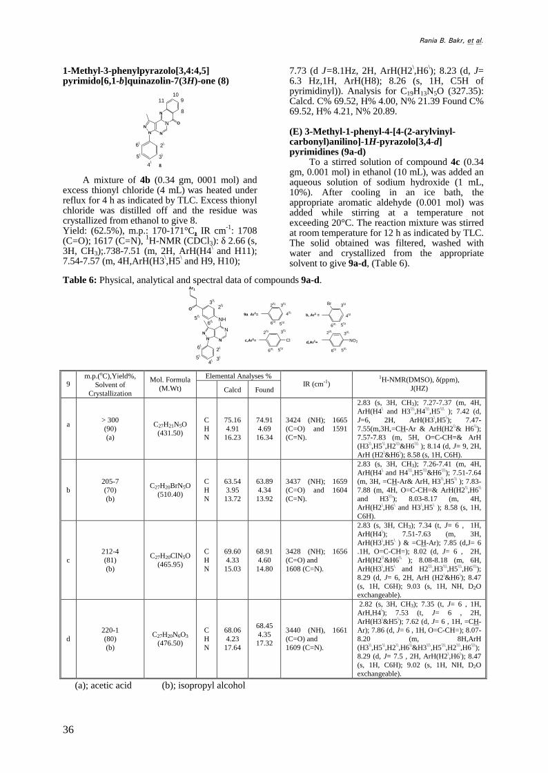
To a stirred solution of compound 4c (0.34gm, 0.001 mol) in ethanol (10 mL), was added anaqueous solution of sodium hydroxide (1 mL,10%). After cooling in an ice bath, theappropriate aromatic aldehyde (0.001 mol) wasadded while stirring at a temperature notexceeding 20°C. The reaction mixture was stirredat room temperature for 12 h as indicated by TLC.The solid obtained was filtered, washed withwater and crystallized from the appropriatesolvent to give 9a-d, (Table 6).
Table 6: Physical, analytical and spectral data of compounds 9a-d.
O
NN
N N
NH
Ar1
Br
Cl NO2
2\
3\4\
5\
6\
3\\
2\\
5\\
6\\
9a Ar1=
d,Ar1=
3\\\
4\\\
5\\\
2\\\
6\\\
b, Ar1 = 4\\\
5\\\
3\\\
6\\\
c,Ar1=
5\\\
3\\\
6\\\
2\\\
5\\\6\\\
2\\\ 3\\\
Elemental Analyses %9
m.p.(оC),Yield%,Solvent of
Crystallization
Mol. Formula(M.Wt) Calcd Found
IR (cm-1)1H-NMR(DMSO), δ(ppm),
J(HZ)
a> 300(90)(a)
C27H21N5O(431.50)
CHN
75.164.91
16.23
74.914.69
16.34
3424 (NH); 1665(C=O) and 1591(C=N).
2.83 (s, 3H, CH3); 7.27-7.37 (m, 4H,ArH(H4\ and H3\\\,H4\\\,H5\\\ ); 7.42 (d,J=6, 2H, ArH(H3\,H5\); 7.47-7.55(m,3H,=CH-Ar & ArH(H2\\& H6\\);7.57-7.83 (m, 5H, O=C-CH=& ArH(H3\\,H5\\,H2\\\&H6\\\ ); 8.14 (d, J= 9, 2H,ArH (H2\&H6\); 8.58 (s, 1H, C6H).
b205-7(70)(b)
C27H20BrN5O(510.40)
CHN
63.543.95
13.72
63.894.34
13.92
3437 (NH); 1659(C=O) and 1604(C=N).
2.83 (s, 3H, CH3); 7.26-7.41 (m, 4H,ArH(H4\ and H4\\\,H5\\\&H6\\\); 7.51-7.64(m, 3H, =CH-Ar& ArH, H3\\,H5\\ ); 7.83-7.88 (m, 4H, O=C-CH=& ArH(H2\\,H6\\
and H3\\\); 8.03-8.17 (m, 4H,ArH(H2\,H6\ and H3\,H5\ ); 8.58 (s, 1H,C6H).
c212-4(81)(b)
C27H20ClN5O(465.95)
CHN
69.604.33
15.03
68.914.60
14.80
3428 (NH); 1656(C=O) and1608 (C=N).
2.83 (s, 3H, CH3); 7.34 (t, J= 6 , 1H,ArH(H4\); 7.51-7.63 (m, 3H,ArH(H3\,H5\ ) & =CH-Ar); 7.85 (d,J= 6.1H, O=C-CH=); 8.02 (d, J= 6 , 2H,ArH(H2\\&H6\\ ); 8.08-8.18 (m, 6H,ArH(H3\,H5\ and H2\\\,H3\\\,H5\\\,H6\\\);8.29 (d, J= 6, 2H, ArH (H2\&H6\); 8.47(s, 1H, C6H); 9.03 (s, 1H, NH, D2Oexchangeable).
d220-1(80)(b)
C27H20N6O3
(476.50)
CHN
68.064.23
17.64
68.454.35
17.32
3440 (NH), 1661(C=O) and1609 (C=N).
2.82 (s, 3H, CH3); 7.35 (t, J= 6 , 1H,ArH,H4\); 7.53 (t, J= 6 , 2H,ArH(H3\&H5\); 7.62 (d, J= 6 , 1H, =CH-Ar); 7.86 (d, J= 6 , 1H, O=C-CH=); 8.07-8.20 (m, 8H,ArH(H3\\,H5\\,H2\\,H6\\&H3\\\,H5\\\,H2\\\,H6\\\);8.29 (d, J= 7.5 , 2H, ArH(H2\,H6\); 8.47(s, 1H, C6H); 9.02 (s, 1H, NH, D2Oexchangeable).
(a); acetic acid (b); isopropyl alcohol

37
3-Methyl-1-phenyl-4-[4-(1-acetyl-5-aryl-4,5-dihydro-1H-pyrazol-3-yl)anilino]-1H-pyazolo[3,4-d]pyrimidines (10a-d)
To a solution of hydrazine hydrate(99.9%, 0.1 mL, 0.002 mol) in glacial aceticacid (5 mL), the appropriate chalcone 9a-d(0.001 mol) was added. The reaction mixture
was heated under reflux for 5 h as indicated byTLC. After cooling, the solution was poured onan ice-cold water. The obtained solid wascollected by filtration, washed with water andcrystallized from ethanol to yield 10a-d,(Table 7).
Table 7: Physical and spectral data of compounds 10a-d.
NNAr1
O
NN
N N
NH
Br
Cl NO2
10a Ar1=2\\
3\\
5\\
6\\
d,Ar1=
3\\\
4\\\
5\\\
2\\\
6\\\
b, Ar1 = 4\\\
5\\\
3\\\
6\\\
c,Ar1=
5\\\
3\\\
6\\\
2\\\
5\\\6\\\
2\\\ 3\\\
2\
3\
6\
5\
4\
Elemental Analyses %10
m.p. оCYield %
Mol. Formula(M.Wt) Calcd Found IR (cm-1)
1H-NMR(DMSO), δ (ppm), J (Hz)
a > 300(60)
C29H25N7O(487.57)
CHN
71.445.17
20.11
71.205.58
20.16
3315 (NH)1652 C=O)1587 C=N).
2.44 (s, 3H, O=C-CH3); 2.60 (s, 3H, CH3); 3.15,3.21 (dd, J1= 4.8 , J2= 4.8 ,1H, CH2 of pyrazoline);3.78, 3.84 (dd, J1= 12 , J2= 12, 1H, CH2 ofpyrazoline); 5.57, 5.61 (dd, J1= 4.5 , J2= 12 , 1H,CH of pyrazoline); 7.17 (d, J= 7.5 , 2H,ArH(H2\\\,H6\\\ ); 7.26-7.38 (m, 4H, ArH(H4\
,H3\\\,H4\\\,H5\\\); 7.55-7.61 (m, 4H, ArH(H3\,H5\,H2\\,H6\\); 7.76 (d, J= 9, 2H, ArH(H3\\,H5\\);8.08 (s, 1H, NH, D2O exchangeable); 8.12 (d, J= 9,2H, ArH(H2\,H6\); 8.53 (s, 1H, C6H).
b 270-1(50)
C29H24BrN7O(566.46)
CHN
61.494.27
17.31
61.394.28
17.00
3438 (NH);1663 C=O);and 1602(C=N).
2.51 (s, 3H, O=C-CH3); 2.70 (s, 3H, CH3); 3.06,3.11 (dd, J1= 4.5 , J2= 4.5 , 1H, CH2 of pyrazoline);3.88, 3.93 (dd, J1= 12.3 , J2= 4.5 , 1H, CH2 ofpyrazoline); 5.91, 5.94 (dd, J1= 4.2, J2= 4.5 , 1H,CH of pyrazoline); 7.06 (d, J= 6 , 1H,ArH(H6\\\);7.11-7.18 (m, 1H, ArH(H5\\\); 7.26-7.36 (m, 2H,ArH(H4\ &H4\\\); 7.46-7.55 (m, 3H, ArH(H3\,H5\)& NH( D2O exchangeable)); 7.60 (d, J= 8.1 , 2H,ArH(H2\\&H6\\); 7.81 (d, J= 8.1, 2H,ArH(H3\\&H5\\); 7.91 (s, 1H, ArH(H2\\\); 8.14 (d, J=9 , 2H, ArH(H2\&H6\ ); 8.55 (s, 1H, C6H).
c 230-1(45)
C29H24ClN7O(522.01)
CHN
66.734.63
18.78
66.715.02
18.68
3320 (NH);1705 (C=O);and 1619(C=N).
2.37 (s, 3H, O=C-CH3); 2.81 (s, 3H, CH3);3.00,3.06 (dd, J1= 4.8 , J2= 4.8 , 1H, CH2 ofpyrazoline); 3.96, 4.06 (dd, J1=12 Hz, J2= 12 , 1H,CH2 of pyrazoline); 5.72, 5.74 (dd, J1= 4.5 , J2= 12 ,1H, CH of pyrazoline); 7.04 (d, J= 7.8 , 2H, ArH(H2\\\,H6\\\ ); 7.24 (d, J= 7.8 , 2H, ArH(H3\\\,H5\\\);7.33-7.35 (m, 1H, ArH(H4\); 7.45-7.50 (m, 2H,ArH(H3\&H5\ ); 7.67 (d, J= 7.8, 2H,ArH(H2\\&H6\\)); 7.89 (d, J= 7.8 , 2H,ArH(H3\\&H5\\); 8.18 (d, J= 8 , 2H, ArH(H2\&H6\
); 8.44 (s, 1H, C6H); 8.91 (s, 1H, NH, D2Oexchangeable).
d 252-4(66),
C29H24N8O3
(532.57)CHN
65.404.54
21.04
65.444.77
21.14
3426 (NH);1655 (C=O);and 1588(C=N).
2.44 (s, 3H, O=C-CH3); 2.81 (s, 3H, CH3); 3.15,3.21 (dd, J1= 4.8 , J2= 4.8 , 1H, CH2 of pyrazoline);3.76, 3.81 (dd, J1= 12 , J2= 12, 1H, CH2 ofpyrazoline); 5.57, 5.60 (dd, J1= 4.2, J2= 4.5 , 1H,CH of pyrazoline); 7.19 (d, J= 9 , 2H,ArH(H2\\\,H6\\\); 7.27-7.32 (m, 3H, ArH(H4\ &H3\\\,H5\\\); 7.49-7.55 (m, 4H, ArH(H3\,H5\
&H2\\,H6\\); 7.83 (d, J= 9 , 2H, ArH(H3\\,H5\\); 8.15(d, J= 9,2H, ArH(H2\,H6\); 8.17 (s, 1H, NH, D2Oexchangeable); 8.57 (s, 1H, C6H).

Rania B. Bakr, et al.
38
3-Methyl-1-phenyl-4-[3-(substitutedaminomethyl)-4-hydroxyanilino]-1H-pyrazolo[3,4-d]pyra- midines (11a-d)
To a solution of 4d (3.17gm, 0.01mol) inabsolute ethanol (10 mL), a mixture ofsecondary amine (0.012 mol) andparaformaldehyde (0.45gm, 0.015 mol) was
added. The mixture was heated under reflux for5 h as indicated by TLC on a steam bath andleft overnight. The solvent was distilled offunder reduced pressure. The solid residue wasdissolved in ethanol, precipitated with water,filtered, dried and crystallized from ethanol togive 11a-d, (Table 8).
Table 8: Physical and spectral data of compounds 11a-d.
HN
NN
N N
OH
R
2\
3\
4\5\
6\
5\\
2\\
6\\
11a, R = -N(CH3)2b, R = -N(C2H5)2
c,R =
d, R = N
ON
Elemental Analyses %11
m.p. (оC)Yield %
Mol. Formula(M.Wt) Calcd. Found
IR (cm-1) 1H-NMR(DMSO), δ (ppm), J (HZ)
a115-8(56) C21H22N6O
(374.45)
CHN
67.365.92
22.44
67.555.82
22.44
3500-3250(OH& NH) and1588 (C=N).
2.69 (s, 3H, CH3); 2.75 (s, 6H, N(CH3)2);3.41 (s, 1H, OH, D2O exchangeable);4.23 (s, 2H, CH2-N); 7.31-7.59 (m, 5H,ArH(H4\,H3\,H5\,& H2\\H5\\); 7.63 (d,J= 6 Hz, 1H, ArH(H6\\); 8.17 (d, J= 9Hz, 2H, ArH(H2\,H6\ ); 8.37 (s, 1H,C6H); 8.80 (s, 1H, NH, D2Oexchangeable).
b101-3
(60), C23H26N6O (402.50)
CHN
68.636.51
20.88
68.396.41
20.90
3414-3102(OH& NH) and1591 (C=N).
1.21 (t, J=7.8 Hz, 6H, 2CH2-CH3); 2.72(s, 3H, CH3); 3.10 (q, J=7.8 , 4H,2CH2CH3); 4.21 (s, 2H, CH2-N); 6.81(d,1H, ArH(H2\\); 7.08-7.51 (m, 5H,ArH(H4\,H3\,H5\& H5\\,H6\\); 8.17 (d, J=8.1, 2H, ArH(H2\,H6\); 8.32 (s, 1H,C6H); 8.70 (s, 1H, NH, D2Oexchangeable); 9.51 (1H, OH, D2Oexchangeable).
c120-2
(42) C23H24N6O2
(416.49)
CHN
66.335.81
20.18
66.695.54
20.31
3393-3250(OH& NH) and1589 (C=N).
1.49 (t, 4H, CH morpholino); 2.73 (s,3H, CH3); 2.98 (t, 4H, CH morpholino);3.62 (1H, OH, D2O exchangeable); 3.91(s, 2H, CH2-N); 7.30-7.51 (m, 5H,ArH(H4\,H3\,H5\ &H2\\ ,H6\\); 8.01 (d,J= 9 , 1H, ArH(H5\\); 8.16 (d, J= 9, 2H,ArH(H2\,H6\ ); 8.32 (s, 1H, C6H); 8.74(s, 1H, NH, D2O exchangeable).
d190-2
(66) C24H26N6O (414.51)
CHN
69.546.32
20.27
69.636.18
19.91
3416-3138(OH& NH) and1610 (C=N).
1.59-1.70 (m, 6H, CH piperidino); 2.64-2.75 (m, 4H, CH piperidino); 2.83 (s, 3H,CH3); 4.00 (s, 2H, CH2-N); 5.40 (s, 1H,NH, D2O exchangeable); 6.90 (d, J= 7.8 ,1H, ArH(H2\\); 7.26-7.50 (m, 4H,ArH(H4\,H3\,H5\&H6\\ amino)); 7.98 (d,J= 9 , 1H, ArH(H5\\); 8.10 (d, J= 4.5 ,2H, ArH(H2\,H6\ ); 8.46 (s, 1H, C6H).

39
Molecular docking: The docking wasperformed using Molecular OperatingEnvironment (MOE) 2008, 10 softwareaccording to the following procedures.• Downloading step: This step includes
downloading of EGFR from the protein databank (PDB code: 1M17) and hydrogen wasclarified hence the downloaded proteins fromPDB do not show any hydrogen.
• The charging step: the protein needs to becharged to calculate the total charge over itand this allows the free energy calculationstep.
• The Surface conversion step: the appearanceof the protein is changed by converting itssurface to molcad or slab surface, whichhelps to see the active site in details, extractthe ligand and modify it.
• Then extraction step: through it the a ligandcould be separated from protienand modifiedto a form that enhances determination of itsbinding affinity.
• Modifying step that includes adding orremoving any atom or group of atoms fromthe ligand.
• The minimization step: that include thegeometrical optimization process that enablesthe ligand before docking step to give us thelowest conformational energy for a ligand.
• The molecular docking step that includesfitting the ligand after extraction, modifyingand minimization into the active site.Finally Minimization of ligand inside theactive site: this step includes selection of theactive site with the ligand to minimize theirconformational energy resulted after ligandbinding process.
• Energy calculation step: for every change tothe ligand, the free energy (ΔE) is used todetermine the binding affinity of each ligandwith the receptor site.
Biology screening
General methodology: Study was operated inNational Cancer institute, CairoUniversity,Egypt.
All reagents and authentic samples usedduring the biology experiment were obtainedthrough (sigma & Aldrichcompany)and are ofanalytical grades.
MCF was grown as monolayer culture inRPM 11640 medium supplemented with 10%fetal bovine serum (FBS) and 1%
Penicillin/Streptomycin. The cell line wasincubated at 37°C 5% CO2 95% air and highhumidity atmosphere in the water jacketedincubator (Revco, GS laboratory equipment,RCO 3000 TVBB, U.S.A). The cell line wasregularly subcultured to be maintained in theexponential growth phase. The sterileconditions were strictly attained by workingunder the equipped laminar flow (MicroflowLaminar flow cabinet, Hamsphire SP 105aa,U.K.).
A- Maintenance of the human tumor cellline
1. A cryotube containing frozen cells wastaken out of the nitrogen container and thenthawed in a water bath at 37°C.
2. The cryotube was opened under strictaseptic conditions and its contents weretransferred into sterile 50 ml disposablefalcon tube supplemented by 5 ml mediumdrop by drop.
3. The tube was incubated for 2h then itscontents were centrifuged at 1200 rpm for10 min.
4. The supernatant was discarded and the cellpellet was suspended and seeded in 5 mlsupplemented medium in T25 Nunclonsterile tissue culture flasks.
5. The cell suspension was incubated andfollowed up daily with replacing thesupplemented medium every 2-3 days.
6. Incubation was continued until a confluentgrowth was achieved and the cells werefreshly subcultured before each experimentto be in the exponential phase of growth.
B- Collection of cells by trypsinization1. The medium was discarded.2. The cell monolayer was washed with 10 ml
phosphate-buffered saline (PBS).3. All the adherent cells were dispersed from
their monolayer by the addition of I mltrypsin solution (0.025% trypsin w/v).
4. The flask was left in incubator till completedetachment of the cells and checked withthe inverted microscope (Olympus 1×70,Tokyo, Japan).
5. Trypsin was inactivated by the addition of 5ml of the supplemented medium containingfetal calf serum (FCS). The trypsin contentwas discarded by centrifugation (BoacoGermany) at 1200 rpm for 10 min. Cells

Rania B. Bakr, et al.
40
were separated in a single suspension bygentle dispersion several times.
C- Determination and counting of viablecells
1. 100 μl of 0.05% trypan blue solution wereadded to 100 μl of the single cellsuspension.
2. The cells were examined under the invertedmicroscope using the haemocytometer.
3. Non stained (viable) cells were counted andthe following equation was used to calculatethe cell count /ml of cell suspension.
4. Viable cells/ml= [Number of cells in 4quarters x 2 (dilution factor) ×104]/4.
5. The cells were then diluted to give therequired concentration of single cellsuspension.
D- Cytotoxicity of the test compounds usingSulphordhodamine-B (SRB) assay
The principleThe cytotoxicity of the prepared compoundswere tested on the MCF cell line determinedusing SRB assay. SRB is a bright pinkaminoxanthene dye with two sulfonic groups. Itis a protein stain that binds to the amino groupsof intracellular protein under mild acidicconditions to provide a sensitive index ofcellular protein content.
Procedure36
The breast cancer cells were seeded in 96-wellmicrolitre plates and left to attach for 24h.Cells were incubated with the testedcompounds at concentration range from 0, 5,12.5, 25 and 50 μg/ml)as well as doxorubicinand incubation was continued for 48 h. After48h treatment, the cells were fixed with 50 μlcold 50% trichloroacetic acid (TCA) for 1h at4°C.Wells were then washed 5 times withwater and stained for 30 min at roomtemperature with 50 μl of 0.4% SRB dissolvedin 1% acetic acid. The wells were then washed4 times with 1% acetic acid. The plates wereair dried and the dye was solubilized with 100μl/well of 10Mm tris base (pH 10.5) for 5 minon a shaker (Orbital Shaker OS, Boaco,Germany) at 1600 rpm. The optical density(O.D) of each well was measuredspectrophotometrically at 564 nm with enzymelinked immunosorbent assay (ELISA)micraplate reader (Tecan Sunrise, Austria). The
mean values for each drug concentration wascalculated.
REFERENCES
1. A. M. Aronov, B. McClain, C. S. Moodyand M. A. Murcko, “Kinase-likenes andkinase-privileged fragments: towardvirtual poly pharmacology”, J. Med.Chem., 51, 1214 (2008).
2. L. v. Peng-Cheng, C. F. Zhou, J. Chen, P.G. Liu, K. R. Wang, W. J. Mao, L. Huan-Qiu., Y. Yang, J. Xiong and H. L. Zhu,“Design, synthesis and biologicalevaluation of thiazolidinone derivatives aspotential EGFR and HER-2 inhibitors”,Bioorg. Med. Chem. Lett., 18, 314 (2010).
3. R. Lin, S. G. Johnson, P. J. Connolly, S.K. Wetter, E. Binnun, T. V. Hughes, W.V. Murray, N. B. Pandey, S. J. Morreno-Mazza, M. Adams, A. R. Fuentes-Pesquera and S. A. Middleton, “Synthesisand evaluation of 2,7-diamino-thiazolo[4,5-d]pyrimidine analogues as antitumorepidermal growth factor receptor tyrosinekinase inhibitors”, ibid., 19, 2333 (2009).
4. “Erlotinib CP358774, NSC718781,OSI774, R1415”, Drugs R&D, 4, 243(2003).
5. J. Smith, “Erlotinib: small-moleculetargeted therapy in the treatment of non-small-cell lung cancer”, ClinicalTherapeutics, 27, 1513 (2005).
6. K. Amine, H. Georgey and F. Awadallah,"EGFR tyrosine kinase targetedcompounds: Synthesis, docking study, andin-vitro antitumor activity of some newquinazoline and benzo[d]isothiazolederivatives", Med. Chem. Res., 20, 1042(2011).
7. R. Riera, P. C. de Soarez, M. E. Puga andM. B. Ferraz, “Lapatinib for treatment ofadvanced or metastasized breast cancer:Systemic review”, Sao Paulo Med J., 127,295 (2009).
8. H. Yamauchi, T. Lafortune and N. T.Ueno, “Lapatinib in the treatment ofbreast cancer”, Clinical Medicine:Therapeutics, 1, 513 (2009).
9. K. Bouchalova, M. Cizkova, K. Cwiertka,R. Trojanec, D. Friedecky and M.Hajduch, “Lapatinib in breast cancer – thepredictive significance of HER1 (EGFR)”,

41
Biomed. Pap. Med. Fac. Uni., PalackyOlomuc Czech Repub., 154, 281(2010).
10. A. F. Burchat, D. J. Calderwood, M.M. Friedman, G. C. Hirst, B. Li, P.Rafferty, K. Ritter and B. S. Skinner"Pyrazolo[3,4-d]pyrimidines containing anextented 3- subsitutent as potant inhibitorsof Lck-a selectivity insight", Bioorg. Med.Chem. Lett., 12, 1687 (2002).
11. D.W.Borhani; D. J. Calderwood, M. M.Friedman, G. C. Hirst, B. Li, Leung, B.McRae, S. Ratnofsky, K. Ritter and W.Waegell “A-420983: a potant, orallyactive inhibitors of LCK with efficacy inmodel of transplant rejection”, ibid., 14,2613 (2004).
12. A. J. Peat, D. Garrido, J. A. Boucheron, S.L. Schweiker, S. H. Dickerson, J. R.Wilson, T. Y. Wang and S. A.Thomson,“Novel GSK-3inhibitors with improvedcellular activity”, ibid., 14, 2127 (2004).
13. L. Revesz, E.Blum, F. E. Di Padova, T.Buhl, R. Feifel, H. Gram, P. Hiestand, U.Manning, U. Neumann and G. Rucklin,"Pyrazoloheteroaryls: Novell p38α MAPkinase inhibiting scaffolds with oralactivity", ibid., 16, 262 (2006).
14. A. Angelucci, S. Schenone, G. L. Gravina,P. Muzi, C. Festuccia, C. Vicentini, M.Botta and M. Bologna, “Pyrazolo[3,4-d]pyrimidines C-Src inhibitors reduceEpidermal growth factor-inducedmigeration in prostate cancer cells”, Eur.J. of Cancer, 42, 2838 (2006).
15. A. M. Gilbert, P. Nowak, N. Brooijmans,M. G. Bursavich, C. Dehnhardt, E. SantosD., L. R. Feldberg, I. Hollander, S. Kim,S. Lombardi, E. Park, A. M. Venkatesanand R. Mallon, “Novel purine andpyrazolo[3,4-d]pyrimidne inhibitors ofp13 kinase–α: Hitto & lea studies”,Bioorg. Med. Chem. Lett., 20, 636 (2010).
16. K. J. Curran, J. C. Verheijen, J. Kaplan,D. J. Richard, L. Toral-Barza, I.Hollander, J. Lucas, S. Aryal-Kaloustian,K. Yu and A. Zask, "Pyrazolopyrimidinesas highly potant and selective ATPcompetative inhibitors of the mammaliantarget of rapamycin (mTOR) optimizationof 1- subsitutent", ibid., 20, 1440 (2010).
17. M. M. Heravi, N. Nami, N. Seifi, H. A.Oskoie and R. Hekmatshoar, “MicrowaveAssisted synthesis of substituted pyra-
zolo[3,4-d]thiopyrimidine”, Phosphorous,Sulfur and Silicon, 181, 591 (2006).
18. A. Davoodnia, M. Rahimizadeh, ShRivadeh, M. Bakavoli and M. Roshani,“Synthesis of new substitutedpyrazolo[3,4-d] pyrimidine-4-ones undermicrowave irradiation”, Indian J.Heterocyclic Chem., 16, 151 (2006).
19. A. Miyashita, Y. Suzuki, K. Iwamoto andT. Higashino, “Catalytic action of azoliumsalts IX1: Synthesis of 6-aroyl-9H-purinesand their analogues by nucleophilicaroylation catalysed by imidazolium orbenzimidazolium salt”, Chem. Pharm.Bull., 46, 390 (1998).
20. S. R. Hubbard, “Protein tyrosine kinases:Autoregulation and small-moleculeinhibition”, Curr. Opin. in Struct. Biology,12, 735 (2002).
21. K. Shen, A. C. Hines, D. Schwarzer, K. A.Pickin and P. A. Cole, “Protein kinasestructure and function analysis withchemical tools”, Biochimica et BiophysicaActa., 65, 1754 (2005).
22. S. Xue-mei and G. J. Lieschke, “Abnormalprotein tyrosine kinase associated withhuman hematological malignancies”,Chin. J. of Canc. Res., 14, 79 (2002).
23. A. A. El-Zarruk and H. W. van den Berg,“The antiproliferative effects of tyrosineinhibitors towards tamoxifen – sensitiveand tamoxifen resistant human breastcancer cell line in relation to theexpression of EGF-R tyrosine receptorsand the inhibition of EGF-R tyrosinekinase”, Cancer Lett., 142, 185 (1999).
24. J. C. Becker, C. Muller-Tidow, H. Serve,W. Domschke and T. Pohle, “Role ofreceptor tyrosine kinases in gastric cancer:New targets for a selective therapy”,World J. Gastroenterol., 12, 3297 (2006).
25. A. Ocana, R. Serrano, R. Calero and A.Pandiella, “Novel tyrosine kinaseinhibitors in the treatment of cancer”,Current Drug Targets, 10, 575 (2009).
26. R. Williams, A. Berndt, S. Miller, W. Honand X. Zhang, “Form and flexability inphosphoinositide 3-kinases”, Biochem.Soc. Trans., 37, 615 (2009).
27. Y. Liu and N. S. Gray, “Rational design ofinhibitors that bind to inactive Kinaseconfirmations”, Nature Chemical Biology,2, 358 (2006).

Rania B. Bakr, et al.
42
28. J. Zhang, P. L. Yang and N. S. Gray,“Targeting cancer with small moleculekinase inhibitors”, Nat. Rev. Cancer, 9, 28(2009).
29. C. Yun, T. J. Boggon, Y. Li, M. S. Woo,H. Greulich, M. Meyerson and M. J. Eck,“Structure of lung Cancer-ative EGFRMutants and inhibitor complexes”, CancerCell, 11, 217 (2007).
30. M. Cherry and D. H. Williams, “Recentkinase inhibitor X-ray structures:Mechanisms and selectivity insights”, Cur.Med. Chem., 11, 663 (2004).
31. S. Whittaker, R. Kirk, R. Hayward, A.Zambon, A. Viros, N. Cantarino, Affolter,A. A. Nourry, D. Niculescu-Duvaz, C.Springer and R. Marais, “Gatakeepermutantions medicate resistance to BRAF-targeted therapies”, Sci. Transl. Med., 2, 1(2010).
32. E. Weisberg, P. W. Manley, S. W. Cowan-Jacob, A. Hochhaus and J. D. Griffin,“Second generation inhibitors ofBCRABL for the treatment of imatinib-resistant chronic mylenopid leukemia”,Nat. Rev. Cancer, 7, 345 (2007).
33. S. Sridhar, L. Seymour and F. A. Shepherd“Inhibitors of epidermal-growth- factorreceptors a review of clinical researchwith a focus on non small-cell lungcancer”, Lancet Oncology, 4, 397 (2003).
34. F. A. Sharma, R. Sharma and T. Tyagi“Receptor tyrosine kinase inhibitors aspotant weapons in war against cancers”,Cur. Pharm. Design, 15, 758 (2009).
35. Website: http://www.rcsb.org/pdb.36. P. Skehan, A. Scudiero, A. Monks, J.
Mcmahon, D. Vistica, J. Warren, S.Bokesch, S. Kenney and M. R. Boyed,“The NCI In-vitro anticancer drugdiscovery screen”, J. Nat. Cancer Inst., 82,1107 (1990).