
Deposits of different origin in the lungs of the 5,300-year-old Tyrolean Iceman
12
Deposits of Different Origin in the Lungs of the 5,300-Year-Old Tyrolean Iceman MARIA A. PABST 1 * AND FERDINAND HOFER 2 1 Institut fu ¨ r Histologie und Embryologie Universita ¨ t Graz, Graz, Austria 2 Forschungsinstitut fu ¨r Elektronenmikroskopie, Technische Universita ¨t Graz, Graz, Austria KEY WORDS analytical electron microscopy; energy-filtering TEM; minerals; O ¨ tztal Alps; threshing residues ABSTRACT Deposits in the lung of the Late Neolithic Tyrolean Iceman were studied with a combination of different methods of analytical electron microscopy. Numerous anthracotic areas with plentiful inhaled soot particles were found in the lung; these most probably derived from open fires in houses. Between the soot particles tiny mineral crystals (mainly muscovite) were identified, which may indicate that the Tyrolean Iceman lived in a muscovite- rich area. Furthermore, illite, quartz, and a plagioclase (andesine), which are also minerals in the crystalline rocks of the O ¨ tztal Alps, were found. Additionally, organic material, which may represent inhaled threshing resi- dues, was present in the anthracotic areas. As threshing residues and seeds in husk also were detected in the Iceman’s belongings, some kind of rustic occupation seems probable. Outside of the anthracotic areas, vivianite and hydroxyapatite crystals were detected. Because of their separate location, and as vivianite is also described in the Iceman’s skin, these minerals seem to have crystallized during his 5,300 years of storage in the high mountains. Am J Phys Anthropol 107:1–12, 1998. r 1998 Wiley-Liss, Inc. In September, 1991, alpinists discovered a well-preserved human mummy in the Ital- ian part of the Tyrolean O ¨ tztal Alps near the Austrian border. The mummy was lying in a depression in the glacial region near the Hauslabjoch at an altitude of 3,210 m (Lip- pert and Spindler, 1991; Henn, 1992; Zis- sernig, 1992). The discovery was a sensa- tion, as this freeze-dried corpse is that of a man who had lived in the Late Neolithic, 5,300 years ago (Seidler et al., 1992). It is one of the oldest human mummies ever found. Along with the mummy, remnants were found of his clothes, made from hide, leather, and grass (Barfield et al., 1992; Spindler, 1995; Wininger, 1995), which also contained pollen grains (Bortenschlager et al., 1992; Groenman van Waateringe, 1995), seeds (Barfield et al., 1992; Spindler, 1995), and insects (Gothe and Scho ¨l, 1992). Addi- tionally, a number of items made of raw material of animal and plant origin, and of stone and copper were detected (Egg, 1992). All these materials reflect the living and environmental conditions of the Tyrolean Iceman, also affectionately called ‘‘O ¨ tzi.’’ For additional information about the liv- ing and environmental conditions of this Late Neolithic individual, we studied depos- its in his lungs. We used a combination of different methods of analytical electron mi- croscopy to obtain comprehensive informa- tion on the elemental composition of organic and amorphous or crystalline inorganic ma- terials in the Iceman’s lungs (Hofer and Pabst, 1998). *Correspondence to: Prof. Dr. Maria Anna Pabst, Department of Histology and Embryology, University of Graz, Harrachgasse 21, A-8010 Graz, Austria. E-mail: [email protected] Received 5 December 1997; accepted 24 April 1998. AMERICAN JOURNAL OF PHYSICAL ANTHROPOLOGY 107:1–12 (1998) r 1998 WILEY-LISS, INC.
Transcript of Deposits of different origin in the lungs of the 5,300-year-old Tyrolean Iceman
Deposits of Different Origin in the Lungs of the 5,300-Year-OldTyrolean Iceman
MARIA A. PABST1* AND FERDINAND HOFER2
1Institut fur Histologie und Embryologie Universitat Graz, Graz, Austria2Forschungsinstitut fur Elektronenmikroskopie, Technische UniversitatGraz, Graz, Austria
KEY WORDS analytical electron microscopy; energy-filteringTEM; minerals; Otztal Alps; threshing residues
ABSTRACT Deposits in the lung of the Late Neolithic Tyrolean Icemanwere studied with a combination of different methods of analytical electronmicroscopy. Numerous anthracotic areas with plentiful inhaled soot particleswere found in the lung; these most probably derived from open fires in houses.Between the soot particles tiny mineral crystals (mainly muscovite) wereidentified, which may indicate that the Tyrolean Iceman lived in a muscovite-rich area. Furthermore, illite, quartz, and a plagioclase (andesine), which arealso minerals in the crystalline rocks of the Otztal Alps, were found.Additionally, organic material, which may represent inhaled threshing resi-dues, was present in the anthracotic areas. As threshing residues and seeds inhusk also were detected in the Iceman’s belongings, some kind of rusticoccupation seems probable. Outside of the anthracotic areas, vivianite andhydroxyapatite crystals were detected. Because of their separate location, andas vivianite is also described in the Iceman’s skin, these minerals seem tohave crystallized during his 5,300 years of storage in the high mountains. AmJ Phys Anthropol 107:1–12, 1998. r 1998 Wiley-Liss, Inc.
In September, 1991, alpinists discovered awell-preserved human mummy in the Ital-ian part of the Tyrolean Otztal Alps near theAustrian border. The mummy was lying in adepression in the glacial region near theHauslabjoch at an altitude of 3,210 m (Lip-pert and Spindler, 1991; Henn, 1992; Zis-sernig, 1992). The discovery was a sensa-tion, as this freeze-dried corpse is that of aman who had lived in the Late Neolithic,5,300 years ago (Seidler et al., 1992). It isone of the oldest human mummies everfound. Along with the mummy, remnantswere found of his clothes, made from hide,leather, and grass (Barfield et al., 1992;Spindler, 1995; Wininger, 1995), which alsocontained pollen grains (Bortenschlager etal., 1992; Groenman van Waateringe, 1995),seeds (Barfield et al., 1992; Spindler, 1995),and insects (Gothe and Schol, 1992). Addi-tionally, a number of items made of raw
material of animal and plant origin, and ofstone and copper were detected (Egg, 1992).All these materials reflect the living andenvironmental conditions of the TyroleanIceman, also affectionately called ‘‘Otzi.’’
For additional information about the liv-ing and environmental conditions of thisLate Neolithic individual, we studied depos-its in his lungs. We used a combination ofdifferent methods of analytical electron mi-croscopy to obtain comprehensive informa-tion on the elemental composition of organicand amorphous or crystalline inorganic ma-terials in the Iceman’s lungs (Hofer andPabst, 1998).
*Correspondence to: Prof. Dr. Maria Anna Pabst, Departmentof Histology and Embryology, University of Graz, Harrachgasse21, A-8010 Graz, Austria.E-mail: [email protected]
Received 5 December 1997; accepted 24 April 1998.
AMERICAN JOURNAL OF PHYSICAL ANTHROPOLOGY 107:1–12 (1998)
r 1998 WILEY-LISS, INC.

MATERIALS AND METHODS
Lung material from the Tyrolean Icemanwas fixed in a mixture of 3% glutaraldehydeand 3% paraformaldehyde in 0.1 M cacodyl-ate buffer for 3 days at 4°C, followed by a2-hour fixation with 2% OsO4 at room tem-perature and embedding in TAAB epoxyresin. Semithin sections were stained withtoluidine blue. Ultrathin sections were cutwith thicknesses ranging from 30 to 60 nmand mounted without additional supportdirectly onto Cu- or Ni-grids. The sectionsused for elemental analyses usually werenot stained with heavy metal compoundsbecause this staining would interfere withsome elements in elemental analysis. Theultrathin sections were characterized by acombination of transmission electron micros-copy (TEM), electron energy loss spectrom-etry (EELS), energy dispersive x-ray spec-troscopy (EDXS), energy filtering TEM(EFTEM), and electron diffraction. The in-strument used for this investigation was aPhilips CM20/STEM equipped with a Gatanimaging filter (GIF) (Krivanek et al., 1991,1992; Gubbens and Krivanek, 1993). Themicroscope was operated at a high tension of200 kV with an LaB6 cathode. The spectraand the images were recorded with theslow-scan CCD camera (De Ruijter, 1995)integrated in the GIF (YAG scintillator crys-tal; 1024 3 1024 pixel array). To collectchemical information (especially on lighterelements), EEL spectra were acquired withthe GIF operated in spectrum mode with aprobe diameter of about 20 nm (TEM imagemode) (Egerton, 1996). The spectra werequantified using procedures described else-where (Hofer, 1991). Additionally, energy-dispersive x-ray spectra were recorded usinga Noran HPGe detector with an ultrathinpolymer window, thus enabling the detec-tion of the elements C to U.
The Gatan imaging filter is a very impor-tant extension of the analytical capabilitiesof analytical electron microscopy because itmakes possible the acquisition of energy-filtered TEM images (Gubbens and Kriv-anek, 1993; Hofer et al., 1995). The mainadvantage of EFTEM is that the two-dimensional distribution of chemical ele-ments can be measured at high spatial
resolution (some nanometers) and with shortacquisition times (Hofer et al., 1995). In aprevious work, we demonstrated the use ofEFTEM elemental mapping for characteriz-ing deposits in human lung tissue (Hoferand Pabst, 1998). The elemental maps canbe obtained by recording an energy-filteredTEM image at the energy of an element-specific ionization edge. However, since thisimage contains an unspecific backgroundone has to subtract this background contribu-tion in order to get the true elemental map.
To obtain an optimal signal-to-noise ratio(SNR) in the elemental maps, the experimen-tal conditions were chosen as previouslyrecommended (Berger and Kohl, 1993; Koth-leitner, 1996). The background contributionto the image intensity was removed by twomethods. For the acquisition of elementalmaps (three-window method), two energy-filtered background images in front of theedge and one image at the ionization edge ofthe element of interest were acquired (Eger-ton, 1996). An extrapolated background im-age was calculated using the power-lawmodel I 5 A.E-r, where I is the intensity, E isthe energy loss, and A and r are two fittingparameters. The background image is sub-tracted from the ionization-edge image, thusproducing the elemental map. If the elementof interest is present only in low concentra-tion, the three-window method producesnoisy images (Krivanek et al., 1993; Hofer etal., 1995). Therefore, we also calculated jumpratio images (two-window method) by divid-ing the post-edge image by a pre-edge image(Johnson, 1979; Hofer et al., 1995).
Electron diffraction was carried out to seeif the deposits were crystalline or amor-phous. The electron diffraction patterns ofthe crystalline particles were used to mea-sure the lattice distances (d-values), whichwere matched with the d-values of knownmineral phases from the powder diffractionfile (International Centre for DiffractionData, Swarthmore, PA, USA). Together withthe elemental composition, which was de-rived from the EDX and EEL spectra, thedeposits could be identified unequivocally.
RESULTS
Light microscopic sections of the lung ofthe Tyrolean Iceman show tissue areas with
2 M.A. PABST AND F. HOFER
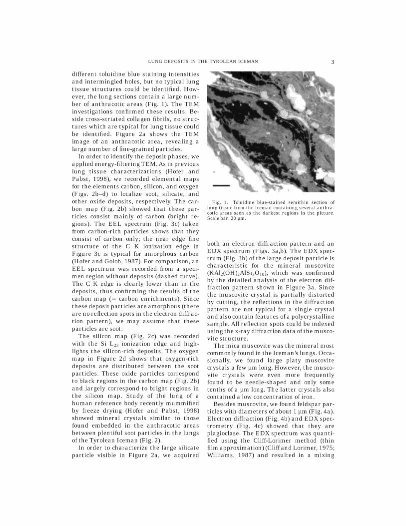
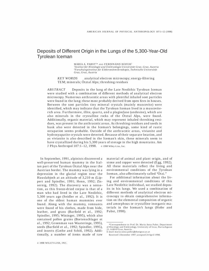
different toluidine blue staining intensitiesand intermingled holes, but no typical lungtissue structures could be identified. How-ever, the lung sections contain a large num-ber of anthracotic areas (Fig. 1). The TEMinvestigations confirmed these results. Be-side cross-striated collagen fibrils, no struc-tures which are typical for lung tissue couldbe identified. Figure 2a shows the TEMimage of an anthracotic area, revealing alarge number of fine-grained particles.
In order to identify the deposit phases, weapplied energy-filtering TEM. As in previouslung tissue characterizations (Hofer andPabst, 1998), we recorded elemental mapsfor the elements carbon, silicon, and oxygen(Figs. 2b–d) to localize soot, silicate, andother oxide deposits, respectively. The car-bon map (Fig. 2b) showed that these par-ticles consist mainly of carbon (bright re-gions). The EEL spectrum (Fig. 3c) takenfrom carbon-rich particles shows that theyconsist of carbon only; the near edge finestructure of the C K ionization edge inFigure 3c is typical for amorphous carbon(Hofer and Golob, 1987). For comparison, anEEL spectrum was recorded from a speci-men region without deposits (dashed curve).The C K edge is clearly lower than in thedeposits, thus confirming the results of thecarbon map (5 carbon enrichments). Sincethese deposit particles are amorphous (thereare no reflection spots in the electron diffrac-tion pattern), we may assume that theseparticles are soot.
The silicon map (Fig. 2c) was recordedwith the Si L23 ionization edge and high-lights the silicon-rich deposits. The oxygenmap in Figure 2d shows that oxygen-richdeposits are distributed between the sootparticles. These oxide particles correspondto black regions in the carbon map (Fig. 2b)and largely correspond to bright regions inthe silicon map. Study of the lung of ahuman reference body recently mummifiedby freeze drying (Hofer and Pabst, 1998)showed mineral crystals similar to thosefound embedded in the anthracotic areasbetween plentiful soot particles in the lungsof the Tyrolean Iceman (Fig. 2).
In order to characterize the large silicateparticle visible in Figure 2a, we acquired
both an electron diffraction pattern and anEDX spectrum (Figs. 3a,b). The EDX spec-trum (Fig. 3b) of the large deposit particle ischaracteristic for the mineral muscovite(KAl2(OH)2AlSi3O10), which was confirmedby the detailed analysis of the electron dif-fraction pattern shown in Figure 3a. Sincethe muscovite crystal is partially distortedby cutting, the reflections in the diffractionpattern are not typical for a single crystaland also contain features of a polycrystallinesample. All reflection spots could be indexedusing the x-ray diffraction data of the musco-vite structure.
The mica muscovite was the mineral mostcommonly found in the Iceman’s lungs. Occa-sionally, we found large platy muscovitecrystals a few µm long. However, the musco-vite crystals were even more frequentlyfound to be needle-shaped and only sometenths of a µm long. The latter crystals alsocontained a low concentration of iron.
Besides muscovite, we found feldspar par-ticles with diameters of about 1 µm (Fig. 4a).Electron diffraction (Fig. 4b) and EDX spec-trometry (Fig. 4c) showed that they areplagioclase. The EDX spectrum was quanti-fied using the Cliff-Lorimer method (thinfilm approximation) (Cliff and Lorimer, 1975;Williams, 1987) and resulted in a mixing
Fig. 1. Toluidine blue-stained semithin section oflung tissue from the Iceman containing several anthra-cotic areas seen as the darkest regions in the picture.Scale bar: 20 µm.
3LUNG DEPOSITS IN THE TYROLEAN ICEMAN
ratio of about 60%:40% between albiteNa(AlSi3O8) and anorthite Ca(Al2Si2O8).Therefore, this phase is most probably ande-sine.
Additionally, platy crystals a few µm insize were found in the anthracotic areas(Fig. 4d). Using electron diffraction (Fig. 4e)and EDX spectrometry (Fig. 4f), these particleswere identified as the clay mineral, illite(K,K1,H3O1)(Al,Fe,Mg)2(OH)2(Si,Al)4(O,OH)10.The distinction between muscovite and illite
was made, as stated above, by the combina-tion of x-ray spectrometry and electron dif-fraction, where the latter method has theadvantage that it enables an unequivocalidentification. All the diffraction patterns ofthe crystalline phases have been completelyindexed and compared with the data of thepowder diffraction file; for a given compoundall reflections had to be identified. Thesedata from muscovite and illite, like the otherinvestigated minerals, have been summa-
Fig. 2. Deposits in lung tissue imaged using the elemental mapping technique of energy-filteringTEM. (a) TEM bright field image of an anthracotic area. (b) Carbon elemental map showing thedistribution of the soot particles. (c) Silicon and (d) oxygen elemental map showing the distribution ofsilicate particles.
4 M.A. PABST AND F. HOFER

rized in tables which have not been includedhere.
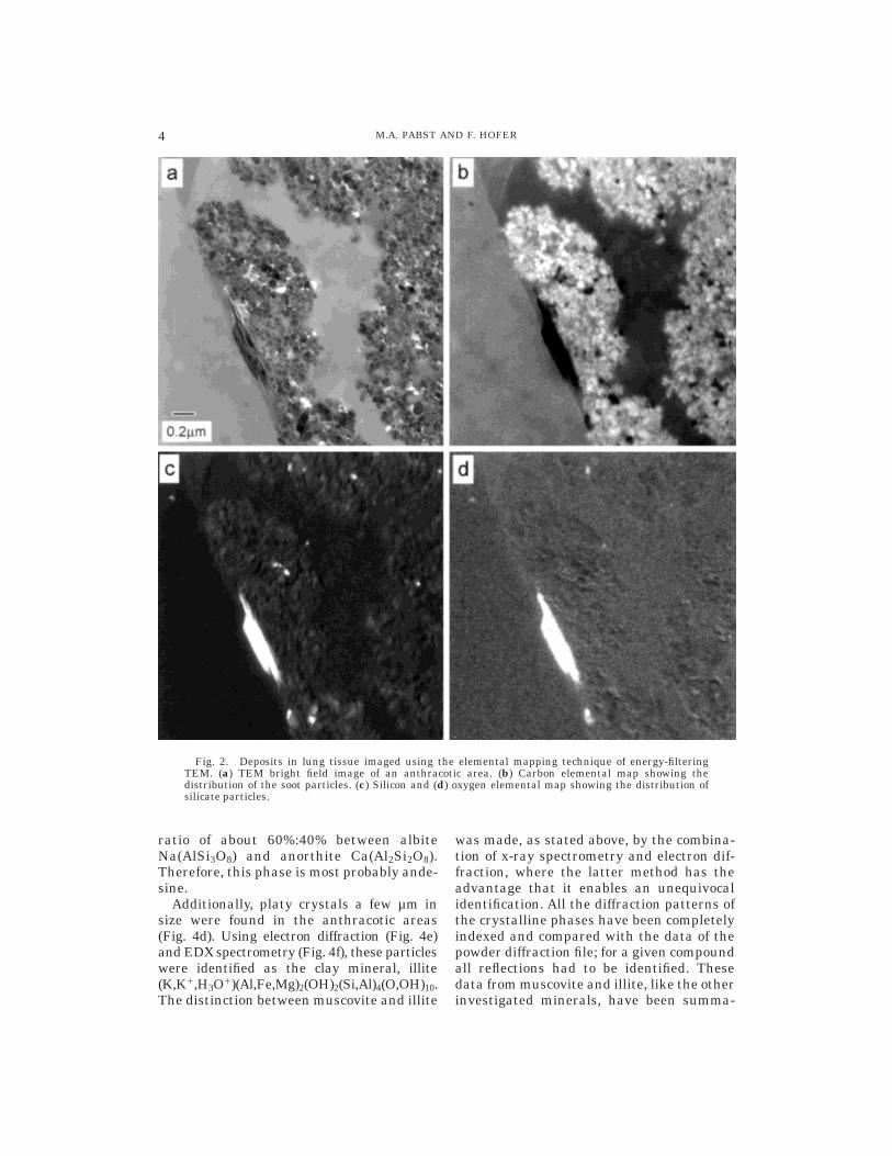
The Tyrolean Iceman also inhaled somesmall quartz particles of some tenths µm insize (Figs. 5a–c).
Not only mineral particles were found inthe anthracotic areas of the Iceman’s lungs.There were also particles exhibiting a wide
range of morphology and size. Figure 5dshows such a particle embedded in an an-thracotic area with a diameter of some tenthsof µm. Electron diffraction revealed that thisparticle is amorphous.
Electron energy loss- and EDX-spectrom-etry showed that this material consistedmainly of carbon and oxygen, sometimes with asmall amount of silicon (Figs. 5e–f). The darkstripes in these particles probably derive fromcutting, as this material seems to be harderthan the surrounding tissue (Fig. 5d).
All the materials described so far werefound in the anthracotic areas, sometimesembedded in and occasionally on the surfaceof the soot aggregates.
Besides these findings, there were alsotwo kinds of crystalline compounds whichwere not found in these anthracotic areas,but were mainly deposited separately in thelung tissue. One of these materials is foundas aggregates of small columnar iron phos-phate crystals (Fig. 6a). EEL spectra, shownin Figure 6c, and electron diffraction pat-terns (Fig. 6b) show that it refers to vivian-ite Fe3(PO4)2 3 8H2O in an oxidized form(Gmelin, 1965). The quantification of theEEL spectrum gives a Fe to P ratio of 0.67and a Fe to O ratio of 0.22; the latter istypical for oxidized vivianite and the nearedge fine structure of the P L23 edge ischaracteristic of phosphate compounds(Hofer and Golob, 1987).
The second kind of crystal found sepa-rately in the lung tissue is hydroxyapatiteCa5(PO4)3OH (Fig. 6d). The crystals werefound as tiny needles; the largest seen areabout 100 nm long and about 7 nm wide.They were found grouped in areas with adiameter of a few µm, where they werearranged in a hexagonal pattern aroundorganic structures (Fig. 6d). This phase wasagain identified with electron diffraction (Fig.6e) and EEL spectrometry (Fig. 6f). Oncemore, the P L23 edge exhibits a fine structurewhich is typical for phosphates (Hofer andGolob, 1987).
These hydroxyapatite crystals were alsofound in round, dense aggregates (Fig. 7), upto 1.8 µm in diameter. In these aggregates,the crystals are found mainly in the form oftiny needles, as described above. Addition
Fig. 3. (a) Electron diffraction pattern of the largemineral particle in Figure 2; the reflection spots havebeen indexed and correspond to the mineral muscovite.(b) EDX spectrum of the large particle shown in Figure 2reveals elemental composition typical for the mineralmuscovite, showing the element specific x-ray lines forAl, Si, K, and O. (c) EEL spectrum of a soot particle(solid) together with the EEL spectrum of a specimenregion without deposits (dashed), showing the element-specific ionization edges for the elements C, N, and O(ionization of the K shells).
5LUNG DEPOSITS IN THE TYROLEAN ICEMAN

Fig. 4. Identification of deposits in an anthracotic area using TEM. (a) TEM bright field image of amineral crystal identified by electron diffraction (b) and EDX-spectrometry (c) as plagioclase (elementspecific x-ray lines for Al, Si, Ca, Na, and O). (d) TEM image of a mineral crystal identified by electrondiffraction (e) and EDX-spectrometry (f) as illite (element-specific x-ray lines for Al, Si, K, Fe, and O).
6 M.A. PABST AND F. HOFER
Fig. 5. Identification of deposits in an anthracotic area using TEM. (a) TEM image of a mineral crystalidentified by electron diffraction (b) and EDX-spectrometry (c) as quartz (element-specific x-ray lines forSi and O). (d) TEM image of an often-found organic inclusion embedded in soot aggregates; the EELspectrum (e) and EDX spectrum (f) of this particle. The spectra reveal the occurrence of carbon, oxygen,and a small amount of silicon. The Pb M edge derives from staining of this section.
7LUNG DEPOSITS IN THE TYROLEAN ICEMAN

Fig. 6. Identification of deposits in lung tissue lo-cated outside of anthracotic areas using TEM. (a) Aggre-gates consisting of small crystals. (b) Electron diffrac-tion and the EEL-spectrum (c) show that the crystalsare vivianite (element-specific ionization edges for P, O,Fe, and C. The background below the P L23 edge wassubtracted to enhance the visibility of the fine structure
typical for phosphates. (d) Hexagonally arranged groupsof nanometer-sized crystals. (e) Electron diffraction and(f) the EEL spectrum show that the crystals are hydroxy-apatite (element-specific ionization edges for P, C, Ca,and O); the background below the PL23 edge is againsubtracted and the net edge is shown on the left side ofthe spectrum.
8 M.A. PABST AND F. HOFER

ally, hydroxyapatite crystals were found inring-shaped structures (Fig. 7) of up to 2–3µm in diameter, but here occurring in theform of round nanometer-sized crystals,which are arranged in concentric layers. Theelemental maps shown in Figure 7 revealthat the visible crystals are all hydroxyapa-tites. The ring seems to fill from the outsideto the center. The ‘‘lumen’’ of the ring is filled
with an electron-lucent organic material con-taining carbon and oxygen.
DISCUSSION
Humans have long been exposed to airpollution. As long as 5,300 years ago theTyrolean Iceman was already inhaling differ-ent kinds of dust. Particulate carbon, mostprobably from open indoor fires, was mainly
Fig. 7. Nanometer-sized hydroxyapatite crystal aggregates located outside of anthracotic areas,arranged in dense aggregates and in ring-like structures. They were characterized using the elementalmapping technique of energy-filtering TEM. (a) TEM bright field image. (b) Calcium elemental map.(c) Phosphorus elemental map. (d) Oxygen elemental map.
9LUNG DEPOSITS IN THE TYROLEAN ICEMAN
responsible for the anthracotic areas in hislung. Lungs of mummies from different cli-matic conditions, from different epochs andmummified in different ways, have beenfound to have anthracotic areas in theirlungs (Brothwell et al., 1969; Zimmerman,1981; Walker et al., 1987; Ascenzi et al.,1996; Hart Hansen and Nordqvist, 1996;Wang, 1996; Zimmerman, 1996). In Egyp-tian mummies (Walker et al., 1987) and inmummies from Xinjiang (China) (Wang,1996) not only soot particles but also double-refracting crystals were identified in thepolarizing microscope, and dust analysesshowed that these crystals were silicates,which were believed to derive from eoliansands (Wang, 1996).
As in Egyptian and Chinese mummies,mineral crystals were also detected in thelungs of the Tyrolean Iceman, in the anthra-cotic areas between the plentiful soot par-ticles. It is not surprising to find muscovitecrystals, as the Otztal Alps, the part of theCentral Alps where the Iceman was foundand where he probably lived, are composedof muscovite-bearing metamorphic rocks, in-cluding large amounts of paragneiss andschists with intercalated muscovite granite-gneiss and two-mica augen-gneiss (Purt-scheller, 1971; Oberhauser, 1980). Mica-richrocks are known to weather easily(Purtscheller, 1971) so that muscovite crys-tals, which are relatively resistant to weath-ering, come free. As muscovite is able tocleave to fine plates, the smallest crystalseasily become airborne and then may beinhaled. Some of these crystals could comedirectly from stones in the fireplace, wherethe heat might liberate them. It is knownthat macrophages in the lung alveoli areable to ingest this material and they areeither coughed up with ingested material ordeposited in the connective tissue of thelung. As there are a lot of muscovite crystalsin the Iceman’s lung tissue and because theinhaled material presumably came from hisliving environment, he may have lived in anarea with mica- (muscovite-) rich rocks. Theillite particles could refer to a muscovite-rich area too, as illite is a hydromica andderives mainly from muscovite by partialhydrolysis, e.g., in mica schist (Rosler, 1984).From the anthropological and archaeologi-
cal points of view, the Tyrolean Iceman mayhave come from the Vintschgau, the east-west Etsch valley south of the Hauslabjoch(Spindler, 1995), probably from the area ofNaturns (Barfield et al., 1992), where LateNeolithic settlements have been found. Thisis an area with muscovite granite-gneissand two-mica augen-gneiss occurrencesnearby. Such rocks contain an abundance ofmuscovite.
Because of their composition of carbonand oxygen, and sometimes silicon, the amor-phous organic particles in the Iceman’s lungscould derive from plant cell wall material.The latter is partly indigestible by alveolarmacrophages because human beings do nothave enzymes to digest special plant cellwall materials. These could then be depos-ited in the anthracotic areas like inorganicmaterial. The particles no longer exhibitspecial cell wall structures, but this could bedue to the long storage or partial digestionby the macrophages. This organic materialin the lungs could be threshing residues, asbotanical analyses of the Iceman’s fur cloth-ing revealed two grains of one-grained wheat(Triticum monococcum) still in the husks(Bortenschlager et al., 1992), and threshedfragments of the same wheat and of Triti-cum sp. were found in his birch-bark vessel(Spindler, 1992). Cell wall material withsimilar composition to that in the lungs ofthe Iceman was also detected in recent wheatthreshing residues (Pabst and Hofer, unpub-lished). These results give further support toSpindler’s (1996) assumption that the Ice-man at least occasionally was in contactwith a grain-growing community. He couldhave helped to harvest and thresh the grainbefore leaving the community for the highmountains, where he may have died inautumn because of an early onset of winter(Spindler, 1996).
As vivianite and hydroxyapatite are foundoutside the anthracotic areas, we assumethat these crystals were not inhaled butwere crystallized after the death of theTyrolean Iceman. Vivianite is also known tobe formed in fossil bones and teeth (Gmelin,1965). Vivianite has crystallized not only inthe lungs but also in some areas on thesurface of the Iceman’s skin, where bluepustules were described (Tiefenbrunner.
10 M.A. PABST AND F. HOFER
1992). These were interpreted as air-oxi-dized vivianite, possibly crystallized on con-tact regions between the body surface andiron-containing material, an interpretationthat was recently verified by chemical analy-ses (Tessadri, 1998). Additionally, iron andphosphate were found using x-ray micro-analysis in ultrathin sections of the Ice-man’s skin (Van der Velden, 1995).
When the Iceman was found in a rockhollow he was partly lying in melted ice(Henn, 1992). As the discovery site in factconsists of iron-containing para/ortho-gneiss(Tessadri, 1998), leached iron ions in themelted ice may have entered the Iceman’slungs through his mouth. The phosphatemay derive from excrement. The rock hollowwhere the Iceman was found seems to be anideal place for humans and animals to shel-ter, and recently chamois and semi-wildsheep were seen there (A. Lippert and G.Patzelt, personal communications) and leftexcrement. Additional excrement in the hol-low comes from large flocks of sheep that forcenturies, perhaps even in the Iceman’stime, were driven in the spring from theVintschgau through the Schnalstal and overpasses to the meadows of the northernOtztal and its side valleys and were drivenback in autumn (Spindler, 1995). There wasalso excrement at the site which may derivefrom the Late Neolithic (Barfield et al.,1992) along with a few old bones. Excrementand bones are rich in phosphates, whichmay have mixed into the melted ice. Duringthe 5,300 years that the Iceman lay in statein the Otztal Alps, the glacier advanced andreceded several times (Spindler, 1995).Probably there were times when the Icemanlay in a slush of ice and water, then under-went freeze-drying when the temperaturedropped. Such conditions would have permit-ted crystallization of the phosphates.
Since iron and phosphates are compo-nents of the human body as well, it is alsopossible that these mineral crystals mayhave originated in the Iceman’s own body.
Apatite crystals similar to those we foundin the Tyrolean Iceman have also been re-ported in an Alaskan mummy (Zimmermanet al., 1971). The hydroxyapatite crystals inthe Iceman could derive from calcium andphosphates from the above-described excre-
ment or bones, as is known from areas withguano deposits (Gmelin, 1965). Calciumcould also come from weathered feldspar atthe site, or could have come from endog-enous calcium and phosphates in the Ice-man’s body. Further evidence of a secondarycrystallization of hydroxyapatite is given byx-ray microanalysis of the Iceman’s skin(Van der Velden et al., 1995), where calciumphosphate was also detected.
In conclusion, it can be said that the lungsof the Tyrolean Iceman contain inorganicand organic materials that were inhaled andso deposited during his lifetime; these mate-rials tell us something about his environ-ment and his work. On the other hand, hislungs provide evidence of secondary crystal-lization that occurred during his 5,300 yearsof storage high in the mountains.
ACKNOWLEDGMENTS
For the opportunity to do this study wethank the Forschungsinstitut furAlpine Vor-zeit, Innsbruck, especially W. Platzer and O.Gaber for information pertaining to thiswork. We thank G. Dohr (Inst. fur Histologieand Embryologie) for making contacts andhelpful discussions, A. Fenninger (Inst. furGeologie und Palaontologie), M. Gailhofer(Inst. fur Pflanzenphysiologie), G. Reibneg-ger (Medizinisch-Chemisches Institut) (allUniv. of Graz), K. Stattegger (Inst. fur Geolo-gie, Univ. Kiel), F. Purtscheller and P. Mir-wald (Inst. fur Mineralogie und Petrogra-phie, Univ. Innsbruck), and A. Lippert (Inst.fur Ur-und Fruhgeschichte, Univ. Wien), G.Patzelt (Inst. fur Hochgebirgsforschung) fordiscussion and helpful suggestions, K. Ir-golic for providing research material, and E.Schoninkle and A. Blaschitz for technicalassistance.
LITERATURE CITED
Ascenzi A, Bianco P, Nicoletti R, Ceccarini G, FornaseriM, Graziani G, Giuliani MR, Rosicarello R, CiuffarellaL, and Granger-Taylor H (1996) The roman mummy ofGrottarossa. In Spindler K, Wilfing H, Rastbichler-Zissernig E, zur Nedden D, Nothdurfter H (eds):Human Mummies (The Man in the Ice, Vol. 3). Wien:Springer, pp 83–92.
Barfield L, Koller E, and Lippert A (1992) Der Zeuge ausdem Gletscher. Wien, Ueberreuter.
Berger A and Kohl H (1993) Optimum imaging param-eters for elemental mapping in an energy filteringtransmission electron microscope. Optik 92:175–193.
Bortenschlager S, Kofler W, Oeggl K, and Schoch W(1992) Erste Ergebnisse der Auswertung der vegetabi-
11LUNG DEPOSITS IN THE TYROLEAN ICEMAN
lischen Reste vom Hauslabjochfund. In Hopfel F,Platzer W, Spindler K (eds): Der Mann im Eis, Vol 1.Innsbruck: University of Innsbruck, pp 307–320.
Brothwell DR, Sandison AT, and Gray PHK (1969)Human biological observations on a Guanche mummywith anthracosis. Am. J. Phys. Anthropol. 30:333–347.
Cliff G and Lorimer GW (1975) The quantitative analy-sis of thin specimens J. Microsc. 103:203–215.
De Ruijter WJ (1995) Imaging properties and applica-tions of slow-scan CCD cameras suitable for electronmicroscopy. Micron 26:247–276.
Egerton RF (1996) Electron Energy-Loss Spectroscopyin the Electron Microscope. New York: Plenum Press.
Egg M (1992) Zur Ausrustung des Toten vom Hauslab-joch, Gem. Schnals (Sudtirol). In Hupfel F, Platzer W,Spindler K (eds): Der Mann im Eis, Vol. 1. Innsbruck:University of Innsbruck, pp 254–272.
Gmelins Handbuch der Anorganischen Chemie, A (1965)Weinheim/Bergstrabe: Verlag Chemie GmbH.
Gothe R and Schol H (1992) Hirschlausfliegen (Diptera,Hippoboscidae: Lipoptena cervi) in den Beifunden derLeiche vom Hauslabjoch. In Hopfel F, Platzer W,Spindler K (eds): Der Mann im Eis, Vol 1. Innsbruck:University of Innsbruck, pp 299–306.
Groenman van Waateringe W (1995) Pollenanalyse alsIndikator fur das Gerbeverfahren bei den Tierfellendes Mannes vom Tiesenjoch. In Spindler K, Rast-bichler-Zissernig E, Wilfing H, zur Nedden D, Noth-durfter H (eds): Der Mann im Eis, Neue Funde undErgebnisse (The Man in the Ice, Vol. 2). Wien: Springer,pp 67–70.
Gubbens AJ and Krivanek OL (1993) Applications of apost-column imaging filter in biology and materialsscience. Ultramicroscopy 51:146–159.
Hart Hansen JP and Nordqvist J (1996) The mummyfind from Qilakitsoq in northwest Greenland. In Spind-ler K, Wilfing H, Rastbichler-Zissernig E, zur NeddenD, Nothdurfter H (eds): Human Mummies (The Manin the Ice, Vol. 3). Wien: Springer, pp 107–121.
Henn R (1992)Auffindung und Bergung der Gletscherlei-che im Jahre 1991. In Hopfel F, Platzer W, Spindler K(eds): Der Mann im Eis, Vol 1. Innsbruck: Universityof Innsbruck, pp 88–91.
Hofer F (1991) Determination of inner-shell cross-sections for EELS-quantification. Microsc. Microanal.Microstruct. 2:215–230.
Hofer F and Golob P (1987) New examples for near-edgefine structures in electron energy-loss spectroscopy.Ultramicroscopy 21:379–384.
Hofer F and Pabst MA (1998) Characterization of depos-its in human lung tissue by a combination of differentmethods of analytical electron microscopy. Micron29:7–15.
Hofer F, Warbichler P, and Grogger W (1995) Imaging ofnanometer-sized precipitates in solids by electronspectroscopic imaging. Ultramicroscopy 59:15–31.
Johnson DE (1979) Energy-loss spectrometry for biologi-cal research. In Hren JH, Goldstein JI, Joy DC (eds):Introduction to Analytical Electron Microscopy. NewYork: Plenum Press, pp 245–258.
Kothleitner G (1996) Beitrage zur quantitativen Nanobe-reichsanalytik mittels EELS und EFTEM im TEM.PhD Thesis, Graz University of Technology, Graz,Austria.
Krivanek OL, Gubbens AJ, and Dellby N (1991) Develop-ments in EELS instrumentation for spectroscopy andimaging. Microsc. Microanal. Microstruct. 2:315–332.
Krivanek OL, Gubbens AJ, Dellby N, and Meyer CE(1992) Design and first applications of a post-columnimaging filter. Microsc. Microanal. Microstruct. 3:187–199.
Krivanek OL, Gubbens AJ, Kundmann MK, and Carpen-ter GC (1993) Elemental mapping with an energy-selecting imaging filter. In Bailey GW, Rieder CL
(eds): Proc. 51st EMSA Meeting. San Francisco: SanFrancisco Press, pp 586–587.
Lippert A and Spindler K (1991) Die Auffindung einerfruhbronzezeitlichen Gletschermumie am Hauslab-joch in den Otztaler Alpen (Gem. Schnals). Archaolo-gie Osterreichs 2:11–17.
Oberhauser R (1980) Der Geologische Aufbau Oster-reichs. Wien: Springer.
Purtscheller F (1971) Otztaler und Stubaier Alpen.Berlin, Stuttgart: Gebr. Borntrager.
Seidler H, Bernhard W, Teschler-Nicola M, Platzer W,zur Nedden D, Henn R, Oberhauser A, and Sjovold T(1992) Some anthropological aspects of the prehistoricTyrolean Ice Man. Science 258:455–457.
Rosler HJ (1984) Lehrbuch der Mineralogie. Leipzig,VEB Deutscher Verlag fur Grundstoffindustrie.
Spindler K (1992) Der Mann im Eis. Sandoz Bulletin 99.Spindler K (1995) Der Mann im Eis. Munchen, Gold-
mann.Spindler K (1996) The iceman’s last weeks. In Spindler
K, Wilfing H, Rastbichler-Zissernig E, zur Nedden D,Nothdurfter H (eds): Human Mummies (The Man inthe Ice, Vol. 3). Wien: Springer, pp 249–263.
Tessadri R (1998) Vivianite from the iceman of theHauslabjoch (Tyrol, Austria): Mineralogical-chemicaldata. In Bortenschlager S (ed): Biogenous Finds Dis-covered Along with the Iceman (The Man in the Ice,Vol. 4). Wien: Springer.
Tiefenbrunner F (1992) Bakterien und Pilze, ein Prob-lem fur unseren altesten Tiroler? In Hopfel F, PlatzerW, Spindler K (eds): Der Mann im Eis, Vol. 1. Inns-bruck: University of Innsbruck, pp 100–107.
Van der Velden E, den Dulk L, Leenders H, DingemansK, van der Bergh M, Weerman M, van der Putte S,Vuzevsky V, and Naafs B (1995) The decorated body ofthe man from Hauslabjoch. In Spindler K, Rastbichler-Zissernig E, Wilfing H, zur Nedden D, Nothdurfter H(eds): Der Mann im Eis, Neue Funde und Ergebnisse(The Man in the Ice, Vol. 2). Wien: Springer, pp275–278.
Walker R, Parsche F, Bierbrier M, and McKerrow JH(1987) Tissue identification and histologic study of sixlung specimens from Egyptian mummies. Am. J.Phys. Anthropol. 72:43–48.
Wang BH (1996) Excavation and preliminary studies ofthe ancient mummies of Xinjiang in China. In Spin-dler K, Wilfing H, Rastbichler-Zissernig E, zur Ned-den D, Nothdurfter H (eds): Human Mummies (TheMan in the Ice, Vol. 3). Wien: Springer, pp 59–69.
Williams DB (1987) PracticalAnalytical Electron Micros-copy in Materials Science, 2nd ed. Mahwah, NJ:Philips Electron Optics Publishing Group.
Wininger J (1995) Die Bekleidung des Eismannes unddie Anfange der Weberei nordlich der Alpen. In Spin-dler K, Rastbichler-Zissernig E, Wilfing H, zur Ned-den D, Nothdurfter H (eds): Der Mann im Eis, NeueFunde und Ergebnisse (The Man in the Ice, Vol. 2).Wien: Springer, pp 119–187.
Zimmerman MR (1996) Mummies of the Arctic regions.In Spindler K, Wilfing H, Rastbichler-Zissernig E, zurNedden D, Nothdurfter H (eds): Human Mummies(The Man in the Ice, Vol. 3). Wien: Springer, pp 83–92.
Zimmerman MR, Yeatman G, and Sprinz H (1971)Examination of an Aleutian mummy. Bull. NY Acad.Med. 47: 80–103.
Zimmerman MR, Trinkaus E, LeMay M, AufderheideAC, Reyman TA, Marrocco GR, Ortel RW, Benitez JT,Laughlin WS, Horne PD, Schultes RE, and CoughlinEA (1981) The paleopathology of an Aleutian mummy.Arch. Pathol. Lab. Med. 105:638–641.
Zissernig E (1992) Der Mann vom Hauslabjoch. Von derEntdeckung bis zur Bergung. In Hopfel F, Platzer W,Spindler K (eds): Der Mann im Eis, Vol. 1. Innsbruck:University of Innsbruck pp 234–244.
12 M.A. PABST AND F. HOFER


