DEPARTMENT OF ZOOLOGY students centric... · OBJECTIVE OF THE LAB Enhance Critical Thinking...
45
DEPARTMENT OF ZOOLOGY (Star Science Department, Conferred by UGC) DEV SAMAJ COLLEGE FOR WOMEN Re-accredated ‘A’ Grade College (2 nd cycle) with 3.75/4 CGPA (Highest in India,2013-14) by NAAC Banglore College of Excellence Status Award by the UGC, New DelhI Ferozpur City, Punjab, India-152002, Hands-on experience via Lab Sessions (State of Art Labs with latest Machinery/ Equipment/ Software)
Transcript of DEPARTMENT OF ZOOLOGY students centric... · OBJECTIVE OF THE LAB Enhance Critical Thinking...

DEPARTMENT OF ZOOLOGY(Star Science Department, Conferred by UGC)
DEV SAMAJ COLLEGE FOR WOMENRe-accredated ‘A’ Grade College (2nd cycle) with 3.75/4 CGPA (Highest in
India,2013-14) by NAAC Banglore
College of Excellence Status Award by the UGC, New DelhIFerozpur City, Punjab, India-152002,
Hands-on experience via Lab Sessions (State of Art Labs with latest Machinery/ Equipment/ Software)

Dept. of ZoologyStar Science Dept Conferred by UGC New Delhi

OBJECTIVE OF THE LAB
Enhance Critical Thinking Learning Skills.
Provide a Broad Overview and Application of Advanced Technology
Develop an understanding of the Physiological and Biochemical
Experiments and its relation with Research and development
Explore the Physical, Morphological, and Physiological
Characteristics of animals in Natural diversity
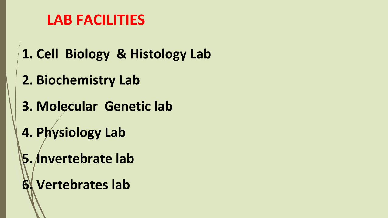
1. Cell Biology & Histology Lab
2. Biochemistry Lab
3. Molecular Genetic lab
4. Physiology Lab
5. Invertebrate lab
6. Vertebrates lab
LAB FACILITIES
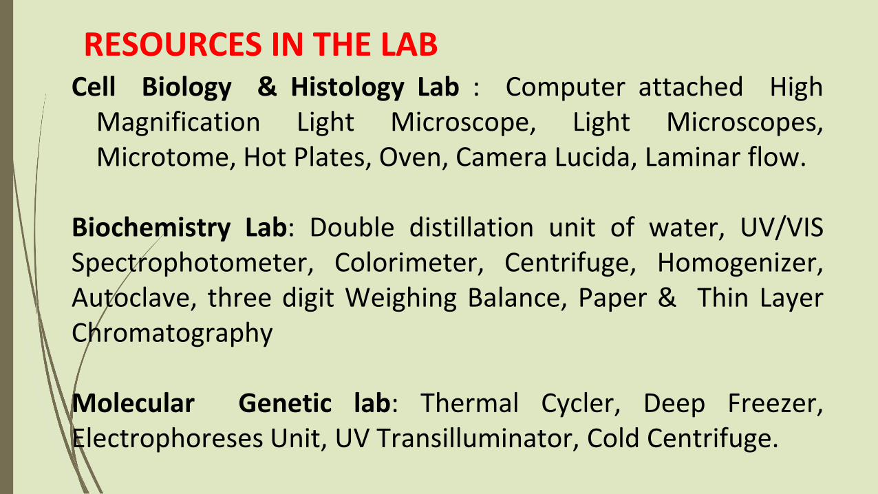
Cell Biology & Histology Lab : Computer attached HighMagnification Light Microscope, Light Microscopes,Microtome, Hot Plates, Oven, Camera Lucida, Laminar flow.
Biochemistry Lab: Double distillation unit of water, UV/VISSpectrophotometer, Colorimeter, Centrifuge, Homogenizer,Autoclave, three digit Weighing Balance, Paper & Thin LayerChromatography
Molecular Genetic lab: Thermal Cycler, Deep Freezer,Electrophoreses Unit, UV Transilluminator, Cold Centrifuge.
RESOURCES IN THE LAB

Continue……
Physiology Lab: Animals storage box, Metabolic study cage,Sphygmomanometer, stethoscope, Hb apparatus, waterbath
Invertebrate lab- Specimen of all Phyla, Staining kits, Insectcollection Net, Insect Collection Box, Permanent stainedslides
Vertebrates lab: Specimen of all Vertebrate classes, Stuffedanimals, preserved animals, desiccators, dissection tray

MICROTOMY
Microtomy is the means bywhich tissues can be sectionedand attached to the surface sothat examination bymicroscopy can take place.
The basic instrument used inmicrotomy is a microtome intowhich cutting tool is clamped.

General Processes
Fixation
Dehydration
Clearing
Embedding
Cutting
Staining

Steps in the processing of tissues
Fixation – preservation of tissues in its original condition.Dehydration – removal of water from tissues.Embedding – infiltration of paraffin wax.Microtomy – preparing thin slices of tissues.Mounting – arranging tissues on slides.Staining – colouring of tissues.

This is the process by which the constituents of cells and tissue are fixed in aphysical and partly also in a chemical state so that they will withstandsubsequent treatment with various reagents with minimum loss ofarchitecture.This is achieved by exposing the tissue to chemical compounds, call fixatives.
Tissues are dehydrated by using increasing strength ofalcohol; e.g. 50%, 70%, 90% and 100%.The duration for which tissues are kept in each strength ofalcohol depends upon the size of tissue, fixative used and typeof tissue; e.g. after fixation in aqueous fixative delicate tissueneed to be dehydrated slowly starting in 50% ethyl alcoholdirectly whereas most tissue specimens may be put into 70%alcohol.
Impregnated tissues are placed in a mould withtheir labels and then fresh melted wax is poured init and allowed to settle and solidify.
Once the block has cooled sufficiently to form asurface skin it should be immersed in cold water tocool it rapidly.
After the block has completely cooled it is cutinto individual blocks and each is trimmed.

Set the wax block on the Hub of Microtome and cut the section with the help of Microtome.
Spread the section on glass slides

Staining
Without staining, the tissue section would remain translucent and we would still have difficulty identifying the relevant cell types and features.
Staining using colored dyes provides a mechanism for introducing contrast.

Staining Cell components such as nucleic acids with a net negative
charge (anionic) stain more readily with basic dyes and are termed basophilic cationic components, such as proteins with many ionized
amino groups, have affinity for acidic dyes and are termed acidophilic.
Examples of basic dyes are toluidine blue, alcian blue, and methylene blue. Hematoxylin , staining behaves like a basic dye basophilic
tissue components.

Tissue Staining
Basophilic Acidophilic
Basophilic
Stain with basic dye [dye+Cl-]
Toluidine blue, methylene blue, hematoxylin, alcian blue
Nucleic acids, some cytoplasmic components (rRNA and rER), glycosaminoglycans and acidic glycoproteins
Stain with acidic dye [Na+dye-]
Orange G, eosin, acid fuschin
Mitochondria, cytoplasm, secretory granules, ECM proteins
H&E Stain(Eosin is the counterstain to Hematoxylin)
Hematoxylin produces a dark blue or purple color,staining DNA in the cell nucleus and other acidicstructures (such as RNA-rich portions of the cytoplasmand the matrix of cartilage).
In contrast, Eosin stains other cytoplasmiccomponents and collagen. In many stainingprocedures certain structures such as nuclei becomevisible, but other parts of cells remain color free.
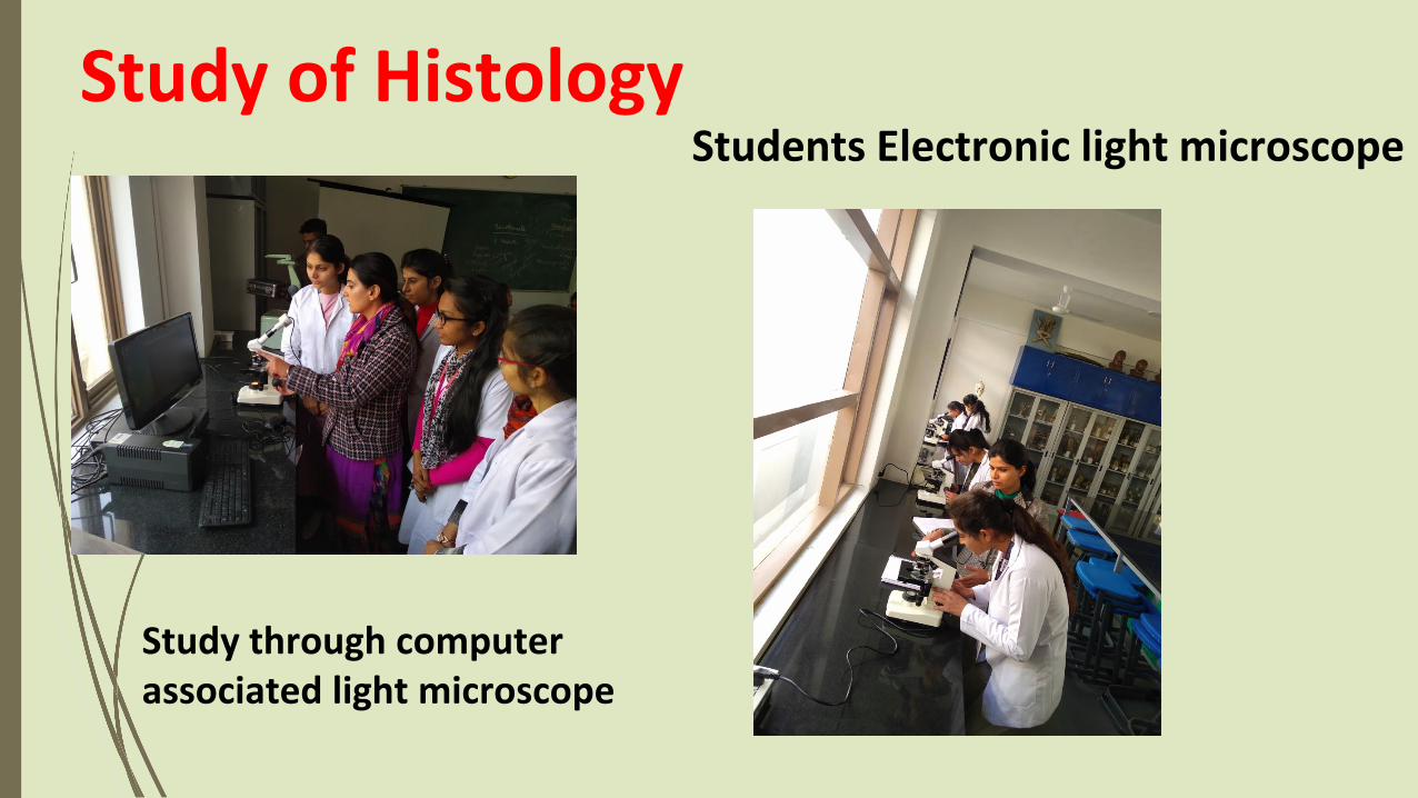
Study of Histology
Study through computer associated light microscope
Students Electronic light microscope

Polymerase Chain ReactionPCR, polymerase chainreaction, is an in-vitrotechnique for amplificationof a region of DNA whosesequence is known orwhich lies between tworegions of known sequence

Primer Selection
Target Selection
Sample Preparation
Critical Steps in PCR
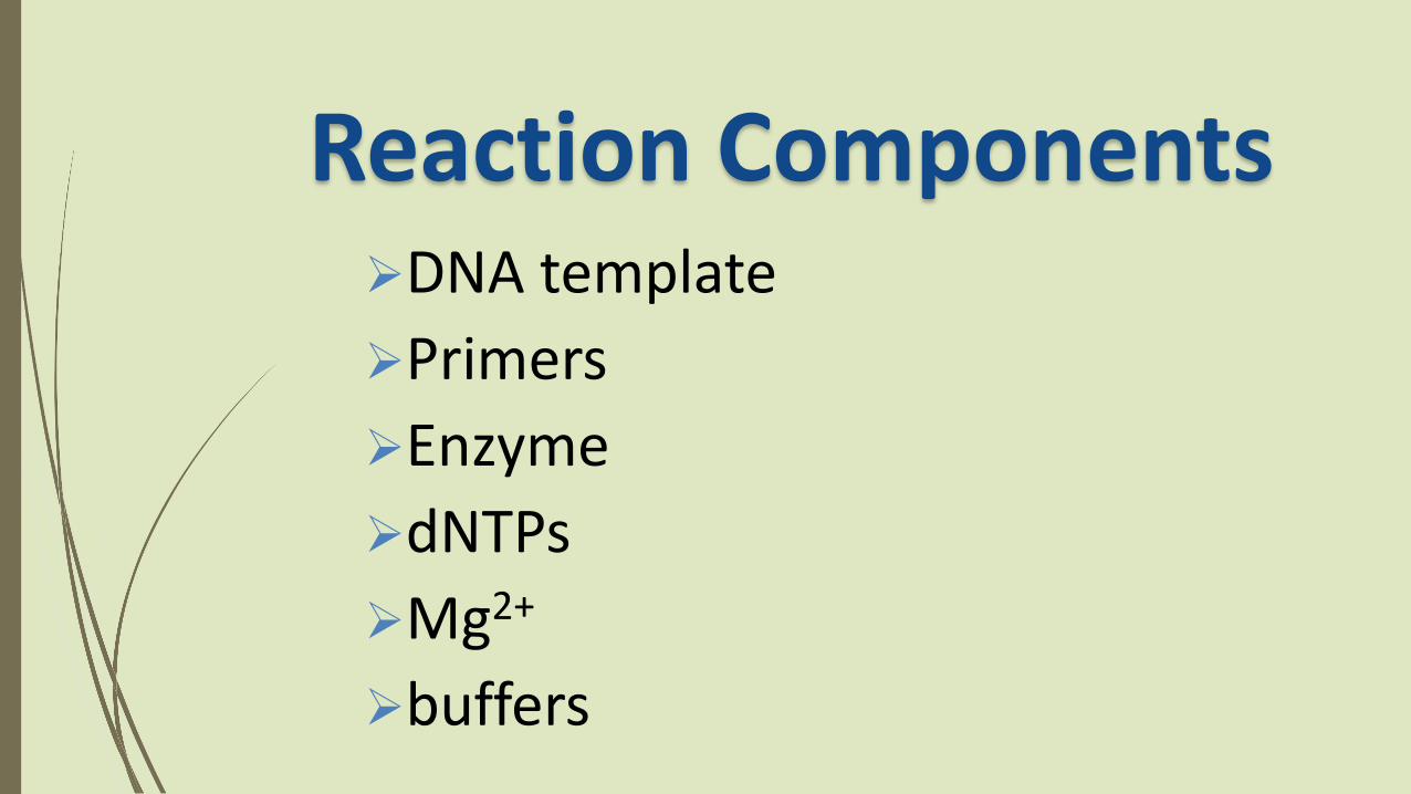
DNA template
Primers
Enzyme
dNTPs
Mg2+
buffers
Reaction Components

Anneal at 55⁰ C
Extension at 72⁰ C
Denature at 94⁰ C
Step Cycle ProgrammeDenaturationTrade off between denaturing DNA and
not denaturing Taq Polymerase
Taq half-life 40min at 95 °, 10min at 97.5°
94° C
AnnealingTrade off between efficient annealing and
specificity
2-5 ° below Tm
ExtensionTemperature optimum for Taq Polymerase
72 °

Sample
DNA Extraction
Addition of premix reagent
Automated Amplification
PCR Protocol
DetectionMutation Detection
SNP Quantification Genotyping
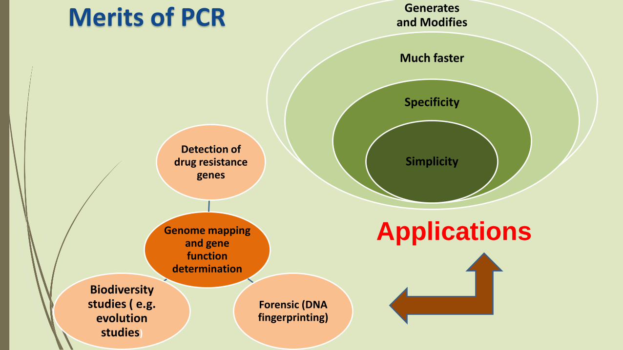
Merits of PCR Generates and Modifies
DNA
Much faster
Specificity
Simplicity
ApplicationsGenome mapping and gene function
determination
Detection of drug resistance
genes
Forensic (DNA fingerprinting)
Biodiversity studies ( e.g.
evolution studies)

Quantitative Proteins Estimation

Specificity and Sensitivity of the Method
Specificity of an assay relates to how good the assay is in discriminating between the requested analyte and interfering substances
Sensitivity of an assay is a measure of how little of the analyte the method can detect

There are many Spectrophotometric method for determining the protein concentration
Bicinchoninicacid method
biuret Lowry BradfordSpectrometric (A280/A260)
There are a wide variety of protein assays available. but each assay has its own advantages and limitationsThe factors that you should consider in choosing a method:
• Sensitivity• The presence of interfering substance• Time available of the assay
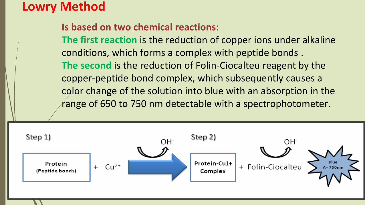
Lowry Method
Is based on two chemical reactions:The first reaction is the reduction of copper ions under alkaline conditions, which forms a complex with peptide bonds .The second is the reduction of Folin-Ciocalteu reagent by the copper-peptide bond complex, which subsequently causes a color change of the solution into blue with an absorption in the range of 650 to 750 nm detectable with a spectrophotometer.

Standard Curve
There is a linear relationship between absorbance and concentration.
The amount of protein in the sample can be estimated using a standard curve of a selected standard protein solution such as (casein ).
Concentration of unknown sample
Standard curve are most commonly used to determine
the concentration of a substance, using serial dilution of
solutions of known concentrations(standard solutions)

Tube Casein Standard Unknown
A
B
C
D
E
F
Set up 7 tubes as follows:
Add 3ml reagent C to all tubes.Mix and let stand at room temperature for 15 min. Add 0.5 ml of Folin-Ciocalteu reagent. (Add this reagent to one tube at a time and immediately after adding it mix well). Let the tubes stand at room temperature for 45 min. 5. Read absorbance at 660 nm against the blank.

Glimpse of Biochemistry lab

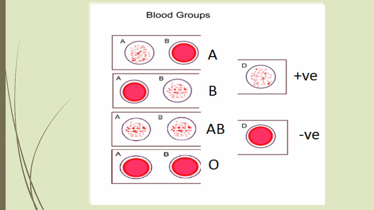
Blood Group
Detection
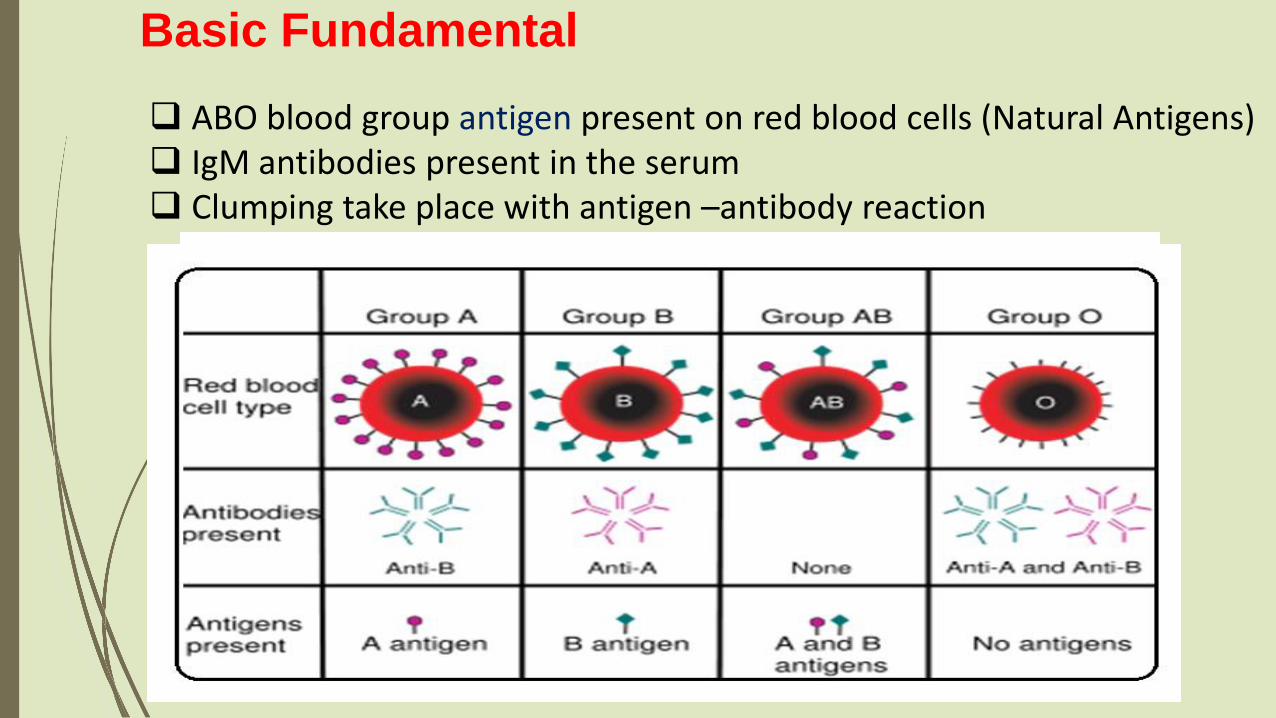
ABO blood group antigen present on red blood cells (Natural Antigens) IgM antibodies present in the serum Clumping take place with antigen –antibody reaction
Basic Fundamental

Common Lab Method- Slide Method
Principle:When red cells are mixed with various
reagent anti-seras (soluble antibody),agglutination will occur on the slides containingcells positive for (possessing the antigen) thecorresponding antigen.
No agglutination will occur when the red cellsdo not contain the corresponding antigen.

Reagents
Anti-A antibodiesAnti-B antibodiesAnti-D
SlidesSterilized NeedlesAlcohol Wooden applicator.

Procedure: 1. On one side of slide labeled anti-A place one drop of
antibody A.2. On the middle of slide labeled anti-B place one drop of
antibody B.3. On last side labeled anti-D place one drop of antibody D4. Place one drop of cells in each antibody containing
circle.5. Carefully mix each solution with a separate applicator
stick.6. Tilt slowly for one minute, then observe for the
agglutination.

Interpretation of the results
Strong agglutination of RBCs in the presence of any ABO grouping reagent constitutes a positive result.
A smooth suspension of RBCs at the end of 2 minutes is a negative result.
Samples that give weak or doubtful reactions should be retested by Tube test ABO grouping


VERTEBRATE LAB SPECMEN
PHYLA/ CLASS
UROCHORDATAPISCES AMPHIBIAREPTILIAAVESMAMMALIA
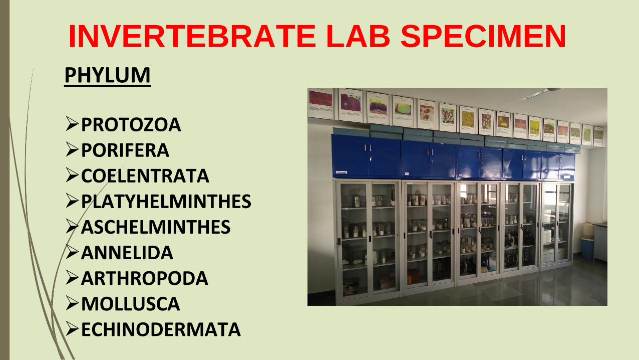
INVERTEBRATE LAB SPECIMENPHYLUM
PROTOZOAPORIFERACOELENTRATAPLATYHELMINTHESASCHELMINTHESANNELIDAARTHROPODAMOLLUSCAECHINODERMATA

RULE OF THE ZOOLOGY LABAlways wear overcoat while working in laboratory. it should be washed at least once a week. Keep the hair and loose garments under check.
Always mop the bench with disinfectant such as 2% phenol before and after use. Be systematic and logical
keep a faithful record of all the experiments and observations. Update it regularly and submit it for evacuation at the end of each exercise.
Always wear gloves if there are cuts on hand or working with hazardous chemicals.
Be familiar about the working of instrument prior to handling it independently keep all the laboratory equipments at their respective place after use.Don’t remove any culture ir any other article from the laboratory.
Continue……
Dispose of infectious material , cultures and contaminated material carefully in container of disinfectant and check its infecting activity regularly.
Dispose of all cultures after autoclaving and pipettes should be placed in disinfectant after use.
Work either using laminar flow chamber or light the burner at least five minutes prior to making any inoculations and work near the burner.
Always discard the disposable and use cotton, match’s stick, paper pieces etc to trash can and never into the sink.

Output of the laboratory Work
Familiar with Modern Tool & Technology
Expert in Handling the Instrument
Trained in Lab Experiment
Able to Perform an Independent Research Project

Gratitude From the Dept. of Zoology