
Decreased Acetylcholine in the Basal Forebrain: Insight to the ... · – Nucleus basalis –...
41
Decreased Acetylcholine in the Basal Forebrain: Insight to the Neurocognitive Deficits in the Subarachnoid Hemorrhage Patient Erol Veznedaroglu, MD Department of Neurosurgery/Division of Cerebrovascular and Endovascular Neurosurgery Thomas Jefferson Hospital for Neuroscience Philadelphia PA
Transcript of Decreased Acetylcholine in the Basal Forebrain: Insight to the ... · – Nucleus basalis –...

Decreased Acetylcholine in the Basal Forebrain: Insight to the Neurocognitive Deficits in the Subarachnoid Hemorrhage
Patient Erol Veznedaroglu, MD
Department of Neurosurgery/Division of Cerebrovascular and Endovascular
Neurosurgery Thomas Jefferson Hospital for Neuroscience Philadelphia PA

Jefferson Hospital for Neuroscience

Collaborators• Elisabeth Van Bockstaele, PhD• John Birknes MD


Subarachnoid Hemorrhage• Verbal Fluency • Short/Long Term Memory• Temporal Lobe Dysfunction• “Global Amnestic Syndrome”

GOS ≠ QOL• 1 year
Excessive fatigueLowered work status
• 4-7 year follow up41% memory problems (affecting ADL)35% daytime sleepiness20% reduced ability to work49% changed personality
Ogden et al Neurosurgery 1997 Jul;41(1):25-34

Etiology?• Blood products• Subarachnoid Blood extravasation does
NOT induce Neurocognitive alterationsGermano et al Acta Neurochir 1998;140(8): 805-11

Vascular Hypothesis• Frontal lobe dysfunction resultant of
focally decreased perfusion and or depressed metabolism…
• Vasospasm NOT indicator of deficitHillis et al J Neurol Neurosurg Psychiatry 2000;69:608-615

Positive Correlates• Higher Fisher grade, frontal hematoma,
intraventricular hemorrhage correlates with significanltly worse neurocognitive outcome – Memory– Concentration– Attention – PerseverationHutter,et al Neurosurgery 1998 Nov;43(5):1054-65

Cont’d• Location of blood as well as amount of
blood on CT correlated with memory dysfunction Acom’s show both long term and short term memory deficitsLarsson et al Acta Neurol Scand 1994 Nov;90(5):331-6Vilkki et al Neurosurgery 1989 Aug;25(2):166-72Mavaddat et al j Neurosurgery 1999 Sep;91(3) 402-7

Rationale: Deficits after ICH• ICH found to cause deficits of higher cortical
functioning– Short-term & long-term memory impairment from
caudate ICH• Fuh et al. 1995
– Deficits of higher-level perceptual functions• Su et al. 2000
– Unilateral sensory neglect & constructional apraxia
• Maeshima et al. 2002

Rationale: ACh-related deficits• Acetycholine implicated in learning and
memory– In particular visuospatial discrimination tasks in
marmosets treated with scopolamine (cholinergic-R blocking agent)
– Ridley et al. 1984• Impairment of memory in lesioned animals
– Lesions to VDB (cholinergic basal forebrain projection to hippocampus) or NBM.
– Ridley et al. 1988, 1989, Barefoot & Ridley 2000

Alzheimer’s Dementia• Recent/ Remote Memory• Learning Memory• Calculation• Abstract Verbal Thinking• Irritability/Concentration
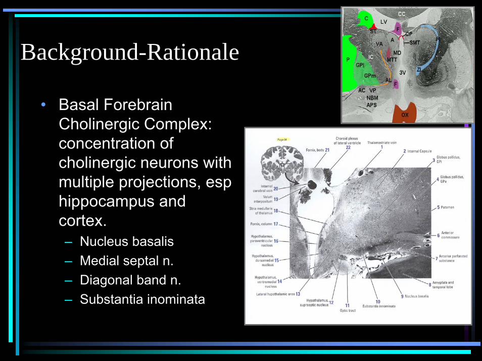
Background-Rationale
• Basal Forebrain Cholinergic Complex: concentration of cholinergic neurons with multiple projections, esp hippocampus and cortex.– Nucleus basalis– Medial septal n.– Diagonal band n.– Substantia inominata

Background-Rationale
• In humans loss of cholinergic cells in n. basalis of Meynert seen with Alzheimer’s dementia.– Beginning of AD hallmarked by memory impairment– No cortical dysfunction (e.g. hemiparesis, sensory
deficit, visual deterioration).• Cholinergic loss implicated in cognitive dysfxn
after TBI

Experimental Design-Immunohistochemistry
• Ab to C-terminus of vesicular acetycholine transporter (VAT)
• Mark expression in perinuclear regions of soma & nerve terminals
• (VAT)-IR shown to be more sensitive, in cell body as well as axon terminal projections.
Methods• 8 male Sprague Dawley Rats
– 3 100µl injections in medial rostral forebrain
– 3 10µl injections in lateral caudal forebrain– 2 saline control injections

Methods• Focal injections of blood microinjected
into the ventral forebrain • Forty-micron thick tissue sections
processed for immunoperoxidase localization of VAcht using the avidin biotin detection method
• Data analysis using acquisition of digital images using Image Pro Software


From: Butcher, Cholinergic Neurons and NetworksThe Rat Nervous System, Paxinos ed., Academic Press


Results-VAT-IR
• >50% reduction in number of VAT-IR cells
• No variation cranial to caudal (44.7% and 47.4% of control respectively)


Vesicular Acetycholine Transporter Labeling Following Arterial Blood Injections
0
20
40
60
80
100
120
140
160
1 2
Experimental Groups
Non-Lesion SideAB Lesion Side

Results-VAT-IR
050
100150200250300350400450500
Saline ICH
# V
AT
-IR
Cel
ls
SalineICH

Experimental Design-Western Blot
• Confirmatory method to illustrate loss of VAT – presumed to represent cholinergic cell loss
or at least loss of function of cholinergic cells.
• Rats sarcrificed at 5 or 6 days post-op and sections of basal forebrain/VP dissolved in extraction buffer.

Results-Western Blot
• A: non-injected side; B: injected side (ICH vs saline)
• Quantitave results pending

Conclusions• Clinical correlation between hypo-
cholinergic state of Alzheimers and subarachnoid hemorrhage patients “Global Amnestic Syndrome”
• Selective loss of acetylcholine in hemorrhage model with anatomic dependence

Phase II• Larger cohort with dose/location variability• Protective effects of Acetylcholinesterase
inhibitors• Recovery of basal forebrain loss after
treatment with Acetylcholinesterase inhibitors• Pre /Post cognitive testing with treatment in
patients (IRB in progress)

Clinical Correlate (In Rats)• Johnny and the Watermaze

Rationale: ICH pathology• ICH results from rupture of
small penetrating arteries.– ACA: medial lenticulostriates– MCA: lateral lenticulostriates– PCA: thalamogeniculates– Basilar: brainstem perferators
• EM studies: bleeding occurs at or near bifurcation of affected arteries, prominent degeneration of media smooth muscle seen.

Rationale: HTN #1 cause of ICH, HTN ICH in basal ganglia• Hypertension (HTN)
leading cause of ICH.• Chronic HTN causes
degenerative changes in vessel wall reduced compliance, increase likelihood rupture.
• Basal Ganglia is most common site for hypertensive ICH

Summary• ICH accounts for 10-20% strokes• BG as frequent site of ICH (result of HTN)• Deficits from ICH of higher cognitive function
– Memory, perceptual functions and others• ACh & basal forebrain cholinergic complex
implicated in learning and memory– Especially visuospatial tasks
• Is there a connection???

Experimental Design• Anatomy of rat brain
– C-Pu– Ventral Pallidum ~
GPi– Basal nucleus
• Cholinergic basal forebrain complex very similar.

Experimental Design• Blood extracted from
femoral artery• Stereotactic injection
of 50-100 µL blood into ventral pallidum
• Rats sacrificed at 5 days

Experimental Design-Immunohistochemistry

Experimental Design-Western Blot
• Confirmatory method to illustrate loss of VAT – presumed to represent cholinergic cell loss
or at least loss of function of cholinergic cells.
• Rats sarcrificed at 5 or 6 days post-op and sections of basal forebrain/VP dissolved in extraction buffer.


