Dan Hicks, DVM, MS, DACVIM (Neurology/Neurosurgery) Veterinary Neurology & Neurosurgical...
37
Dan Hicks, DVM, MS, DACVIM (Neurology/Neurosurgery) Veterinary Neurology & Neurosurgical Specialists
-
Upload
dominique-colleton -
Category
Documents
-
view
240 -
download
3
Transcript of Dan Hicks, DVM, MS, DACVIM (Neurology/Neurosurgery) Veterinary Neurology & Neurosurgical...

Dan Hicks, DVM, MS, DACVIM (Neurology/Neurosurgery)
Veterinary Neurology & Neurosurgical Specialists

1. Clinical features
2. Anatomic localization
3. Differential diagnosis
4. Plan
Clinical Neurology:
A
B

Differential Diagnoses: dependent on where, when (acuity), what (signalment)
Exam tells us where not what!
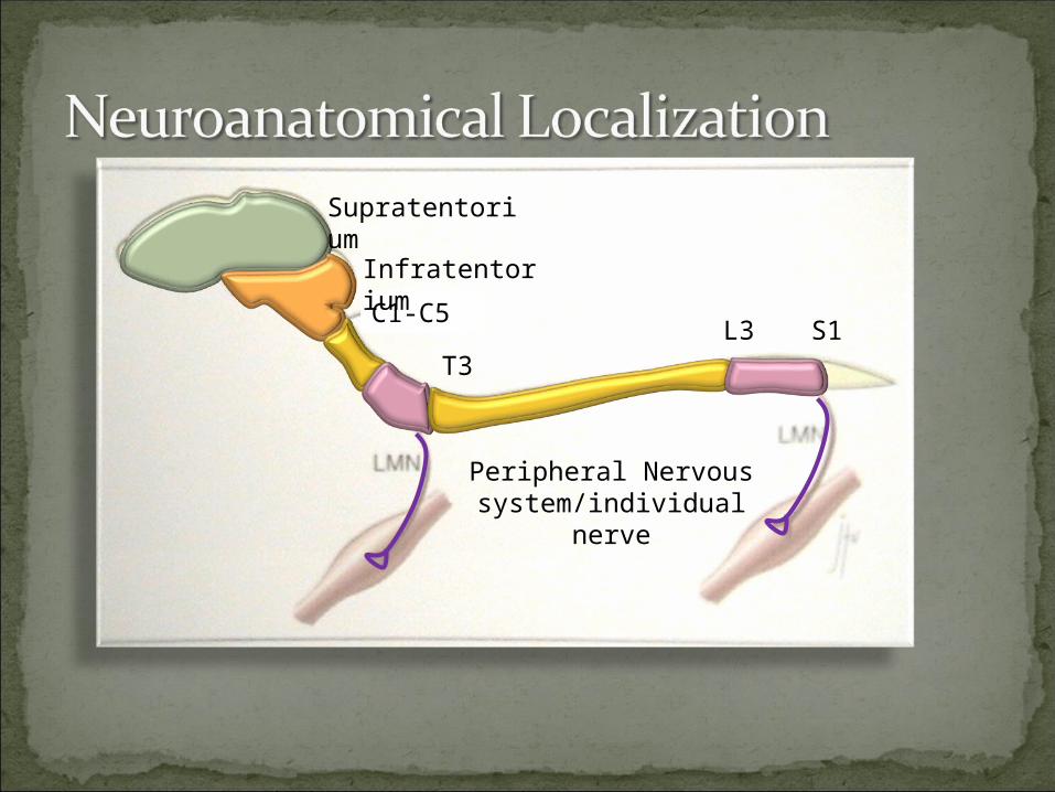
T3L3 S1
C1-C5
Infratentorium
Supratentorium
Peripheral Nervous system/individual nerve


1. Paresis/Paralysis – hopping is normal (brisk) if voluntary movement remains
2. Hypo to atonic3. Hypo to areflexic4. Neurogenic mm atrophy

Ability to support wt decreasedShort stride (step-distance)Bunny-hoppingCollapsingFine mm tremors (orthostatic tremors)Not ataxic (not proprioceptive disorder)

Paresis: delays in gait generation, hopping is particularly slow
Spasticity: release of antigravity mm from inhibition
Hypereflexia/tonia/crossed extensor: release of inhibition
Basis for decrebrate rigidity (lesion b/n rostral and caudal colliculi


Signs explained by focal lesion?Think “structural” problem – compression or
inflammationGeneralized lower motor neuron?
Think botulism, tick paralysis, or Coon hound paralysis


“Structural” lesions - Imaging
MRI CT

Radiographs:Osseous lesions
Fracture/luxationsDiskospondylitisOsteoproliferation/osteolytic diseases
“In-direct” evaluationChest films in older dogs?Screening for metallic objects


InvasiveSeizures

On CT, bone is ALWAYS WHITEBone is black or dark on MRIn bone window CT, trabecular pattern
of bone is evidentIn soft tissue window CT, bone is bright
whiteIn a T1-weighted MRI image, fat is
WHITE. CSF is darkIn a T2-weighted image, CSF is WHITE

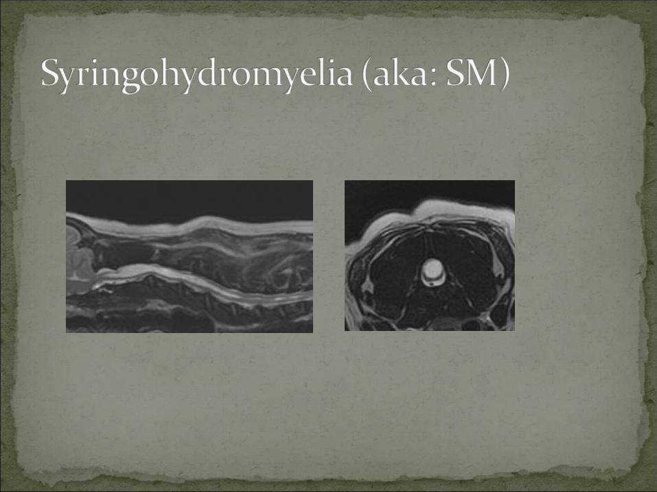
Normal cordHerniated Disk
•Great bone detail•Soft-tissue detail: moderate


Disk ruptur
e
Spinal lymphoma


Age of advanced imagingAdvancement in neurosurgeryGreat, but…$$$$Concomitant illness
Consider neurologic scoreLess severe signs = higher success rate
50% improve or recovery without surgeryWarning for deep pain negative

1.“Pain and Pee”pain control speeds healing times, improves
efficacy of cage rest and rehab2. CAGE REST
Allows injury annulus fibrosus to heal and inflammation to subside
Minimizes additional traumaMinimizes volume of extruded disk materialTry sedative if pet is anxious
RehabFocal spinal injections

Length of cage rest not well studiedTraditionally, 4-6 weeks (time for ligamentous
healing)Shorter times may be just as goodEarly controlled movement (rehab) may
promote healing

Prevent bed soresMonitor of UTIHome rehab exercises
Most owners enjoy participating in pet’s recovery
Profession rehab

OpioidsNSAIDSGabapentinAmantidineFocal injections
Prazosin Phenyoxybenzamine Diazepam
**Bladder expression or catheterization may be needed
BLADDER

Possible benefits:Reducing inflammation of injured cordAltering propagation of inflammatory
mediatorsSide effects:
GI ulcerationPneumoniaUTILonger hospital stays (Levine, 2007)

One study: Dogs receiving steroids had lower “quality of life” index scores than those not receiving steroids (Levine 2007)
Currently no scientific evidence supporting use in IVDD
True answer still remains