f. CN 4: lateral rotation of bulb - Delaware Valley Academy...
33
Guide to Neurolocalization William Bush, VMD, DACVIM (Neurology) Introduction In clinical neurology disease of segments of the nervous system have distinct clinical signs. A neuroanatomic diagnosis occurs when a constellation of clinical signs indicate there is a lesion within a segment of the nervous system. These segments include the brain, spinal cord, and peripheral nerve/muscle. The brain can be further usefully divided into the Forebrain (cerebral hemispheres and thalamus), Brainstem (midbrain to medulla), Cerebellum and Vestibular System Forebrain (Cerebral & Thalamic dysfunction) A patient with a right forebrain or thalamic lesion may act confused, compulsively circle to the right and have diminished to absent postural reactions on left with a normal gait, and an absent menace on left with normal pupil light response. This is outlined below with a brief explanation. 1. Seizure - synchronized discharges are generated by the grey matter of the cerebral cortex. Disease in the cerebral cortex or thalamus or the connection between these structures can cause seizure. Seizures only originate from the cerebral hemispheres and thalamus. 2. Altered mental status - these phenomena probably revolve around an altered perception. a. Aggression b. Inappropriate urination and defecation c. Dementia, disorientation, lethargy, and if bilateral and severe disease- stupor, coma d. Head pressing, aggression, pacing, hyperexcitability e. Restless at night and sleeping during the day 3. Normal gait- the generators of gait are below the thalamus 4. Compulsive pacing - patient may continuously propel itself forward despite having obstacles in their path. 5. Circling towards the side of the lesion - with right side lesion the ability to perceive stimulus from left side maybe lost. The patient with a right side lesion only perceives information on the right side of the body and therefore may circle right or have a head turn to the right. 6. Contralateral postural, sensory, and menace deficits - revealed by examination: a. Poor/Absent postural reactions - the proprioceptive information is relayed to the ipsilateral thalamus and then crosses to the opposite somatosensory cortex. b. Hypalgesia - information about pain and sensation also cross to the opposite thalamus and ascend to the opposite cortex. c. Poor/Absent menace – visual information crosses at the optic chiasm and is ultimately projected via the thalamus to the opposite occipital cortex. The response to light does not involve the thalamus or cerebrum thus pupil size and light response are normal. d. Brainstem (midbrain to medulla) A patient with a brainstem lesion is often dull, stuporous or even comatose depending on the severity of the lesion. Gait exam often shows weakness along with ipsilateral postural reaction deficits may also be noted depending on the level of the lesion. 1. Depression, stupor, coma – the reticular activating system located in brainstem 2. Abnormal gait and posture (dysfunction is ipsilateral to lesion) a. Paresis and ataxia b. Poor postural reactions c. Increased to normal tone and reflex These deficits occur because of a lesion within the white matter tracts affecting both descending motor and ascending proprioceptive tracts (see discussion below) 3. Cranial nerve deficits at level of lesion a. CN 12, 10, 9 : poor gag, dysphagia, laryngeal paralysis, megesophagus b. CN 8 : head tilt, nystagmus, positional strabismus, rolling or tight circles c. CN 7 : absent blink, droopy ear and lip, no response to noxious stimulus other than pulling head away or withdrawing globe d. CN 5: abnormal sensation over face, poor jaw tone, atrophy in muscles of mastication e. CN 6: no retraction of the globe, medial strabismus
Transcript of f. CN 4: lateral rotation of bulb - Delaware Valley Academy...

Guide to Neurolocalization William Bush, VMD, DACVIM (Neurology)
Introduction In clinical neurology disease of segments of the nervous system have distinct clinical signs. A neuroanatomic diagnosis occurs when a constellation of clinical signs indicate there is a lesion within a segment of the nervous system. These segments include the brain, spinal cord, and peripheral nerve/muscle. The brain can be further usefully divided into the Forebrain (cerebral hemispheres and thalamus), Brainstem (midbrain to medulla), Cerebellum and Vestibular System
Forebrain (Cerebral & Thalamic dysfunction) A patient with a right forebrain or thalamic lesion may act confused, compulsively circle to the right and have diminished to absent postural reactions on left with a normal gait, and an absent menace on left with normal pupil light response. This is outlined below with a brief explanation.
1. Seizure - synchronized discharges are generated by the grey matter of the cerebral cortex. Disease in the cerebral cortex or thalamus or the connection between these structures can cause seizure. Seizures only originate from the cerebral hemispheres and thalamus.
2. Altered mental status - these phenomena probably revolve around an altered perception. a. Aggression b. Inappropriate urination and defecation c. Dementia, disorientation, lethargy, and if bilateral and severe disease- stupor, coma d. Head pressing, aggression, pacing, hyperexcitability e. Restless at night and sleeping during the day
3. Normal gait- the generators of gait are below the thalamus 4. Compulsive pacing - patient may continuously propel itself forward despite having obstacles in
their path. 5. Circling towards the side of the lesion - with right side lesion the ability to perceive stimulus
from left side maybe lost. The patient with a right side lesion only perceives information on the right side of the body and therefore may circle right or have a head turn to the right.
6. Contralateral postural, sensory, and menace deficits - revealed by examination: a. Poor/Absent postural reactions - the proprioceptive information is relayed to the ipsilateral
thalamus and then crosses to the opposite somatosensory cortex. b. Hypalgesia - information about pain and sensation also cross to the opposite thalamus
and ascend to the opposite cortex. c. Poor/Absent menace – visual information crosses at the optic chiasm and is ultimately
projected via the thalamus to the opposite occipital cortex. The response to light does not involve the thalamus or cerebrum thus pupil size and light response are normal.
d. Brainstem (midbrain to medulla) A patient with a brainstem lesion is often dull, stuporous or even comatose depending on the severity of the lesion. Gait exam often shows weakness along with ipsilateral postural reaction deficits may also be noted depending on the level of the lesion.
1. Depression, stupor, coma – the reticular activating system located in brainstem 2. Abnormal gait and posture (dysfunction is ipsilateral to lesion)
a. Paresis and ataxia b. Poor postural reactions c. Increased to normal tone and reflex
These deficits occur because of a lesion within the white matter tracts affecting both descending motor and ascending proprioceptive tracts (see discussion below) 3. Cranial nerve deficits at level of lesion
a. CN 12, 10, 9 : poor gag, dysphagia, laryngeal paralysis, megesophagus b. CN 8 : head tilt, nystagmus, positional strabismus, rolling or tight circles c. CN 7 : absent blink, droopy ear and lip, no response to noxious stimulus other than
pulling head away or withdrawing globe d. CN 5: abnormal sensation over face, poor jaw tone, atrophy in muscles of mastication e. CN 6: no retraction of the globe, medial strabismus

f. CN 4: lateral rotation of bulb g. CN 3: ptosis, ventrolateral strabismus, dilated and not responsive pupil (3,4,6) connected
to vestibular nuclei and responsible for oculocephalic reflex
Cerebellum A patient with only cerebellar lesion is bright and responsive with preserved strength and minimal postural reaction deficits. However, they have a characteristic high stepping gait, intention tremor, and occasionally a delay in the menace response. Vestibular signs are often noted with cerebellar disease and maybe manifested as head tilt, nystagmus, and/or positional strabismus.
1. Intention tremor – cerebellum is responsible for smoothing out movement 2. Wide-based stance 3. Abnormal gait - high stepping, hypermetric or over-reaching gait, hypometria possible but more
difficult to see 4. No paresis - dogs with pure cerebellar lesions are not weak as the cerebellum coordinates but
does not initiate gait 5. Delayed or exaggerated postural reaction - cerebellum is the integrator of proprioceptive
information 6. Menace deficit - ipsilateral menace deficit as this response coordinated through cerebellum
Vestibular Disease The vestibular system is responsible for the sense of balance. This system includes receptors (semicircular canals) in the inner ear, the connecting nerve and nerve root, and the 4 nuclei nestled in the brainstem around the 4th ventricle. Peripheral vestibular disease is from involvement of the receptor system, nerve, or nerve roots. Central vestibular disease is generated from lesions that involve the vestibular nuclei, portions of the cerebellum, or less commonly the high cervical region. It is very useful to be able to distinguish central from peripheral disease because the diagnostic work-up and prognosis are so different. As you might imagine there is some overlap in the clinical signs of peripheral and central disease, however, there are some distinguishing features of central vestibular disease.
Signs with peripheral vestibular disease: • Head tilt (usually about 20 degrees) • Leaning, rolling, tight circles opposite side of the head tilt • Nystagmus fast phase opposite side of head tilt and rate > 60 beats per minute • Positional strabismus on the same side as head tilt
Signs with central vestibular disease: • Dull or depressed • Absent or extreme head tilt often on same side as other deficits • Nystagmus - vertical, change in direction of the fast phase, fast phase towards head tilt • Cranial nerve deficits other than Facial nerve or Horner’s syndrome • Abnormal gait (high stepping, side stepping left and right, side step toward head tilt,
spinal cord ataxia) • Postural reaction deficits • Neck pain can be seen with many diseases of the brainstem
*If overall clinical presentation is different, opposite or paradoxical to what you would expect from a typical peripheral case then highly suspect central disease. Some examples would be an extreme head tilt without nystagmus, side stepping towards the side of the head tilt, or a waxing and waxing progressive course of disease. ** Note that acutely both peripheral and central vestibular patients may have postural reaction deficits. Dogs with central disease tend to stay the same or get worse versus dogs with idiopathic or reversible peripheral disease will often start to get better in the following 24 hours. Clinical Signs Distinguishing Central from Peripheral Vestibular Disease

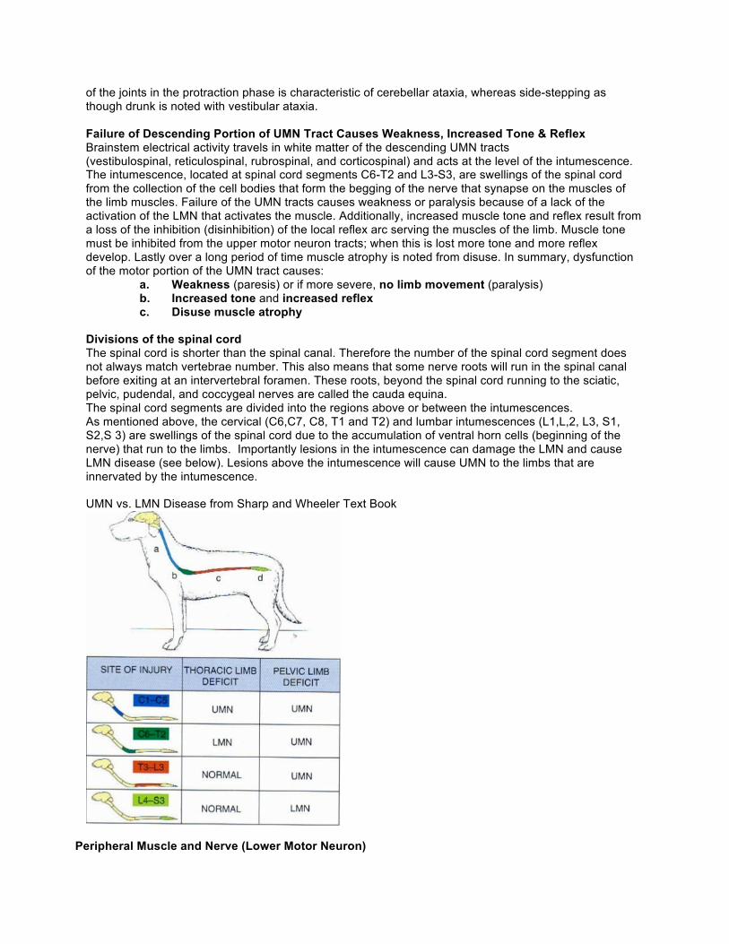
Spinal Cord The divisions of the spinal cord with distinct clinical signs include cervical disease (C1-C5), cervical intumescence (C6-T2), thoracolumbar (T3-L3) and lumbar intumescence / cauda equine (L3-cauda equine) UMN vs LMN Gait generation is primarily a brainstem phenomenon. The upper motor neuron (UMN) system starts in the brainstem and relays information to and from the lower motor neuron system (LMN) consisting of the muscle and nerve. The axonal portions of the UMN neurons descend within the white matter of the spinal cord until synapsing on the LMN cell body located in the grey matter of the spinal cord. As previously stated, the LMN starts in the grey matter of the spinal cord and synapses on the muscle triggering muscle depolarization and contraction. Failure of Ascending Portion of UMN Tract Cause Ataxia and Postural Deficit A lesion of the white matter can cause sensory dysfunction (ataxia, postural deficit) when the ascending tracts are affected and motor dysfunction (weakness to paralysis) when the descending tracts are affected. The ascending tracts (to the cerebellum and contralateral cortex) provide information about limb position- this is called proprioception. When ascending proprioceptive information cannot reach the cerebellum and the somatosensory cortex then the brain cannot determine where the limb is located in space leading to ataxia and postural deficit. When a gait is referred to as ‘ataxic’ it means that an observer can’t consistently predict where the limb will land at the end of the protraction phase. Ataxia means without order. To say a gait is disordered or the animal is ataxic, may mean the patient is long-strided, limbs are too narrow or cross midline, limbs are too wide or circumduct, interfere or all of the above. When we perform postural reactions (hopping, paw flip test, tactile placing) we are testing the patient’s ability to receive information from the proprioceptors and then make the proper adjustments. The loss of this ascending information provides for an abnormal gait with the following characteristics.
a. Long-strided gait - patient does not know where limb is so can be slow to initiate protraction phase of gait.
b. Limbs cross midline - patient does not know where limb is during protraction phase of gait so it may take a course towards midline instead of straight forward
c. Interference – one limb may hit the limb on the opposite side d. Knuckling – the patient does not know that the dorsum instead of the palmer or plantar
surface of the paw is touching the ground e. Circumduction or abduction during protraction phase. f. Limbs too close together or too far apart g. Delayed to absent postural reactions
The ataxia described here is referred to as a proprioceptive or spinal cord ataxia, however, vestibular and cerebellar lesions can also cause ataxia with different characteristic. High stepping where there is flexion
Observation Brainstem / Central Nerve / Peripheral Mentation Dull Normal Gait Weak, spinal cord ataxia, side
step to side of head tilt, side step left and right
Side step, lean or tight circles towards head tilt
Postural Reaction Delayed or absent Normal Head tilt Absent or extreme (> 30 degree) Present and about 20% off
midline Cranial Nerve Deficits Yes Facial, Horner’s tract permissible Nystagmus Vertical
Fast phase towards head tilt Changing direction of fast phase Fewer than 60 beats per minute
Horizontal or/and rotary Fast phase opposite head tilt More than 60 beats per minute
Positional Strabismus Present without head tilt Ventral on side of head tilt Neck pain Maybe present Absent Typical progression of clinical signs
Progressive, wax and wane Sudden onset improving
of the joints in the protraction phase is characteristic of cerebellar ataxia, whereas side-stepping as though drunk is noted with vestibular ataxia. Failure of Descending Portion of UMN Tract Causes Weakness, Increased Tone & Reflex Brainstem electrical activity travels in white matter of the descending UMN tracts (vestibulospinal, reticulospinal, rubrospinal, and corticospinal) and acts at the level of the intumescence. The intumescence, located at spinal cord segments C6-T2 and L3-S3, are swellings of the spinal cord from the collection of the cell bodies that form the begging of the nerve that synapse on the muscles of the limb muscles. Failure of the UMN tracts causes weakness or paralysis because of a lack of the activation of the LMN that activates the muscle. Additionally, increased muscle tone and reflex result from a loss of the inhibition (disinhibition) of the local reflex arc serving the muscles of the limb. Muscle tone must be inhibited from the upper motor neuron tracts; when this is lost more tone and more reflex develop. Lastly over a long period of time muscle atrophy is noted from disuse. In summary, dysfunction of the motor portion of the UMN tract causes:
a. Weakness (paresis) or if more severe, no limb movement (paralysis) b. Increased tone and increased reflex c. Disuse muscle atrophy
Divisions of the spinal cord The spinal cord is shorter than the spinal canal. Therefore the number of the spinal cord segment does not always match vertebrae number. This also means that some nerve roots will run in the spinal canal before exiting at an intervertebral foramen. These roots, beyond the spinal cord running to the sciatic, pelvic, pudendal, and coccygeal nerves are called the cauda equina. The spinal cord segments are divided into the regions above or between the intumescences. As mentioned above, the cervical (C6,C7, C8, T1 and T2) and lumbar intumescences (L1,L,2, L3, S1, S2,S 3) are swellings of the spinal cord due to the accumulation of ventral horn cells (beginning of the nerve) that run to the limbs. Importantly lesions in the intumescence can damage the LMN and cause LMN disease (see below). Lesions above the intumescence will cause UMN to the limbs that are innervated by the intumescence. UMN vs. LMN Disease from Sharp and Wheeler Text Book
Peripheral Muscle and Nerve (Lower Motor Neuron)
The nerve that innervates a muscle of the limb starts within the spinal cord. The parts of the nerve include: a. Cell body - these are the large ventral horn cells. b. Nerve roots - these exit the spinal cord and merge to form a numbered spinal nerve c. Spinal nerve – the numbered nerves exit via intervertebral foramen and merge at a
plexus d. Peripheral nerve - leave the plexus as a named e. Endplate or synapse – named nerve ends at nerve terminal where will release
acetylcholine into the synapse with the muscle leading to muscle depolarization, calcium release, and muscle contraction.
A lesion in any part of the described system will cause what are called lower motor neuron signs. The muscle is also included in this system as muscle disease, endplate disease, nerve disease, nerve root disease, and ventral horn cell disease can all present with similar clinical signs.
a. Short-stided, choppy gait, or lameness - the nerve or muscle damage causes less muscle fibers to be working so overall the limb can only travel a short distance.
b. No ataxia - some sensory information reaches the spinal cord and this information reaches cerebellum and contralateral cortex.
c. Less muscle tone and less reflex - the loss of nerve or muscle means fewer muscle fibers are working.
d. Rapid loss of muscle mass - neurogenic atrophy can cause significant muscle loss in only 5-7 days. This stands in contrast to disuse atrophy which is an upper motor neuron phenomenon, slower, and generally less severe.
Upper Motor Neuron (Spinal Cord) and Lower Motor Neuron (Muscle / Nerve) Disease
Observation Upper Motor Neuron Lower Motor Neuron Gait Long-strided Short-strided Ataxia Yes No Postural Deficit Yes No Tone and Reflex Increased Decreased Muscle Atrophy No Yes
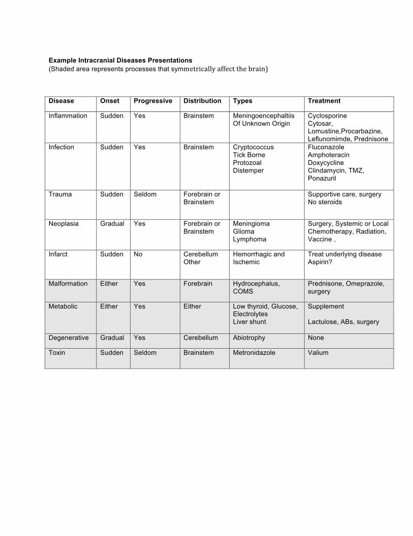
Example Intracranial Diseases Presentations (Shaded area represents processes that symmetrically affect the brain)
Disease Onset Progressive Distribution Types Treatment
Inflammation
Sudden Yes Brainstem
Meningoencephaltiis Of Unknown Origin
Cyclosporine Cytosar, Lomustine,Procarbazine, Leflunomimde, Prednisone
Infection
Sudden Yes Brainstem Cryptococcus Tick Borne Protozoal Distemper
Fluconazole Amphoteracin Doxycycline Clindamycin, TMZ, Ponazuril
Trauma
Sudden Seldom Forebrain or Brainstem
Supportive care, surgery No steroids
Neoplasia
Gradual Yes Forebrain or Brainstem
Meningioma Glioma Lymphoma
Surgery, Systemic or Local Chemotherapy, Radiation, Vaccine ,
Infarct
Sudden No Cerebellum Other
Hemorrhagic and Ischemic
Treat underlying disease Aspirin?
Malformation Either Yes Forebrain Hydrocephalus, COMS
Prednisone, Omeprazole, surgery
Metabolic
Either Yes Either Low thyroid, Glucose, Electrolytes Liver shunt
Supplement Lactulose, ABs, surgery
Degenerative Gradual Yes Cerebellum Abiotrophy None
Toxin Sudden Seldom Brainstem Metronidazole Valium

Seizure-like Episodes: Startling Results of EEG Video Case StudiesWilliam Bush, VMD, DACVIM (Neurology)
Introduction Discerning whether episodes of abnormal behavior, movement or sudden changes in awareness are epileptic seizure or seizure-like can be very difficult. There are many disease processes that can mimic the symptoms of epileptic seizure but not respond anti-epileptic drug (AED). Falsely identifying these processes as seizure will expose the patient to AED side-effects, set false expectations for the owner and delay or preclude the diagnosis of a potentially serious or life-threatening condition. Conversely failure to recognize an event as an epileptic seizure can delay diagnosis of an underlying cause and the application of life preserving AED. This talk will describe our experience discerning seizure from seizure-like episodes using EEG.
ElectroencephalographyIn our clinic, an EEG is the amplified and filtered electrical activity of the cerebral cortex recorded from subcutaneous scalp electrodes and displayed on a laptop screen. The EEG is interpreted vie remote viewing by an expert in both human and animal EEG. Very typically dexmedetomidine is used for the implantation of the electrodes and to eliminate muscle artifact. We have an intramural certification program for out veterinary technicians to help standardize our EEG records. In human medicine electroencephalography (EEG) is used to help distinguish epileptic seizure from movement disorders, psychogenic events and metabolic disease. Additionally, and perhaps more importantly, EEG is used to identify non-convulsive seizure and status epilepticus and then monitor efficacy of AED therapy. This talk will describe our use of EEG for the same purposes.
What happens during a seizure?A seizure is fundamentally an electrical event in the brain which are associated with an easily identified symptoms. During a seizure, a group of neurons synchronizes and depolarizes / repolarizes autonomously and spreads within that hemisphere of the brain due to failure of spatial containment. This hypersynchronous electrical activity then crosses to the other hemisphere capturing the entire brain. Most often the focus is then contained and there is a return to normal activity. Before the seizure or in the pre-ictal state, as the electrical focus is developing and spreading, the patient may experience abnormal visual, auditory, physical, or autonomic nervous system abnormalities. This might be manifested as staring off into space, searching a room, restlessness, clingy behavior, fly biting, circling, odd vocalization, a limb becoming stiff or rhythmically moving, elevated heart rate, dilated pupils, salivation, vomiting. Next the seizure or ictus may be more readily noticed as the focus captures both hemispheres the patient loses consciousness and inhibition of the brainstem motor tracts manifested as the head being arched back and often stiffness of all 4 limbs. The hypersynchronous or rhythmic nature of the electrical focus can be noted as paddling or all 4 limbs. A failure to control and regulate the breathing can manifest as apnea and paradoxical breathing where the diaphragm and intercostal muscles are not working together. Perturbations in the autonomic nervous system can lead to bradycardia or tachycardia, profuse salivation, urination, defecation, miosis or mydriasis, and piloerection. During the seizure there is unregulated discharge of neurotransmitters resulting in excitotoxcity, temporary neuronal dysfunction, and potentially neuronal necrosis. In the post-ictal period or acclimation period a patient can appear confused, blind, weak, side-step and look drunk.Although there can be much variation, typically the pre-ictal period is usually seconds to a few minutes, the seizure about 1-2 minutes and the post-ictal period about 20 minutes. When enough symptoms follow this time course of events in the correct time-course then the clinician will conclude the event was a seizure. As a point of reference the International League Against Epilepsy (ILAE) has defined seizure in human medicine as a transient occurrence of signs, symptoms, or both due to abnormal excessive or synchronous activity in the brain. In a recent paper the inclusion criteria for seizure was when 3 of the 4 of the following symptoms enumerated below were observed during an event. In the absence of EEG during the event it was assumed that if these observations were made then the symptoms were due to abnormal excessive or synchronous activity in the brain.
1) Salivation, urination or defecation

2) Tonic or tonic-clonic posture or movements or rhythmic contractions of facial or appendicular muscles3) Decreased responsiveness intra-ictally4) Postictal phase in which abnormal behavior or mental state was noted
What disease processes can mimic seizure?As mentioned above movement disorders, metabolic disease and psychogenic events can be mistaken for seizure (see Table 1). Interestingly about 1/3 of humans admitted to epilepsy units for in-hospital continuous video EEG monitoring are determined to have psychogenic non-epileptic spells (PNES). It can be debated whether this occurs in veterinary medicine but the fact remains that often events are misclassified as seizure (false positive). Several video cases with EEG will be presented during this talk.
Electrical Seizure An electrical seizure is defined as ictal discharges consisting of a rhythmic pattern with definitive evolution in frequency, amplitude and/or morphology persisting for at least 10 seconds Electrical seizure can occasionally manifest as convulsions (generalized tonic-clonic) with patient flailing on its side, paddling all 4 limbs or holding the limbs, head and neck in rigid extension. However, continuous EEG studies in hospitalized humans demonstrate that most electrical seizure are non-convulsive. A non-convulsive seizure (NCS) is defined as a seizure where there is no overt convulsive movements. Another term used for non-convulsive seizure is complex partial seizure where there is only an acute alteration in consciousness. NCS is more common than convulsive seizure in people and cats, and potentially dogs as well. When an electrical seizure lasts for more than 30 minutes it is almost always non-convulsive and termed non-convulsive status epilepticus (NCSE).
Treatment and Prognosis with Non-convulsive seizure and Non-convulsive Status EpilepticusConvulsive seizure with time and/or once treated often present with much more subtle signs. Multiple human studies have shown a high incidence of electrical seizure and electrical status epilepticus after what appeared to be successful treatment of convulsive seizure. Electrical seizure and status epilepticus can present as a mild twitching, abrupt changes in vital signs, slight decrease in mentation, or coma and in human medicine continuous video EEG monitoring is thought to be required to diagnose this condition.In these cases, the presence of electrical seizure is significantly and independently associated with higher mortality rates and loss of function.
BVNS Retrospective Results from 86 EEG CasesIn a retrospective study of 75 dogs and 11 cats from the BVNS EEG database 15/86 (17%) had electrical seizure or electrical status epilepticus. Of these 13/15 (87%) had non-convulsive seizure with subtle findings like twitching, changes in temperature or respiratory rate, altered mentation – some patients were also comatose. Younger patients were at significant risk for electrical seizure and cats were at significant risk for electrical status epilepticus. It was more common for electrical seizure or electrical status epilepticus patients to have had a seizure within 8 hours, history of cluster seizure, facial / ear twitching, and a structural brain problem, but none of these associations were statistically significant. Mortality rates were 41% in the electrical seizure / electrical status group and 21% in the non-electrical seizure group. These findings are all similar to those done in human ICUs. Several video cases with EEG studies of patients with electrical seizure and electrical status epilepticus will be presented in the talk.

Table 1. Disease Processes with Seizure-like Appearance
Disease Processes Where Signs Mimic Seizure♣ Cataplexy, narcolepsy, REM sleep disorder
♣ Vestibular episodes
♣ Panic attack / Psychogenic non-epileptic spells
♣ Metabolic / toxic event
♣ Episodes of neuromuscular disease or encephalitis
♣ Myoclonus
♣ Breed associated movement disorders
♣ Exercise induced collapse
♣ Cervical muscle spasm
♣ Head bobbing / Tremor syndromes
♣ Feline hyperesthesia syndrome
♣ Intermittent decerebrate/decerebellate rigidity
♣ Chiari malformation / syringomyelia associated episodes
♣ Atlanto – Axial subluxation
♣ Syncope
♣ Lip smacking / neck extension associated with esophageal disease
All references available upon request – selected references are below
DeLorenzo RJ, et al. Persistent NCSE after control of CSE. Epilepsia 1998; 39(8): 833-840Foreman B, et al. Epilepsy Emergencies: Diagnosis and Management. Neurol Clin 2012; 30: 11-41Raith K, et al. Continuous EEG monitoring of SE in dogs and cats: 10 patients (2004 -2005). J Vet Emerg Crit Care. 2010; 20(4): 446 -455Schreiber JM, et al. Continuous Video EEG for Patients with Acute Encephalopathy in Pediatric Intensive Care Unit. Neurocrit Care (2012) 17: 31-38Schwartz M, et al. Assessment of the prevalence and clinical features of cryptogenic epilepsy in dogs: 45 cases (2003-2011). Journ Amer Vet Med Assoc Vol 242, No 5, March 1, 2013Rueegg SJ, et al. Diagnosis and treatment of NCSE in ICU setting. Current Treatment Options in Neurology 2003; 5: 93-110
Idiopathic vs Structural Epilepsy - Clinical Guidelines for Making this Vital Distinction William Bush, VMD, DACVIM (Neurology)
IntroductionSome studies show more than 1 in 20 dogs will suffer from recurrent seizure in their lifetime. When a client presents a recent onset seizure patient they are keenly interested in the diagnosis and prognosis along with best course of action. Some cases will be of unknown or genetic cause (idiopathic) and others will have a specific (structural) cause for the seizure. The diagnostic plan, prognosis and treatment plan can be very different between dogs with an unknown cause for their seizure and dogs with a structural problem (brain tumor, encephalitis, stroke, malformation). Considering the age of onset, breed, weight, historical and neurological exam findings are crucial in estimating the likelihood that there is a structural cause for the seizure. This talk will discuss the current terminology and rational for grouping seizure by their underlying cause and frequency and then discuss how to make the distinction between structural versus idiopathic epilepsy.
Epilepsy vs. Reactive Seizure Epilepsy generally means recurrent seizure, however in humans after just one seizure you can be considered epileptic if the seizure is associated with an enduring alteration of the brain that increases the likelihood of seizure. Reactive seizures occur when the brain is normal but reacting to an extra-cranial toxic or metabolic insult.
Epilepsy TerminologyIn 1989 the International League Against Epilepsy (ILAE) distinguished 3 etiologies of epilepsy which were then adopted in veterinary medicine. Idiopathic or primary epilepsy is diagnosed if no underlying cause can be determined other than a possible hereditary predisposition. Symptomatic epilepsy is a consequence of an identifiable brain disorder. Cryptogenic (probable symptomatic) epilepsy a heritable cause is not likely and an underlying pathologic change in the brain suspected but not proven. In 2005 these terms for epilepsy were changed by the ILAE to genetic, structural and unknown cause and now these are the terms used in published veterinary literature.Genetic epilepsy can be diagnosed when the prevalence in a breed exceeds that of the general population. Making this distinction is important because certain breeds may have a particularly severe form of genetic epilepsy. For example in the Border Collie survival from seizure onset is 2 years with a 94% rate of cluster seizure and 53% rate of status epilepticus. Conversely genetic epilepsy in the Lagotto Ramagnolo starts at 6 weeks of age and resolves by 16 weeks of age. Structural epilepsy is diagnosed when there is a physical disruption of the brain from a malformation, infection, inflammation, stroke or brain tumor. Epilepsy of unknown cause is diagnosed when a cause for the seizure has not been determined.
Classification by Seizure FrequencyProgression of disease and a worse prognosis is often indicated when seizure becomes more frequent. Therefore applying other terms for more frequent or longer seizures is valuable. A cluster seizure is noted there are 2 or more seizure within 24 hours and acute repetitive seizure is 2 or more seizure within 5-12 hours. Status epilepticus (SE) is present when the seizure lasts 5 minutes or longer, 2 seizures where the patient is unable to respond to commands or walk between seizure, or patient having a seizure at presentation. SE may not respond to initial treatment with Benzodiazepine, Phenobarbital and/or Levetiracetam at which time it is called refractory status epilepticus (RSE). In these cases electroencephalography (EEG) often shows continued seizure activity despite few to no physical manifestation of the seizure, a condition called non-convulsive status epilepticus (NCSE). SE and NCSE have an associated 25 and 50% mortality rate in human and veterinary medicine.
Age of OnsetA recent study of dogs 7 or older at time of first seizure that had a MRI determined that 79% of dogs had structural epilepsy and 21% had cryptogenic epilepsy (now called seizure of unknown origin). Furthermore when the dogs were 10 or older at seizure onset there was an 87% chance of an abnormal MRI showing a structural cause for the seizure. In the dogs with structural epilepsy, 72% had a brain tumor with stroke and encephalitis being the next most common causes of seizure. At

other end of spectrum, dogs younger than 6 months of age are very likely to have a genetic or seizure of unknown cause.
BreedGenetic epilepsy and epilepsy of unknown cause is the most prevalent diagnosis in dogs between 6 months and 7 years of age. However, within this age group encephalitis in young dogs and prevalent in many small breeds (Pug, Chihuahua, Yorkshire terrier, Maltese, Westie, Dachshund, Minature poodle, Shih Tzu, others). Therefore in young, small breed dogs encephalitis should be highly suspected as the cause of seizure, especially when seizure are clustered, progressive over a few weeks to a few months or there are examination or behavioral changes. A recent study showed a statistically higher incidence of brain tumors in the breeds Golden Retriever, Boxers, French Bulldog, Rat Terrier and Boston Terriers. Increasing age and weight were also correlated with higher rates of brain tumor. Therefore in these breeds and dogs > 15 kg, a recent onset seizure when 5 or older should raise a high suspicion for brain tumor.
BehaviorIn dogs with seizure from structural brain disease the seizure can be the only symptom, however there are often subtle behavioral changes. When these behavioral changes are noted in a seizure patient then this should raise suspicion for a structural brain problem. These include inappropriate defecation, inappropriate urination, not greeting the owners, restless at night, sleeping more in the day, irritability, not playing, and aggression.
Exam FindingsSeizure is generated from lesions in the forebrain or thalamus. Lesions in this area can cause patients to circle towards the side of the lesion and have contralateral menace and postural deficits. Since strength and gait are generated from the brainstem, a focal forebrain lesion would not be expected to cause weakness or ataxia. If a patient has a unilateral menace deficit with normal pupillary light responses and normal palpebral response then a contralateral forebrain mass lesion should be suspected. Similarly if the gait is normal but there is a unilateral postural deficit (paw flip test, tactile placing, hopping) then a contralateral forebrain lesion should be suspected. Lastly, while in the exam room if a patient circles to only one side then a forebrain lesion is very likely and will be located on the side towards which they are circling. In a recent study of dogs and cats where only neck pain was noted almost 10% had only a focal brain tumor. The presence of neck pain in a seizure patient should suggest there is a structural cause of the seizure. However an abnormal exam is not always noted and about 30% of patients with a mass lesion will have a normal neurological exam.
ConclusionYour client expects a sense of the diagnosis, treatment plan and prognosis when they present with a pet with recent onset seizure. Prior to starting AED or/and referral for MRI and neurological consultation, you can make an accurate guess as to the diagnosis by considering age, breed, weight, historical findings and then performing a 5 minute neurological examination.
Selected ReferencesDe Lahunta AD, Glass E, eds. Veterinary Neuroanatomy and Clinical Neurology. 3rdedit, Saunders Elseveir, St. Loius, 2009Monteiro R, et al. Canine idiopathic epilepsy: prevalence, risk factors and outcome associated with CS and SE. Journ Small Anim Pract 2012; 53: 526-530Schwartz M, et al. Assessment of the prevalence and clinical features of cryptogenic epilepsy in dogs: 45 cases (2003-2011). JAVMA 2013; 242 (5): 651Song RB, et al. Postmortem evaluation of 435 cases of intracranial neoplasia in dogs and relationship with breed, age and body weight. J Vet Intern Med 2013; 27: 1143-1152

Acute and Chronic Seizure Management: When & How to Treat While Optimizing New Generation Anti-Epileptic Drugs
William Bush, VMD, DACVIM (Neurology)
IntroductionOptimal management of seizure disorders is important because seizures are common, potentially life threatening, and very upsetting to the other family members. There are several important questions that a veterinarian must ask during every seizure evaluation. One, is the described or perhaps videotaped event actually seizure. Two, is there an underlying genetic, structural or metabolic cause that can be diagnosed and treated more specifically than just treating the symptom of seizure. Three, when and how should I start treatment an antiepileptic drug (AED).
Treatment ChallengesAbout 30% of epileptic dogs will be refractory or drug resistant. There are variable definitions for this but many dogs continue to have seizure despite having been on more than one AED with trough serum concentrations in the reference range. Cluster seizure (CS) is defined as more than 1 seizure within 24 hours, and status epilepticus (SE) is defined as a 5 minute or longer seizure, 2 seizures without becoming normal in between or seizing at presentation. These are common problems in dogs with idiopathic epilepsy (genetic, unknown cause) with reported rates of CS at 41-94% and SE at 53-59%. Furthermore, in the Border Collie the average life expectancy after the first seizure is 2 years with cluster seizure and status epilepticus being significant risk factors for euthanasia. Therefore the veterinarian often needs to develop a treatment plan for maintenance AED therapy, switching / transitioning AED therapy, and at home and in hospital treatments for cluster seizure (CS) and status epilepticus (CS).
When to start an AED AED should be initiated when there is a structural cause for the seizure, severe first seizure or post-ictal period or owner’s preference is to reduce the chance of another seizure. When the seizures are sporadic and likely from genetic or unknown causes then I would recommend starting an AED after 1 or 2 seizures in 6 to 12 months. The rationale is four-fold. One, AED likely reduces the chance of a life-threatening seizure or SE. Secondly, there is very good experimental and some clinical evidence in people to suggest that having a seizure sets-up or facilitates connections in the brain that reduce the seizure threshold. In other words, every seizure can make it a little easier to have another seizure. We know that about 1/3 of veterinary patients with primary epilepsy are difficult to control and delayed treatment may allow a particular patient to be in this category. Thirdly, a recent study surveying owners of dogs with seizure revealed, not surprisingly, that the most acceptable seizure frequency was not once per month, but no seizure. Another study of dogs on bromide or/and phenobarbital found owners reasonably satisfied with seizures less often than every 3 months. Owners have come to the veterinarian not to be told seizures are harmless and that 1 seizure per a month is acceptable, but to have the seizure disorder treated with the goal being no more seizure. Lastly, the balance between side-effect, risk of organ failure, ease of administration and cost vs. efficacy will determine when an AED is applied. Recent, popular AEDs like Zonisamide and Levetiracetam have few side-effects, little risk, low cost in generic form and can be given twice a day. These medications have been shown to be effective as add-on medications and clinical experience in human and veterinary patients suggest they are effective for monotherapy as well.
Prophylactic AED therapy In our clinic patients with lesions from tumor or encephalitis in the cerebrum and sometimes thalamus are often placed on one of the newer AED to try to prevent a first seizure. The perception of benefit in reducing the chance of a seizure is thought to outweigh risks, cost, side-effect and inconvenience of giving AED.
Maintenance TherapyMaintenance therapy is the application of an AED on a daily basis to reduce or eliminate seizures.There are no placebo controlled or even crossover studies done in veterinary medicine to determine the effectiveness or side-effects of a sole AED (monotherapy) for this purpose.. There have been studies to compare efficacy and side-effect of the traditional AEDs: phenobarbital and bromide, but
not with a cross-over design. Multiple studies have been conducted where a newer generation AED (Pregabalin, Levetiracetam, Zonisamide, Topirimate) have been added-on to traditional AED phenobarbital therapy resulting in at least a 50% reduction in seizure frequency. However, when Levetiracetam was studied as an add-on to phenobarbital and bromide in a placebo controlled, randomized, crossover design, a significant reduction in seizure frequency was not observed but the quality of life was thought better on Levetiracetam relative to placebo. Regardless, when and what AED to apply in the clinical setting remains uncertain or controversial. Some reasonable guidelines for seizure management are to use one medication at a time, determine serum concentrations prior to adding-on or abandoning an AED and pick medications with best efficacy to side-effect ratio. Table 2 is a summary of the AED that are used in our clinic.
Placebo effectWhen there was a meta-analysis of three prospective, placebo controlled AED studies it was noted that among the 28 total dogs that 79% had fewer seizure and 29% had fewer than 50% while being treated with placebo. For the 3 trials evaluated, the average reduction in seizures during placebo administration relative to baseline was 26%. The authors concluded their findings were important because open label studies in dogs that aim to assess efficacy of antiepileptic drugs might inadvertently overstate their results and that there is a need for more placebo-controlled trials in veterinary medicine.
When to Change AEDSide effects and lack of efficacy can prompt the need to change AEDs. Studies show that only about 70% of dogs are well controlled on an AED and fewer than half of the dogs on phenobarbital and/or bromide are seizure-free without adverse medication related side-effects. Treating with multiple AED may be beneficial because of broader range of mechanisms and synergy, however side-effects can be additive and determining which AED is effective if difficult when using multiple AED. Therefore AEDs often need to be switched instead of added.
Transitioning AEDAbrupt cessation or missed doses of AED is a common cause of seizure and SE in humans. This may be less of a concern in dogs – only 6% of SE cases in one study resulted from low AED. Regardless, tapering the dose prior to stopping is recommended and the risk of seizure can be further reduced if at least one AED is maintained in the therapeutic range during transition. Generally I recommend adding on the new AED for 1 week then in the following 5 days reduce the dose of the old AED by 50%, then in the following 5 days reduce the frequency of old AED to once a day and then stop old AED. If marked sedation, ataxia or weakness ensue in the first week then the taper of the old AED or just the stopping is recommended. If there is a marked increase in seizure frequency or severity on the new AED then a return to the former AED and/or substitution / addition of a new, different AED is recommended.
Rescue or Pulse AED Therapy – Oral TherapyAdditional or different, oral or parental AED therapy to control cluster seizure or status epilepticus is called rescue therapy. Oral rescue therapy is appropriate if time to next seizure is an hour or greater which will give time for AED to start to reach a useful serum concentration, for example Levetiracetam takes about 81 minutes to reach maximal serum concentration. A recent double-blind, placebo controlled, crossover pilot study of 6 outpatient dogs with idiopathic epilepsy and cluster seizure being treated with maintenance doses of bromide and phenobarbital was performed with Levetiracetam 30 mg/kg, PO, Q8 h (or placebo) given after first seizure and for 24 hours after last seizure. There were statistically fewer cluster seizure in the study group and the authors concluded Levetiracetam pulse therapy for cluster seizure is probably effective. Because most patients are already on Levetiracetam or Zonisamide, the author often uses Gabapentin (10-30 mg/kg, Q8 H, and/or Clorazepate (1/2 to 2 mg/kg, Q 8 h) for pulse therapy – given after the first seizure and continued for 24 hours after the last seizure. Phenobarbital is used commonly for CS and SE and is given until seizure stop and then stopped – the dose is 6-10 mg/kg after every seizure up to total dose of 40 mg/kg. Acepromazine at 0.5 to 1 mg/kg, PO, up to every 6 hours, can be useful to reduce post-ictal confusion and prevent stress-induced seizure. Bromide is avoided for pulse therapy due to side-effects and long elimination half-life. Lastly, Recent EEG evidence suggests from dogs suggest that

seizure are not random events and that forecasting seizure is possible. Therefore while therapy can be initiated after a seizure, it can potentially be administered before a seizure, as many owners think they can predict when a seizure will occur. To discern the side-effects of an AED used for pulse therapy separate from the influence of the post-ictal state or additional maintenance medication, I advise owners to try the novel AED between seizures and before it is used in the rescue scenario.
Parenteral Rescue TherapyIntranasal, subcutaneous, intramuscular and rectal AED administration have been advocated when patient unable to swallow and/or when rapid cessation of seizure activity is required and intravenous route not available (at home or ambulance therapy). Subcutaneous Levetiracetam 60 mg/kg will reach therapeutic concentrations in 15 minutes or less and last for 7 hours and currently authors at home therapy of choice. The same dose, undiluted can be given as intravenous bolus to rapidly achieve useful serum concentrations without causing any sedation. Diazepam solution at 2 mg/kg per rectum is also advised, however an intranasal injection of 0.5 mg/kg reaches more rapid, more consistent and longer lasting serum concentrations. Midazolam 0.2 mg/kg intramuscular or intranasal can also be recommended. Phenobarbital is commonly used by the author for CS and SE at 8-10 mg/kg doses up to total doses of 60-70 mg/kg, provided the systolic blood pressure is greater than 90 mmHg. Rectal valium suppository formulations have unfavorable absorption and are not recommended for emergency treatment of seizure.
AED MonitoringSerum drug concentrations can be monitored for many of the AED – see Table 1. The author will assess serum concentrations when starting a new AED in a difficult to control patient, when toxicity is suspected at a relatively low dose, or before abandoning an AED because there is poor seizure control. Another important consideration is that phenobarbital will increase metabolism of both Levetiracetam and Zonisamide such that the serum concentrations maybe 50% lower than expected. Therefore serum concentrations of these AEDs are recommended whenever they are added-on to phenobarbital. Lastly, since liver, kidney, bone marrow, immune, and urinary calculi problems are possible as a consequence of AED therapy, biochemistry, complete blood cell count, urinalysis and physical examination are recommended at minimum every 6 to 12 months based on AED therapy and patient’s needs.
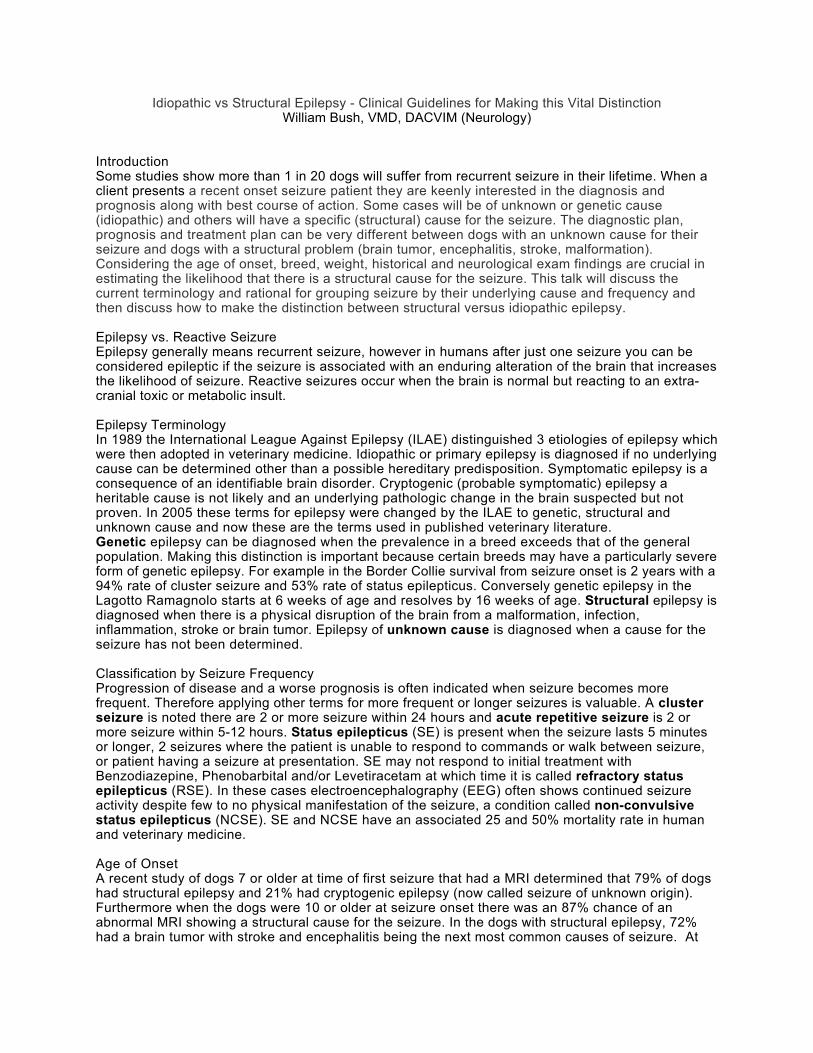
Table 1. Maintenance AED Therapy in Dogs
Drug Dose Side Effect Scale
Primary Side Effect Reported Toxicity / Dysfunction
Levetiracetam* 25-50 mg/kgPO, Q8 h (or Q 12 for extended release
1 Ataxia, sedation None
Zonisamide* 5-10 mg/kgPO Q 12 h
2 Decreased eating, ataxia, sedation
Liver, Kidney
Gabapentin 10-30 mg/kgPO Q 8 h
2 Sedation None
Pregabalin 2-4 mg/kgPO Q 12 H
2 Sedation None
Phenobarbital* 2-6 mg/kgPO Q 12 H
4 Ataxia, polydipsia, polyphagia, weight gain, polyuria, sedation, weakness
Liver, bone marrow, skin, endocrine
Bromide * 25-50 mg/kgPO Q 24
5 Ataxia, diarrhea, polydipsia, polyphagia, weight gain, sedation, vomiting, weakness
Esophagus, pancreas, stomach, panniculus
Felbamate 10-60 mg/kgPO Q8
1 Tremors (rare) Liver, bone marrow, lacrimal gland (KCS)
Topiramate 5-10 Mg/kgPO Q 8-12 H
1 Sedation Urinary calculi
Clorazepate ½ to 2 mg/kgPO Q 12 H
3 Ataxia, sedation, weakness, polyphagia
None
*Indicates serum drug monitoring recommended
Selected References Bentley RT, et al. A pilot study of Levetiracetam pulse therapy for canine cluster seizures. ACVIM Abstract, 2014Boothe DM, et al. Comparison of phenobarbital with bromide as a first-choice antiepileptic drug for treatment of epilepsy in dogs.J Am Vet Med Assoc. 2012;240(9):1073-83. Chang Y, et al. Idiopathic epilepsy in dogs: owners' perspectives on management with phenobarbitone and/or potassium bromide. J Small Anim Pract. 2006;47(10):574-81Cook AK, et al. Renal tubular acidosis associated with zonisamide therapy in a dog. J Vet Intern Med. 2011;(6):1454-7Dewey CW, et al. Pregabalin as an adjunct to phenobarbital, potassium bromide, or a combination of phenobarbital and potassium bromide for treatment of dogs with suspected idiopathic epilepsy. J Am Vet Med Assoc. 2009;235(12):1442-9.Dewey CW. Anticonvulsant therapy in dogs and cats. Vet Clin North Am Small Anim Pract. 2006 ;36(5):1107-27 Gaskill CL, et al. Pancreatitis associated with potassium bromide/phenobarbital combination therapy in epileptic dogs.Can Vet J. 2000 Jul;41(7):555-8.Govendir M, et al. Improving seizure control in dogs with refractory epilepsy using gabapentin as an adjunctive agent. Aust Vet J. 2005;83(10):602-8.Hulsmeyer V, et al. Epilepsy in Border Collie: clinical manifesations, outcome, and mode of inheritace. J Vet Intern Med 2010; 24: 171-178Hardy BT, et al. Double-masked, placebo-controlled study of intravenous levetiracetam for the treatment of status epilepticus and acute repetitive seizures in dogs. J Vet Intern Med. 2012;26(2):

334-40.Kluger EK, et al. Serum triglyceride concentration in dogs with epilepsy treated with phenobarbital or with phenobarbital and bromide. J Am Vet Med Assoc. 2008;233(8):1270-7.Kube SA, et al.Dyskinesia associated with oral phenobarbital administration in a dog. J Vet Intern Med. 2006;20(5):1238-40.Miller ML, et al. Apparent acute idiosyncratic hepatic necrosis associated with zonisamide administration in a dog. J Vet Intern Med. 201;25(5):1156-60. Müller PB, et al. Effects of long-term phenobarbital treatment on the thyroid and adrenal axis and adrenal function tests in dogs.J Vet Intern Med. 2000;14(2):157-64.Muñana KR,et al. Placebo effect in canine epilepsy trials. J Vet Intern Med. 2010 Jan-Feb;24(1):166-70.Muñana KR,et al. Evaluation of levetiracetam as adjunctive treatment for refractory canine epilepsy: a randomized, placebo-controlled, crossover trial. J Vet Intern Med. 2012;26(2):341-8Nichols ES, et al. Bromide toxicosis secondary to renal insufficiency in an epileptic dog. J Am Vet Med Assoc. 1996 Jan 15;208(2):231-3.Platt SR, et al. Treatment with gabapentin of 11 dogs with refractory idiopathic epilepsy. Vet Rec. 2006;23-30;159(26):881-4.Rossmeisl JH, et al. Clinical signs, risk factors, and outcomes associated with bromide toxicosis (bromism) in dogs with idiopathic epilepsy.J Am Vet Med Assoc. 2009;234(11):1425-31Ruehlmann D, et al. Treatment of partial seizures and seizure-like activity with felbamate in six dogs. J Small Anim Pract. 2001;42(8):403-8.Schwartz M, et al. Possible drug-induced hepatopathy in a dog receiving zonisamide monotherapy for treatment of cryptogenic epilepsy. J Vet Med Sci. 2011;73(11):1505-8. Schwartz M et al. Assessment of the prevalence and clinical features of cryptogenic epilepsy in dogs: 45 cases (2003-2011) J Am Vet Med Assoc. 2013 Mar 1;242(5):651-7Streeter AJ, et al. Pharmacokinetics and bioavailability of topiramate in the beagle dog. Drug Metab Dispos. 1995;23(1):90-3Tobias KM, et al. A retrospective study on the use of acepromazine maleate in dogs with seizures. J Am Anim Hosp Assoc. 2006;42(4):283-9.von Klopmann T, et al. Prospective study of zonisamide therapy for refractory idiopathic epilepsy in dogs. J Small Anim Pract. 2007;48(3):134-8.Yohn SE, et al. Bromide toxicosis (bromism) in a dog treated with potassium bromide for refractory seizures. J Am Vet Med Assoc. 1992;201(3):468-70.
Central vs. Peripheral Vestibular Disease: A Matter of Life and Death William Bush, VMD, DACVIM (Neurology)
IntroductionThe vestibular system provides information to the brainstem and somatosensory cortex regarding head position, acceleration, and deceleration and provides us with our sense of balance. Clinical signs of dysfunction include side-stepping as though drunk, abnormal head or eye position and spontaneous eye movement. Examination of the patient will allow an assessment of whether the dysfunction is from the nerve and therefore peripheral to the brain or from the brainstem or central. This distinction is critical because central diseases are often life-threatening unless identified and treated, whereas peripheral disease often improves on its own or with minor intervention. There are many causes of peripheral and central vestibular disease but special attention should be given to meningoencephalitis of unknown etiology (MUE) because it is common and often lethal if not treated promptly. This talk will discuss common and distinguishing features of central and peripheral vestibular disease, common causes for diseases in each location and available treatments and prognosis for MUE.
Vestibular Anatomy and FunctionMovement of endolymph over the hair cells of the receptors of the inner ear (semicircular canal, saccule, and utriculus) provides input to the vestibular nerve. The cell bodies for the vestibular nerve are located in 4 paired nuclei located within the brainstem nestled around the fourth ventricle and choroid plexus. The receptor apparatus the detects acceleration, deceleration as well as the static position of the head. There are many outputs from the vestibular nuclei:
1) Vestibular system controls eye position and coordinated movement by synapsing on cranial nerve 3, 4, and 6 via the medial longitudinal fasiculus (MLF). The generation of physiological nystagmus by moving the head left and right is called the vestibulo-ocular reflex. This reflex relies on structures deep within the brainstem and when abnormal and not related to drug therapy, there is an indication of severe brainstem dysfunction.
2) The vestibulospinal tract connects the vestibular nuclei with the nerve and muscle and will increase extensor tone to support the body against gravity during movement
3) Vestibular system has projections via the caudal cerebellar peduncle to the cerebellum which functions to coordinate movement of the eyes, neck, trunk, and limbs in relation to movement of the head as well as static head position.
4) Vestibular influences on the vomiting center in the reticular formation of the brainstem account for the motion sickness often noted in people and possibly in dogs with vestibular dysfunction.
5) There is a conscious awareness or perception of balance and equilibrium and although the pathway is not currently well defined, there is a thalamic relay of information to the somatosensory cortex.
Besides the receptors of the inner ear there are visual and proprioceptive inputs into the vestibular system. Blindfolding a vestibular patient and then lifting them off the floor often increase the sense of poor balance. Also, congenitally blind patients often have spontaneous nystagmus.
Central vs. Peripheral Vestibular DiseasePeripheral vestibular disease has a fairly consistent clinical presentation. A useful tool to think about central disease is that dogs whose clinical signs do not look like they peripheral likely have central disease. Please see Table 1.
Peripheral Vestibular DiseasePeripheral vestibular disease typically has a sudden onset and can be associated with vomiting at its onset. Patients have rotary or horizontal nystagmus at a rate of 60 beats per minute or greater and a head tilt of about 20 degrees from midline. The nystagmus can change from rotary to horizontal but its fast phase should remain opposite the direction of the head tilt. Persistent weakness and postural deficit are not noted and after a few hours of acclimating these dogs are bright and responsive and able to ambulate. These patients may lean, side-step or rarely roll in the same direction as the head tilt. The three most common causes of peripheral vestibular disease are infection of the middle ear

extending into the inner ear’s bony labyrinth that contains the vestibular receptors (OTMI), the old dog peripheral vestibular or idiopathic vestibular syndrome (dogs typically older than 5, cats of any age), and the low thyroid state, especially when the cholesterol is elevated.
Central Vestibular DiseaseOne specific example of central disease is called paradoxical vestibular disease because the signs are different or opposite of what would be expected for peripheral disease. In this syndrome, the lesion is within the brain in the caudal cerebellar peduncle or floculonodular lobe of the cerebellum and the head tilt is opposite the side of the lesion. Some clinical signs of non-peripheral or central vestibular disease include dull mentation, side- stepping/leaning towards head tilt, sway back and forth, hypermetria, tremors, weakness, non-ambulation, postural deficit, nystagmus at a rate under 60 beats per minute, extreme head tilt, cranial nerve deficits besides those associated with Facial nerve and Horner’s tract (commonly seen with OTMI). Common causes of central vestibular disease include neoplasia like meningioma (larger breeds), meningoencephaltiis of unknown etiology (MUE), and infarcts of the cerebellum (larger breeds). Please see Table 2.
ConclusionVestibular disease is a common presenting complaint and assessing the disease to be central or peripheral provides the owner with the best sense of the appropriate diagnostic plan, treatment and prognosis. Having the image of a typical peripheral case in your mind and comparing all cases against this image can allow for best determination of the likelihood of central disease. Prompt treatment of the diseases that cause central vestibular signs is essential for a good outcome.
Table 1. Clinical Signs of Disease in the Central or Peripheral Vestibular System
Observation Central Disease Peripheral Disease
Mentation Dull Normal
Gait Side step opposite head tiltHypermetriaWeakness
Side-steps towards side of lesion
Postural Reactions Delayed or absent Normal
Head Tilt Absent or extreme 20 Degrees
Cranial Nerve Deficits Any +/- Facial, +/- Horner’s tract
Nystagmus Vertical or positional (chronic)Fast phase towards lesionFewer than 10 beats/second
Rotary or horizontal Fast phase away from lesionGreater than 60 beats/minute
Positional Strabismus Ventral on side of head tiltDorsal opposite head tilt
Ventral on side of head tilt
Neck Pain Yes No

Table 2. Categories of Disease that Cause Central or Peripheral Vestibular Disease
Category Central Diseases Peripheral Diseases
Malformation Rostrocerebellar fluid accumulationCaudal occipital malformation syndrome (COMS)Hydrocephalus
Congenital vestibular disease
Metabolic Hypothyroidism (± infarction) HypothyroidismNeoplastic Primary intracranial neoplasms
Metastatic neoplasmsPrimary aural neoplasiaVestibular neurofibroma
Infectious & Inflammatory
Viral: Canine distemper virus, Feline infectious peritonitisBacterial: Abscess, Rocky mountain spotted fever, Ehrlichiosis, Bartonellosis, AnaplasmosisProtozoal: Toxoplasmosis, NeosporosisMycotic: Cryptococcosis, Blastomycosis, othersNon-infectious (MUE)
Otitis media interna (OMI)Nasal- and otopharyngeal polypsIdiopathic vestibular disease (vestibular neuronitis)
Trauma Brainstem trauma Inner ear traumaToxic Metronidazole Ototoxic drugs (systemic and
topical)Vascular Cerebrovascular disease
Selected ReferencesDe Lahunta A. Veterinary Neuroanatomy and Clinical Neurology. 2nd Edit, W.B. Saunders, Philadelphia, 1983De Lahunta AD, Glass E. Veterinary Neuroanatomy and Clinical Neurology. St. Louis: Saunders Elsevier, 2009Rossmeisl JH, Vestibular Disease in Dogs and Cats. Vet Clin Small Anim 40 (2010) 81-100Troxel MT, et al. Signs of neurologic dysfunction in dogs with central versus peripheral vestibular disease. J Am Vet Med Assoc 2005; 227 (4): 570-4

Common and Deadly: Inflammatory Disease of the Brain, Spinal Cord, and MeningesWilliam Bush, VMD, DACVIM (Neurology)
IntroductionWhen there are inflammatory cells within the brain, spinal cord or meninges then the terms encephalitis, myelitis, and meningitis are used, respectively. When inflammation is in more than one area the terms are combined like with meningoencephalomyelitis. The inflammation in these cases if most often from a non-infectious, unknown etiology and therefore called meningoencephalomyelitis of unknown etiology (MUE). The signs of the disease are specific to the location of the inflammation and most cases respond well to aggressive immune suppression. This talk discusses the terminology, definition / histopathology, common presentations, treatment and prognosis for different manifestations of non-infectious inflammatory disease within the nervous system
TerminologyMUE is an umbrella term for necrotizing encephalitis (NE) and granulomatous meningoencephalomyelitis (GME). Necrotizing encephalitis implies death of neurons within the brain from inflammation and is further subdivided into Pug dog encephalitis or necrotizing meningoencephalitis (NME) and Yorkshire terrier encephalitis or necrotizing leukoencephalitis (NLE). MUE has replaced these terms since multiple breeds have been identified with these disease and the prognosis, testing and treatment protocols are similar. Steroid responsive meningitis-arteritis (SRMA) is another non-infectious inflammatory disease that typically involves only the meninges – this disease will be discussed separately at end of this talk.
Definitions and SignalmentsNLE was first described in the Yorkshire terrier. NLE is a progressive disease with an acute or chronic onset where there is necrosis of the white matter that with time, can coalesce into cavities or holes in the brain. The grey matter and spinal cord are spared in this disease. The other form of NE, NME first described in the Pug dog has since been noted in many other small breeds like the Maltese, Brussels Griffon, Colon du Tulear, Shih Tzu, and Papillion. NME is typically an acute onset and rapidly progressive disease of the both the grey and white matter of the cerebrum, with only 25 % of cases showing any multifocal or brainstem signs. Because the cerebrum is so commonly affected, seizure is common clinical sign - 94% of Pugs with this disease have seizure. GME is common and may account for up to 25% of canine CNS disease – it is an acute onset, progressive and potentially fatal disease. Unlike NE, the disease can manifest in the cerebrum, brainstem and spinal cord – 8 % of all cases present with only spinal cord signs. Histopathologically GME is noted most often in the white matter as perivascular infiltrates of rounds cells (plasma cells, lymphocytes and occasionally lymphoblasts) - these can coalesce to form tumors (unlike NE where the lesions coalesce to form cavities within the brain) (SJSR). Female, small breed dogs like the Miniature poodle, Maltese, Dachshund, Westie, and Chihuahua are commonly affected. Most dogs with GME are 4-8 years of age, whereas with NE most dogs are under 4 years of age. The take home point is that MUE should be suspected in small breed under 8 years of age with acute onset of brain and less commonly spinal cord signs.
Signs of Disease with MUEThe signs of disease are specific to the region of the brain that is involved. Most cases of MUE presents with multifocal clinical representing a mixture of forebrain and brainstem signs which can include altered mentation, visual deficits, central vestibular signs, proprioceptive placing and hopping deficits and seizures. In one report, 8% of cases has only spinal cord signs (weakness, paralysis, ataxia).
What is the cause of MUE?The causes of MUE is thought to be from a genetic predisposition coupled with environmental exposures leading to a pathologic immune response. For instance, the histopathological differences in NE may result from minor differences among breeds, modifying genes, or variations in antigenic exposure. Breed predispositions indicate there is a heritable component to development of MUE. In
the Pug, heritability has been proven and a strong association demonstrated between affected dogs with single nucleotide polymorphism within the dog’s leucocyte antigen (DLA) complex II region located on chromosome 12. The authors point out that this same association is made in human multiple sclerosis (MS) patients and that NME in the Pug dog maybe a good model for the less common acute variant forms of MS. Recent work in Maltese with NME show risk loci on chromosome 4 and 15. MUE has been associated with viral diseases like Borna virus, West Nile, Canine parainfluenza, and Encephalomyocarditis virus, Canine herpes virus-1, Parvovirus, Porcine herpes virus-1, Bunya- and Polyomaviruses. Additionally, DNA from E. Coli, Mycoplasma canis. and Bartonella vinsonii subsp berkhoffii have been identified in sporadic cases of MUE and a recent report shows DNA from Anaplama phagocytephilum in 4/23 cases SRMA. These pathogens are not thought to be direct cause of the disease but according to the “Hit-and-Run Hypothesis” work in tandem with genetic and other environmental factors (vaccination?) to generate an autoimmune response, perhaps through molecular mimicry. Autoimmune disease is likely in MUE because the CSF and serum of dogs with MUE contain an anti-astrocyte autoantibodies against glial fibrillary acidic protein (GFAP) which is an intermediate filament protein important in astrocyte function. Recent work has shown that the active cellular proliferation is thought to occur within the CNS lesion (and not from a migration from outside the CNS) and is assisted by matrix metalloprotineases (MMPs). MMPs are enzymes necessary for migration of leukocytes into the CNS or CSF and MMP-9 is elevated in some dogs with MUE. Other work by Dr. Mariani has also shown elevations in many interleukins necessary for lymphocyte proliferation and trafficking into tissue. However, to date there is no useful serum or CSF biomarker to assist in the diagnosis or treatment of MUE.Lastly, since some cases of MUE lesions contain small amount of lymphoblasts and some are truly shown to be lymphoma at the time of histopathology, it is theorized that MUE is a lymphoproliferative disorder with features of both inflammation and neoplasia. Further support for this claim is the marked clinical responses of certain cases to chemotherapy.
MUE DiagnosisAn MUE diagnosis is based on clinical suspicion from the signalment and disease progression, and then MRI, CSF and infectious disease testing. It can be difficult ruling-out infection because of inaccurate test results and the fact that there are not tests for all known pathogens. For example, we had a suspected MUE whose necropsy revealed a high burden of an unknown protozoal agent. Complicating things further is not all cases will have an abnormal MRI and between 12-25% of MUE cases will have normal CSF analysis. In cervical spinal cord MUE, MRI of the paraspinal cervical muscles with STIR sequence in MUE is often abnormal (78% sensitivity) and rarely abnormal in normal controls (92% specificity) – because CSF results can be normal in cases of spinal cord MUE about 10% of the time, this sequence is important in suspected cases of cervical and maybe intracranial MUE.
Pursing Infectious EtiologyWhen the CSF is abnormal in a MUE cases, less than 10% of cases will have a predominantly neutrophilic CSF analysis. Therefore a neutrophilic pleocytosis should alert clinician to a possible infection rather than MUE. Typical testing when searching for infection could include PCR, serology and rarely cultures for protozoal, rickettsial, fungal, bacterial, and viral diseases. In the Mid-Atlantic region of the USA, we typically test the CSF via PCR for distemper virus, serology for Toxoplasmosis gondi, Neospora caninum and potentially Sarcocystis neurona, and antigen testing for Cryptoccocus sp. as well as whole blood PCR testing for vector borne disease. Failure to improve while on antibiotics or a relapse of signs when prednisone is reduced while on antibiotic therapy is often the last step in ruling-out infection and committing to multimodal immune suppressive therapy (see below). Brain biopsy has been reported and occasionally performed in our clinic however, the procedure has risks, costs, may yield false negative or positive results and may not change the course of treatment. A recent paper describing needle guided brain biopsy had 82% of cases achieved a specific diagnosis with a 6% indirect mortality rate and 29% incidence of transient side

effects (stupor, seizure, weakness and loss of proprioception).
MUE TreatmentInitial testing often reveals inflammation but does not clearly delineate between non-infectious and infectious inflammation. To address a possible infection antimicrobial therapy (clindamycin 15 mg/kg, BID, minocycline 10 mg/kg, BID +/- Fluconazole 10 mg/kg, BID) if often started while waiting for infectious disease test results. Prednisone 0.5 mg/kg, BID is also started and if signs are progressive and severe additional immune suppression could be considered with chemotherapy (Cytosine arabinoside, Lomustine, Procarbazine) and/or immune modulation with (Cyclosporine and less commonly Leflunomide, Azathioprine, or Mycophenolate). Radiation therapy has also been reported to have a positive influence of the disease course with MUE. There are many important and unanswered clinical questions revolving around what is best immunosuppressive protocol and when it is advised to stop therapy.
Steroid Alone are InsufficientIn a meta-analysis of MUE cases the median survival for dogs treated with corticosteroid plus any other immune suppressant protocol ranged from 240 to 590 days (n=96) compared with corticosteroid alone where range of median survival was 28 to 357 days ( n= 43). A recent retrospective study evaluating different glucocorticoid protocols (no other immune therapy) showed survival times ranging from 2 to 2065 days – and the authors concluded that an 18 week schedule of sole prednisone therapy can be used to treat MUE. However, multiple other authors conclude that treating with immune suppressants other than prednisone will improve control of the immune condition, improve survival times, and improve quality of life for the patient by reducing steroid associated side-effects (polydipsia, polyuria, polyphagia, muscle loss, urinary tract infection, hepatotoxicity, etc.). However, which immune suppressive protocol is best in not known and there is a desperate need for randomized, blinded, controlled, prospective study of MUE to assess current and future therapies that could include (intravenous immunoglobulin, plasma exchange and even anti-viral therapy). Once remission or improvement is achieved it can be difficult to know when to taper steroid and other immune suppressive therapy. In our experience, tapering medication can lead to relapses with poor outcomes in dogs that had a normal neurological exam. A recent paper showed that follow-up CSF analysis at 3 months can predict relapse and that tapering medication in dogs with an abnormal MRI always lead to relapse. Repeating these tests when they were previously abnormal is advised prior to the tapering or elimination of immune suppressive therapy.
Prognosis Comparing studies is difficult due to different inclusion criteria, therapy, treatment endpoints, and lack of a prospective, controlled study. There is a recent prospective study of 39 MUE dogs treated with prednisone and then Cytosine arabinoside that provides insight into the prognosis with MUE. 13/39 (33%) died in the first 72 hours and 22/39 (56%) died within the first 52 days and the study had an overall mean survival time of 26 days (range 0-2250 days). The remaining 17 dogs that lived beyond 52 days had survivals that ranged from 562 to 2241 days (median 1616 days). Overall 12/39 (31%) dogs returned to normal and 7/39 (18%) were normal without treatment. These results can best be summarized by saying MUE can have an acute and fatal presentation up to 33% of the time and if alive at 8 weeks then survival time jumps to a median of 4 and ½ years. Among the dogs that survive more than 8 weeks, most return to normal and some can be off medication altogether.
Prognostic IndicatorsOne paper demonstrated that signs of high intracranial pressure (foramen magnum herniation, loss of cerebral sulci) was associated with a higher mortality. Multifocal disease and seizure have been inconsistently reported as negative prognostic indicators in MUE. A recent abstract suggested that focal brainstem disease carried best prognosis in MUE.
Seizure and the Role of Electroencephalography (EEG) in MUENon-convulsive seizure and non-convulsive status epilepticus (NCSE) can be present and only

detectable by using EEG. In pediatrics, continuous EEG is used in patients with encephalitis, seizure and altered mentation to identify non-convulsive seizure and non-convulsive status epilepticus. Children with non-convulsive seizure have a poor outcome compared to those with the same diseases without non-convulsive seizure. We have also documented NCSE in MUE and believe that identifying and treating NCSE would improve outcome in MUE cases. NCSE should be highly suspected in MUE patients with seizure as part of the presenting complaint plus altered mentation, twitching of the ears or eyelids, sudden changes in temperature or respiratory rate, or unexplained coma. If referral for EEG is not possible, I recommend treating with Levetiracetam 60 mg/kg, IV and then potentially phenobarbital at doses of 20-40 mg/kg, divided into 6-8 mg/kg boluses until there are no abnormal movements or paroxysmal changes in vital parameters.
Steroid Responsive Meningitis-Arteritis (SRMA)SRMA is a systemic immune disorder characterized by inflammatory lesions of the meninges and associated arteries. This disease can occur in any breed but the Bernese Mountain Dog, Boxer, Beagle, German Short and Wire Haired pointers, Weimaraner are over-represented. Clinical signs typically start at 10 months of age with a range of 6-18 months, however it has been reported in dogs as old as 7 years of age. Although histopathological changes have been noted in the heart, mediastinum, thyroid and there is an association with immune mediated polyarthritis - the clinical signs are from the meningitis (Webb). Clinical sign include neck pain and lethargy, not eating, and fever. Typical exam findings include stiff neck, short- strided gait, neck and back pain on palpation and spontaneous yelping-out or with movement. Misdiagnosis can occur because this is a sporadic disease, with non-specific, waving and waning signs that are often initially responsive to antibiotics and NSAID therapy. The diagnosis is made when inflammation is noted on CSF analysis, with most cases having a severe neurotrophilic pleocytosis. The major differential diagnosis for these clinical signs is diskospondylitis which would be best identified with MRI. A peripheral neutrophilia and elevated globulin count are inconsistently findings whereas serum C reactive protein (CRP) is elevated in all cases. CRP is an excellent biomarker for this disease because it drops to normal with resolution of disease and increases with relapse. Serum and CSF IgA concentration are increased indicating this is an immune disease, however IgA is a poor biomarker because it remains elevated, even in remission. Treatment involves high dose steroid therapy and when there is an incomplete response, relapse, or intolerable corticosteroid side effects then other immune modulators can be added-on (Azathioprine, Cyclosporine, Mycophenolate). A chronic form of the disease has been reported where there is weakness and ataxia in all 4 limbs and a mononuclear CSF analysis - it has been suggested this develops from late recognition of the disease or inadequate treatment (premature taper of therapy, too little immune suppression). Most cases return to normal. Relapse can occur in 20-60% of the cases – a higher CRP at 4 weeks is associated with multiple relapses. Although treatment durations of up to 20 months have been reported – most cases require about 6-10 months of therapy.
Conclusion MUE is common and should be expected in any small breed dog with acute and/or progressive brain or spinal cord signs. Once diagnosed with MUE the prognosis is guarded but might be able to be improved by treating with multiple immune suppressive medications as well as anti-epileptic drugs. A return to normal is certainly possible as well as clinical remission. More study is desperately needed to determine the benefit of different treatment protocols, especially ones where multiple immune suppressive medication are given early in the course of the disease. SRMA is another example of immune mediated disease observed in medium to large breeds at about 10 months of age – if recognized early in the disease course the prognosis is excellent.
Selected ReferencesCizinauskas S, et al. Long-term treatment with SRMA: clinical, lab, and therapeutic results. Journ Small Anim Pract 2000; 41: 295-301Flegel T, et al. Diagnostic yield and adverse effects of MRI-guided free-hand brain biopsy through a mini-burr hole in dogs with encephalitis. J Vet Intern Med 2012; 26: 969

Granger N, et al. Clinical findings and treatment in non-infectous meningoencephalomyelitis in dogs: a systemic review of 457 published cases from 1962-2008. The Veterinary Journal 2010; 184: 290-297Higginbotham MJ, et al. Noninfectious CNS disease in dogs. Compendium 2007 : 488Lowrie M, et al. MUO investigations of prognostic factors and outcome using a standard treatment protocol. Veterinary Record 2013Lowrie M, et al. SRMA: a prospective study of potential disease markers, prednisone treatment and long-term outcome in 20 dogs (2006-2008). J Vet Intern Med 2009; 23: 862-870Mariani C. Canine meningoencephalitis: what do we know?. ACVIM Proceedings 2014Schatzberg SJ. Idiopathic and granulomatous and necrotizing inflammatory disorders of the CNS. Vet Clin Small Anim 2010; 40: 101-120Tipold A, et al. An update on steroid responsive meningitis-arteritis. Journ Small Anim Pract 2010; 51: 150-154

Upper Motor Neuron vs. Lower Motor Neuron Disease Never Miss Again After This Video Case Presentation
William Bush, VMD, DACVIM (Neurology)
IntroductionThe ability to examine a patient and determine where in the body the disease is located is critical to determining the cause, best therapy and prognosis. Weakness is a common presenting complaint and can result from spinal cord (upper motor neuron - UMN) or nerve and muscle disease (lower motor neuron-LMN). Understanding the function of the UMN and LMN system will enhance the accuracy of lesion localization and therefore determination of the diagnostic testing and prognosis. This talk will discuss the function of the UMN and LMN system and then how to assess these systems via examination of a patient’s gait, postural responses, and reflex testing. Secondly we will discuss assessing the cutaneous trunci reflex and for focal pain to assist in lesion localization and honing the list of possible the spinal cord diseases. Lastly we will discuss evaluating the palpebral response and laryngeal / pharyngeal / esophageal function in LMN disease.
Tonic Gamma Loop MechanismMuscle tone refers to the intrinsic tension of the muscle when supporting the body against gravity while standing, shifting weight from limb to limb, jumping or performing stairs – but how does this occur? The tonic gamma loop mechanism (TGLM) is intrinsic to the LMN system and understanding TGLM physiology offers insight into how gait is generated and why UMN or LMN disease cause alterations in muscle tone and reflex. When we jump down from a height and our knees start to bend or buckle there is a sudden stretch of the quadriceps muscles and stimulation of stretch receptors (neuromuscular spindle) within these muscles. These receptors then stimulate the sensory portion of the femoral nerve which then directly synapse and stimulates the motor portion (alpha motor neuron) of the femoral nerve. This causes the quadriceps muscle (which is innervated by the femoral nerve) to contract and prevent your knees from buckling. When the patella tendon is artificially simulated with a reflex hammer it fools the body into thinking there is a sudden heavy load on the quadriceps muscle (like jumping down), the TGLM is stimulated and the knee jerks.
Gait GenerationAs mentioned above, when information from the neuromuscular spindle of the TGLM returns to the spinal cord it directly synapses on the alpha motor neuron or motor portion of the same muscle in which the spindle is located. However, the information also stimulates inhibitory interneurons that then reduce activity or tone in the antagonistic muscle group. Therefore when there is contraction of the quadriceps there will be reduced tone in the hamstring or flexor group. The modulation of flexion is performed by the phasic gamma loop or flexor gammas loop. The UMN system acts on the tonic and then phasic gamma loop mechanisms to generate extension and then flexion of the leg by activating this intrinsic reflex mechanisms and therefore generate gait.
UMN Lesions Influence the TGLMIn our example above, gravity lengthen the quadriceps muscle and stretches the neuromuscular spindle, which then via a direct synaptic connection, stimulates the femoral nerve. This causes contraction of the quadriceps muscle which then causes more stretching of the neuromuscular spindle and more contraction of the quadriceps muscle. This system, if not modulated would lead to dramatic increases in muscle tone and reflex. The UMN system modulates or controls the TGLM and therefore controls muscle tone and reflex. Disease of the UMN lesion can cause increased tone and reflex. Examination of dogs with UMN spinal cord disease often reveals increased tone because there is resistance to flexion of the stifle. This stiffness can also manifest in the protraction phase of the gait and appear as swinging out of the limb (circumduction) or a long-strided gait. Furthermore, brainstem lesion (where the UMN tracts start) can lead to opisthotonus also known as decerebrate rigidity where the head, neck and limbs are held in rigid extension.
Reflex TestingA reflex is something that occurs automatically or spontaneously without influence from the cerebrum whereas a reaction or response requires the unconscious participation of the cerebrum. In reflex testing there is a sensory stimulus that runs into the spinal cord or brainstem and then an immediate

spinal cord or brainstem mediated response. For example, stretching the patella tendon with a pleximeter (reflex hammer) causes a sudden, intense stimulation of the stretch receptors within the femoral nerve, in essence simulating what would happen if we jumped down from a large height. Immediately the muscles innervated by the femoral nerve contact and the knee jerks. An absence of reflex often means there is a lesion of the motor or sensory portion of the femoral nerve or severe disease of the quadriceps muscle. If there is an increase in reflex (exaggerated, clonus) then there is a failure of UMN system to control this reflex.
Upper Motor Neuron SystemThe UMN system primarily starts in the brainstem. The axons from this collection of neurons run within the white matter of the spinal cord and synapses in the ventral horn of the spinal cord to activate the peripheral nerve (LMN). This system activates the LMN to generate gait and modulates or controls tone and reflex by influencing the tonic gamma loop mechanism. A lesion of the descending or motor component of the UMN system results in paresis (weakness), paralysis, increased reflex and increased muscle tone. A lesion of the ascending or sensory system causes a disordered gait and postural deficits (see below).
Lower Motor Neuron SystemThe LMN system starts within the spinal cord where the cell bodies are grouped in the grey matter of the spinal cord within the ventral horn at the intumescence (swelling) located at spinal cord segments C6-T2 and L3-S3. The numbered nerves then run to the brachial or lumbar plexus and then exit as named nerves that will then innervate specific muscles. The LMN generates muscle tone and with a lesion there is weakness, paralysis and loss of muscle tone and reflex. The LMN system also carries sensory information from receptors in the joints and skin into to the dorsal horn of the spinal cord and this is eventually relayed via the UMN system to the cerebellum and somatosensory cortex via the spinocerebellar and spinothalamic tract, respectively.
Spinal Cord Ataxia and Postural ReactionsA complete lesion of the UMN system causes no movement or paralysis and an increase in muscle tone (spastic paralysis). A partial lesion will cause only weakness or paresis but the movement will be ataxic. Ataxia means disorder. The absence of ascending information reaching the brain can result in a loss of self- reception (proprioception) and consequently spinal cord or proprioceptive ataxia and slow postural reactions. Spinal cord ataxia can take the form of a long-strided gait, the limbs can circumduct, cross midline, and interfere with each other - occasionally causing the patient to trip or fall. In addition the patient might stand on the dorsal surface of the paw or stand with limbs too close, too far apart or with limbs crossed. Besides observation of the gait, testing of the postural reactions (paw flip test, hopping, tactile placing) also assesses the function of the UMN system. The postural reactions will be delayed to absent with an UMN lesion.
LMN LesionsA complete lesion of the LMN system causes paralysis with an absence of muscle tone (flaccid paralysis). An incomplete lesion causes weakness and the patient will have a short-strided or choppy gait as though they are walking on egg shells. Importantly, incomplete LMN lesions do not cause significant disruption of the sensory system. Therefore LMN lesions do not cause ataxia. Furthermore, if the patient’s weight is properly supported the postural reactions will be normal. Please see Table 1.
C6-T2 Spinal Cord A lesion that involves the white matter of the spinal cord at C6-T2 will cause UMN signs to the pelvic limbs. The pelvic limbs will have increased tone and reflex, reduced postural reactions, weakness and ataxia. A lesion of the grey matter in this area will generate LMN signs to the thoracic limbs manifested as a short-strided gait, preserved postural reactions and no ataxia, reduced reflex, and neurogenic muscle atrophy. The long-strided, stiff and ataxic gait in the pelvic limbs is much different than the short-strided gait of the thoracic limbs and sometimes referred to as a two engine gait.
T3-L3 Spinal Cord and the Cutaneous Trunci ReflexDisease between the two intumescences is called T3-L3 spinal cord disease and results in upper

motor neuron disease to the pelvic limbs. The presence of a cut-off or cessation of the cutaneous trunci reflex can indicate the level of the spinal cord lesion. The input for the reflex is stimulation of dorsolateral cutaneous receptors. Once a stimulus is registered the information then ascends in the spinal cord where it synapses motor neurons at the level of spinal cord segment C8 -T2. These nerves form the lateral thoracic nerve that causes contraction of the cutaneous trunci muscle. Functionally a pinch of the skin with hemostats should stimulate contraction of the entire cutaneous trunci muscle along the entire flank of the patient. With a thoracolumbar spinal cord lesion, pinching of the skin behind the lesion will not result in twitching of the skin and thus there appears to be a cut-off of this reflex. A cut-off in the cutaneous trunci reflex indicates the lesion is about 2 vertebral bodies cranial to the cut-off. Furthermore, following surgery movement of the cut-off caudally predicts recovery while movement cranially predicts myelomalacia.
Lumbar Intumescence and Nerve Root Disease (Lumbosacral Syndrome)Disease of the spinal column or spinal cord/nerve roots from the L5 to S1vertebrae can generate LMN signs to the pelvic limbs, fecal and urinary incontinence as well as paralysis of the tail. These signs can overlap and be mistaken for osteoarthritis of the hip or stifle. A sciatic lesion can be the cause of an increased patella reflex as a consequence of losing strength and tone to the antagonist of stifle extension, this is called a pseudo-hyperpatella reflex and should not be mistaken for an UMN reflex. A reduction of the patella reflex can help localize lesion to L3-L4 vertebral bodies and would not be expected with disease from L5 – S1 vertebrae. The patella reflex can be absent in otherwise healthy middle-age and older dogs, presumably from degeneration of the sensory portion of the femoral nerve.
Pain AssessmentDiseases of the nerve and muscle (LMN disease) are typically not painful, however many spinal cord diseases are associated with pain. Determining the patient is painful at a specific location can direct diagnostic testing and also hone the list of possible causes of disease – for instance intervertebral disk disease, neoplasia, and diskospondylitis are typically painful whereas ischemic myelopathy (fibrocartilaginous emboli) and acute, non-compressive nucleus pulposus extrusions are often non-painful, especially after the first 24 hours. Neck pain is often suspected when patient spontaneously yelps out but there is no gait or posture deficits, intermittent thoracic limb lameness (root signature), or stiff neck or decreased range of motion is noted. Palpating muscle spasm laterally at level of transverse process, pain with manipulation or ventral process of C6, or resistance to range of motion can also indicate neck pain. Mid-back pain is often suspected with kyphosis, stiffness and when slow to sit or rise. Palpating and applying pressure to dorsal processes while putting pressure / palpating the ventrum and palpating muscle / rib heads at level of transverse process often allow for detection of back pain. Lumbosacral pain is suspected with abnormal tail carriage and when patient is slow to sit and rise. Pain can often be detected with rectal palpation of the lumbosacral junction (or spondylosis at L7-S1), tail extension or by applying pressure to muscle between dorsal process of L7 and S1. Hip extension will not differentiate back from hip pain. However, hip pain can be discerned by slowly elevating the femoral head about 3-5 mm from acetabulum by lifting up on the medial surface of the femur while the patient is in lateral recumbency.
Cranial Nerve Exam in LMN DiseaseLMN disease can affect cranial nerves when there is a polymyositis, polyneuropathy, or disease of the neuromuscular junction (Myasthenia gravis). When LMN disease is suspected then a few physical examination maneuvers can be helpful. Firstly listen to the patient’s breathing – a respiratory stridor can indicate weakness of neuromuscular system that abducts the vocal folds. Gagging can indicate pharyngeal weakness or incoordination and misdirection of saliva into the airway. Pneumonia may be present from laryngeal or pharyngeal dysfunction or from megesophagus – listen for a soft, moist cough and carefully auscultation the lungs. Thoracic radiographs are indicated in dogs with suspected LMN disease to assess for megesophagus, aspiration pneumonia and other pathology. Secondly, assess temporalis muscle mass because marked atrophy can indicate a lesion of the mandibular nerve. Lastly, repeated stimulation of the medical canthus of the eye should provoke a prompt and complete blink response – incomplete blinking or an absent blink indicates there is neuromuscular disease.

Table 1. Distinguishing Characteristics of UMN and LMN Disease
UMN LMNGait Characteristic Long strides Short stridesAtaxia Yes NoPostural Deficit Yes NoTone & Reflex Increased DecreasedAtrophy No YesSpinal Pain Often Seldom
Selected ReferencesBraund KG. Clinical Syndromes in Veterinary Neurology. 2ed. St. Louis, MO, Mosby, 1994De Lahunta A. Veterinary Neuroanatomy and Clinical Neurology. 2nd Edit, W.B. Saunders, Philadelphia, 1983De Lahunta AD, Glass E. Veterinary Neuroanatomy and Clinical Neurology. St. Louis: Saunders Elsevier, 2009Gutierrez-Quintana R, et al. The cutaneous trunci reflex for localizing and grading thoracolumbar spinal cord injuries in dogs. J Small Anim Pract. 2012;53(8):470-5. Muguet-Chanoit AC, et al. The cutaneous trunci muscle reflex: a predictor of recovery in dogs with acute thoracolumbar myelopathies caused by intervertebral disc extrusions. Vet Surg. 2012; 41(2):200-6. Sharp NJ, Wheeler SJ: Small Animal Spinal Disorders. 2ed. Philadelphia, PA, ElsevierMosby, 2005
The Diagnosis and Treatment of the 5 Disk DiseasesWhy is MRI so Important?
William Bush, VMD, DACVIM (Neurology)
IntroductionThe spinal cord connects the brain to the neuromuscular system which is responsible for locomotion and respiration. The spinal cord is protected by a long bony box that is segmented to allow for improved axial movements. The segments of the bony box (vertebrae) are cushioned by the intervertebral disk. The intervertebral disk (IVD) contains a jello-like substance called nucleus pulposus (embryonic notochord) that is wrapped by concentric layers of ligamentous material (annulus fibrosus). The IVD is located below and adjacent to the spinal cord and pathology of the IVD puts the spinal cord at risk for infection, infarction, and compression all of which can lead weakness, paralysis, ataxia, pain, and incontinence.
Role of Magnetic Resonance Imaging (MRI)MRI characteristics are based on the proton or water content of different tissue allowing for detection of even mild pathology in the soft tissue and to a lesser extent bone. Because MRI directly images the spinal cord it is superior to X-ray based technology like radiography, myelography and CT scanning. MRI consistently demonstrates superior accuracy in the diagnosis of spinal cord disease. Additionally, MRI is better at predicting outcome than assessment of neurological function at presentation. MRI is currently essential to the practice of high quality veterinary neurology.
Hansen Type I Disk DiseaseHansen Type I disease occurs when the degenerated annulus fibrosus loses tensile strength and tears allowing the degenerated, dehydrated nucleus pulposus to extrude or herniate and co-occupy the spinal canal with the spinal cord. The stretching of the meninges, nerve root and annulus fibrosus tear generating pain and the spinal cord compression can cause weakness, paralysis and incontinence. Prognosis and the choice of medical vs. surgical management is made by combining information about the neurological grade and MRI findings. This disease is prevalent in 3-6 year-old dachshunds and many other small breed dogs and 6-9 year-old large breed dogs like the German Shepherd, Rottweiler, Dalmatians and mixed-breeds. Type I disk disease has a very good to excellent prognosis but surgery is often required when there is persistent pain and/or weakness with significant spinal cord compression. Remarkably, in paraplegic dogs with Type I disease that have hemilaminectomy, MRI is a greater predictor of outcome than nociceptive or deep pain status.
Hansen Type II DiseaseHansen Type II disease is present when there are micro-tears and bulging of the annulus fibrosus and compression of the spinal cord, meninges and nerve roots. The disease is most common in larger breed dogs in the low cervical spine and lumbosacral junction. In the low cervical spine, Type II disk extrusion is an important factor in cervical spondylomyelopathy and is also known by disk associated wobbler’s syndrome (DAWS). In DAWS the outcome with surgery is better than with medical management although there is no significant difference in life expectancy with these 2 treatments. Lumbosacral disk disease can mimic hip or stifle disease but unlike these other conditions, it can lead to urinary or fecal incontinence. Success rates with surgery are generally with LS surgery unless incontinence is already present. Therefore the distinction between lumbosacral Type II disk extrusion and orthopedic disease is an important. Nerve pain (and not orthopedic disease) can be distinguished from orthopedic disease via palpation of lateral muscles just cranial and lateral to the wings of the ilium, between the L7 and S1 dorsal spinous process, ventral surface of L7 and S1 via rectal, or with elevation of the tail. Pain with hip extension can indicate nerve compression or joint pain. However, hip pain can be discerned by slowly elevating the femoral head about 3-5 mm from acetabulum by lifting up on the medial surface of the femur while the patient is laterally recumbent

Acute Non-Compressive Nucleus Pulposus Extrusion (ANNPE)ANNPE is sometimes referred to as Type III disk disease or low volume high velocity disk extrusion. In this disease a small amount of nucleus pulposus (NP) ruptures at a high velocity through a small tear in the dorsal annulus fibrosus leading to edema, malacia, and/or hemorrhage of the spinal cord and epidural fat but minimal to no compression of the spinal cord. ANNPE has a peracute onset and associated with activity or a traumatic event and seen more commonly in medium to large breed, male dogs, especially Labrador retrievers and mixed breeds. About 2/3 of the patients in one study returned to walking and when the T2 weighted cross sectional MRI showed less than 90% of the spinal cord to be affected, 93% of the dogs regained function. Similar to Type I disk disease, the prognosis is more strongly correlated to MRI findings than admission neurological grade or nociceptive status. Many patients have difficulty urinating over the short-term and are treated with phenoxybenzamine or prazosin and diazepam to reduce smooth and skeletal muscle tone, respectively. Urinary catheterization can be a useful way of managing these patients in the first 2-3 days while the disease improves and the phenoxybenzamine has time to take maximal effect. Monitoring the urinalysis and bacterial culture and sensitivity are advised because the resulting urethral inflammation can frustrate useful urination. Exercise restriction, rehab therapy and pain medication as indicated are hallmarks of therapy. Pain medication in the short term and rehab therapy may improve outcome.
Fibrocartilaginous Emboli (FCE)FCE or ischemic myelopathy occur when the NP obstructs blood flow within a spinal cord arteriole leading to necrosis of the spinal cord from loss of blood flow (infarct). The onset and progression of clinical signs are very similar to ANNPE although dogs with FCE are rarely painful. The middle aged Miniature Schnauzer, Shetland sheepdog and Labrador are at higher risk for FCE. The overall recovery rate is about 84% but 100% of dogs will improve if the T2 weighted MRI cross sectional lesion is less than 67% and the length of the lesion is less than 2 vertebral bodies. Despite the fact that the MRI can be normal about 20% of the time, MRI is the most accurate test and the strongest predictor of outcome in dogs with FCE. The same concerns exist for micturition with ANNPE and FCE. Rehab therapy instituted early in disease process may improve recovery rate.
DiskospondylitisDiskospondylitis is an uncommon infectious and therefore inflammatory condition of the intervertebral disk and surrounding bony endplate, soft tissue, and meninges. Increasing age, male, large breed dogs are at higher risk and the Great Dane, Boxer and Labrador are thought to be predisposed to this disease. Pain, lethargy, not eating well and low grade fever are often noted. This can progress to weakness, paralysis and incontinence if there is spinal cord compression from empyema, disk extrusion, fracture or subluxation. Diagnostic evaluation can be frustrating as neutrophilia, monocytosis and hypergloblinemia are inconsistently elevated – a C reactive protein maybe a sensitive indicator of inflammation from diskospondylitis. Radiographic are often initially normal in this disease, MRI far more sensitive and can help determine degree of spinal cord compression and requirement for surgery. The typical bacteria implicated are Staphylococcus, Streptococcus, E.Coli and less commonly the zoonotic agent Brucella canis or fungal agents. Antimicrobial therapy is ideally based on a culture from the urine, blood, and/or affected interspace or spinal canal. However, even this combination of testing does not always produce a specific pathogen and sensitivity profile. Empiric therapy often recommended with cephalosporin, fluroquinolone or/and clindamycin. Pain management is often with NSAIDs, however, despite the concern for immune suppression, the author prefers a tapering course of anti-inflammatory doses of glucocorticoids plus pain modulators. Surgery to decompress the affected spinal cord and nerve roots is often very useful in improving outcome, perhaps by enhancing the delivery of antibiotic to the nervous tissue and surrounding structures. The overall prognosis for this disease is thought to be fair to good with mortality rates of about 30%. Early detection, absence of systemic disease, a better neurological grade, non-fungal and non-Brucella cases, and a good response to initial therapy are positive indicators for surviving this disease.
Conclusion
Pain, weakness and ataxia are common presenting complaints in veterinary medicine. IVD pathology

is commonly implicated as the cause of the clinical signs. A presumptive diagnosis can often be established by considering breed, age of onset, progression but MRI is best test for establishing definitive diagnosis, prognosis and the requirement for surgery.
Table 1. Clinical Features of 5 Disk Diseases
Type I Type II ANNPE FCE Diskospondylitis
Definition Annulus tear, nucleus pulposus in spinal canal
Annulus bulge, microtears
Small annulus tear, low volume, high velocity
Spinal cord stroke
Infection vertebral endplate, disk, soft tissue
Signalment
3-6, Chondrodystrophic 6-8, Large Breeds
>6 yrsLarge Breeds
6 yrs, medium to large, Lab, Border collie
6 yrs, Sheltie, Schnauzer, Lab
Male, Great Dane, Boxer, Labrador, risk up with age
Onset Progress
Sudden, progressive with periods of rapid progression
Slowly progressive
Peracute, can progress in first 24 hours
Peracute, can progress in first 24 hours
Progressive, wax and wane
Painful Yes, episodically very painful, muscle spasm
Mild pain, limits mobility
Moderate pain, improves in 24 hours
No Painful to episodically very painful
Preferred Location
Neck, TL junction
low neck, low back
Over disk space
Intumescence
C6-7, Mid-thoracic, LS
Signs Paresis to paralysis
Weak, incontinent, tail down (LS)
Paresis to paralysis, often one side worse
Paresis to paralysis, often one side worse
Painful, sick, weak
Diagnosis MRI especially in deep pain negative
MRI MRI required MRI required MRI most sensitive, CRP
Treating Surgery often required, exercise restrictions, NSAIDs
Exercise restrictions, surgery, NSAIDs
No surgery, NSAIDs, exercise restrictions
No surgery, NSAIDs, exercise restrictions
Long term antibiotics, surgery, pain meds, NSAID >Steroid
Prognosis Dependent on neurological grade and MRI findings but generally good to excellent
Selected ReferencesType I Disk DiseaseAikawa T, et al. Long-term neurologic outcome of hemilaminectomy and disk fenestration for treatment of dogs with thoracolumbar intervertebral disk herniation: 831 cases (2000-2007). J Am Vet Med Assoc. 2012;241(12):1617-26.Bos AS. Clinical usefulness of MRI and myelography in the diagnosis of intervertebral disc extrusion in dogs. University of Guelph. 2008: 113-49.Bray JP, Burbidge HM. The canine intervertebral disk: part one: structure and function. J Am Anim Hosp Assoc 1998; 34: 55-63.

Brisson, B. Intervertebral disc disease in dogs. Vet Clin Small Anim. 2010; 40:829-858.Ito D, et al. Prognostic value of magnetic resonance imaging in dogs with paraplegia caused by thoracolumbar intervertebral disc extrusion: 77 cases (2000-2003). J Am Vet Med Assoc. 2005; 227: 1454-60.Levine JM, et al. Evaluation of the success of medical management for presumptive thoracolumbar intervertebral disk herniation in dogs. Vet Surg.2007; 36: 482-91.Naude SH, et. al. Association of preoperative magnetic resonance imaging findings with surgical features in dachshunds with thoracolumbar intervertebral disc extrusion. J Am Vet Med Assoc. 2008; 232: 702-8.Okada M, et. al. Magentic resonance imaging features and clinical signs associated with presumptive and confirmed progressive myelomalacia in dogs: 12 cases (1997-2008). J Am Vet Med Assoc. 2010; 237: 1160-1165.
Type II IVDDe Lahunta AD, Glass E. Small animal spinal cord disease. In: De Lahunta AD, Glass E, eds. Veterinary Neuroanatomy and Clinical Neurology. St. Louis: Saunders Elsevier, 2009; 243-284.Levine GJ, Levine JM, Walker MA, et al. Evaluation of the association between spondylosis deformans and clinical signs of intervertebral disk disease in dogs: 172 cases (1999-2000): J Am Vet Med Assoc 2006;228(1):96-100.De Risio L, Sharp NJ, Olby NJ, et al. Predictors of outcome after dorsal decompressive laminectomy for degenerative lumbosacral stenosis in dogs: 69 cases (1987-1997): J Am Vet Med Assoc 2001;219(5):624-628.Danielsson F, Sjostrom L. Surgical treatment of degenerative lumbosacral stenosis in dogs. Vet Surg 1999;28(2):91-98.
ANNPE / FCEDe Risio L, et al. Fibrocartilaginous Embolic Myelopathy in Small Animals. Vet Clin Small Anim 40 (2010) 859-869De Risio L, et al. Association of clinical and magnetic resonance imaging findings with outcome in dogs suspected to have ischemic myelopathy: 50 cases (2000-2006) J Am Vet Med Assoc.2008 233 (1) 2008Cauzinille L, et al. FCE in 75 dogs: clinical findings and factors influencing outcome. Journ Small Anim Pract 2003; (44): 76De Risio L, et al. Association of clinical and magnetic resonance imaging findings with outcome in dogs with presumptive ANNPE: 42 cases (2000-2007). J Am Vet Med Assoc. 2008; (4): 234
DiskospondylitisBathen-Noethen, et al. Concentrations of acute-phase proteins in dogs with steroid responsive meningitis-arteritis. J Vet Intern Med. 2008;22(5):1149-56. Burkert BA, et al. Signalment and clinical features of diskospondylitis in dogs: 513 cases (1980-2001). J J Am Vet Med Assoc 2005;227 (2): 268-75Gorgi A, et al. Diskospondylitis in Dogs. Standards of Care: Emergency and Critical Care Medicine 2007 (95):11-15Harris JM, et al. Clinical features and magnetic resonance imaging characteristics of diskospondylitis in dogs: 23 cases (1997-2010). J Am Vet Med Assoc 2013;242 (3): 359-65Tipold A, et al. Inflammatory disease of the spine in small animals. Vet Clin Small Anim 2010; 40 871-879
William Bush, VMD, DACVIM (Neurology) Dr. Bush was an English major at College of the Holy Cross and a naval officer prior to graduating near the top of his class at the University of Pennsylvania Veterinary School. He then completed a rotating internship in medicine and surgery at NC State University. Dr. Bush then returned to University of Pennsylvania for a residency in Neurology where he earned research and teaching awards. After finishing his residency, he settled into the DC Metro area and practiced locally until launching Bush Veterinary Neurology Service in 2005. He currently works with 8 other board certified neurologist / neurosurgeons and directs an internship and residency program in neurology / neurosurgery. He lives with his wife, two children, 3 dogs and 3 cats in Montgomery County, MD.