Cytology: Cells & mitosis - Welcome to Biology! - Mt. SAC-...
38
Cytology: Cells & mitosis
-
Upload
vuongthien -
Category
Documents
-
view
217 -
download
1
Transcript of Cytology: Cells & mitosis - Welcome to Biology! - Mt. SAC-...

Cytology:
Cells & mitosis

Introduction
• Cells are the structural “building blocks” of
all plants and animals.
• Cells are produced by the division of pre-
existing cells.
• Cells are the smallest structural units that
perform all vital functions.

Introduction
• Cells fall into two categories:
• Sex cells (germ cells or reproductive
cells), which are the sperm in males and
oocyte in females.
• Somatic cells are all of the other cells in
the body that are not sex cells.

Cytology- The study of cells
light microscopy
(respiratory tract)
LM 400 TEM 2400 SEM 14,000
Transmission electron microscopy
(intestinal tract)
Scanning electron
microscopy
(respiratory tract)

Cells are diverse in structure & function
Smooth
muscle
cell
Blood
cells
Bone
cell
Oocyte Sperm
Neuron in
brain
Fat cell
Cells lining
intestinal tract

Anatomy of a Typical
Cell Microvilli
Secretory
vesicles
Cytosol
Lysosome
Centrosome Centriole
Chromatin
Nucleoplasm
Nucleolus
Nuclear envelope
surrounding nucleus
Cytoskeleton
Plasmalemma
Golgi apparatus
Mitochondrion
Peroxisome
Nuclear pores
Smooth
endoplasmic
reticulum Rough
endoplasmic
reticulum Fixed ribosomes
Free ribosomes

The Study of Cell Structures
CYTOPLASM
CYTOSOL
PLASMALEMMA
ORGANELLES
NONMEMBRANOUS ORGANELLES
MEMBRANOUS ORGANELLES
THE CELL
• Cytoskeleton
• Microvilli
• Centrioles
• Cilia
• Flagella
• Ribosomes
• Mitochondria
• Nucleus
• Endoplasmic
reticulum
• Golgi apparatus
• Lysosomes
• Peroxisomes

Cell Anatomy

Cell Anatomy
The Plasma membrane
Phospholipid bilayer
• polar heads = hydrophillic ( face inside and outside of the cell);
• greasy lipid parts = hydrophobic (found in the middle of the membrane layer)
• Proteins and cholesterol are also found embedded in the membrane
• Four major functions of the cell membrane can be described:
– Physical Isolation
– Regulation of exchange with the environment
– Sensitivity
– Structural support

Glycolipids
of glycocalyx Phospholipid
bilayer
Integral protein
with channel
Hydrophobic
tails
Gated
channel
Cholesterol
Peripheral
proteins Hydrophilic
heads
Cytoskeleton
(Microfilaments) 2 nm CYTOPLASM
The plasmalemma
The phospholipid bilayer
Hydrophobic
tails
Hydrophilic
heads
Cholesterol
Integral
glycoproteins
EXTRACELLULAR FLUID
The Plasma membrane

The Plasma membrane
Glycolipids
of glycocalyx Phospholipid
bilayer
Integral protein
with channel
Hydrophobic
tails
Gated
channel
Cholesterol
Peripheral
proteins Hydrophilic
heads
Cytoskeleton
(Microfilaments) 2 nm CYTOPLASM
Integral
glycoproteins
EXTRACELLULAR FLUID
Cholesterol serves to stabilize the membrane structure & maintain its fluidity;
Carbohydrates found on outside surface of cell ex. receptors

The Plasma membrane
The phospholipid bilayer
Hydrophobic
tails
Hydrophilic
heads
Cholesterol

Movement across
Membrane
Plasmalemma Channel
protein
CYTOPLASM
EXTRACELLULAR
FLUID
Lipids, lipid-soluble molecules, and soluble gases (O2 and CO2) can diffuse across the lipid bilayer of the plasmalemma.
Water, small water- soluble molecules, and ions diffuse through membrane channels.
Large molecules that cannot fit through the membrane channels and cannot diffuse through the membrane lipids can only cross the plasmalemma when transported by a carrier mechanism.
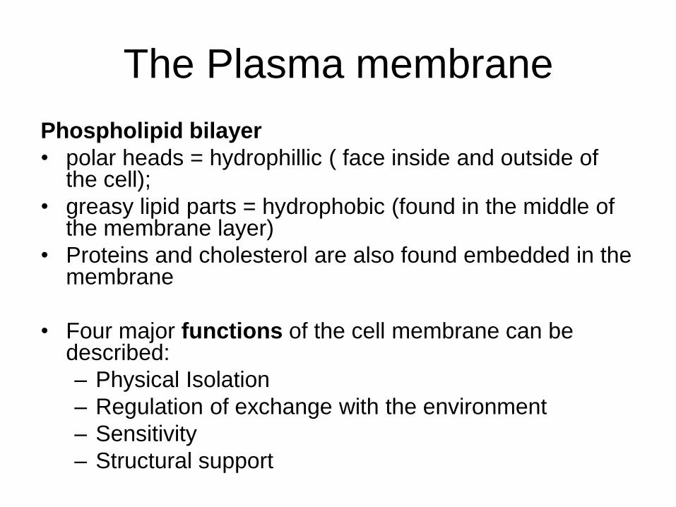
Cytoplasm: Cytosol and
Organelles • The cytoplasm is the general term for the
material inside the cell.
• The cytosol is the intracellular fluid.
– high in potassium ions.
– contains high concentrations of proteins.
• Organelles are structures within the cytoplasm each with a specific structure and function. – Non-membranous organelles
– membranous organelles

Non-Membranous organelles
• Cytoskeleton
• Microvilli
• Centrosomes
• Cilia
• Ribomes

The Cytoskeleton
A SEM image of the microfilaments
and microvilli of an intestinal cell
Microtubules in a living cell, as
seen after special fluorescent
labeling
LM 3200
SEM 30,000
Microvilli
Microfilaments
Plasmalemma
Terminal web
Mitochondrion
Intermediate
filaments
Endoplasmic
reticulum
Microtubule
Secretory
vesicle
Microfilaments can
produce movement of a
portion of a cell
Ex. Microvilli increases
surface area for
absorption
Intermediate filaments
provide strength & stability
Microtubules- hollow tubes
that transport organelles
(think roads)
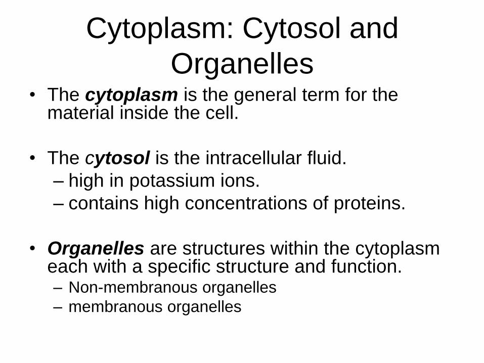
Centrioles and Cilia made by microtubules
Microtubules
Basal body
Plasmalemma
Microtubules
Power stroke Return stroke
TEM 240,000
Centrioles
direct the movement of
chromosomes
in cell division;
a pair = centrosomes
Cilia usually
move mucus
along cell
surface
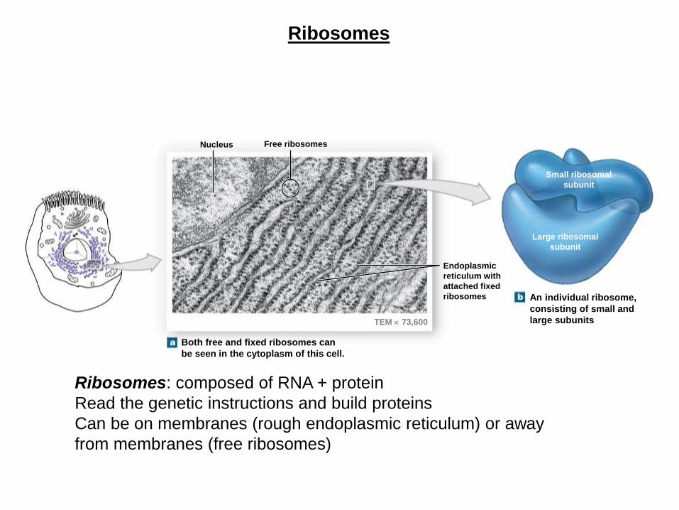
Ribosomes
Nucleus Free ribosomes
Endoplasmic
reticulum with
attached fixed
ribosomes
Small ribosomal
subunit
Large ribosomal
subunit
TEM 73,600
Both free and fixed ribosomes can
be seen in the cytoplasm of this cell.
An individual ribosome,
consisting of small and
large subunits
Ribosomes: composed of RNA + protein
Read the genetic instructions and build proteins
Can be on membranes (rough endoplasmic reticulum) or away
from membranes (free ribosomes)

Membranous organelles
• Mitochondria
• Nucleus
• Endoplasmic reticulum
• Golgi apparatus
• Lysosome
• Peroxisome
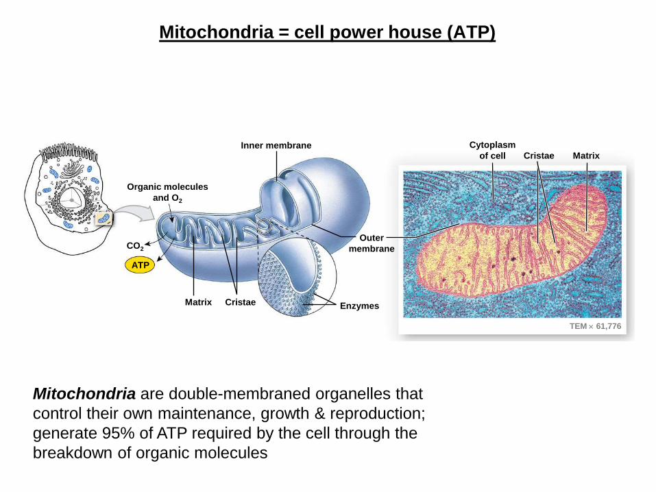
Mitochondria = cell power house (ATP)
Inner membrane
Organic molecules
and O2
CO2
ATP
Matrix Cristae Enzymes
Outer
membrane
TEM 61,776
Cytoplasm
of cell Cristae Matrix
Mitochondria are double-membraned organelles that
control their own maintenance, growth & reproduction;
generate 95% of ATP required by the cell through the
breakdown of organic molecules

The Nucleus = control center
Perinuclear
space
Nucleoplasm
Chromatin
Nucleolus
Nuclear envelope
Nuclear pores
TEM showing important nuclear structures
TEM 4828 The Nucleus is the control center of the cell
-Control of metabolism
-Storage and processing of genetic info
-Control of protein synthesis Parts:
-Nucleolus
-Nuclear envelope
-Nuclear pores

Chromosome vs. Chromatin
Nucleus of nondividing cell
Chromatin in nucleus
Dividing cell Visible chromosome
Supercoiled
region
Nucleosome
Histones DNA double
helix
In cells that are not
dividing, the
nucleosomes are loosely
coiled, forming a tangle
of fine filaments known
as chromatin.
Chromosomes (in
dividing cells): condense
their DNA to protect it
during Division; tightly
coiled
Chromatin (in non-diving cells) : the
form when the DNA is not in chromosomes
(DNA is being used); loosely coiled

Endoplasmic Reticulum (ER)
• The endoplasmic reticulum (ER) has four major functions:
– Synthesis of all classes of macromolecules
– Storage of the manufactured molecules
– Transport of substances from one area of the cell to another
– Enzymes in the lumen of the ER provide detoxification.
• Rough and Smooth ER

The Endoplasmic Reticulum: Rough and Smooth ER
Ribosomes
Cisternae
Rough endoplasmic
reticulum with
fixed (attached)
ribosomes
Free
ribosomes
Smooth
endoplasmic
reticulum
Endoplasmic
Reticulum
TEM 11,000
Rough ER- synthesizes
proteins and phospholipids;
transport vesicles deliver proteins
to the golgi apparatus
Smooth ER- synthesizes
other lipids; stores calcium;
detoxification
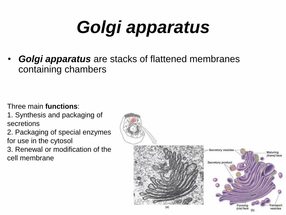
• Golgi apparatus are stacks of flattened membranes containing chambers
Golgi apparatus
Three main functions:
1. Synthesis and packaging of
secretions
2. Packaging of special enzymes
for use in the cytosol
3. Renewal or modification of the
cell membrane

Golgi apparatus
1. Proteins to be released from the cell are sorted into secretory vesicles, and released from the plasma membrane (exocytosis).
2. Phospholipids are added to renew the cell membrane when vesicles fuse.

Lysosome
• Lysosomes are vesicles filled with digestive
enzymes.
• Three main functions:
1. Lysosomes fuse with and recycle damaged
organelles.
2. Lysosomes fuse with phagosomes to digest
solid materials (damaged organelles or of
pathogens).
3. Lysosomes sometimes rupture resulting in
autolysis.

Figure 2.17 Lysosomal Functions
Waste products and debris are then ejected from the
cell when the vesicle fuses with the plasma membrane.
Endocytosis
Extracellular
solid or fluid
As digestion
occurs, nutrients
are reabsorbed for
recycling.
Primary
lysosomes
contain
inactive
enzymes.
As the materials
or pathogens are
broken down,
nutrients are
absorbed.
Golgi
apparatus
Function 1: A primary
lysosome may fuse with
the membrane of another
organelle, such as a
mitochondrion, forming a
secondary lysosome.
Function 2: A secondary
lysosome may also form
when a primary lysosome
fuses with a vesicle
containing fluid or solid
materials from outside the
cell.
Function 3: The lysosomal
membrane breaks down
following injury to, or death
of, the cell. The digestive
enzymes then attack the
cytoplasm in a destructive
process known as
autolysis. For this reason
lysosomes are sometimes
called “suicide packets.”
Peroxisome
• Peroxisomes are vesicles containing degradative enzymes
• smaller than lysosomes
• Two main functions:
• Breakdown organic compounds into hydrogen peroxide water
• Absorb and breakdown fatty acids
• Ex. most abundant in liver cells

Membrane flow 1. Nucleus: DNA
information Creates copy of DNA information (RNA) which travels to RER
2. RER uses RNA instructions to synthesize the protein
3. Protein is sent to Golgi for processing in a vesicle
4. Protein is modified in Golgi for secretion and placed into a secretory vesicle
5. Protein is released from the secretory vesicle via exocytosis

Intercellular attachment
There are three major types
of cell junctions:
• Communicating (gap)
junctions –found in heart
& smooth muscle
• Adhering (tight) junctions
- found in epithelium
• Anchoring junctions
(desmosomes)- found in
heart muscle and found
in layered epithelium
Tight junction
Anchoring junction

The Cell Life Cycle
INTERPHASE
THE CELL
CYCLE
MITOSIS AND CYTOKINESIS (See Figure 2.21)
Indefinite period G0
Specialized cell functions
G1 Normal cell functions plus cell growth, duplication of organelles, protein synthesis
G2 Protein synthesis
S DNA
replication, synthesis
of histones
M
Mitosis
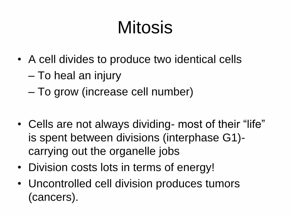
• A cell divides to produce two identical cells
– To heal an injury
– To grow (increase cell number)
• Cells are not always dividing- most of their “life”
is spent between divisions (interphase G1)-
carrying out the organelle jobs
• Division costs lots in terms of energy!
• Uncontrolled cell division produces tumors
(cancers).

Stages of cell’s life cycle:
(PMAT) Interphase- (between divisions, can’t see chromosomes)
• G1- cell is not ready to divide, carries out normal functions
• S- cell commits to divide, and copies all the DNA
• G2- cell prepares for division, and generates more lipids and Proteins
Mitosis- Division of genetic information (division of the nucleus): think P.M.A.T. (“Passed My Anatomy Test”)
– Prophase- preparations (preliminary steps)- package chromosomes
– Metaphase- chromosomes line up in the middle of the cell
– Anaphase- chromosomes separate and the two halves move apart to opposite sides of the cell
– Telophase – chromosomes are surrounded by membranes to form two nuclei
Cytokinesis- Division of the cell into two identical cells

DNA Replication
KEY
Adenine
Guanine
Cytosine
Thymine
Segment 2
DNA polymerase
DNA nucleotide
DNA
polymerase
Segment 1
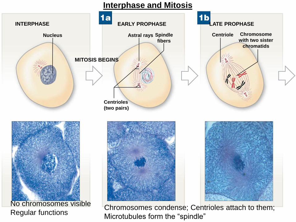
Interphase and Mitosis
INTERPHASE
MITOSIS BEGINS
EARLY PROPHASE LATE PROPHASE
Nucleus
Centrioles
(two pairs)
Astral rays Spindle
fibers
Centriole Chromosome
with two sister
chromatids
No chromosomes visible
Regular functions Chromosomes condense; Centrioles attach to them;
Microtubules form the “spindle”
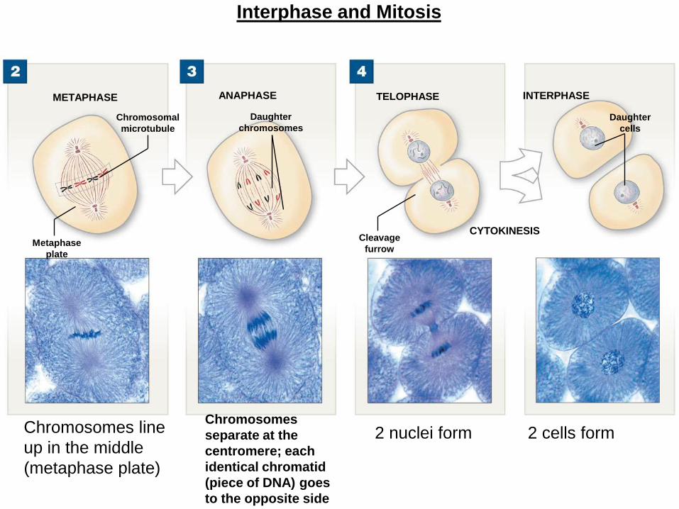
METAPHASE ANAPHASE TELOPHASE INTERPHASE
CYTOKINESIS Metaphase
plate
Chromosomal
microtubule
Daughter
chromosomes
Cleavage
furrow
Daughter
cells
Chromosomes line
up in the middle
(metaphase plate)
Chromosomes
separate at the
centromere; each
identical chromatid
(piece of DNA) goes
to the opposite side
2 nuclei form 2 cells form
Interphase and Mitosis