Cyclophilin Inhibitors Block Arterivirus Replication by
11
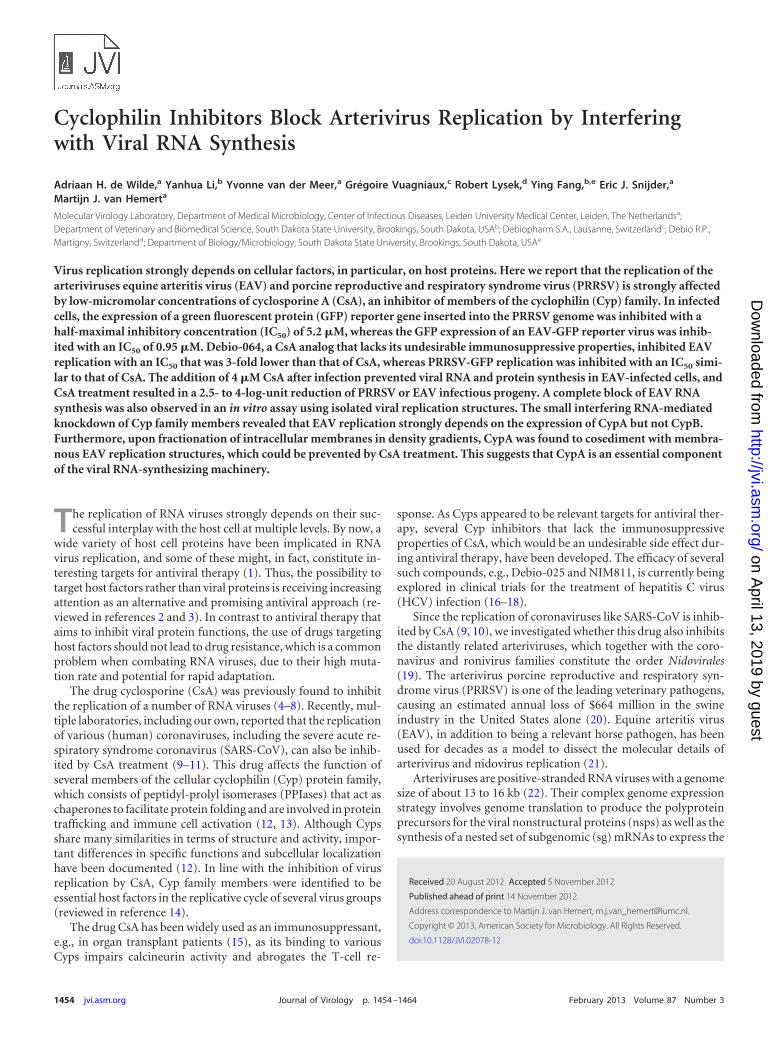
Cyclophilin Inhibitors Block Arterivirus Replication by Interfering with Viral RNA Synthesis Adriaan H. de Wilde, a Yanhua Li, b Yvonne van der Meer, a Grégoire Vuagniaux, c Robert Lysek, d Ying Fang, b,e Eric J. Snijder, a Martijn J. van Hemert a Molecular Virology Laboratory, Department of Medical Microbiology, Center of Infectious Diseases, Leiden University Medical Center, Leiden, The Netherlands a ; Department of Veterinary and Biomedical Science, South Dakota State University, Brookings, South Dakota, USA b ; Debiopharm S.A., Lausanne, Switzerland c ; Debio R.P., Martigny, Switzerland d ; Department of Biology/Microbiology, South Dakota State University, Brookings, South Dakota, USA e Virus replication strongly depends on cellular factors, in particular, on host proteins. Here we report that the replication of the arteriviruses equine arteritis virus (EAV) and porcine reproductive and respiratory syndrome virus (PRRSV) is strongly affected by low-micromolar concentrations of cyclosporine A (CsA), an inhibitor of members of the cyclophilin (Cyp) family. In infected cells, the expression of a green fluorescent protein (GFP) reporter gene inserted into the PRRSV genome was inhibited with a half-maximal inhibitory concentration (IC 50 ) of 5.2 M, whereas the GFP expression of an EAV-GFP reporter virus was inhib- ited with an IC 50 of 0.95 M. Debio-064, a CsA analog that lacks its undesirable immunosuppressive properties, inhibited EAV replication with an IC 50 that was 3-fold lower than that of CsA, whereas PRRSV-GFP replication was inhibited with an IC 50 simi- lar to that of CsA. The addition of 4 M CsA after infection prevented viral RNA and protein synthesis in EAV-infected cells, and CsA treatment resulted in a 2.5- to 4-log-unit reduction of PRRSV or EAV infectious progeny. A complete block of EAV RNA synthesis was also observed in an in vitro assay using isolated viral replication structures. The small interfering RNA-mediated knockdown of Cyp family members revealed that EAV replication strongly depends on the expression of CypA but not CypB. Furthermore, upon fractionation of intracellular membranes in density gradients, CypA was found to cosediment with membra- nous EAV replication structures, which could be prevented by CsA treatment. This suggests that CypA is an essential component of the viral RNA-synthesizing machinery. T he replication of RNA viruses strongly depends on their suc- cessful interplay with the host cell at multiple levels. By now, a wide variety of host cell proteins have been implicated in RNA virus replication, and some of these might, in fact, constitute in- teresting targets for antiviral therapy (1). Thus, the possibility to target host factors rather than viral proteins is receiving increasing attention as an alternative and promising antiviral approach (re- viewed in references 2 and 3). In contrast to antiviral therapy that aims to inhibit viral protein functions, the use of drugs targeting host factors should not lead to drug resistance, which is a common problem when combating RNA viruses, due to their high muta- tion rate and potential for rapid adaptation. The drug cyclosporine (CsA) was previously found to inhibit the replication of a number of RNA viruses (4–8). Recently, mul- tiple laboratories, including our own, reported that the replication of various (human) coronaviruses, including the severe acute re- spiratory syndrome coronavirus (SARS-CoV), can also be inhib- ited by CsA treatment (9–11). This drug affects the function of several members of the cellular cyclophilin (Cyp) protein family, which consists of peptidyl-prolyl isomerases (PPIases) that act as chaperones to facilitate protein folding and are involved in protein trafficking and immune cell activation (12, 13). Although Cyps share many similarities in terms of structure and activity, impor- tant differences in specific functions and subcellular localization have been documented (12). In line with the inhibition of virus replication by CsA, Cyp family members were identified to be essential host factors in the replicative cycle of several virus groups (reviewed in reference 14). The drug CsA has been widely used as an immunosuppressant, e.g., in organ transplant patients (15), as its binding to various Cyps impairs calcineurin activity and abrogates the T-cell re- sponse. As Cyps appeared to be relevant targets for antiviral ther- apy, several Cyp inhibitors that lack the immunosuppressive properties of CsA, which would be an undesirable side effect dur- ing antiviral therapy, have been developed. The efficacy of several such compounds, e.g., Debio-025 and NIM811, is currently being explored in clinical trials for the treatment of hepatitis C virus (HCV) infection (16–18). Since the replication of coronaviruses like SARS-CoV is inhib- ited by CsA (9, 10), we investigated whether this drug also inhibits the distantly related arteriviruses, which together with the coro- navirus and ronivirus families constitute the order Nidovirales (19). The arterivirus porcine reproductive and respiratory syn- drome virus (PRRSV) is one of the leading veterinary pathogens, causing an estimated annual loss of $664 million in the swine industry in the United States alone (20). Equine arteritis virus (EAV), in addition to being a relevant horse pathogen, has been used for decades as a model to dissect the molecular details of arterivirus and nidovirus replication (21). Arteriviruses are positive-stranded RNA viruses with a genome size of about 13 to 16 kb (22). Their complex genome expression strategy involves genome translation to produce the polyprotein precursors for the viral nonstructural proteins (nsps) as well as the synthesis of a nested set of subgenomic (sg) mRNAs to express the Received 20 August 2012 Accepted 5 November 2012 Published ahead of print 14 November 2012 Address correspondence to Martijn J. van Hemert, [email protected]. Copyright © 2013, American Society for Microbiology. All Rights Reserved. doi:10.1128/JVI.02078-12 1454 jvi.asm.org Journal of Virology p. 1454 –1464 February 2013 Volume 87 Number 3 on April 13, 2019 by guest http://jvi.asm.org/ Downloaded from
Transcript of Cyclophilin Inhibitors Block Arterivirus Replication by
Cyclophilin Inhibitors Block Arterivirus Replication by Interferingwith Viral RNA Synthesis
Adriaan H. de Wilde,a Yanhua Li,b Yvonne van der Meer,a Grégoire Vuagniaux,c Robert Lysek,d Ying Fang,b,e Eric J. Snijder,a
Martijn J. van Hemerta
Molecular Virology Laboratory, Department of Medical Microbiology, Center of Infectious Diseases, Leiden University Medical Center, Leiden, The Netherlandsa;Department of Veterinary and Biomedical Science, South Dakota State University, Brookings, South Dakota, USAb; Debiopharm S.A., Lausanne, Switzerlandc; Debio R.P.,Martigny, Switzerlandd; Department of Biology/Microbiology, South Dakota State University, Brookings, South Dakota, USAe
Virus replication strongly depends on cellular factors, in particular, on host proteins. Here we report that the replication of thearteriviruses equine arteritis virus (EAV) and porcine reproductive and respiratory syndrome virus (PRRSV) is strongly affectedby low-micromolar concentrations of cyclosporine A (CsA), an inhibitor of members of the cyclophilin (Cyp) family. In infectedcells, the expression of a green fluorescent protein (GFP) reporter gene inserted into the PRRSV genome was inhibited with ahalf-maximal inhibitory concentration (IC50) of 5.2 �M, whereas the GFP expression of an EAV-GFP reporter virus was inhib-ited with an IC50 of 0.95 �M. Debio-064, a CsA analog that lacks its undesirable immunosuppressive properties, inhibited EAVreplication with an IC50 that was 3-fold lower than that of CsA, whereas PRRSV-GFP replication was inhibited with an IC50 simi-lar to that of CsA. The addition of 4 �M CsA after infection prevented viral RNA and protein synthesis in EAV-infected cells, andCsA treatment resulted in a 2.5- to 4-log-unit reduction of PRRSV or EAV infectious progeny. A complete block of EAV RNAsynthesis was also observed in an in vitro assay using isolated viral replication structures. The small interfering RNA-mediatedknockdown of Cyp family members revealed that EAV replication strongly depends on the expression of CypA but not CypB.Furthermore, upon fractionation of intracellular membranes in density gradients, CypA was found to cosediment with membra-nous EAV replication structures, which could be prevented by CsA treatment. This suggests that CypA is an essential componentof the viral RNA-synthesizing machinery.
The replication of RNA viruses strongly depends on their suc-cessful interplay with the host cell at multiple levels. By now, a
wide variety of host cell proteins have been implicated in RNAvirus replication, and some of these might, in fact, constitute in-teresting targets for antiviral therapy (1). Thus, the possibility totarget host factors rather than viral proteins is receiving increasingattention as an alternative and promising antiviral approach (re-viewed in references 2 and 3). In contrast to antiviral therapy thataims to inhibit viral protein functions, the use of drugs targetinghost factors should not lead to drug resistance, which is a commonproblem when combating RNA viruses, due to their high muta-tion rate and potential for rapid adaptation.
The drug cyclosporine (CsA) was previously found to inhibitthe replication of a number of RNA viruses (4–8). Recently, mul-tiple laboratories, including our own, reported that the replicationof various (human) coronaviruses, including the severe acute re-spiratory syndrome coronavirus (SARS-CoV), can also be inhib-ited by CsA treatment (9–11). This drug affects the function ofseveral members of the cellular cyclophilin (Cyp) protein family,which consists of peptidyl-prolyl isomerases (PPIases) that act aschaperones to facilitate protein folding and are involved in proteintrafficking and immune cell activation (12, 13). Although Cypsshare many similarities in terms of structure and activity, impor-tant differences in specific functions and subcellular localizationhave been documented (12). In line with the inhibition of virusreplication by CsA, Cyp family members were identified to beessential host factors in the replicative cycle of several virus groups(reviewed in reference 14).
The drug CsA has been widely used as an immunosuppressant,e.g., in organ transplant patients (15), as its binding to variousCyps impairs calcineurin activity and abrogates the T-cell re-
sponse. As Cyps appeared to be relevant targets for antiviral ther-apy, several Cyp inhibitors that lack the immunosuppressiveproperties of CsA, which would be an undesirable side effect dur-ing antiviral therapy, have been developed. The efficacy of severalsuch compounds, e.g., Debio-025 and NIM811, is currently beingexplored in clinical trials for the treatment of hepatitis C virus(HCV) infection (16–18).
Since the replication of coronaviruses like SARS-CoV is inhib-ited by CsA (9, 10), we investigated whether this drug also inhibitsthe distantly related arteriviruses, which together with the coro-navirus and ronivirus families constitute the order Nidovirales(19). The arterivirus porcine reproductive and respiratory syn-drome virus (PRRSV) is one of the leading veterinary pathogens,causing an estimated annual loss of $664 million in the swineindustry in the United States alone (20). Equine arteritis virus(EAV), in addition to being a relevant horse pathogen, has beenused for decades as a model to dissect the molecular details ofarterivirus and nidovirus replication (21).
Arteriviruses are positive-stranded RNA viruses with a genomesize of about 13 to 16 kb (22). Their complex genome expressionstrategy involves genome translation to produce the polyproteinprecursors for the viral nonstructural proteins (nsps) as well as thesynthesis of a nested set of subgenomic (sg) mRNAs to express the
Received 20 August 2012 Accepted 5 November 2012
Published ahead of print 14 November 2012
Address correspondence to Martijn J. van Hemert, [email protected].
Copyright © 2013, American Society for Microbiology. All Rights Reserved.
doi:10.1128/JVI.02078-12
1454 jvi.asm.org Journal of Virology p. 1454–1464 February 2013 Volume 87 Number 3
on April 13, 2019 by guest
http://jvi.asm.org/
Dow
nloaded from
structural proteins (23). The viral nsps, presumably together withvarious host factors, are thought to assemble into membrane-as-sociated replication and transcription complexes (RTCs) thatdrive viral RNA synthesis (for recent reviews, see references 22 and24). Arterivirus RNA synthesis was reported to be associated witha virus-induced network of endoplasmic reticulum (ER)-derivedmembrane structures, including large numbers of double-mem-brane vesicles (25). Many arteriviral proteins were found to beassociated with these membrane structures, on which viral RNAsynthesis was found to depend (26, 27). However, thus far, theidentity and role of proviral host factors involved in the replicativecycle of arteriviruses have remained largely unexplored.
Using EAV and PRRSV, our studies on the inhibition of nidovirusreplication by CsA have now been extended to arteriviruses and ex-plored the mechanism of action of the compound in more detail. Weshow that low-micromolar concentrations of CsA can fully block ar-terivirus RNA synthesis and that the nonimmunosuppressive cyclo-philin inhibitor Debio-064 is an even more potent inhibitor. Thesecompounds probably exert their effect through their inhibition ofCypA, as RNA interference (RNAi)-mediated knockdown of CypAstrongly affected EAV RNA synthesis and CypA was found to cosedi-ment with EAV replication structures.
MATERIALS AND METHODSCell culture, infection, and virus titration. BHK-21 (28), Vero E6 (29),and MARC-145 (30) cells were cultured as described previously. 293/ACE2 cells (31) were cultured in Dulbecco’s modified Eagle’s medium(DMEM; Lonza) supplemented with 8% fetal calf serum, 100 U/ml ofpenicillin, 100 �g/ml of streptomycin, 2 mM L-glutamine, and 12 �g/mlblasticidin (PAA Laboratories). A cell culture-adapted derivative of theEAV Bucyrus isolate (32) and green fluorescent protein (GFP)-expressingrecombinant EAV (33) were used to infect monolayers of BHK-21, VeroE6, and 293/ACE2 cells at a multiplicity of infection (MOI) of 5, as de-scribed previously (28, 29). MARC-145 cells were infected with a GFP-expressing recombinant PRRSV (SD01-08-GFP) at an MOI of 0.1, as pre-viously described (34). EAV titers in cell culture supernatants weredetermined by plaque assay on BHK-21 cells (28), whereas PRRSV titerswere determined by fluorescent focus assay (FFA) on MARC-145 cells, asdescribed previously (35). For half-maximal inhibitory concentration(IC50) determinations, cells were grown in black 96-well plates (Greiner),infected with EAV-GFP or PRRSV-GFP, and treated with compounds inoctuplet. GFP reporter expression was quantified by measuring the fluo-rescence in a 96-well plate reader, using an excitation wavelength of 485nm and an emission wavelength of 535 nm.
Antibodies and drugs. Rabbit polyclonal antibodies against CypA(Abcam), CypB (Abcam), and calnexin (BD), a goat polyclonal antiserumagainst GAPDH (glyceraldehyde-3-phosphate dehydrogenase; SantaCruz), and a mouse monoclonal antibody (MAb) against �-actin (Sigma)were used. Rabbit antisera recognizing the EAV replicase subunits nsp3(36) and nsp9 (27) and the EAV membrane (M) protein (29) and a MAbagainst the EAV nucleocapsid (N) protein (37) have been described pre-viously. The cyclophilin inhibitors CsA (Sigma) and Debio-064 (Debio-pharm, Switzerland) were dissolved in dimethyl sulfoxide (DMSO). CsAwas stored as a 50-mg/ml stock at �20°C, and Debio-064 was stored as a10 mM stock at 4°C in aliquots for single use. The IC50 of inhibitors wascalculated with GraphPad Prism (version 5) software using a nonlinearregression model.
Immunofluorescence microscopy. EAV-infected or mock-infectedBHK-21 cells, grown on coverslips at 39.5°C, were fixed with 3% paraform-aldehyde in phosphate-buffered saline (PBS), permeabilized with 0.1%Triton X-100, and processed for immunofluorescence microscopy as de-scribed previously (26). Specimens were examined with a Zeiss Axioskop
2 fluorescence microscope with an Axiocam HRc camera and Zeiss Axio-vision (version 4.4) software.
Western blot analysis. After SDS-PAGE, proteins were transferred toHybond-LFP membranes (GE Healthcare) by semidry blotting. Mem-branes were blocked with 1% casein in PBS containing 0.1% Tween 20(PBST) and were incubated with anti-nsp3 (1:2,000), anti-nsp9 (1:2,000),anti-M (1:2,000), anti-N (1:10,000), anti-CypA (1:1,000), anti-CypB (1:2,000), or anti-�-actin (1:50,000) antiserum diluted in PBST with 0.5%casein. Biotin-conjugated swine anti-rabbit IgG (1:2,000) or goat anti-mouse IgG (1:1,000) antibodies (Dako) and Cy3-conjugated mouse anti-biotin (1:2,500) antibodies were used for detection. Blots were scannedwith a Typhoon 9410 imager (GE Healthcare) and analyzed withImageQuant TL software.
Isolation of EAV RTC-containing replication structures and in vitroRNA synthesis assays. EAV replication structures were isolated fromBHK-21 or Vero E6 cells, and in vitro RNA synthesis assays were per-formed essentially as described previously (27). In short, approximately1 � 108 EAV-infected BHK-21 or Vero E6 cells were harvested at 6 or 7 hpostinfection (p.i.), and cells were lysed to obtain a postnuclear superna-tant (PNS) (27). A standard in vitro RNA synthesis assay mixture con-tained 20 �l of PNS (the equivalent of 6 � 104 cells) from EAV-infectedBHK-21 cells, 5 �l of an inhibitor solution, or 5 �l of RTC dilution buffer(control). Following gel electrophoresis, 32P-labeled reaction productswere analyzed by denaturing agarose gel electrophoresis and by exposinga PhosphorImager screen directly to the dried gel, after which screenswere scanned with a Typhoon 9410 imager (GE Healthcare), and incor-poration of label was quantified using ImageQuant TL software.
Density gradient fractionation. Subcellular fractionation of PNS wasperformed in continuous 0 to 30% OptiPrep density gradients in RTCdilution buffer. The gradients were prepared in 13.2 ml Ultra-Clear cen-trifugation tubes (Beckman Coulter) using a Gradient Master gradientformer (Biocomp). One milliliter of PNS from Vero E6 cells was carefullyloaded on top of the preformed gradient. After centrifugation for 17 h at48,000 � g in an SW41 rotor at 4°C, the gradient was fractionated into0.5-ml fractions. The density of each fraction was determined with a re-fractometer (GETI).
Metabolic labeling of viral RNA synthesis. Labeling of viral RNA with[3H]uridine was performed essentially as described previously (38).Briefly, at 4.5 h p.i., 4 � 105 EAV-infected BHK-21 cells in 4-cm2 disheswere given medium containing 10 �g/ml actinomycin D (ActD; Sigma-Aldrich) and either 4 �M CsA or 0.01% DMSO as a solvent control. After1 h, viral RNA synthesis was labeled by adding 100 �Ci of [3H]uridine tothe medium. The 3H-labeled RNAs were isolated, separated in denaturingagarose gels, and visualized by fluorography. To verify that equal amountsof total RNA were loaded, the gel was hybridized with a 32P-labeled oli-gonucleotide probe (5=-TTCACGCCCTCTTGAACTCTCTCTTC-3=)recognizing 28S rRNA, as described previously (27).
RNA interference. ON-TARGETplus smart-pool small interferingRNA (siRNA) duplexes (Dharmacon) against CypA (PPIA; catalog no.L-004979-04) and CypB (PPIB; catalog no. L-004606-00) were used tosilence CypA and CypB expression in 293/ACE2 cells. A nontargetingsiRNA (D-001810-10) was used as a control, and a GAPDH-targetingsiRNA (D-001830-10) was used to monitor transfection and knockdownefficiency. Stock solutions of 2 �M were prepared by dissolving siRNAs in1� siRNA buffer (Dharmacon). For transfection of cells in 96-well clus-ters, 1 � 104 293/ACE2 cells per well were transfected with a 100-�lmixture containing 100 nM siRNA, 0.2 �g DharmaFECT1 transfectionreagent (Dharmacon), Opti-MEM reduced-serum medium (Invitrogen),and antibiotic-free culture medium, according to the manufacturers’ in-structions. For cells in 12-well clusters, 600-�l transfection mixtures wereused. Medium was replaced at 24 h posttransfection (p.t.) by antibiotic-free culture medium, and at 48 h p.t., cells were infected with EAV-GFP orwild-type (wt) EAV. Duplicate cultures were used either to prepare lysatesto analyze protein expression levels or to monitor cell viability using a
Cyclophilin Inhibitors Block Arterivirus Replication
February 2013 Volume 87 Number 3 jvi.asm.org 1455
on April 13, 2019 by guest
http://jvi.asm.org/
Dow
nloaded from
CellTiter 96 AQueous nonradioactive cell proliferation assay (Promega),according to the manufacturer’s instructions.
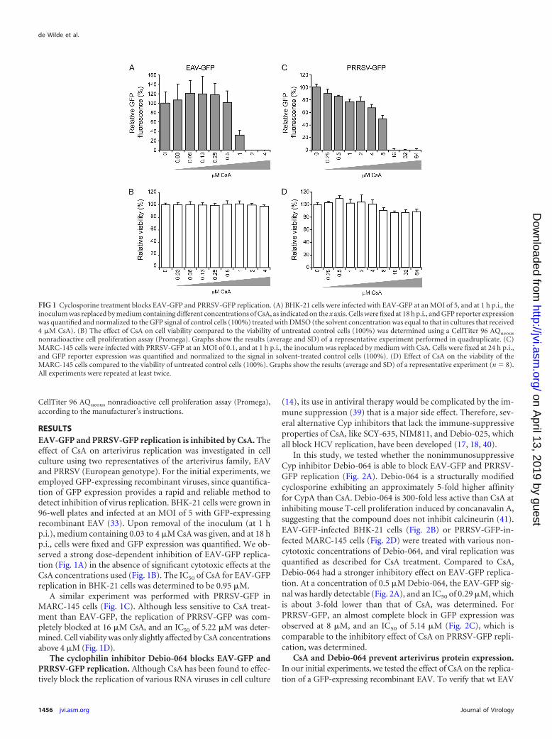
RESULTSEAV-GFP and PRRSV-GFP replication is inhibited by CsA. Theeffect of CsA on arterivirus replication was investigated in cellculture using two representatives of the arterivirus family, EAVand PRRSV (European genotype). For the initial experiments, weemployed GFP-expressing recombinant viruses, since quantifica-tion of GFP expression provides a rapid and reliable method todetect inhibition of virus replication. BHK-21 cells were grown in96-well plates and infected at an MOI of 5 with GFP-expressingrecombinant EAV (33). Upon removal of the inoculum (at 1 hp.i.), medium containing 0.03 to 4 �M CsA was given, and at 18 hp.i., cells were fixed and GFP expression was quantified. We ob-served a strong dose-dependent inhibition of EAV-GFP replica-tion (Fig. 1A) in the absence of significant cytotoxic effects at theCsA concentrations used (Fig. 1B). The IC50 of CsA for EAV-GFPreplication in BHK-21 cells was determined to be 0.95 �M.
A similar experiment was performed with PRRSV-GFP inMARC-145 cells (Fig. 1C). Although less sensitive to CsA treat-ment than EAV-GFP, the replication of PRRSV-GFP was com-pletely blocked at 16 �M CsA, and an IC50 of 5.22 �M was deter-mined. Cell viability was only slightly affected by CsA concentrationsabove 4 �M (Fig. 1D).
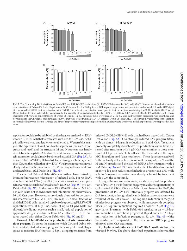
The cyclophilin inhibitor Debio-064 blocks EAV-GFP andPRRSV-GFP replication. Although CsA has been found to effec-tively block the replication of various RNA viruses in cell culture
(14), its use in antiviral therapy would be complicated by the im-mune suppression (39) that is a major side effect. Therefore, sev-eral alternative Cyp inhibitors that lack the immune-suppressiveproperties of CsA, like SCY-635, NIM811, and Debio-025, whichall block HCV replication, have been developed (17, 18, 40).
In this study, we tested whether the nonimmunosuppressiveCyp inhibitor Debio-064 is able to block EAV-GFP and PRRSV-GFP replication (Fig. 2A). Debio-064 is a structurally modifiedcyclosporine exhibiting an approximately 5-fold higher affinityfor CypA than CsA. Debio-064 is 300-fold less active than CsA atinhibiting mouse T-cell proliferation induced by concanavalin A,suggesting that the compound does not inhibit calcineurin (41).EAV-GFP-infected BHK-21 cells (Fig. 2B) or PRRSV-GFP-in-fected MARC-145 cells (Fig. 2D) were treated with various non-cytotoxic concentrations of Debio-064, and viral replication wasquantified as described for CsA treatment. Compared to CsA,Debio-064 had a stronger inhibitory effect on EAV-GFP replica-tion. At a concentration of 0.5 �M Debio-064, the EAV-GFP sig-nal was hardly detectable (Fig. 2A), and an IC50 of 0.29 �M, whichis about 3-fold lower than that of CsA, was determined. ForPRRSV-GFP, an almost complete block in GFP expression wasobserved at 8 �M, and an IC50 of 5.14 �M (Fig. 2C), which iscomparable to the inhibitory effect of CsA on PRRSV-GFP repli-cation, was determined.
CsA and Debio-064 prevent arterivirus protein expression.In our initial experiments, we tested the effect of CsA on the replica-tion of a GFP-expressing recombinant EAV. To verify that wt EAV
FIG 1 Cyclosporine treatment blocks EAV-GFP and PRRSV-GFP replication. (A) BHK-21 cells were infected with EAV-GFP at an MOI of 5, and at 1 h p.i., theinoculum was replaced by medium containing different concentrations of CsA, as indicated on the x axis. Cells were fixed at 18 h p.i., and GFP reporter expressionwas quantified and normalized to the GFP signal of control cells (100%) treated with DMSO (the solvent concentration was equal to that in cultures that received4 �M CsA). (B) The effect of CsA on cell viability compared to the viability of untreated control cells (100%) was determined using a CellTiter 96 AQueous
nonradioactive cell proliferation assay (Promega). Graphs show the results (average and SD) of a representative experiment performed in quadruplicate. (C)MARC-145 cells were infected with PRRSV-GFP at an MOI of 0.1, and at 1 h p.i., the inoculum was replaced by medium with CsA. Cells were fixed at 24 h p.i.,and GFP reporter expression was quantified and normalized to the signal in solvent-treated control cells (100%). (D) Effect of CsA on the viability of theMARC-145 cells compared to the viability of untreated control cells (100%). Graphs show the results (average and SD) of a representative experiment (n � 8).All experiments were repeated at least twice.
de Wilde et al.
1456 jvi.asm.org Journal of Virology
on April 13, 2019 by guest
http://jvi.asm.org/
Dow
nloaded from
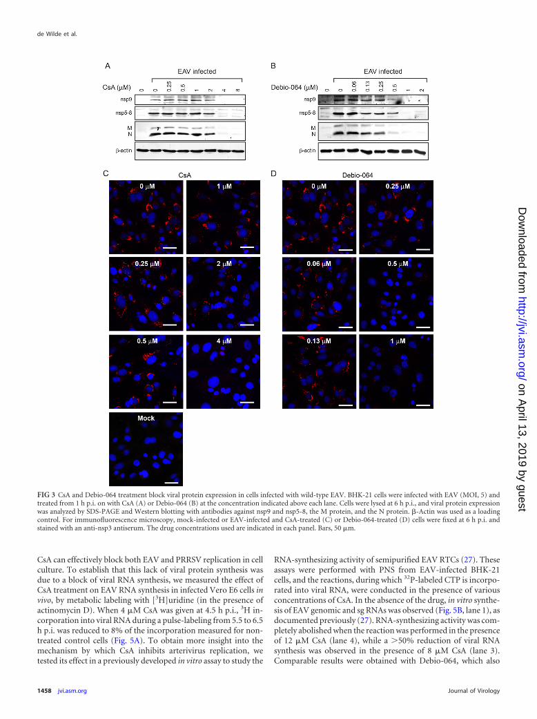
replication could also be inhibited by the drug, we analyzed wt EAV-infected BHK-21 cells that were treated with 0.25 to 8 �M CsA. At 6 hp.i., cells were lysed and lysates were subjected to Western blot anal-ysis. The expression of viral nonstructural proteins (the nsp5-8 pre-cursor and nsp9) and the structural M and N proteins was hardlydetectable after 4 �M CsA treatment, while a clear reduction in pro-tein expression could already be observed at 2 �M CsA (Fig. 3A). Asobserved for EAV-GFP, Debio-064 had a stronger inhibitory effectthan CsA on the replication of wt EAV. Viral protein expression wasclearly reduced in the presence of 0.5�M the drug and became almostundetectable at 1 �M Debio-064 (Fig. 3B).
The effect of CsA and Debio-064 was further characterized byimmunofluorescence microscopy of infected cells. For wt EAV,double-stranded RNA (dsRNA) (data not shown) and viral pro-teins were undetectable after a dose of 4 �M CsA (Fig. 3C) or 1 �MDebio-064 (Fig. 3D). In the case of PRRSV-GFP-infected MARC-145 cells (data not shown), maximal inhibition was observed at a16 �M CsA dose. However, as previously observed for coronavi-rus-infected Vero E6, 17Cl1, or Huh7 cells (9), a small fraction ofthe MARC-145 cells remained capable of supporting PRRSV-GFPreplication, even at high CsA doses of up to 64 �M (data notshown). We did not observe such a small, residual population ofapparently drug-insensitive cells in EAV-infected BHK-21 cul-tures treated with either CsA or Debio-064 (Fig. 3C and D).
CsA and Debio-064 block the production of arterivirus infec-tious progeny. To assess the extent to which CsA and Debio-064treatment affected infectious progeny titers, we performed plaqueassays to measure EAV titers at 12 h p.i. using supernatants from
infected (MOI, 5) BHK-21 cells that had been treated with CsA orDebio-064 (Fig. 4A). CsA strongly reduced EAV progeny titers,with an almost 4-log-unit reduction at 4 �M CsA. Treatmentprobably completely abolished virus production, as the titers ob-served after treatment with 4 �M CsA were similar to those mea-sured at 1 h p.i., which likely reflected the remainder of the high-MOI inoculum used (data not shown). These data correlated wellwith the barely detectable expression of the nsp5-8, nsp9, and theM and N proteins and the lack of dsRNA after treatment with 4�M CsA (Fig. 3A and C). Treatment with Debio-064 also resultedin an �4-log-unit reduction of infectious progeny at 2 �M, whilea 2- to 3-log-unit reduction was already achieved by treatmentwith 1 �M the compound.
Using a fluorescent focus assay, we also analyzed the produc-tion of PRRSV-GFP infectious progeny in culture supernatants ofCsA-treated MARC-145 cells at 24 h p.i. As observed for EAV, theproduction of PRRSV-GFP infectious progeny was affected byCsA treatment, although significantly higher concentrations wererequired. At 16 �M CsA, an �1.5-log-unit reduction in the yieldof infectious progeny was observed, while an apparently completeblock (2.5-log-unit reduction) required a dose of 32 �M (Fig. 4B,gray bars). Treatment with Debio-064 resulted in an �1.5-log-unit reduction of infectious progeny at 16 �M and an �2.5-log-unit reduction of infectious progeny at 32 �M (Fig. 4B, whitebars), which are comparable to the reduction in PRRSV progenyobserved upon CsA treatment.
Cyclophilin inhibitors affect EAV RNA synthesis both invivo and in vitro. The above-described experiments showed that
FIG 2 The CsA analog Debio-064 blocks EAV-GFP and PRRSV-GFP replication. (A) EAV-GFP-infected BHK-21 cells (MOI, 5) were incubated with variousconcentrations of Debio-064 from 1 h p.i. onwards. Cells were fixed at 18 h p.i., and GFP reporter expression was quantified and normalized to the GFP signalof control cells (100%) that were treated with DMSO (the solvent concentration was equal to that in medium containing 4 �M Debio-064). (B) Effect ofDebio-064 on BHK-21 cell viability compared to the viability of untreated control cells (100%). (C) PRRSV-GFP-infected MARC-145 cells (MOI, 0.1) wereincubated with various concentrations of Debio-064 from 1 h p.i. onwards. Cells were fixed at 24 h p.i., and GFP reporter expression was quantified andnormalized to the GFP signal of control cells (100%) that were treated with DMSO. (D) Effect of Debio-064 on MARC-145 cell viability compared to the viabilityof control cells (100%). Results (average and SD) of a representative experiment performed in quadruplicate are shown, and all experiments were repeated at leasttwice.
Cyclophilin Inhibitors Block Arterivirus Replication
February 2013 Volume 87 Number 3 jvi.asm.org 1457
on April 13, 2019 by guest
http://jvi.asm.org/
Dow
nloaded from
CsA can effectively block both EAV and PRRSV replication in cellculture. To establish that this lack of viral protein synthesis wasdue to a block of viral RNA synthesis, we measured the effect ofCsA treatment on EAV RNA synthesis in infected Vero E6 cells invivo, by metabolic labeling with [3H]uridine (in the presence ofactinomycin D). When 4 �M CsA was given at 4.5 h p.i., 3H in-corporation into viral RNA during a pulse-labeling from 5.5 to 6.5h p.i. was reduced to 8% of the incorporation measured for non-treated control cells (Fig. 5A). To obtain more insight into themechanism by which CsA inhibits arterivirus replication, wetested its effect in a previously developed in vitro assay to study the
RNA-synthesizing activity of semipurified EAV RTCs (27). Theseassays were performed with PNS from EAV-infected BHK-21cells, and the reactions, during which 32P-labeled CTP is incorpo-rated into viral RNA, were conducted in the presence of variousconcentrations of CsA. In the absence of the drug, in vitro synthe-sis of EAV genomic and sg RNAs was observed (Fig. 5B, lane 1), asdocumented previously (27). RNA-synthesizing activity was com-pletely abolished when the reaction was performed in the presenceof 12 �M CsA (lane 4), while a �50% reduction of viral RNAsynthesis was observed in the presence of 8 �M CsA (lane 3).Comparable results were obtained with Debio-064, which also
FIG 3 CsA and Debio-064 treatment block viral protein expression in cells infected with wild-type EAV. BHK-21 cells were infected with EAV (MOI, 5) andtreated from 1 h p.i. on with CsA (A) or Debio-064 (B) at the concentration indicated above each lane. Cells were lysed at 6 h p.i., and viral protein expressionwas analyzed by SDS-PAGE and Western blotting with antibodies against nsp9 and nsp5-8, the M protein, and the N protein. �-Actin was used as a loadingcontrol. For immunofluorescence microscopy, mock-infected or EAV-infected and CsA-treated (C) or Debio-064-treated (D) cells were fixed at 6 h p.i. andstained with an anti-nsp3 antiserum. The drug concentrations used are indicated in each panel. Bars, 50 �m.
de Wilde et al.
1458 jvi.asm.org Journal of Virology
on April 13, 2019 by guest
http://jvi.asm.org/
Dow
nloaded from
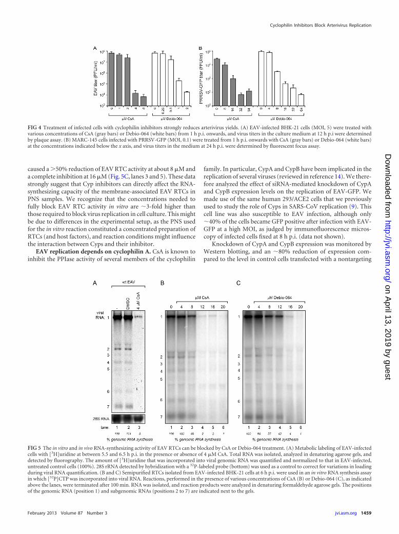
caused a �50% reduction of EAV RTC activity at about 8 �M anda complete inhibition at 16 �M (Fig. 5C, lanes 3 and 5). These datastrongly suggest that Cyp inhibitors can directly affect the RNA-synthesizing capacity of the membrane-associated EAV RTCs inPNS samples. We recognize that the concentrations needed tofully block EAV RTC activity in vitro are �3-fold higher thanthose required to block virus replication in cell culture. This mightbe due to differences in the experimental setup, as the PNS usedfor the in vitro reaction constituted a concentrated preparation ofRTCs (and host factors), and reaction conditions might influencethe interaction between Cyps and their inhibitor.
EAV replication depends on cyclophilin A. CsA is known toinhibit the PPIase activity of several members of the cyclophilin
family. In particular, CypA and CypB have been implicated in thereplication of several viruses (reviewed in reference 14). We there-fore analyzed the effect of siRNA-mediated knockdown of CypAand CypB expression levels on the replication of EAV-GFP. Wemade use of the same human 293/ACE2 cells that we previouslyused to study the role of Cyps in SARS-CoV replication (9). Thiscell line was also susceptible to EAV infection, although only�40% of the cells became GFP positive after infection with EAV-GFP at a high MOI, as judged by immunofluorescence micros-copy of infected cells fixed at 8 h p.i. (data not shown).
Knockdown of CypA and CypB expression was monitored byWestern blotting, and an �80% reduction of expression com-pared to the level in control cells transfected with a nontargeting
FIG 4 Treatment of infected cells with cyclophilin inhibitors strongly reduces arterivirus yields. (A) EAV-infected BHK-21 cells (MOI, 5) were treated withvarious concentrations of CsA (gray bars) or Debio-064 (white bars) from 1 h p.i. onwards, and virus titers in the culture medium at 12 h p.i were determinedby plaque assay. (B) MARC-145 cells infected with PRRSV-GFP (MOI, 0.1) were treated from 1 h p.i. onwards with CsA (gray bars) or Debio-064 (white bars)at the concentrations indicated below the x axis, and virus titers in the medium at 24 h p.i. were determined by fluorescent focus assay.
FIG 5 The in vitro and in vivo RNA-synthesizing activity of EAV RTCs can be blocked by CsA or Debio-064 treatment. (A) Metabolic labeling of EAV-infectedcells with [3H]uridine at between 5.5 and 6.5 h p.i. in the presence or absence of 4 �M CsA. Total RNA was isolated, analyzed in denaturing agarose gels, anddetected by fluorography. The amount of [3H]uridine that was incorporated into viral genomic RNA was quantified and normalized to that in EAV-infected,untreated control cells (100%). 28S rRNA detected by hybridization with a 32P-labeled probe (bottom) was used as a control to correct for variations in loadingduring viral RNA quantification. (B and C) Semipurified RTCs isolated from EAV-infected BHK-21 cells at 6 h p.i. were used in an in vitro RNA synthesis assayin which [32P]CTP was incorporated into viral RNA. Reactions, performed in the presence of various concentrations of CsA (B) or Debio-064 (C), as indicatedabove the lanes, were terminated after 100 min. RNA was isolated, and reaction products were analyzed in denaturing formaldehyde agarose gels. The positionsof the genomic RNA (position 1) and subgenomic RNAs (positions 2 to 7) are indicated next to the gels.
Cyclophilin Inhibitors Block Arterivirus Replication
February 2013 Volume 87 Number 3 jvi.asm.org 1459
on April 13, 2019 by guest
http://jvi.asm.org/
Dow
nloaded from
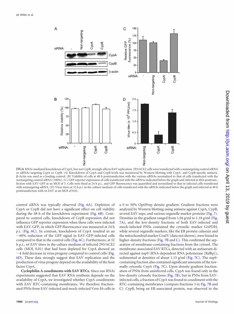
control siRNA was typically observed (Fig. 6A). Depletion ofCypA or CypB did not have a significant effect on cell viabilityduring the 48 h of the knockdown experiment (Fig. 6B). Com-pared to control cells, knockdown of CypB expression did notinfluence GFP reporter expression when these cells were infectedwith EAV-GFP, in which GFP fluorescence was measured at 24 hp.i. (Fig. 6C). In contrast, knockdown of CypA resulted in an�60% reduction of the GFP signal in EAV-GFP-infected cellscompared to that in the control cells (Fig. 6C). Furthermore, at 32h p.i., wt EAV titers in the culture medium of infected 293/ACE2cells (MOI, 0.01) that had been depleted for CypA showed an�4-fold decrease in virus progeny compared to control cells (Fig.6D). These data strongly suggest that EAV replication and theproduction of virus progeny depend on the availability of the hostfactor CypA.
Cyclophilin A cosediments with EAV RTCs. Since our RNAiexperiments suggested that EAV RNA synthesis depends on theavailability of CypA, we investigated whether CypA cosedimentswith EAV RTC-containing membranes. We therefore fraction-ated PNSs from EAV-infected and mock-infected Vero E6 cells in
a 0 to 30% OptiPrep density gradient. Gradient fractions wereanalyzed by Western blotting using antisera against CypA, CypB,several EAV nsps, and various organelle marker proteins (Fig. 7).Densities in the gradient ranged from 1.04 g/ml to 1.18 g/ml (Fig.7A), and the low-density fractions of both EAV-infected andmock-infected PNSs contained the cytosolic marker GAPDH,while several organelle markers, like the ER protein calnexin andthe mitochondrial marker CoxIV (data not shown), were found inhigher-density fractions (Fig. 7B and C). This confirmed the sep-aration of membrane-containing fractions from the cytosol. Themembrane-associated EAV RTCs, detected with an antiserum di-rected against nsp9 (RNA-dependent RNA polymerase [RdRp]),sedimented at densities of about 1.15 g/ml (Fig. 7C). The nsp9-containing fraction also contained significant amounts of the nor-mally cytosolic CypA (Fig. 7C). Upon density gradient fraction-ation of PNSs from uninfected cells, CypA was found only in thelow-density cytosolic fractions (Fig. 7B), but in PNSs from EAV-infected cells, a fraction of CypA was found to cosediment with theRTC-containing membranes (compare fractions 3 in Fig. 7B andC). CypB, being an ER-associated protein, was observed in the
FIG 6 RNAi-mediated knockdown of CypA, but not CypB, strongly affects EAV replication. 293/ACE2 cells were transfected with a nontargeting control siRNAor siRNAs targeting CypA or CypB. (A) Knockdown of CypA and CypB levels was monitored by Western blotting with CypA- and CypB-specific antisera.�-Actin was used as a loading control. (B) Viability of cells at 48 h posttransfection with the various siRNAs normalized to that of cells transfected with thenontargeting control siRNA (100%). (C) GFP reporter expression of cells transfected with the siRNAs indicated below the graph and infected at 48 h posttrans-fection with EAV-GFP at an MOI of 5. Cells were fixed at 24 h p.i., and GFP fluorescence was quantified and normalized to that in infected cells transfectedwith nontargeting siRNA. (D) Virus titers at 32 h p.i. in the culture medium of cells transfected with the siRNAs indicated below the graph and infected at 48 hposttransfection with wt EAV at an MOI of 0.01.
de Wilde et al.
1460 jvi.asm.org Journal of Virology
on April 13, 2019 by guest
http://jvi.asm.org/
Dow
nloaded from
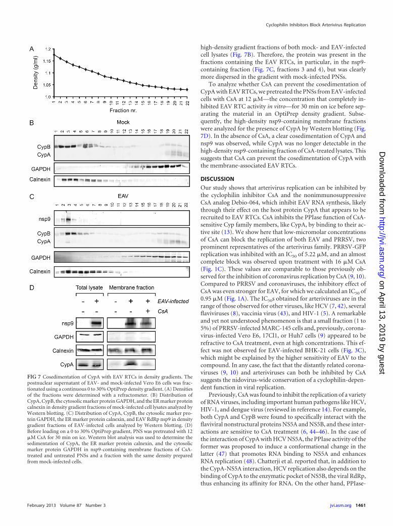
high-density gradient fractions of both mock- and EAV-infectedcell lysates (Fig. 7B). Therefore, the protein was present in thefractions containing the EAV RTCs, in particular, in the nsp9-containing fraction (Fig. 7C, fractions 3 and 4), but was clearlymore dispersed in the gradient with mock-infected PNSs.
To analyze whether CsA can prevent the cosedimentation ofCypA with EAV RTCs, we pretreated the PNSs from EAV-infectedcells with CsA at 12 �M—the concentration that completely in-hibited EAV RTC activity in vitro—for 30 min on ice before sep-arating the material in an OptiPrep density gradient. Subse-quently, the high-density nsp9-containing membrane fractionswere analyzed for the presence of CypA by Western blotting (Fig.7D). In the absence of CsA, a clear cosedimentation of CypA andnsp9 was observed, while CypA was no longer detectable in thehigh-density nsp9-containing fraction of CsA-treated lysates. Thissuggests that CsA can prevent the cosedimentation of CypA withthe membrane-associated EAV RTCs.
DISCUSSION
Our study shows that arterivirus replication can be inhibited bythe cyclophilin inhibitor CsA and the nonimmunosuppressiveCsA analog Debio-064, which inhibit EAV RNA synthesis, likelythrough their effect on the host protein CypA that appears to berecruited to EAV RTCs. CsA inhibits the PPIase function of CsA-sensitive Cyp family members, like CypA, by binding to their ac-tive site (13). We show here that low-micromolar concentrationsof CsA can block the replication of both EAV and PRRSV, twoprominent representatives of the arterivirus family. PRRSV-GFPreplication was inhibited with an IC50 of 5.22 �M, and an almostcomplete block was observed upon treatment with 16 �M CsA(Fig. 1C). These values are comparable to those previously ob-served for the inhibition of coronavirus replication by CsA (9, 10).Compared to PRRSV and coronaviruses, the inhibitory effect ofCsA was even stronger for EAV, for which we calculated an IC50 of0.95 �M (Fig. 1A). The IC50s obtained for arteriviruses are in therange of those observed for other viruses, like HCV (7, 42), severalflaviviruses (8), vaccinia virus (43), and HIV-1 (5). A remarkableand yet not understood phenomenon is that a small fraction (1 to5%) of PRRSV-infected MARC-145 cells and, previously, corona-virus-infected Vero E6, 17Cl1, or Huh7 cells (9) appeared to berefractive to CsA treatment, even at high concentrations. This ef-fect was not observed for EAV-infected BHK-21 cells (Fig. 3C),which might be explained by the higher sensitivity of EAV to thecompound. In any case, the fact that the distantly related corona-viruses (9, 10) and arteriviruses can both be inhibited by CsAsuggests the nidovirus-wide conservation of a cyclophilin-depen-dent function in viral replication.
Previously, CsA was found to inhibit the replication of a varietyof RNA viruses, including important human pathogens like HCV,HIV-1, and dengue virus (reviewed in reference 14). For example,both CypA and CypB were found to specifically interact with theflaviviral nonstructural proteins NS5A and NS5B, and these inter-actions are sensitive to CsA treatment (6, 44–46). In the case ofthe interaction of CypA with HCV NS5A, the PPIase activity of theformer was proposed to induce a conformational change in thelatter (47) that promotes RNA binding to NS5A and enhancesRNA replication (48). Chatterji et al. reported that, in addition tothe CypA-NS5A interaction, HCV replication also depends on thebinding of CypA to the enzymatic pocket of NS5B, the viral RdRp,thus enhancing its affinity for RNA. On the other hand, PPIase-
FIG 7 Cosedimentation of CypA with EAV RTCs in density gradients. Thepostnuclear supernatant of EAV- and mock-infected Vero E6 cells was frac-tionated using a continuous 0 to 30% OptiPrep density gradient. (A) Densitiesof the fractions were determined with a refractometer. (B) Distribution ofCypA, CypB, the cytosolic marker protein GAPDH, and the ER marker proteincalnexin in density gradient fractions of mock-infected cell lysates analyzed byWestern blotting. (C) Distribution of CypA, CypB, the cytosolic marker pro-tein GAPDH, the ER marker protein calnexin, and EAV RdRp nsp9 in densitygradient fractions of EAV-infected cells analyzed by Western blotting. (D)Before loading on a 0 to 30% OptiPrep gradient, PNS was pretreated with 12�M CsA for 30 min on ice. Western blot analysis was used to determine thesedimentation of CypA, the ER marker protein calnexin, and the cytosolicmarker protein GAPDH in nsp9-containing membrane fractions of CsA-treated and untreated PNSs and a fraction with the same density preparedfrom mock-infected cells.
Cyclophilin Inhibitors Block Arterivirus Replication
February 2013 Volume 87 Number 3 jvi.asm.org 1461
on April 13, 2019 by guest
http://jvi.asm.org/
Dow
nloaded from
defective CypA failed to interact with HCV NS5B (49), suggestingthat the isomerase activity of CypA is an essential factor in theinteraction with NS5B that promotes HCV replication. Liu et al.showed that the binding of CypA to NS5B mediates the properfolding of enzymatically active NS5B and facilitates the incorpo-ration of the latter into replication complexes. The interactionbetween CypA and NS5B can be inhibited by CsA (50). In addi-tion, Kaul et al. showed that the development of resistance againstthe Cyp inhibitor Debio-025 involved mutations (V2440A andV2440L) in HCV NS5B that are close to the NS5A/NS5B cleavagesite. These are thought to delay processing of the NS5A/NS5Bjunction, thus extending the time during which the CypA bindingsite in NS5B is accessible (51). As a result, smaller amounts ofCypA would suffice to mediate the proper folding of NS5B and itsincorporation into replication complexes. Similar functions wereattributed to CypB, since the interaction between CypB and NS5Bwas also found to be essential for RNA binding by NS5B and forHCV replication as a whole (52). Furthermore, Japanese enceph-alitis virus replication depends on the binding of CypB to NS4Aand on CypB isomerase activity (6). For a number of RNA viruses,CypA was found to be incorporated into newly formed virions,although the functional relevance of this finding remains to beaddressed in more detail (4, 53, 54). CypA also interacts with theSARS-CoV N protein (55), suggesting that the protein could beincorporated into virions (55), although coronavirus N proteinshave also been implicated in viral RNA synthesis (56, 57) and areassociated with intracellular replication structures (58).
We show here that expression of CypA is required for efficientEAV replication, as siRNA-mediated knockdown of CypA drasti-cally reduced EAV-GFP replication (Fig. 6), while targeting CypBor Cyp40 (Fig. 6 and data not shown) had no effect. The impor-tance of the normally cytosolic CypA was further substantiated byits cosedimentation with RTC-containing membrane structuresin the high-density gradient fractions of EAV-infected cell lysates.In such gradients, the sedimentation of the ER marker proteincalnexin was essentially similar when comparing infected andmock-infected PNSs (Fig. 7B and C). Furthermore, CsA treatmentwas able to prevent the sedimentation of CypA to the part of thegradient that also contained the EAV RTCs, following their bio-chemical isolation from infected cells (Fig. 7D), and the in vitroRNA-synthesizing activity of such replication structures wasfound to be inhibited by CsA and Debio-064 (Fig. 5B and C). Thedistribution of CypB appeared to be less dispersed in gradientscontaining infected cell lysates than in those containing mock-infected lysates, even though EAV-GFP replication was not af-fected by the siRNA-mediated knockdown of CypB levels (Fig. 6).This suggests that although the subcellular localization of CypBmight be affected by the extensive EAV-driven modification ofintracellular membranes (25), this does not have a measurableeffect on virus replication. By using fluorescence microscopy, co-localization of CypA and viral RTCs could not be observed, pre-sumably because the fraction of CypA that localizes to replicationstructures is too small (data not shown). Interestingly, we previ-ously could not measure an effect on SARS-CoV replication whenCypA or CypB expression was (largely) silenced (9) in the same293/ACE2 cells used here for our EAV studies. The �20% residualCyp expression that remained after siRNA-mediated knockdownmay have been sufficient to support normal SARS-CoV replica-tion, whereas it appears to have been insufficient to support the
efficient replication of the apparently more sensitive EAV, in linewith the higher sensitivity of this arterivirus to CsA treatment.
As reported for HCV (48–50), the association of CypA with theEAV replication structures suggests the existence of a functional—presumably PPIase activity-dependent—interaction that is essen-tial for virus replication. This member of the cyclophilin proteinfamily appears to directly promote the RNA-synthesizing activityof the EAV RTC (Fig. 5). Based on studies that analyzed bindingsites for CypA using a set of 40 potential CypA-inhibiting peptides(59), we identified several potential CypA binding sites in EAVnsp10, the viral helicase protein. A functionally important inter-action with such a key enzyme in arterivirus RNA synthesis couldcertainly explain that efficient EAV replication depends on theavailability of sufficient CypA. Clearly, at this moment, we cannotexclude (direct or indirect) interactions with any of the other viralproteins, including—in analogy to HCV (49)—the viral RdRpsubunit (nsp9). In line with the ideas regarding the influence ofCypA on the RNA-binding capacity of HCV NS5A and NS5B,EAV RTC-associated CypA may be involved in the proper foldingor activation of viral enzymes and/or their binding to viral RNA,which might directly affect their function in RNA synthesis.
CsA analogs like Debio-025, NIM811, and SCY635, which havean increased affinity for Cyps and lack the undesired immunosup-pressive effect of CsA (17, 18, 40), can be considered promisingantiviral compounds, as they could block HCV replication almostcompletely and resistance to these compounds does not easilydevelop compared to the ease of resistance development by inhib-itors directly targeting viral enzymes (60). In our study, we com-pared the inhibition of EAV and PRRSV replication by CsA withthat caused by the nonimmunosuppressive CsA analog Debio-064. For Debio-064, we obtained an IC50 of 0.32 �M, 3-fold lowerthan that of CsA (Fig. 2), which is in line with Debio-064’s higheraffinity for Cyps. We also observed an inhibitory effect of Debio-064 on PRRSV-GFP replication, although in contrast to EAV, itsIC50 was similar to that of CsA. Therefore, not only do more po-tent (nonimmunosuppressive) CsA analogs constitute a promis-ing class of molecules for the treatment of viral infections, but also,these compounds are valuable research tools for mechanistic stud-ies into the role of cyclophilins in the replication of nidovirusesand other positive-stranded RNA viruses.
ACKNOWLEDGMENTS
We thank Johan Neyts for helpful discussions and Corrine Beugeling forexcellent technical assistance.
This research was supported in part by TOP grant 700.57.301 from theCouncil for Chemical Sciences of The Netherlands Organization for Sci-entific Research (NWO-CW) and by the European Union Seventh Frame-work Programme (FP7/2007-2013) under SILVER grant agreement no.260644.
REFERENCES1. Nagy PD, Pogany J. 2012. The dependence of viral RNA replication on
co-opted host factors. Nat. Rev. Microbiol. 10:137–149.2. Kellam P. 2006. Attacking pathogens through their hosts. Genome Biol.
7:201. doi:10.1186/gb-2006-7-1-201.3. Schwegmann A, Brombacher F. 2008. Host-directed drug targeting of
factors hijacked by pathogens. Sci. Signal. 1:re8. doi:10.1126/scisignal.129re8.
4. Bose S, Mathur M, Bates P, Joshi N, Banerjee AK. 2003. Requirementfor cyclophilin A for the replication of vesicular stomatitis virus New Jer-sey serotype. J. Gen. Virol. 84:1687–1699.
5. Briggs CJ, Ott DE, Coren LV, Oroszlan S, Tozser J. 1999. Comparison
de Wilde et al.
1462 jvi.asm.org Journal of Virology
on April 13, 2019 by guest
http://jvi.asm.org/
Dow
nloaded from
of the effect of FK506 and cyclosporin A on virus production in H9 cellschronically and newly infected by HIV-1. Arch. Virol. 144:2151–2160.
6. Kambara H, Tani H, Mori Y, Abe T, Katoh H, Fukuhara T, Taguwa S,Moriishi K, Matsuura Y. 2011. Involvement of cyclophilin B in the rep-lication of Japanese encephalitis virus. Virology 412:211–219.
7. Nakagawa M, Sakamoto N, Enomoto N, Tanabe Y, Kanazawa N,Koyama T, Kurosaki M, Maekawa S, Yamashiro T, Chen CH, ItsuiY, Kakinuma S, Watanabe M. 2004. Specific inhibition of hepatitis Cvirus replication by cyclosporin A. Biochem. Biophys. Res. Commun.313:42– 47.
8. Qing M, Yang F, Zhang B, Zou G, Robida JM, Yuan Z, Tang H, Shi PY.2009. Cyclosporine inhibits flavivirus replication through blocking theinteraction between host cyclophilins and viral NS5 protein. Antimicrob.Agents Chemother. 53:3226 –3235.
9. de Wilde AH, Zevenhoven-Dobbe JC, van der Meer Y, Thiel V, Naray-anan K, Makino S, Snijder EJ, van Hemert MJ. 2011. Cyclosporin Ainhibits the replication of diverse coronaviruses. J. Gen. Virol. 92:2542–2548.
10. Pfefferle S, Schopf J, Kogl M, Friedel CC, Muller MA, Carbajo-LozoyaJ, Stellberger T, von Dall’armi E, Herzog P, Kallies S, Niemeyer D, DittV, Kuri T, Zust R, Pumpor K, Hilgenfeld R, Schwarz F, Zimmer R,Steffen I, Weber F, Thiel V, Herrler G, Thiel HJ, Schwegmann-WesselsC, Pohlmann S, Haas J, Drosten C, von Brunn A. 2011. The SARS-coronavirus-host interactome: identification of cyclophilins as target forpan-coronavirus inhibitors. PLoS Pathog. 7:e1002331. doi:10.1371/journal.ppat.1002331.
11. Tanaka Y, Sato Y, Osawa S, Inoue M, Tanaka S, Sasaki T. 2012.Suppression of feline coronavirus replication in vitro by cyclosporin A.Vet. Res. 43:41.
12. Barik S. 2006. Immunophilins: for the love of proteins. Cell. Mol. Life Sci.63:2889 –2900.
13. Davis TL, Walker JR, Campagna-Slater V, Finerty PJ, Paramanathan R,Bernstein G, MacKenzie F, Tempel W, Ouyang H, Lee WH, Eisen-messer EZ, Dhe-Paganon S. 2010. Structural and biochemical character-ization of the human cyclophilin family of peptidyl-prolyl isomerases.PLoS Biol. 8:e1000439. doi:10.1371/journal.pbio.1000439.
14. Nagy PD, Wang RY, Pogany J, Hafren A, Makinen K. 2011. Emergingpicture of host chaperone and cyclophilin roles in RNA virus replication.Virology 411:374 –382.
15. Tedesco D, Haragsim L. 2012. Cyclosporine: a review. J. Transplant.2012:230386. doi:10.1155/2012/230386.
16. Flisiak R, Feinman SV, Jablkowski M, Horban A, Kryczka W, Paw-lowska M, Heathcote JE, Mazzella G, Vandelli C, Nicolas-Metral V,Grosgurin P, Liz JS, Scalfaro P, Porchet H, Crabbe R. 2009. Thecyclophilin inhibitor Debio 025 combined with PEG IFNalpha2a signifi-cantly reduces viral load in treatment-naive hepatitis C patients. Hepatol-ogy 49:1460 –1468.
17. Flisiak R, Horban A, Gallay P, Bobardt M, Selvarajah S, Wiercinska-Drapalo A, Siwak E, Cielniak I, Higersberger J, Kierkus J, AeschlimannC, Grosgurin P, Nicolas-Metral V, Dumont JM, Porchet H, Crabbe R,Scalfaro P. 2008. The cyclophilin inhibitor Debio-025 shows potent anti-hepatitis C effect in patients coinfected with hepatitis C and human im-munodeficiency virus. Hepatology 47:817– 826.
18. Lawitz E, Godofsky E, Rouzier R, Marbury T, Nguyen T, Ke J, HuangM, Praestgaard J, Serra D, Evans TG. 2011. Safety, pharmacokinetics,and antiviral activity of the cyclophilin inhibitor NIM811 alone or in com-bination with pegylated interferon in HCV-infected patients receiving 14days of therapy. Antiviral Res. 89:238 –245.
19. de Groot RJ, Cowley JA, Enjuanes L, Faaberg KS, Perlman S, Rottier PJ,Snijder EJ, Ziebuhr J, Gorbalenya AE. 2012. Order of Nidovirales, p785–795. In King A, Adams M, Carstens E, Lefkowitz EJ (ed), Virus tax-onomy, the 9th Report of the International Committee on Taxonomy ofViruses. Academic Press, New York, NY.
20. Holtkamp DJ, Kliebenstein JB, Neumann EJ, Zimmerman JJ, Rotto H,Yoder TK, Wang C, Yeske P, Mowrer C, Haley C. 2011. Abstr. 2011 Int.PRRS Symp., Chicago, IL, abstr. 86.
21. Snijder EJ, Spaan WJM. 2007. Arteriviruses, p 1337–1355. In Knipe DM,et al. (ed), Fields virology, 5th ed. Lippincott Williams & Wilkins, Phila-delphia, PA.
22. Gorbalenya AE, Enjuanes L, Ziebuhr J, Snijder EJ. 2006. Nidovirales:evolving the largest RNA virus genome. Virus Res. 117:17–37.
23. Pasternak AO, Spaan WJ, Snijder EJ. 2006. Nidovirus transcription: howto make sense? J. Gen. Virol. 87:1403–1421.
24. Fang Y, Snijder EJ. 2010. The PRRSV replicase: exploring the multi-functionality of an intriguing set of nonstructural proteins. Virus Res.154:61–76.
25. Knoops K, Barcena M, Limpens RW, Koster AJ, Mommaas AM, SnijderEJ. 2012. Ultrastructural characterization of arterivirus replication struc-tures: reshaping the endoplasmic reticulum to accommodate viral RNAsynthesis. J. Virol. 86:2474 –2487.
26. van der Meer Y, van Tol H, Krijnse Locker J, Snijder EJ. 1998. ORF1a-encoded replicase subunits are involved in the membrane association ofthe arterivirus replication complex. J. Virol. 72:6689 – 6698.
27. van Hemert MJ, de Wilde AH, Gorbalenya AE, Snijder EJ. 2008. The invitro RNA synthesizing activity of the isolated arterivirus replication/transcription complex is dependent on a host factor. J. Biol. Chem. 283:16525–16536.
28. Nedialkova DD, Gorbalenya AE, Snijder EJ. 2010. Arterivirus Nsp1modulates the accumulation of minus-strand templates to control therelative abundance of viral mRNAs. PLoS Pathog. 6:e1000772. doi:10.1371/journal.ppat.1000772.
29. de Vries AA, Chirnside ED, Horzinek MC, Rottier PJ. 1992. Structuralproteins of equine arteritis virus. J. Virol. 66:6294 – 6303.
30. Kim HS, Kwang J, Yoon IJ, Joo HS, Frey ML. 1993. Enhanced replica-tion of porcine reproductive and respiratory syndrome (PRRS) virus in ahomogeneous subpopulation of MA-104 cell line. Arch. Virol. 133:477–483.
31. Kamitani W, Narayanan K, Huang C, Lokugamage K, Ikegami T, Ito N,Kubo H, Makino S. 2006. Severe acute respiratory syndrome coronavirusnsp1 protein suppresses host gene expression by promoting host mRNAdegradation. Proc. Natl. Acad. Sci. U. S. A. 103:12885–12890.
32. Bryans JT, Crowe ME, Doll ER, McCollum WH. 1957. Isolation of afilterable agent causing arteritis of horses and abortion by mares; its dif-ferentiation from the equine abortion (influenza) virus. Cornell Vet. 47:3– 41.
33. van den Born E, Posthuma CC, Knoops K, Snijder EJ. 2007. Aninfectious recombinant equine arteritis virus expressing green fluorescentprotein from its replicase gene. J. Gen. Virol. 88:1196 –1205.
34. Fang Y, Rowland RR, Roof M, Lunney JK, Christopher-Hennings J,Nelson EA. 2006. A full-length cDNA infectious clone of North Americantype 1 porcine reproductive and respiratory syndrome virus: expression ofgreen fluorescent protein in the Nsp2 region. J. Virol. 80:11447–11455.
35. Sun Z, Li Y, Ransburgh R, Snijder EJ, Fang Y. 2012. Nonstructuralprotein 2 of porcine reproductive and respiratory syndrome virus inhibitsthe antiviral function of interferon-stimulated gene 15. J. Virol. 86:3839 –3850.
36. Pedersen KW, van der Meer Y, Roos N, Snijder EJ. 1999. Open readingframe 1a-encoded subunits of the arterivirus replicase induce endoplas-mic reticulum-derived double-membrane vesicles which carry the viralreplication complex. J. Virol. 73:2016 –2026.
37. MacLachlan NJ, Balasuriya UB, Hedges JF, Schweidler TM, McCollumWH, Timoney PJ, Hullinger PJ, Patton JF. 1998. Serologic response ofhorses to the structural proteins of equine arteritis virus. J. Vet. Diagn.Invest. 10:229 –236.
38. Knoops K, Swett-Tapia C, van den Worm SH, Te Velthuis AJ, KosterAJ, Mommaas AM, Snijder EJ, Kikkert M. 2010. Integrity of the earlysecretory pathway promotes, but is not required for, severe acute respira-tory syndrome coronavirus RNA synthesis and virus-induced remodelingof endoplasmic reticulum membranes. J. Virol. 84:833– 846.
39. Schreiber SL, Crabtree GR. 1992. The mechanism of action of cyclospo-rin A and FK506. Immunol. Today 13:136 –142.
40. Hopkins S, Dimassimo B, Rusnak P, Heuman D, Lalezari J, Sluder A,Scorneaux B, Mosier S, Kowalczyk P, Ribeill Y, Baugh J, Gallay P. 2012.The cyclophilin inhibitor SCY-635 suppresses viral replication and in-duces endogenous interferons in patients with chronic HCV genotype 1infection. J. Hepatol. 57:47–54.
41. Wenger R, Mutter M, Garrouste P, Lysek R, Turpin O, Vuagniaux G,Nicolas V, Novaroli Zanolari L, Crabbé R. November 2009. Cycloun-decadepsipeptide compounds and use of said compounds as a medica-ment. Patent WO 2010/052559.
42. Watashi K, Hijikata M, Hosaka M, Yamaji M, Shimotohno K. 2003.Cyclosporin A suppresses replication of hepatitis C virus genome in cul-tured hepatocytes. Hepatology 38:1282–1288.
43. Damaso CR, Keller SJ. 1994. Cyclosporin A inhibits vaccinia virus repli-cation in vitro. Arch. Virol. 134:303–319.
44. Chatterji U, Lim P, Bobardt MD, Wieland S, Cordek DG, Vuagniaux G,
Cyclophilin Inhibitors Block Arterivirus Replication
February 2013 Volume 87 Number 3 jvi.asm.org 1463
on April 13, 2019 by guest
http://jvi.asm.org/
Dow
nloaded from
Chisari F, Cameron CE, Targett-Adams P, Parkinson T, Gallay PA.2010. HCV resistance to cyclosporin A does not correlate with a resistanceof the NS5A-cyclophilin A interaction to cyclophilin inhibitors. J. Hepa-tol. 53:50 –56.
45. Fernandes F, Ansari IU, Striker R. 2010. Cyclosporine inhibits a directinteraction between cyclophilins and hepatitis C NS5A. PLoS One5:e9815. doi:10.1371/journal.pone.0009815.
46. Gaither LA, Borawski J, Anderson LJ, Balabanis KA, Devay P, JobertyG, Rau C, Schirle M, Bouwmeester T, Mickanin C, Zhao S, Vickers C,Lee L, Deng G, Baryza J, Fujimoto RA, Lin K, Compton T, WiedmannB. 2010. Multiple cyclophilins involved in different cellular pathways me-diate HCV replication. Virology 397:43–55.
47. Coelmont L, Hanoulle X, Chatterji U, Berger C, Snoeck J, Bobardt M,Lim P, Vliegen I, Paeshuyse J, Vuagniaux G, Vandamme AM, Barten-schlager R, Gallay P, Lippens G, Neyts J. 2010. DEB025 (alisporivir)inhibits hepatitis C virus replication by preventing a cyclophilin A inducedcis-trans isomerisation in domain II of NS5A. PLoS One 5:e13687. doi:10.1371/journal.pone.0013687.
48. Foster TL, Gallay P, Stonehouse NJ, Harris M. 2011. Cyclophilin Ainteracts with domain II of hepatitis C virus NS5A and stimulates RNAbinding in an isomerase-dependent manner. J. Virol. 85:7460 –7464.
49. Chatterji U, Bobardt M, Selvarajah S, Yang F, Tang H, Sakamoto N, Vuag-niaux G, Parkinson T, Gallay P. 2009. The isomerase active site of cyclophilin Ais critical for hepatitis C virus replication. J. Biol. Chem. 284:16998–17005.
50. Liu Z, Yang F, Robotham JM, Tang H. 2009. Critical role of cyclophilinA and its prolyl-peptidyl isomerase activity in the structure and functionof the hepatitis C virus replication complex. J. Virol. 83:6554 – 6565.
51. Kaul A, Stauffer S, Berger C, Pertel T, Schmitt J, Kallis S, Zayas M,Lohmann V, Luban J, Bartenschlager R. 2009. Essential role of cyclophi-lin A for hepatitis C virus replication and virus production and possiblelink to polyprotein cleavage kinetics. PLoS Pathog. 5:e1000546. doi:10.1371/journal.ppat.1000546.
52. Watashi K, Ishii N, Hijikata M, Inoue D, Murata T, Miyanari Y,Shimotohno K. 2005. Cyclophilin B is a functional regulator of hepatitis Cvirus RNA polymerase. Mol. Cell 19:111–122.
53. Castro AP, Carvalho TM, Moussatche N, Damaso CR. 2003. Redistri-bution of cyclophilin A to viral factories during vaccinia virus infectionand its incorporation into mature particles. J. Virol. 77:9052–9068.
54. Liu X, Sun L, Yu M, Wang Z, Xu C, Xue Q, Zhang K, Ye X, KitamuraY, Liu W. 2009. Cyclophilin A interacts with influenza A virus M1 proteinand impairs the early stage of the viral replication. Cell. Microbiol. 11:730 –741.
55. Luo C, Luo H, Zheng S, Gui C, Yue L, Yu C, Sun T, He P, Chen J, ShenJ, Luo X, Li Y, Liu H, Bai D, Yang Y, Li F, Zuo J, Hilgenfeld R, Pei G,Chen K, Shen X, Jiang H. 2004. Nucleocapsid protein of SARS corona-virus tightly binds to human cyclophilin A. Biochem. Biophys. Res. Com-mun. 321:557–565.
56. Almazan F, Galan C, Enjuanes L. 2004. The nucleoprotein is required forefficient coronavirus genome replication. J. Virol. 78:12683–12688.
57. Schelle B, Karl N, Ludewig B, Siddell SG, Thiel V. 2006. Nucleocapsidprotein expression facilitates coronavirus replication. Adv. Exp. Med.Biol. 581:43– 48.
58. van der Meer Y, Snijder EJ, Dobbe JC, Schleich S, Denison MR, SpaanWJ, Krijnse Locker J. 1999. Localization of mouse hepatitis virus non-structural proteins and RNA synthesis indicates a role for late endosomesin viral replication. J. Virol. 73:7641–7657.
59. Pang X, Zhang M, Zhou L, Xie F, Lu H, He W, Jiang S, Yu L, Zhang X.2011. Discovery of a potent peptidic cyclophilin A inhibitor Trp-Gly-Pro.Eur. J. Med. Chem. 46:1701–1705.
60. Puyang X, Poulin DL, Mathy JE, Anderson LJ, Ma S, Fang Z, Zhu S, LinK, Fujimoto R, Compton T, Wiedmann B. 2010. Mechanism of resis-tance of hepatitis C virus replicons to structurally distinct cyclophilin in-hibitors. Antimicrob. Agents Chemother. 54:1981–1987.
de Wilde et al.
1464 jvi.asm.org Journal of Virology
on April 13, 2019 by guest
http://jvi.asm.org/
Dow
nloaded from