
Cyclophilin E Functions as a Negative Regulator to Influenza Virus ...
12
Cyclophilin E Functions as a Negative Regulator to Influenza Virus Replication by Impairing the Formation of the Viral Ribonucleoprotein Complex Zengfu Wang 1,2 , Xiaoling Liu 1 , Zhendong Zhao 1,2 , Chongfeng Xu 1,2 , Ke Zhang 1,2 , Caiwei Chen 1,2 , Lei Sun 1 , George F. Gao 1,2,3 , Xin Ye 1,2,3 , Wenjun Liu 1,2,3 * 1 Center for Molecular Virology, Chinese Academy of Sciences Key Laboratory of Pathogenic Microbiology and Immunology, Institute of Microbiology, Chinese Academy of Sciences, Beijing, China, 2 Graduate University of Chinese Academy of Sciences, Beijing, China, 3 China-Japan Joint Laboratory of Molecular Immunology and Molecular Microbiology, Institute of Microbiology, Chinese Academy of Sciences, Beijing, China Abstract Background: The nucleoprotein (NP) of influenza A virus is a multifunctional protein that plays a critical role in the replication and transcription of the viral genome. Therefore, examining host factors that interact with NP may shed light on the mechanism of host restriction barriers and the tissue tropism of influenza A virus. Here, Cyclophilin E (CypE), a member of the peptidyl-propyl cis-trans isomerase (PPIase) family, was found to bind to NP and inhibit viral replication and transcription. Methodology/Principal Findings: In the present study, CypE was found to interact with NP but not with the other components of the viral ribonucleoprotein complex (vRNP): PB1, PB2, and PA. Mutagenesis data revealed that the CypE domain comprised of residues 137–186 is responsible for its binding to NP. Functional analysis results indicated that CypE is a negative regulator in the influenza virus life cycle. Furthermore, knock-down of CypE resulted in increased levels of three types of viral RNA, suggesting that CypE negatively affects viral replication and transcription. Moreover, up-regulation of CypE inhibited the activity of influenza viral polymerase. We determined that the molecular mechanism by which CypE negatively regulates influenza virus replication and transcription is by interfering with NP self-association and the NP-PB1 and NP-PB2 interactions. Conclusions/Significance: CypE is a host restriction factor that inhibits the functions of NP, as well as viral replication and transcription, by impairing the formation of the vRNP. The data presented here will help us to better understand the molecular mechanisms of host restriction barriers, host adaptation, and tissue tropism of influenza A virus. Citation: Wang Z, Liu X, Zhao Z, Xu C, Zhang K, et al. (2011) Cyclophilin E Functions as a Negative Regulator to Influenza Virus Replication by Impairing the Formation of the Viral Ribonucleoprotein Complex. PLoS ONE 6(8): e22625. doi:10.1371/journal.pone.0022625 Editor: Paul Digard, University of Cambridge, United Kingdom Received March 16, 2011; Accepted June 27, 2011; Published August 24, 2011 Copyright: ß 2011 Wang et al. This is an open-access article distributed under the terms of the Creative Commons Attribution License, which permits unrestricted use, distribution, and reproduction in any medium, provided the original author and source are credited. Funding: This work was supported by the National Basic Research Program (973) of China (2011CB504705), Chinese Academy of Sciences Innovation projects (KSCX2-YW-N-054, KSCX2-YW-R-158), the National Natural Science Foundation of China (30972185, 30901073), and the National Key Technologies Research and Development Program of China (2010BAD04B01). Wenjun Liu, George F Gao, and Xin Ye are the principal investigators of the Innovative Research Group of the National Natural Science Foundation of China (NSFC, Grant No. 81021003). The funders had no role in study design, data collection and analysis, decision to publish, or preparation of the manuscript. Competing Interests: The authors have declared that no competing interests exist. * E-mail: [email protected] Introduction Influenza virus belongs to the orthomyxoviruses and has a genome comprised of eight different negative strand RNAs varying in length from approximately 900 to 2,500 nucleotides [1]. Among the components of the influenza virion, the viral ribonucleoprotein complexes (vRNPs) are the viral core because they are indepen- dent units responsible for the replication and transcription of each virus segment. The native vRNPs are formed by single-stranded RNA, multiple monomers of the nucleoprotein (NP) and a single copy of the polymerase, a heterotrimer composed by the PB1, PB2, and PA subunits [2]. The NP of influenza A virus, encoded by segment 5, is a 498-amino acid polypeptide rich in arginine, glycine, and serine residues. In the virion, the NP forms the protein scaffold to package the helical genomic vRNPs [3,4,5]. The NP protein has multiple functions during the virus life cycle, and plays a critical role in influenza virus replication and transcription [6]. It interacts with viral macromolecules (including PB2, PB1, M1, and itself), displays high RNA binding activity, and binds to cellular factors to accomplish various functions, suggesting that NP is a key adapter molecule during the influenza virus infection cycle. NP directly binds to two subunits of the viral RNA-dependent RNA polymerase, PB1 and PB2, but not with PA, both in virus- infected cells and recombinant systems in the absence of viral RNA [7,8]. The cryo-electron microscopy structure of purified biologically active recombinant vRNPs shows that NP monomers form a nonameric ring, indicating that the homo-oligomerization of NP plays an important role in maintaining the vRNP structure for influenza virus replication and transcription [4,9,10]. Although PLoS ONE | www.plosone.org 1 August 2011 | Volume 6 | Issue 8 | e22625
Transcript of Cyclophilin E Functions as a Negative Regulator to Influenza Virus ...

Cyclophilin E Functions as a Negative Regulator toInfluenza Virus Replication by Impairing the Formationof the Viral Ribonucleoprotein ComplexZengfu Wang1,2, Xiaoling Liu1, Zhendong Zhao1,2, Chongfeng Xu1,2, Ke Zhang1,2, Caiwei Chen1,2, Lei
Sun1, George F. Gao1,2,3, Xin Ye1,2,3, Wenjun Liu1,2,3*
1 Center for Molecular Virology, Chinese Academy of Sciences Key Laboratory of Pathogenic Microbiology and Immunology, Institute of Microbiology, Chinese Academy
of Sciences, Beijing, China, 2 Graduate University of Chinese Academy of Sciences, Beijing, China, 3 China-Japan Joint Laboratory of Molecular Immunology and Molecular
Microbiology, Institute of Microbiology, Chinese Academy of Sciences, Beijing, China
Abstract
Background: The nucleoprotein (NP) of influenza A virus is a multifunctional protein that plays a critical role in thereplication and transcription of the viral genome. Therefore, examining host factors that interact with NP may shed light onthe mechanism of host restriction barriers and the tissue tropism of influenza A virus. Here, Cyclophilin E (CypE), a memberof the peptidyl-propyl cis-trans isomerase (PPIase) family, was found to bind to NP and inhibit viral replication andtranscription.
Methodology/Principal Findings: In the present study, CypE was found to interact with NP but not with the othercomponents of the viral ribonucleoprotein complex (vRNP): PB1, PB2, and PA. Mutagenesis data revealed that the CypEdomain comprised of residues 137–186 is responsible for its binding to NP. Functional analysis results indicated that CypE isa negative regulator in the influenza virus life cycle. Furthermore, knock-down of CypE resulted in increased levels of threetypes of viral RNA, suggesting that CypE negatively affects viral replication and transcription. Moreover, up-regulation ofCypE inhibited the activity of influenza viral polymerase. We determined that the molecular mechanism by which CypEnegatively regulates influenza virus replication and transcription is by interfering with NP self-association and the NP-PB1and NP-PB2 interactions.
Conclusions/Significance: CypE is a host restriction factor that inhibits the functions of NP, as well as viral replication andtranscription, by impairing the formation of the vRNP. The data presented here will help us to better understand themolecular mechanisms of host restriction barriers, host adaptation, and tissue tropism of influenza A virus.
Citation: Wang Z, Liu X, Zhao Z, Xu C, Zhang K, et al. (2011) Cyclophilin E Functions as a Negative Regulator to Influenza Virus Replication by Impairing theFormation of the Viral Ribonucleoprotein Complex. PLoS ONE 6(8): e22625. doi:10.1371/journal.pone.0022625
Editor: Paul Digard, University of Cambridge, United Kingdom
Received March 16, 2011; Accepted June 27, 2011; Published August 24, 2011
Copyright: � 2011 Wang et al. This is an open-access article distributed under the terms of the Creative Commons Attribution License, which permitsunrestricted use, distribution, and reproduction in any medium, provided the original author and source are credited.
Funding: This work was supported by the National Basic Research Program (973) of China (2011CB504705), Chinese Academy of Sciences Innovation projects(KSCX2-YW-N-054, KSCX2-YW-R-158), the National Natural Science Foundation of China (30972185, 30901073), and the National Key Technologies Research andDevelopment Program of China (2010BAD04B01). Wenjun Liu, George F Gao, and Xin Ye are the principal investigators of the Innovative Research Group of theNational Natural Science Foundation of China (NSFC, Grant No. 81021003). The funders had no role in study design, data collection and analysis, decision topublish, or preparation of the manuscript.
Competing Interests: The authors have declared that no competing interests exist.
* E-mail: [email protected]
Introduction
Influenza virus belongs to the orthomyxoviruses and has a
genome comprised of eight different negative strand RNAs varying
in length from approximately 900 to 2,500 nucleotides [1]. Among
the components of the influenza virion, the viral ribonucleoprotein
complexes (vRNPs) are the viral core because they are indepen-
dent units responsible for the replication and transcription of each
virus segment. The native vRNPs are formed by single-stranded
RNA, multiple monomers of the nucleoprotein (NP) and a single
copy of the polymerase, a heterotrimer composed by the PB1,
PB2, and PA subunits [2]. The NP of influenza A virus, encoded
by segment 5, is a 498-amino acid polypeptide rich in arginine,
glycine, and serine residues. In the virion, the NP forms the
protein scaffold to package the helical genomic vRNPs [3,4,5].
The NP protein has multiple functions during the virus life cycle,
and plays a critical role in influenza virus replication and
transcription [6]. It interacts with viral macromolecules (including
PB2, PB1, M1, and itself), displays high RNA binding activity, and
binds to cellular factors to accomplish various functions, suggesting
that NP is a key adapter molecule during the influenza virus
infection cycle.
NP directly binds to two subunits of the viral RNA-dependent
RNA polymerase, PB1 and PB2, but not with PA, both in virus-
infected cells and recombinant systems in the absence of viral
RNA [7,8]. The cryo-electron microscopy structure of purified
biologically active recombinant vRNPs shows that NP monomers
form a nonameric ring, indicating that the homo-oligomerization
of NP plays an important role in maintaining the vRNP structure
for influenza virus replication and transcription [4,9,10]. Although
PLoS ONE | www.plosone.org 1 August 2011 | Volume 6 | Issue 8 | e22625
the NP of influenza A virus displays high affinity for RNA but little
or no sequence specificity, it cannot protect RNA from RNase
digestion [4,10,11]. NP can bind any type of RNA longer than 15
nucleotides and displays no higher binding affinity for influenza
virus-specific RNA sequences [12].
Several host factors interact with NP, such as importin-a, F-actin,
exportin 1 (CRM1), and nuclear factor 90 (NF90) [13,14,15,16].
The interaction between NP and importin a is thought to be a
determinant of the host range of influenza A virus [14]. Moreover,
the interaction between PB2 and NP with different importin-aisoforms governs the host adaptation or cell tropism of influenza A
virus [17,18]. NP further mediates the nuclear import of vRNPs
through its interaction with importin a [15]. The interaction
between NP and F-actin causes modulation of NP localization in the
nucleus of infected cells [13], and NP nuclear export is mediated by
a direct interaction between NP and CRM1 [19].
The above host factors are only associated with NP nuclear
shuttling, while another host factor, NF90, is involved in regulating
the replication and transcription of influenza A virus. NF90
negatively regulates viral replication and transcription during the
early phase of infection by directly interacting with the NP of
influenza A virus [16]. Recently, a cellular deubiquitinase, USP11,
was found to inhibit the RNA replication of influenza A virus by
specifically deubiquitinating NP [20].
Many of the cyclophilins, such as cyclophilin A (CypA) and
cyclophilin B (CypB), interact with various viral proteins and are
involved in viral life cycles. However, the mechanisms by which
cyclophilins are involved in viral life cycles are often unclear.
CypA interacts with the gag protein of human immunodeficiency
virus (HIV), package in HIV virions and benefit for virus
replication [21,22]. In addition, CypA is as an essential cofactor
for hepatitis C virus infection by mediating viral cyclosporine
resistance [23]. CypA also interacts with the M1 protein of
influenza A virus and inhibits virus replication during the early
stage of viral infection [24,25]. The interaction between CypB and
NS5B is essential for hepatitis C virus replication [26].
In the present study, CypE, a nuclear and RNA-binding
cyclophilin [27], was identified to interact with NP. Several lines of
evidences indicate that CypE serves as a host restriction factor that
targets the functions of NP. Moreover, we found that the
molecular mechanism by which CypE negatively regulates
influenza virus replication and transcription occurs by impairing
the formation of the vRNP.
Results
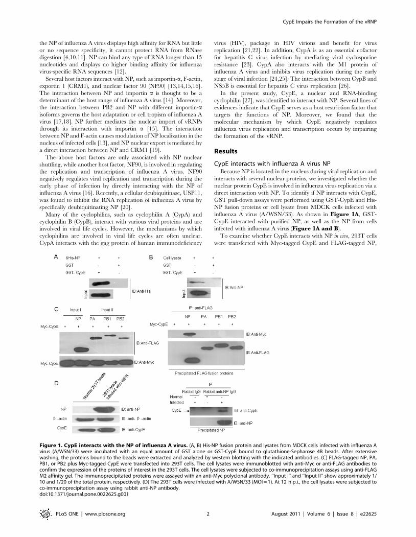
CypE interacts with influenza A virus NPBecause NP is located in the nucleus during viral replication and
interacts with several nuclear proteins, we investigated whether the
nuclear protein CypE is involved in influenza virus replication via a
direct interaction with NP. To identify if NP interacts with CypE,
GST pull-down assays were performed using GST-CypE and His-
NP fusion proteins or cell lysate from MDCK cells infected with
influenza A virus (A/WSN/33). As shown in Figure 1A, GST-
CypE interacted with purified NP, as well as the NP from cells
infected with influenza A virus (Figure 1A and B).
To examine whether CypE interacts with NP in vivo, 293T cells
were transfected with Myc-tagged CypE and FLAG-tagged NP,
Figure 1. CypE interacts with the NP of influenza A virus. (A, B) His-NP fusion protein and lysates from MDCK cells infected with influenza Avirus (A/WSN/33) were incubated with an equal amount of GST alone or GST-CypE bound to glutathione-Sepharose 4B beads. After extensivewashing, the proteins bound to the beads were extracted and analyzed by western blotting with the indicated antibodies. (C) FLAG-tagged NP, PA,PB1, or PB2 plus Myc-tagged CypE were transfected into 293T cells. The cell lysates were immunoblotted with anti-Myc or anti-FLAG antibodies toconfirm the expression of the proteins of interest in the 293T cells. The cell lysates were subjected to co-immunoprecipitation assays using anti-FLAGM2 affinity gel. The immunoprecipitated proteins were assayed with an anti-Myc polyclonal antibody. ‘‘Input I’’ and ‘‘Input II’’ show approximately 1/10 and 1/20 of the total protein, respectively. (D) The 293T cells were infected with A/WSN/33 (MOI = 1). At 12 h p.i., the cell lysates were subjected toco-immunoprecipitation assay using rabbit anti-NP antibody.doi:10.1371/journal.pone.0022625.g001
CypE Impairs the Formation of the vRNP
PLoS ONE | www.plosone.org 2 August 2011 | Volume 6 | Issue 8 | e22625

PB1, PB2, or PA, and the cell lysates were subjected to
immunoprecipitation analysis. The data demonstrate that Myc-
tagged CypE only interacted with FLAG-tagged NP and not PA,
PB1, or PB2 (Figure 1C). To further confirm the interaction
between NP and CypE, we performed binding assays to determine
if endogenous CypE interacts with NP during viral infection. As
shown in Figure 1D, NP bound to endogenous CypE, indicating
that CypE could affect virus replication through its interaction
with NP.
To determine which domain of CypE is responsible for binding
to NP, we generated several truncated mutants of GST-CypE
(Figure 2A and B). GST pull-down assays were then performed
using GST-CypE and its truncated mutants. As shown in
Figure 2B and C, among the CypE mutants, only the truncation
of CypE between residues 137–186 in the PPIase domain was
sufficient and required for its interaction with NP. These data
suggest that residues 137–186 in the CypE PPIase domain
comprise the critical region for NP binding.
As is well known, NP localizes to the nucleus and/or cytoplasm
during different infection stages. Accordingly, the intracellular co-
localization of CypE and NP was examined during viral infection
at 2 h intervals. HeLa cells were infected with A/WSN/33 and
immunostained with anti-CypE and anti-NP antibodies. The
immunofluorescence data revealed that both CypE and NP
co-localize in the nucleus from 4 to 8 h post-infection (p.i.), while
NP is predominantly located in the cytoplasm (and there is
correspondingly less co-localization of CypE and NP in the
nucleus) at 10 h p.i. (Figure 3). These data suggest that the
interaction between CypE and NP occurs in the early phase of
viral replication.
CypE negatively regulated influenza A virus replicationTo investigate the biological effect of CypE on influenza A virus
replication, we analyzed virus replication in the 293T and A549
cell lines where CypE was knocked down by RNA interference. As
shown in Figure 4A and Figure S1, the level of NP was
significantly increased (,2.5-fold) in the CypE knock-down cells
compared to the control cells. The virus titer was also increased
,2.5-fold in endogenous CypE-depleted cells (Figure 4B and
Figure S1). These results indicate that knock-down of endoge-
nous CypE favors the replication of influenza A virus. To further
analyze the function of CypE on viral replication, 293T cells were
transfected with Myc-CypE or Myc-CypE D137–186, followed by
infection with A/WSN/33. Consistently, the NP level was greatly
decreased in CypE over-expressing cells but not in CypE D137–
186 over-expressing cells (Figure 4C), suggesting that the antiviral
function of CypE is dependent on its binding to NP. In addition,
the virus titer in CypE over-expressing cells was decreased
Figure 2. Mapping the domain of CypE responsible for the interaction with NP. (A) CypE has two functional domains: an RNA-bindingdomain at its N-terminus (aa 1–84) and a PPIase domain at its C-terminus (aa 136–275). (B) The binding regions on CypE for NP were confirmed byGST pull-down assays. ‘‘NP binding’’ summarizes the results of the GST pull-down assays. (C) GST pull-down assays were performed using CypE and itstruncation mutants. ‘‘Input’’ shows 1/20 of the total protein included in each binding reaction. (D) Coomassie brilliant blue (CBB) staining patterns forthe pull-down proteins are shown.doi:10.1371/journal.pone.0022625.g002
CypE Impairs the Formation of the vRNP
PLoS ONE | www.plosone.org 3 August 2011 | Volume 6 | Issue 8 | e22625

approximately twofold compared to the control cells (Figure 4D).
Taken together, it is clear that CypE exhibits an inhibitory effect
on viral replication.
To examine the effect of CypE on the infectivity of influenza
virus, 293T cells were transfected with pCMV-Myc-CypE for 12 h
and then infected with A/WSN/33. The cells were subsequently
immunostained with anti-NP and anti-Myc antibodies. As shown in
Figure 4E, a portion of NP translocated into the cytoplasm from
the nucleus in the control cells at 8 h p.i., though most of the NP was
still located in the nucleus and co-localized with over-expressed
CypE. However, the amount of NP in the CypE over-expressing
cells was much less than that in the control cells (Figure 4E a–f).Furthermore, the relative susceptibility of 293T cells to viral
infection was reduced to ,10% in CypE over-expressing cells
compared to control cells (Figure 4F). These data indicate only a 2-
or 2.5-fold difference in virus growth in the presence or absence of
CypE. In fact, the role of CypE in influenza virus replication may be
significant if 100% of the transfected cells were CypE over-
expressing or knocked-down, but the inhibitory efficiency of CypE
was not comparable to that of anti-influenza drugs.
Figure 3. CypE co-localizes with NP during influenza virus infection. The localization of CypE and NP was determined byimmunofluorescence assays using anti-CypE and anti-NP polyclonal antibodies from 2 to 10 h p.i. at 2 h intervals. Scale bar: 10 mm.doi:10.1371/journal.pone.0022625.g003
CypE Impairs the Formation of the vRNP
PLoS ONE | www.plosone.org 4 August 2011 | Volume 6 | Issue 8 | e22625

CypE inhibits the transcription and replication ofinfluenza virus
To further understand the effect of CypE on the replication
and/or transcription of influenza A virus, three types of RNA
levels (viral RNA (vRNA), complementary RNA (cRNA), and
mRNA) of the NP and M1 genes were detected with or without
CypE knock-down by quantitative real-time PCR 4 and 8 h after
viral infection. The amount of the three types of RNA increased
(,5–12 fold) in the CypE knock-down cells compared to the
silencing control cells (Figure 5A). In contrast, levels of the three
types of RNA decreased (,4–8 fold) in the CypE over-expressing
cells compared to control cells (Figure S2). The present data
indicate that CypE is a negative regulator both at the replication
and transcription levels in the viral life cycle.
To examine if CypE affects the activity of influenza virus
polymerase, 293T cells were transfected with Myc-CypE and the
vRNP genes (NP, PB1, PB2 and PA) derived from A/WSN/33,
along with a reporter plasmid containing non-coding sequence
from the NS segment of the influenza A virus genome and the
luciferase gene driven by the PolI promoter. We found that the
vRNP activity was decreased in the CypE over-expressing cells
compared to the control cells, indicating that the over-expression
of CypE inhibits viral replication and transcription by impairing
vRNP activity. Additionally, the inhibitory effect of CypE on
vRNP activity occurred in a dose-dependent manner (Figure 5B).
Though the CypE D137–186 mutant had a minor negative effect
on virus polymerase activity, this difference was not significant
compared to the control by T-test.
Figure 4. CypE inhibits influenza A virus replication. (A–D) As described in the Materials and Methods, 293T cells were transfected with si-CypE, Myc-CypE, or Myc-CypE D136–187 plasmid and then infected with influenza virus A/WSN/33 at an MOI of 0.1. The cell lysates were analyzed bywestern blotting with anti-CypE, anti-b-actin, and anti-NP antibodies (A, C). The media were collected, and the viral titers were measured (B, D). Errorbars represent the standard deviation (SD). (E) Over-expression of CypE decreases the infectivity of influenza virus. The 293T cells were transfectedwith pCMV-Myc empty plasmid or pCMV-Myc-CypE. At 12 h p.t., the cells were infected with A/WSN/33 virus (MOI = 1). At 6 and 8 h p.i., the cells werefixed and then stained with anti-NP antibody and anti-Myc antibody, respectively. Scale bars: 10 mm. (F) The 293T cells were transfected with pCMV-Myc empty plasmid or pCMV-Myc-CypE, together with pEGFP-N1 (10:1). At 12 h p.t., the cells were infected with A/WSN/33 virus (MOI = 1) for 4 h andthen stained for NP. The percent of red color (TRITC stain) in EGFP-expressing cells was calculated, and this was the infectivity of influenza virus. Thedata represent the means of three independent experiments. Error bars represent the SD. *, p,0.05. **; and p,0.01.doi:10.1371/journal.pone.0022625.g004
CypE Impairs the Formation of the vRNP
PLoS ONE | www.plosone.org 5 August 2011 | Volume 6 | Issue 8 | e22625

CypE impairs the formation of vRNPIt has been reported that the oligomerization of NP is critical for
the replication and transcription of influenza A virus [28,29,30].
To analyze whether CypE affects the oligomerization of NP, 293T
cells were transfected with both Myc- and FLAG-tagged NP
expression plasmids, together with Myc-tagged CypE or CypE
D137–186. Cell lysates were then immunoprecipitated with FLAG
antibody and immunoblotted with Myc antibody to detect
precipitated Myc-NP (Figure 6). We found that CypE inhibited
NP self-association in a dose-dependent manner, while the CypE
D137–186 mutant had very little influence on the NP-NP
interaction (Figure 6). Thus, CypE blocks the formation of
vRNP by inhibiting NP self-association.
Because the interaction between NP and polymerase proteins is
critical for regulating the switch of vRNA synthesis from
transcription to replication [8], it is important to understand if
CypE affects the NP-PB1 and NP-PB2 interactions. To examine
this, 293T cells were transfected with Myc-tagged NP and FLAG-
tagged PB1 or PB2, together with Myc-CypE or Myc-CypE
D137–186. Cell lysates were then immunoprecipitated with anti-
FLAG antibody followed by immunoblotting with anti-Myc
antibody. The data demonstrate that CypE impaired the
interaction of NP with both PB1 and PB2 (Figure 7). These
results further supported the deduction that CypE inhibits the
replication and transcription of influenza A virus by impairing the
formation of vRNP.
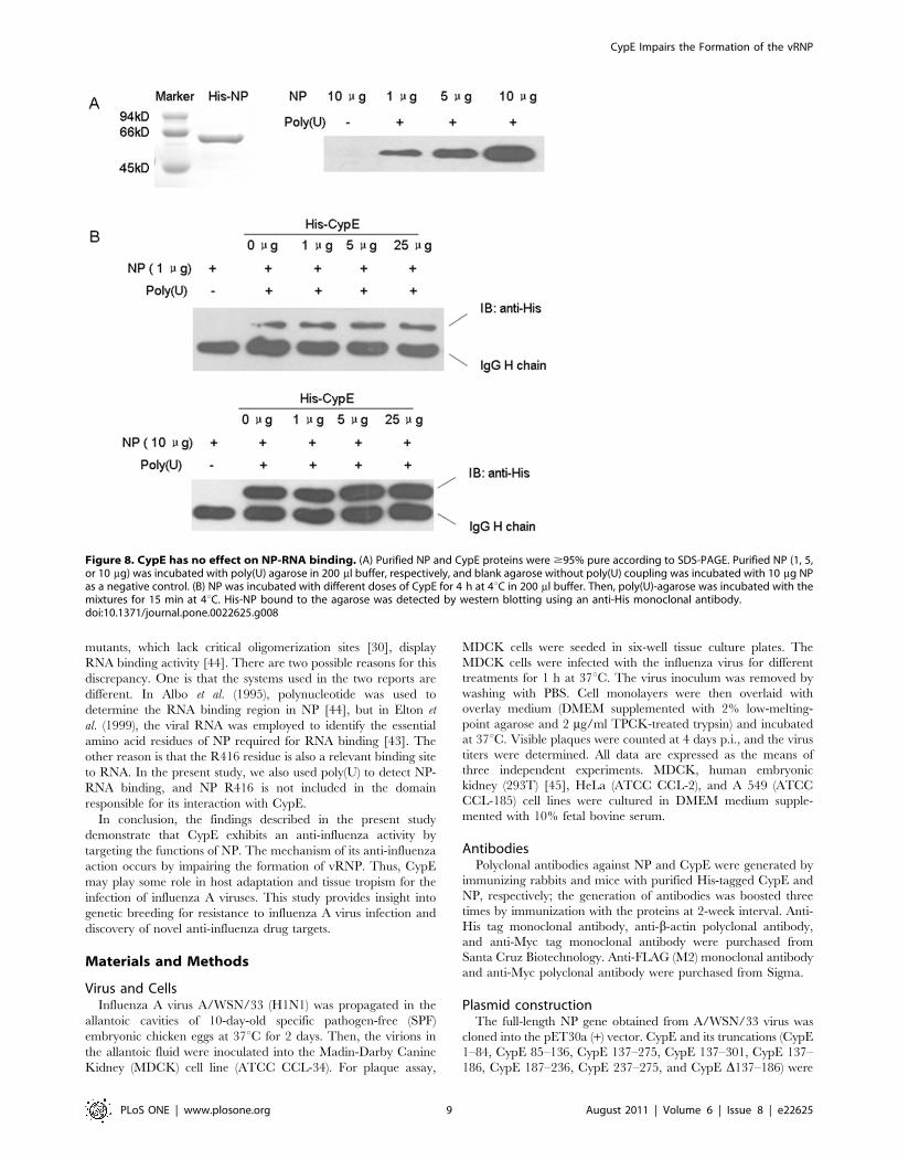
In addition, we examined whether CypE affects the binding of
NP to RNA. His-tagged NP was incubated with poly(U)-agarose in
the presence or absence of CypE, and then, the NP bound to the
poly(U)-agarose was detected by immunoblotting with anti-His
Figure 5. CypE inhibits the transcription and replication of influenza virus. (A) The A549 cells were treated with si-CypE or si-control for48 h and then infected with influenza virus A/WSN/33 at an MOI of 1. Three types of RNA levels of the NP and M1 genes were analyzed with orwithout CypE-knock-down by quantitative real-time PCR after viral infection (4 and 8 h). Western blot to confirm the knock-down of CypE expressionin A549 cells. Error bars represented the standard error of the mean (SEM). (B) All polymerase complex component plasmids and the luciferase gene,along with different doses of CypE and Myc-CypE D136–187 plasmids, were transfected into 293T cells, respectively. The luciferase activity in triplicatecultures was estimated and compared at 36 h p.t.. *, p,0.05; and **, p,0.01.doi:10.1371/journal.pone.0022625.g005
CypE Impairs the Formation of the vRNP
PLoS ONE | www.plosone.org 6 August 2011 | Volume 6 | Issue 8 | e22625

antibody. As shown in Figure 8, excessive CypE had no effect on
the interaction of NP with RNA.
Discussion
As is well known, hemagglutinin (HA) plays a critical role in
receptor binding and membrane fusion during the entry stage of
influenza A virus infection, and thus, the virus is able to access cells
that display matched receptors on their surface to initiate infection.
Though both SAa2, 6-Gal and SAa2, 3-Gal receptors are present
on cells in many organs of birds, pigs, and humans [31,32,33,34],
the susceptibility of different host species to influenza A virus varies
greatly, suggesting the existence of inhibitory mechanisms beyond
the sialic acid receptor specificity. It has been hypothesized that as-
yet undetermined host restriction factors could be involved in
mediating cellular resistance to viral replication, reducing the
efficiency of inter-species transmission. Thus, the identification of
host factors with anti-influenza effects on viral replication may
better provide insight into viral host adaptation. To date, several
host factors have been identified that interact with the viral
proteins/complexes and inhibit the replication of influenza A
virus, implicating them as host barriers to limit viral replication
[16,24,35,36,37,38,39]. Here, CypE was also proven to be an
influenza A virus inhibitory factor.
It has been reported that the nuclear import of vRNPs is
mediated through the interaction between NP and importin-a[15]. Moreover, the interaction between importin-a and PB2 or
NP is a determinant of virus host range, and different importin-aisoforms play critical and distinct roles in host adaptation
[14,18,40]. On the contrary, the current study demonstrated that
the over-expression of CypE delays the translocation of NP from
the nucleus to the cytoplasm (Figure 4E), suggesting that CypE
may play an important role in mediating vRNP export through its
interaction with NP. Our latest findings demonstrated that human
CypE displays different binding affinities to NPs from several
subtypes of influenza A viruses (data not shown). This finding
further supports the conclusion that CypE is a restriction factor to
viral infection and also demonstrates that CypE may play a role in
viral host adaptation. Likewise, it will be important to investigate
whether the tissue distribution of CypE is associated with the tissue
tropism of influenza A virus. These findings will help us to better
understand the molecular mechanisms of host restriction barriers,
host adaptation and tissue tropism of influenza A virus.
It has been reported that CypA inhibits the replication of
influenza A virus in the absence of its PPIase activity [24]. Here,
we demonstrated that the antiviral function of CypE is also
independent of its PPIase activity because the CypE R191A/
W257A mutant, which is defective in PPIase activity [41], also
equally inhibited the replication of influenza A virus (Figure S3).
Furthermore, our mutagenesis data indicate that the key sites of
PPIase activity (R191A and W257A) are not included in the CypE
137–186 domain responsible for its binding to NP. These data
suggest that CypE may play roles in cells infected with influenza A
virus that are independent of its PPIase activity.
NP forms oligomers as a purified recombinant protein, in
virions and in infected cells [5,30]. Furthermore, when the critical
oligomerization sites (E339A, R416A, and D402–428) are
substituted or deleted, all three mutants only form monomers
Figure 6. CypE inhibits NP self-association. pCDNA3-FLAG-NP and pCMV-Myc-NP, together with different doses of pCMV-Myc-CypE or Myc-CypE D136–187 plasmids, were transfected into 293T cells. At 48 h p.t, the cell lysates were immunoblotted with anti-Myc or anti-FLAG antibodies toconfirm the expression of the proteins of interest in the 293T cells. Co-immunoprecipitation assays were then performed using anti-FLAG M2 affinitygel. The immunoprecipitated proteins were assayed with an anti-Myc antibody. ‘‘Input’’ shows ,1/10 of the total protein. *, p,0.05.doi:10.1371/journal.pone.0022625.g006
CypE Impairs the Formation of the vRNP
PLoS ONE | www.plosone.org 7 August 2011 | Volume 6 | Issue 8 | e22625

[30]. Moreover, the E339A and R416A NP mutants lost the ability
to support the vRNP activity, and these mutants result in growth
defects in mutant viruses [29]. These previous reports indicate that
NP oligomerization is a fundamental and necessary step for viral
replication and transcription. In the present study, immunopre-
cipitation assays were employed to demonstrate that the over-
expression of CypE interferes with NP self-association (Figure 6B),
suggesting that the over-expression of CypE inhibits the initiation
of viral replication and transcription. A similar method was also
used to examine the effect of A20 on the dimerization of IRF-3
[42]. According to the binding domains of NP to CypE (Figure
S4), the critical E339 residue for NP oligomerization is included in
the domain responsible for CypE binding.
Elton et al. (1999) demonstrate that the NP R416A mutant,
which only forms monomers [30], exhibits little or no observable
RNA binding activity [43], suggesting that proper self-association
is important for binding to RNA. However, the present study
indicates that NP is bound to CypE and, therefore, not
polymerized but retains RNA binding activity. It seems that there
is a conflict between our report and Elton et al. (1999), but our
results are similar to another report about NP-RNA binding [44].
Indeed, it has been shown that the NP D256–339 and D378–460
Figure 7. CypE impairs the interaction of NP with PB1 and PB2. The pCDNA3-FLAG-PB1 or pCDNA3-FLAG-PB2 and pCMV-Myc-NP plasmids,along with pCMV-Myc-CypE (0, 0.3, and 0.9 mg) or Myc-CypE D136–187 (0.9 mg), were transfected into 293T cells, respectively. At 48 h p.t, the cellswere lysed, immunoblotted with anti-Myc or anti-FLAG antibodies, and subjected to co-immunoprecipitation assays. The immunoprecipitated NPwas detected with anti-Myc antibody. ‘‘Input’’ shows ,1/20 of the total protein. *, p,0.05.doi:10.1371/journal.pone.0022625.g007
CypE Impairs the Formation of the vRNP
PLoS ONE | www.plosone.org 8 August 2011 | Volume 6 | Issue 8 | e22625
mutants, which lack critical oligomerization sites [30], display
RNA binding activity [44]. There are two possible reasons for this
discrepancy. One is that the systems used in the two reports are
different. In Albo et al. (1995), polynucleotide was used to
determine the RNA binding region in NP [44], but in Elton et
al. (1999), the viral RNA was employed to identify the essential
amino acid residues of NP required for RNA binding [43]. The
other reason is that the R416 residue is also a relevant binding site
to RNA. In the present study, we also used poly(U) to detect NP-
RNA binding, and NP R416 is not included in the domain
responsible for its interaction with CypE.
In conclusion, the findings described in the present study
demonstrate that CypE exhibits an anti-influenza activity by
targeting the functions of NP. The mechanism of its anti-influenza
action occurs by impairing the formation of vRNP. Thus, CypE
may play some role in host adaptation and tissue tropism for the
infection of influenza A viruses. This study provides insight into
genetic breeding for resistance to influenza A virus infection and
discovery of novel anti-influenza drug targets.
Materials and Methods
Virus and CellsInfluenza A virus A/WSN/33 (H1N1) was propagated in the
allantoic cavities of 10-day-old specific pathogen-free (SPF)
embryonic chicken eggs at 37uC for 2 days. Then, the virions in
the allantoic fluid were inoculated into the Madin-Darby Canine
Kidney (MDCK) cell line (ATCC CCL-34). For plaque assay,
MDCK cells were seeded in six-well tissue culture plates. The
MDCK cells were infected with the influenza virus for different
treatments for 1 h at 37uC. The virus inoculum was removed by
washing with PBS. Cell monolayers were then overlaid with
overlay medium (DMEM supplemented with 2% low-melting-
point agarose and 2 mg/ml TPCK-treated trypsin) and incubated
at 37uC. Visible plaques were counted at 4 days p.i., and the virus
titers were determined. All data are expressed as the means of
three independent experiments. MDCK, human embryonic
kidney (293T) [45], HeLa (ATCC CCL-2), and A 549 (ATCC
CCL-185) cell lines were cultured in DMEM medium supple-
mented with 10% fetal bovine serum.
AntibodiesPolyclonal antibodies against NP and CypE were generated by
immunizing rabbits and mice with purified His-tagged CypE and
NP, respectively; the generation of antibodies was boosted three
times by immunization with the proteins at 2-week interval. Anti-
His tag monoclonal antibody, anti-b-actin polyclonal antibody,
and anti-Myc tag monoclonal antibody were purchased from
Santa Cruz Biotechnology. Anti-FLAG (M2) monoclonal antibody
and anti-Myc polyclonal antibody were purchased from Sigma.
Plasmid constructionThe full-length NP gene obtained from A/WSN/33 virus was
cloned into the pET30a (+) vector. CypE and its truncations (CypE
1–84, CypE 85–136, CypE 137–275, CypE 137–301, CypE 137–
186, CypE 187–236, CypE 237–275, and CypE D137–186) were
Figure 8. CypE has no effect on NP-RNA binding. (A) Purified NP and CypE proteins were $95% pure according to SDS-PAGE. Purified NP (1, 5,or 10 mg) was incubated with poly(U) agarose in 200 ml buffer, respectively, and blank agarose without poly(U) coupling was incubated with 10 mg NPas a negative control. (B) NP was incubated with different doses of CypE for 4 h at 4uC in 200 ml buffer. Then, poly(U)-agarose was incubated with themixtures for 15 min at 4uC. His-NP bound to the agarose was detected by western blotting using an anti-His monoclonal antibody.doi:10.1371/journal.pone.0022625.g008
CypE Impairs the Formation of the vRNP
PLoS ONE | www.plosone.org 9 August 2011 | Volume 6 | Issue 8 | e22625

cloned into the pGEX-6p-1 vector. For co-immunoprecipitation,
confocal, and luciferase assays, the NP gene was cloned into the
pcDNA3-FLAG and pCMV-Myc vectors, respectively; the full-
length CypE gene and CypE D137–186 were cloned into pCMV-
Myc. The expression plasmids for the PA, PB1, and PB2 genes from
A/WSN/33 virus were generated by cloning into the pCDNA3-
FLAG vector.
GST pull-down assaysGST-fused CypE and its truncations fused were purified using
Sepharose 4B-glutathione (GE Healthcare). His-tagged NP and
200 ml lysate from MDCK cells infected with A/WSN/33 virus
were incubated with equal amounts of GST alone and GST-CypE
bound to glutathione-Sepharose 4B beads in binding buffer (1%
NP-40, 150 mM NaCl, 20 mM HEPES (pH 7.4), 10% glycerol,
and 1 mM EDTA) with protease inhibitor cocktail (Roche). The
beads were washed five times with washing buffer (1% NP-40,
300 mM NaCl, 20 mM HEPES (pH 7.4), 10% glycerol, and
1 mM EDTA) with protease inhibitor cocktail. After extensive
washing, the NP bound to the beads was extracted and analyzed
by western blotting with an anti-His monoclonal antibody or an
anti-NP polyclonal antibody.
Co-immunoprecipitation assaysThe 293T cells were transfected with different plasmids using
lipofectamine 2000 (Invitrogen). At 48 h post-transfection (p.t.),
the cells were lysed in binding buffer supplemented with protease
inhibitor cocktail. The cell lysates were immunoblotted with anti-
Myc or anti-FLAG antibody to confirm the expression of the genes
of interest in the 293T cells, and then, the co-immunoprecipitation
assays were performed using anti-FLAG M2 affinity gel. The
immunoprecipitated proteins were assayed with anti-Myc poly-
clonal antibody. For the binding assay between endogenous CypE
and NP during influenza virus infection, 293T cells were infected
with A/WSN/33 (MOI = 1). At 12 h p.i., the cell lysates were
subjected to co-immunoprecipitation assays using rabbit anti-NP
antibody.
Immunofluorescence assaysHeLa cells were washed three times with PBS, fixed in 4%
paraformaldehyde for 30 min at room temperature, permeabilized
with 0.5% Triton X-100 in PBS (PBST) for 20 min, and then
incubated for 1 h with anti-CypE rabbit antiserum and anti-NP
mouse antiserum. After washing with PBST, the cells were
incubated for 1 h with the secondary antibodies. The cells were
then observed under a Leica confocal microscope.
For influenza virus infectivity assays, 293T cells were transfected
with 1 mg/well pCMV-Myc-CypE plasmid or 1 mg/well pCMV-
Myc empty vector (as a control) and pEGFP-N1 (0.1 mg/well). At
12 h p.t., the cells were infected with influenza virus A/WSN/33
at an MOI of 1. At 6 and 8 h p.i., the cells were treated as
described above and then stained with anti-Myc and anti-NP
antibodies. The cells were observed under a Leica confocal
microscope.
RNA interference assaysThe affective siRNA duplexes for CypE were si-CypE: 59-
ATTGTGGTTTGTGAAATCACCGCCC-39 (Invitrogen). The
293T or A549 cells were transfected with si-CypE or si-Control
according to manufacturer’s instructions using RNAiMax trans-
fection reagent (Invitrogen). At 48 h p.t., the cells were infected
with influenza A virus A/WSN/33. The efficiency of CypE knock-
down was confirmed by western blotting with the indicated
antibodies. The virus titers in the media were measured by plaque
assay.
Quantitative real-time PCR assaysTransfected A549 cells were harvested, and total RNA was
extracted for quantitative real-time PCR assays. We used
published primers (vRNA (59-AGCGAAAGCAGG-39 and 59-
AGCAAAAGCAGG-39), cRNA (59-AGTAGAAACAAGG -39),
and mRNA (oligo (dT))) to detect the vRNA, cRNA, and mRNA
for reverse transcription reactions [46]. The real-time PCR
primers were as follows: WSN M1 (forward, 59-TCTGATC-
CTCTCGTCATTGCAGCAA-39; reverse, 59-AATGACCATC-
GTCAACATCCACAGC-39) [35]; and WSN NP (forward, 59-
TGGCACTCCAATTTGAATGATG-39; reverse, 59-TCCAT-
TCCTGTGCGAACAAG-39) [47]. GAPDH mRNA served as
an internal control: (forward, 59-GGTGGTCTCCTCTGACTT-
CAACA-39; and reverse, 59-GTTGCTGTAGCCAAATTCG-
TTGT-39), as described in [48]. The PCR program was 95uCfor 30 s followed by 40 cycles of 94uC for 5 s and 60uC for 30 s,
and dissociation curve analysis of amplification products was
performed at the end of each PCR reaction to confirm that only
one PCR product was amplified and detected. Each sample was
run in triplicate along with the internal control gene. Data analysis
of real time PCR was performed with Rotor Gene 6000 Series
Software (Corbett).
Luciferase reporter assays for influenza polymerasecomplex activity
All polymerase complex component plasmids were co-trans-
fected with a luciferase reporter plasmid that contained non-
coding sequence from the NS segment of the influenza A virus
genome and the luciferase gene that was driven by the PolI
promoter into 293T cells. At the same time, pCMV-Myc empty
vector (0.45 mg), different doses of CypE plasmid (0.05, 0.15, and
0.45 mg), and pCMV-CypE D137–186 (0.45 mg) were also
transfected into 293T cells, respectively. At 36 h p.t., cell lysates
were prepared using a luciferase assay kit (Promega), and the
relative activities with different doses of CypE were compared.
Plasmid pCMV-b-galactosidase, which expresses b-galactosidase,
was co-transfected as an internal control for data normalization.
NP-RNA binding assaysHis-tagged NP and CypE were purified using Ni-NTA affinity
agarose. NP was incubated with different doses of CypE (1, 5, and
25 mg) for 4 h at 4uC in a Tris-HCl buffer (pH 7.4) containing
1 U/ml RNase inhibitor. Then, equimolar amount of poly(U)
agarose was incubated with the mixtures for 15 min at 4uC. At the
same time, an equivalent amount of anti-FLAG M2 agarose was
added to every binding reaction as an internal control. After
washing extensively, the His-NP bound to the agarose was
detected by western blotting using an anti-His monoclonal
antibody.
Supporting Information
Figure S1 A549 cells were transfected with si-CypE orsi-control and then infected with influenza virus A/WSN/33 at an MOI of 0.1. The cell lysates were analyzed by
western blotting with the indicated antibodies (A), and the viral
titers of the media were measured by plaque assay (B). **, p,0.01.
(TIF)
Figure S2 293T cells were transfected with 1 mg CypEand 1 mg pCMV-Myc vector as a control, and then they
CypE Impairs the Formation of the vRNP
PLoS ONE | www.plosone.org 10 August 2011 | Volume 6 | Issue 8 | e22625

were infected with A/WSN/33 (MOI = 1). The cRNA,
vRNA, and mRNA levels of the NP and M1 genes were analyzed
by quantitative real-time PCR after 2, 4, and 8 h p.i.. Error bars
represented the SEM.
(TIF)
Figure S3 293T cells were transfected with FLAG-CypEor FLAG-CypE R191A/W257A plasmid and then infectedwith influenza virus A/WSN/33 at an MOI of 0.1. The cell
lysates was analyzed by western blotting with the corresponding
antibodies (A). The media were collected, and the viral titers were
measured (B). Error bars represented the SD. *, p,0.05.
(TIF)
Figure S4 FLAG-tagged NP plus CypE or its truncationswere transfected into 293T cells. The co-immunoprecipita-
tion assays were performed using anti-FLAG M2 affinity gel. The
immunoprecipitated proteins were assayed with an anti-Myc
polyclonal antibody. ‘‘Input’’ shows ,1/20 of the total protein.
(TIF)
Author Contributions
Conceived and designed the experiments: WJL ZFW. Performed the
experiments: ZFW XLL ZDZ CFX KZ. Analyzed the data: ZFW CWC
LS. Contributed reagents/materials/analysis tools: XLL ZDZ. Wrote the
paper: ZFW GFG XY WJL.
References
1. Palese P (1977) The genes of influenza virus. Cell 10: 1–10.
2. Murti KG, Webster RG, Jones IM (1988) Localization of RNA polymerases on
influenza viral ribonucleoproteins by immunogold labeling. Virology 164:
562–566.
3. Kingsbury DW, Jones IM, Murti KG (1987) Assembly of influenza
ribonucleoprotein in vitro using recombinant nucleoprotein. Virology 156:
396–403.
4. Pons MW, Schulze IT, Hirst GK, Hauser R (1969) Isolation and character-
ization of the ribonucleoprotein of influenza virus. Virology 39: 250–259.
5. Ruigrok RW, Baudin F (1995) Structure of influenza virus ribonucleoprotein
particles. II. Purified RNA-free influenza virus ribonucleoprotein forms
structures that are indistinguishable from the intact influenza virus ribonucleo-
protein particles. J Gen Virol 76(Pt 4): 1009–1014.
6. Portela A, Digard P (2002) The influenza virus nucleoprotein: a multifunctional
RNA-binding protein pivotal to virus replication. J Gen Virol 83: 723–734.
7. Medcalf L, Poole E, Elton D, Digard P (1999) Temperature-sensitive lesions in
two influenza A viruses defective for replicative transcription disrupt RNA
binding by the nucleoprotein. J Virol 73: 7349–7356.
8. Biswas SK, Boutz PL, Nayak DP (1998) Influenza virus nucleoprotein interacts
with influenza virus polymerase proteins. J Virol 72: 5493–5501.
9. Kingsbury DW, Webster RG (1969) Some Properties of Influenza Virus
Nucleocapsids. J Virol 4: 219–225.
10. Coloma R, Valpuesta JM, Arranz R, Carrascosa JL, Ortin J, et al. (2009) The
structure of a biologically active influenza virus ribonucleoprotein complex.
PLoS Pathog 5: e1000491.
11. Duesberg PH (1969) Distinct subunits of the ribonucleoprotein of influenza
virus. J Mol Biol 42: 485–499.
12. Yamanaka K, Ishihama A, Nagata K (1990) Reconstitution of influenza virus
RNA-nucleoprotein complexes structurally resembling native viral ribonucleo-
protein cores. J Biol Chem 265: 11151–11155.
13. Digard P, Elton D, Bishop K, Medcalf E, Weeds A, et al. (1999) Modulation of
nuclear localization of the influenza virus nucleoprotein through interaction with
actin filaments. J Virol 73: 2222–2231.
14. Gabriel G, Herwig A, Klenk HD (2008) Interaction of polymerase subunit PB2
and NP with importin alpha1 is a determinant of host range of influenza A virus.
PLoS Pathog 4: e11.
15. O’Neill RE, Jaskunas R, Blobel G, Palese P, Moroianu J (1995) Nuclear import
of influenza virus RNA can be mediated by viral nucleoprotein and transport
factors required for protein import. J Biol Chem 270: 22701–22704.
16. Wang P, Song W, Mok BW, Zhao P, Qin K, et al. (2009) Nuclear factor 90
negatively regulates influenza virus replication by interacting with viral
nucleoprotein. J Virol 83: 7850–7861.
17. Boivin S, Hart DJ (2011) Interaction of the influenza A virus polymerase PB2 C-
terminal region with importin {alpha} isoforms provides insights into host
adaptation and polymerase assembly. J Biol Chem doi:10.1074/jbc.M111.226225.
18. Gabriel G, Klingel K, Otte A, Thiele S, Hudjetz B, et al. (2011) Differential use
of importin-alpha isoforms governs cell tropism and host adaptation of influenza
virus. Nat Commun 2: 156.
19. Elton D, Simpson-Holley M, Archer K, Medcalf L, Hallam R, et al. (2001)
Interaction of the influenza virus nucleoprotein with the cellular CRM1-
mediated nuclear export pathway. J Virol 75: 408–419.
20. Liao TL, Wu CY, Su WC, Jeng KS, Lai MM (2010) Ubiquitination and
deubiquitination of NP protein regulates influenza A virus RNA replication.
EMBO J 29: 3879–3890.
21. Franke EK, Yuan HE, Luban J (1994) Specific incorporation of cyclophilin A
into HIV-1 virions. Nature 372: 359–362.
22. Luban J, Bossolt KL, Franke EK, Kalpana GV, Goff SP (1993) Human
immunodeficiency virus type 1 Gag protein binds to cyclophilins A and B. Cell
73: 1067–1078.
23. Yang F, Robotham JM, Nelson HB, Irsigler A, Kenworthy R, et al. (2008)
Cyclophilin A is an essential cofactor for hepatitis C virus infection and the
principal mediator of cyclosporine resistance in vitro. J Virol 82: 5269–5278.
24. Liu X, Sun L, Yu M, Wang Z, Xu C, et al. (2009) Cyclophilin A interacts with
influenza A virus M1 protein and impairs the early stage of the viral replication.
Cell Microbiol 11: 730–741.
25. Xu C, Meng S, Liu X, Sun L, Liu W (2011) Chicken cyclophilin A is an
inhibitory factor to influenza virus replication. Virol J 7: 372.
26. Watashi K, Ishii N, Hijikata M, Inoue D, Murata T, et al. (2005) Cyclophilin B is a
functional regulator of hepatitis C virus RNA polymerase. Mol Cell 19: 111–122.
27. Mi H, Kops O, Zimmermann E, Jaschke A, Tropschug M (1996) A nuclear
RNA-binding cyclophilin in human T cells. FEBS Lett 398: 201–205.
28. Chan WH, Ng AK, Robb NC, Lam MK, Chan PK, et al. (2010) Functional
analysis of the influenza virus H5N1 nucleoprotein tail loop reveals amino acids
that are crucial for oligomerization and ribonucleoprotein activities. J Virol 84:
7337–7345.
29. Li Z, Watanabe T, Hatta M, Watanabe S, Nanbo A, et al. (2009) Mutational
analysis of conserved amino acids in the influenza A virus nucleoprotein. J Virol
83: 4153–4162.
30. Ye Q, Krug RM, Tao YJ (2006) The mechanism by which influenza A virus
nucleoprotein forms oligomers and binds RNA. Nature 444: 1078–1082.
31. Kuchipudi SV, Nelli R, White GA, Bain M, Chang KC, et al. (2009) Differences
in influenza virus receptors in chickens and ducks: Implications for interspecies
transmission. J Mol Genet Med 3: 143–151.
32. Kogure T, Suzuki T, Takahashi T, Miyamoto D, Hidari KI, et al. (2006) Human
trachea primary epithelial cells express both sialyl(alpha2–3)Gal receptor for
human parainfluenza virus type 1 and avian influenza viruses, and sialyl(alpha2–
6)Gal receptor for human influenza viruses. Glycoconj J 23: 101–106.
33. Shinya K, Ebina M, Yamada S, Ono M, Kasai N, et al. (2006) Avian flu:
influenza virus receptors in the human airway. Nature 440: 435–436.
34. Nelli RK, Kuchipudi SV, White GA, Perez BB, Dunham SP, et al. (2010)
Comparative distribution of human and avian type sialic acid influenza receptors
in the pig. BMC Vet Res 6: 4.
35. Watanabe K, Fuse T, Asano I, Tsukahara F, Maru Y, et al. (2006) Identification
of Hsc70 as an influenza virus matrix protein (M1) binding factor involved in the
virus life cycle. FEBS Lett 580: 5785–5790.
36. Li S, Min JY, Krug RM, Sen GC (2006) Binding of the influenza A virus NS1
protein to PKR mediates the inhibition of its activation by either PACT or
double-stranded RNA. Virology 349: 13–21.
37. Min JY, Krug RM (2006) The primary function of RNA binding by the
influenza A virus NS1 protein in infected cells: Inhibiting the 29–59 oligo (A)
synthetase/RNase L pathway. Proc Natl Acad Sci U S A 103: 7100–7105.
38. Nemeroff ME, Barabino SM, Li Y, Keller W, Krug RM (1998) Influenza virus
NS1 protein interacts with the cellular 30 kDa subunit of CPSF and inhibits
39end formation of cellular pre-mRNAs. Mol Cell 1: 991–1000.
39. Opitz B, Rejaibi A, Dauber B, Eckhard J, Vinzing M, et al. (2007) IFNbeta
induction by influenza A virus is mediated by RIG-I which is regulated by the
viral NS1 protein. Cell Microbiol 9: 930–938.
40. Boivin S, Hart DJ (2011) Interaction of the influenza A virus polymerase PB2 C-
terminal region with importin {alpha} isoforms provides insights into host
adaptation and polymerase assembly. J Biol Chem.
41. Wang T, Yun CH, Gu SY, Chang WR, Liang DC (2005) 1.88 A crystal
structure of the C domain of hCyP33: a novel domain of peptidyl-prolyl cis-trans
isomerase. Biochem Biophys Res Commun 333: 845–849.
42. Saitoh T, Yamamoto M, Miyagishi M, Taira K, Nakanishi M, et al. (2005) A20 is a
negative regulator of IFN regulatory factor 3 signaling. J Immunol 174: 1507–1512.
43. Elton D, Medcalf L, Bishop K, Harrison D, Digard P (1999) Identification of
amino acid residues of influenza virus nucleoprotein essential for RNA binding.
J Virol 73: 7357–7367.
44. Albo C, Valencia A, Portela A (1995) Identification of an RNA binding region
within the N-terminal third of the influenza A virus nucleoprotein. J Virol 69:
3799–3806.
45. DuBridge RB, Tang P, Hsia HC, Leong PM, Miller JH, et al. (1987) Analysis of
mutation in human cells by using an Epstein-Barr virus shuttle system. Mol Cell
Biol 7: 379–387.
CypE Impairs the Formation of the vRNP
PLoS ONE | www.plosone.org 11 August 2011 | Volume 6 | Issue 8 | e22625
46. Liang Y, Huang T, Ly H, Parslow TG, Liang Y (2008) Mutational analyses of
packaging signals in influenza virus PA, PB1, and PB2 genomic RNA segments.J Virol 82: 229–236.
47. Goodman AG, Smith JA, Balachandran S, Perwitasari O, Proll SC, et al. (2007)
The cellular protein P58IPK regulates influenza virus mRNA translation andreplication through a PKR-mediated mechanism. J Virol 81: 2221–2230.
48. Lai JP, Yang JH, Douglas SD, Wang X, Riedel E, et al. (2003) Quantification of
CCR5 mRNA in human lymphocytes and macrophages by real-time reverse
transcriptase PCR assay. Clin Diagn Lab Immunol 10: 1123–1128.
CypE Impairs the Formation of the vRNP
PLoS ONE | www.plosone.org 12 August 2011 | Volume 6 | Issue 8 | e22625