Cutaneous Side-effects of EGFR Inhibitors and Their Management
10
Indexed by the US National Library of Medicine and PubMed Volume 16 • Number 1 • January 2011 ALSO IN THIS ISSUE: Current Effective Topical Therapies in the Management of Psoriasis (page 4) & Update on Drugs (page 8) The epidermal growth factor receptor (EGFR), a 170-kd transmembrane glycoprotein, is a member of the type 1 receptor tyrosine kinase (TK) family. The EGFR is physiologically expressed in epithelial tissues and hair follicles, where it contributes to epidermal proliferation, differentiation, and hair growth. In addition, EGFR is overexpressed in many solid tumors, where it is involved in tumor growth, cell proliferation, apoptosis, angiogenesis, cell motility, and metastasis. 1 Preclinical and clinical studies have shown that inhibiting EGFR is a valid strategy in anticancer therapy. 1,2 Different strategies for EGFR inhibition have been described, 3 two of which entered routine clinical use: EGFR-targeting monoclonal antibodies (MoAbs) bind specifically to the extracellular domain of the receptor and competitively inhibit ligand binding, 1 and tyrosine kinase inhibitors suppress EGFR signaling at the intracellular domain of the receptor. 1 Experimental data exist for EGFR ligand toxin and EGFR immunotoxin conjugates. These novel drugs are composed of an EGFR ligand or EGFR-binding antibody and a cytotoxic agent. Finally, antisense oligonucleotides specific for the EGFR or EGFR ligand messenger ribonucleic acids (RNAs) may decrease EGFR expression, thus resulting in inhibition of proliferation and induction of apoptosis. As of July 2010, there are two monoclonal antibodies (cetuximab, Erbitux ® and panitumumab, Vectibix ® ) and one receptor tyrosine kinase inhibitor (Gefitinib, Iressa ® ) that are currently licensed for clinical use in many countries. 1 Gefitinib is a historic tyrosine kinase inhibitor that did not show significant survival benefit. 4 Lapatinib (Tykerb ® /Tyverb ® ) and canertinib (CI-1033) are currently developed tyrosine kinase inhibitors. 4-5 Lapatinib is under investigation for the second-line treatment of metastatic colorectal cancer, whereas canertinib is being studied for progressive, recurrent, locally advanced or metastatic non-small cell lung carcinoma, and metastatic breast cancers. 6,7 The safety profile of EGFR-inhibitors is characterized by a class effect comprised of unique skin reactions, including acneiform rash, xerosis, eczema, paronychia, and changes in the hair and nails. 8 Hyperpigmentation, trichomegaly, and telangiectasia are less commonly seen. EGFR inhibitor-induced urticarial and anaphylactoid reactions are frequently seen in the US, but these are rarely encountered in Europe. 9-10 Here, we review the diagnostic procedures and current treatment options for the more common side-effects of EGFR inhibitors. 4,8,11-16 Acneiform Eruptions Acneiform eruptions are the earliest and most characteristic side- effect of EGFR inhibition. The incidence may be as high as 75% to 100% of cases. 2,4,8,14,16-19 Evidence suggests that severity of the skin rash might be a surrogate marker determining clinical outcome of patients receiving EGFR inhibitor treatment. 1,19-21 The eruption usually occurs after 1 week of treatment. The characteristic distribution pattern is similar to that of acne vulgaris, but there are no comedones present. Although these eruptions are considered a class effect from EGFR inhibition, antibody-induced eruptions tend to be more severe than TK1 induced skin changes. 4 The pathogenesis of acneiform eruptions caused by EGFR is not yet fully understood. Histopathological analysis showed a Cutaneous Side-effects of EGFR Inhibitors and Their Management Laura Maximiliane Ehmann, MD, Thomas Ruzicka, MD and Andreas Wollenberg, MD Department of Dermatology and Allergology, Ludwig-Maximilian-University of Munich, Munich, Germany ABSTRACT Epidermal growth factor receptor (EGFR) inhibitors are an increasingly important treatment option for metastasized cancer in patients. In addition to the pivotal role of EGFR in the development and progression of malignant tumors, EGFR is also important for proliferation and differentiation of the human epidermis and hair follicles. As a consequence, cutaneous side-effects are frequently observed during cancer therapy with EGFR inhibitors. During the first few weeks of treatment, acneiform eruptions are the earliest common side-effect. Xerosis and fissures are complications appearing in later treatment phases. Paronychia and alterations in hair growth are less common and generally seen after a longer period of treatment. We present an overview of the various cutaneous side- effects associated with EGFR inhibition and discuss their respective therapeutic options. Key words: anti-EGFR, epidermal growth factor receptor inhibitors, cutaneous side-effects, acneiform eruption, paronychia, xerosis, management
Transcript of Cutaneous Side-effects of EGFR Inhibitors and Their Management

I n d e x e d b y t h e U S N a t i o n a l L i b r a r y o f M e d i c i n e a n d P u b M e dV o l u m e 1 6 • N u m b e r 1 • J a n u a r y 2 0 1 1
ALSO IN THIS ISSUE: Current Effective Topical Therapies in the Management of Psoriasis (page 4)& Update on Drugs (page 8)
The epidermal growth factor receptor (EGFR), a 170-kd transmembrane glycoprotein, is a member of the type 1 receptor tyrosine kinase (TK) family. The EGFR is physiologically expressed in epithelial tissues and hair follicles, where it contributes to epidermal proliferation, differentiation, and hair growth. In addition, EGFR is overexpressed in many solid tumors, where it is involved in tumor growth, cell proliferation, apoptosis, angiogenesis, cell motility, and metastasis.1 Preclinical and clinical studies have shown that inhibiting EGFR is a valid strategy in anticancer therapy.1,2
Different strategies for EGFR inhibition have been described,3 two of which entered routine clinical use: EGFR-targeting monoclonal antibodies (MoAbs) bind specifically to the extracellular domain of the receptor and competitively inhibit ligand binding,1 and tyrosine kinase inhibitors suppress EGFR signaling at the intracellular domain of the receptor.1 Experimental data exist for EGFR ligand toxin and EGFR immunotoxin conjugates. These novel drugs are composed of an EGFR ligand or EGFR-binding antibody and a cytotoxic agent. Finally, antisense oligonucleotides specific for the EGFR or EGFR ligand messenger ribonucleic acids (RNAs) may decrease EGFR expression, thus resulting in inhibition of proliferation and induction of apoptosis.
As of July 2010, there are two monoclonal antibodies (cetuximab, Erbitux® and panitumumab, Vectibix®) and one receptor tyrosine kinase inhibitor (Gefitinib, Iressa®) that are currently licensed for clinical use in many countries.1 Gefitinib is a historic tyrosine kinase inhibitor that did not show significant survival benefit.4 Lapatinib (Tykerb®/Tyverb®) and canertinib (CI-1033) are
currently developed tyrosine kinase inhibitors.4-5 Lapatinib is under investigation for the second-line treatment of metastatic colorectal cancer, whereas canertinib is being studied for progressive, recurrent, locally advanced or metastatic non-small cell lung carcinoma, and metastatic breast cancers.6,7
The safety profile of EGFR-inhibitors is characterized by a class effect comprised of unique skin reactions, including acneiform rash, xerosis, eczema, paronychia, and changes in the hair and nails.8 Hyperpigmentation, trichomegaly, and telangiectasia are less commonly seen. EGFR inhibitor-induced urticarial and anaphylactoid reactions are frequently seen in the US, but these are rarely encountered in Europe.9-10 Here, we review the diagnostic procedures and current treatment options for the more common side-effects of EGFR inhibitors.4,8,11-16
Acneiform EruptionsAcneiform eruptions are the earliest and most characteristic side-effect of EGFR inhibition. The incidence may be as high as 75% to 100% of cases.2,4,8,14,16-19 Evidence suggests that severity of the skin rash might be a surrogate marker determining clinical outcome of patients receiving EGFR inhibitor treatment.1,19-21 The eruption usually occurs after 1 week of treatment. The characteristic distribution pattern is similar to that of acne vulgaris, but there are no comedones present. Although these eruptions are considered a class effect from EGFR inhibition, antibody-induced eruptions tend to be more severe than TK1 induced skin changes.4
The pathogenesis of acneiform eruptions caused by EGFR is not yet fully understood. Histopathological analysis showed a
Cutaneous Side-effects of EGFR Inhibitors and Their Management
Laura Maximiliane Ehmann, MD, Thomas Ruzicka, MD and Andreas Wollenberg, MDDepartment of Dermatology and Allergology, Ludwig-Maximilian-University of Munich, Munich, Germany
ABSTRACT
Epidermal growth factor receptor (EGFR) inhibitors are an increasingly important treatment option for metastasized cancer in patients. In addition to the pivotal role of EGFR in the development and progression of malignant tumors, EGFR is also important for proliferation and differentiation of the human epidermis and hair follicles. As a consequence, cutaneous side-effects are frequently observed during cancer therapy with EGFR inhibitors. During the first few weeks of treatment, acneiform eruptions are the earliest common side-effect. Xerosis and fissures are complications appearing in later treatment phases. Paronychia and alterations in hair growth are less common and generally seen after a longer period of treatment. We present an overview of the various cutaneous side-effects associated with EGFR inhibition and discuss their respective therapeutic options.
Key words: anti-EGFR, epidermal growth factor receptor inhibitors, cutaneous side-effects, acneiform eruption, paronychia, xerosis, management

• Editor: Dr. Stuart Maddin • Volume 16, Number 1 • January 20112
neutrophilic suppurative infiltrate in the dermis, particularly involving the follicular infundibula. The follicles are frequently enlarged and sometimes obstructed by excess keratinocytes. The sebaceous glands are usually not affected. No consistent changes in the cutaneous microflora have been found.4
Grading of acneiform eruptions is performed with two different scoring systems, depending if quick severity classification or sensitive follow-up of treatment success is the goal of grading. The oncological classification, National Cancer Institute Common Toxicity Criteria for Adverse Events, version 4.0, allows for a quick severity estimation of skin toxicity reactions. It provides a clinical score with five grades of severity.4 The dermatological skin score (WoMoScore) is a sensitive dermatologic scoring system for the long-term assessment of acneiform skin rashes that has been used in our department since 2006.22 The final WoMoScore is calculated from body involvement, facial involvement, and clinical grading of erythema, papulation, postulation, scaling, and crusts, providing a clinical score ranging from 0 to 100.22 Mild skin changes score up to 20, moderate cases range between 20 and 40, whereas severe acneiform eruptions exceed a WoMoScore of 40.20
Treatment of Acneiform EruptionsTreatment of mild acneiform eruptions mostly involves conventional topical medications used for acne vulgaris, such as metronidazole, erythromycin, and clindamycin.4,8
Topical combination therapy with nadifloxacin 1% cream and prednicarbate 0.25% cream is our standard regimen for moderate acneiform eruptions.22 An uncontrolled, open label, follow-up study involving 29 cancer patients with cetuximab-induced acneiform eruptions initially demonstrated clinical efficacy with significant reduction of the WoMoScore after 1, 2 and 6 weeks of therapy.22 Both drugs were selected because nadifloxacin has antimicrobial as well as immunomodulatory effects on the antigen-presenting function of Langerhans cells and keratinocytes,23 and prednicarbate is a well established anti-inflammatory topical corticosteroid with an improved risk-benefit ratio.24
Preclinical data suggest that topical application of the potent phosphatase inhibitor menadione (vitamin K3) might rescue the inhibition of EGFR and downstream signaling molecules in the skin of mice receiving systemic EGFR inhibitors erlotinib (Tarceva®) or cetuximab.25 In a non-randomized study with 30 patients, acneiform eruptions were reduced significantly by the topical use of a cream containing urea and 0.1% K1 vitamin (Reconval K1®).26
Oral tetracyclines have been used for the treatment of acne vulgaris for more than 50 years27 because of their anti-inflammatory and immunomodulatory properties.28-30 These broad-spectrum polyketide antibiotics reduce neutrophilic chemotaxis and inhibit the production of proinflammatory cytokines and matrix metalloproteinase 9.29 Some recent studies investigated the benefit of prophylactic tetracycline for EGFR-induced acneiform eruptions.31-32 Tetracycline-treated patients reported less itching, burning, stinging, and other subjective symptoms compared with placebo. However, prophylactic tetracyclines did not lower the total incidence of the rash.31-32 Tetracyclines are clearly effective, but they may lead to unwanted systemic effects that are not encountered with topical treatment.
Our first choice for severe acneiform eruptions is early treatment with a combination of low dose oral retinoid, topical nadifloxacin, and topical prednicarbate.21-22 We limit the use of this highly effective “triple therapy” to the first 2-3 months of EGFR-inhibitor treatment.21 Some authors have raised the issue of increased xerosis or reduced anticancer properties of the EGFR-inhibitors,5,10,11 whereas others see a potential synergistic effect of this combinatorial approach.11
Management of acneiform skin eruptions is influenced by the level of dermatological background of the treating physician.33 Dermato-oncologists are using oral isotretinoin more frequently, compared with oncologists, and are delaying EGFR inhibitor treatment less frequently because of skin toxicities.33 An interdisciplinary approach in cooperation with dermatologists is highly recommended to improve patient treatment.8,33
ParonychiaEGFR inhibitor-induced paronychia is seen in about 10% to 15% of all treated patients and may be quite painful, adversely affecting their quality of life.12 It generally does not develop during the first 6 weeks of treatment. Patients frequently present with involvement of the great toenails. Fingernail involvement may lead to significant functional impairment. Periungual abscesses and pyogenic granuloma may develop in some cases.4,8,15 A recent study showed that a wide variety of Gram-positive and Gram-negative bacteria, as well as Candida albicans, may be cultured from the nail lesions.15
Initial stages of paronychia should be treated with topical antiseptic measures and application of antiseptic or antibiotic ointments. Antiseptic treatment should be continued after systemic therapy has been initiated. Systemic treatment of paronychia is recommended for all painful or infected lesions, as there is a risk for the development of erysipelas, deep panaritium, and tendon sheath phlegmon. Calculated antibiotic treatment of paronychia is recommended with oral cephalosporines, but oral fluoroquinolones may also be used, especially if Gram-negative infection is suspected. Surgical intervention may be necessary in selected cases, such as ingrown toe nails.4,8,15
XerosisXerosis is also considered a class effect of EGFR inhibitors, as it affects most treated patients to a variable degree. Xerosis typically presents as dry, scaly, itchy skin that can be found on any part of the human body. Some patients may also experience dryness of vaginal and perineal regions. Xerosis may progress to chronic asteatotic eczema and become infected with Staphylococcus aureus or herpes simplex virus. Severe cases of pulpitis sicca (dry skin on the tips of the fingers and toes) with painful rhagades have also been described.4,8
First-line treatment of xerosis is the liberal use of emollients, which should be started within the first days of initiating EGFR inhibitor treatment. This prophylactic approach is safe and effective, and may prevent the onset of eczema. 4,8
RhagadesPainful fissures on the tips of fingers and toes, on the nailfolds, and especially over the interphalangeal joints may develop as a consequence of excessively dry skin. Fissures are less common
• Editor: Dr. Stuart Maddin • Volume 16, Number 1 • January 2011 3
late phase reactions from therapy with EGFR inhibitors. They occur in about 25% of patients and are characterized by pain, severe tenderness, and poor healing tendency.4,8 Fissures are challenging to treat. Silver nitrate solution, creams containing urea under plastic occlusion, and topical antibiotics can be used. Liquid cyanoacrylate glue may be tried for recurrent cases.4,8
Further Cutaneous Side-effectsTrichomegaly, with or without additional hypertrichosis, may develop after 2 to 5 months of continuous anti-EGFR treatment. Trichomegaly may cause visual discomfort, but can be effectively treated by trimming the eye lashes. The higher expression of EGFR in the outer root sheath of the hair follicles may be causative for this infrequent but characteristic side-effect.4,8,12
Hyperpigmentation may appear after several months of EGFR inhibitor therapy. As bleaching creams have not been shown to be effective, patients must wait for the hyperpigmentation to fade over several months.4,12
Telangiectasias may occur together with acneiform eruptions and follow the same pattern on the face and trunk. These telangiectasias vanish with time, but often leave some degree of hyperpigmentation.4,12
Painful aphthae and larger erosions may appear on the oral mucosa, lips, and nose, as well as on the anal and genital mucosa. Localized measures that are pain-relieving and soothing will provide considerable symptomatic relief.
Patients must be warned to stay out of the sun, because photosensitivity is increased during therapy with EGFR inhibitors and quite severe acneiform reactions can be triggered by UV exposure. Consequently, the prophylactic use of sunscreens is highly recommended to prevent these solar-induced reactions.
ConclusionEGFR inhibitors are associated with a unique group of class-specific cutaneous toxicities, which include acneiform eruptions, paronychia, xerosis, hyperpigmentation, trichomegaly, and telangiectasia. As the use of EGFR inhibitors increases, a growing number of cutaneous side-effects will be treated by dermatologists. The discomfort caused by the cutaneous toxicities can reduce compliance with anti-EGFR therapy. For this reason, consequent treatment and psycho-dermatological support are very important. Almost all side-effects are temporary and will resolve several weeks after discontinuation of EGFR inhibitor therapy. It is import to educate patients about these side-effects, in order to increase compliance.
References1. Vallbohmer D, Lenz HJ. Epidermal growth factor receptor as a target for chemotherapy.
Clin Colorectal Cancer 5(Suppl 1):S19-27 (2005 Apr).2. Cunningham D, Humblet Y, Siena S, et al. Cetuximab monotherapy and cetuximab
plus irinotecan in irinotecan-refractory metastatic colorectal cancer. N Engl J Med 351(4):337-45 (2004 Jul 22).
3. Mendelsohn J, Baselga J. The EGF receptor family as targets for cancer therapy. Oncogene 19(56):6550-65 (2000 Dec 27).
4. Hu JC, Sadeghi P, Pinter-Brown LC, et al. Cutaneous side effects of epidermal growth factor receptor inhibitors: clinical presentation, pathogenesis, and management. J Am Acad Dermatol 56(2):317-26 (2007 Feb).
5. Saltz LB, Meropol NJ, Loehrer PJ, Sr., et al. Phase II trial of cetuximab in patients with refractory colorectal cancer that expresses the epidermal growth factor receptor. J Clin Oncol 22(7):1201-8 (2004 Apr 1).
6. Burris HA, 3rd, Taylor CW, Jones SF, et al. A phase I and pharmacokinetic study of oral lapatinib administered once or twice daily in patients with solid malignancies. Clin Cancer Res 15(21):6702-8 (2009 Nov 1).
7. Arora A, Scholar EM. Role of tyrosine kinase inhibitors in cancer therapy. J Pharmacol Exp Ther 315(3):971-9 (2005 Dec).
8. Wollenberg A, Kroth J, Hauschild A, et al. [Cutaneous side effects of EGFR inhibitors--appearance and management]. Dtsch Med Wochenschr 135(4):149-54 (2010 Jan).
9. Chung KY, Shia J, Kemeny NE, et al. Cetuximab shows activity in colorectal cancer patients with tumors that do not express the epidermal growth factor receptor by immunohistochemistry. J Clin Oncol 23(9):1803-10 (2005 Mar 20).
10. Segaert S, Tabernero J, Chosidow O, et al. The management of skin reactions in cancer patients receiving epidermal growth factor receptor targeted therapies. J Dtsch Dermatol Ges 3(8):599-606 (2005 Aug).
11. Gutzmer R, Werfel T, Kapp A, et al. [Cutaneous side effects of EGF-receptor inhibition and their management]. Hautarzt 57(6):509-13 (2006 Jun).
12. Klein E, Tietze J, Wollenberg A. Unerwünschte kutane arzneimittelwirkungen von EGF-rezeptor-antagonisten und deren behandlung. Allergo J 15:559-65 (2006).
13. Bierhoff E, Seifert HW, Dirschka T. [Cutaneous lesions due to inhibition of epidermal growth factor receptor]. Pathologe 27(1):53-6 (2006 Feb).
14. Busam KJ, Capodieci P, Motzer R, et al. Cutaneous side-effects in cancer patients treated with the antiepidermal growth factor receptor antibody C225. Br J Dermatol 144(6):1169-76 (2001 Jun).
15. Eames T, Grabein B, Kroth J, et al. Microbiological analysis of epidermal growth factor receptor inhibitor therapy-associated paronychia. J Eur Acad Dermatol Venereol 24(8):958-60 (2010 Aug).
16. Molinari E, De Quatrebarbes J, Andre T, et al. Cetuximab-induced acne. Dermatology 211(4):330-3 (2005).
17. Burtness B, Goldwasser MA, Flood W, et al. Phase III randomized trial of cisplatin plus placebo compared with cisplatin plus cetuximab in metastatic/recurrent head and neck cancer: an Eastern Cooperative Oncology Group study. J Clin Oncol 23(34):8646-54 (2005 Dec 1).
18. Segaert S, Van Cutsem E. Clinical signs, pathophysiology and management of skin toxicity during therapy with epidermal growth factor receptor inhibitors. Ann Oncol 16(9):1425-33 (2005 Sep).
19. Eames T, Kroth J, Flaig MJ, et al. Perifollicular xanthomas associated with epidermal growth factor receptor inhibitor therapy. Acta Derm Venereol 90(2):202-3 (2010 Mar).
20. Wollenberg A, Moosmann N, Klein E, et al. A tool for scoring of acneiform skin eruptions induced by EGF receptor inhibition. Exp Dermatol 17(9):790-2 (2008 Sep).
21. Wollenberg A, Moosmann N, Kroth J, et al. [Therapy of severe cetuximab-induced acneiform eruptions with oral retinoid, topical antibiotic and topical corticosteroid]. Hautarzt 58(7):615-8 (2007 Jul).
22. Katzer K, Tietze J, Klein E, et al. Topical therapy with nadifloxacin cream and prednicarbate cream improves acneiform eruptions caused by the EGFR-inhibitor cetuximab - A report of 29 patients. Eur J Dermatol 20(1):82-4 (2010 Jan-Feb).
23. Murata K, Sugita K, Kobayashi M, et al. Nadifloxacin downmodulates antigen-presenting functions of epidermal Langerhans cells and keratinocytes. J Dermatol Sci 42(2):91-9 (2006 May).
24. Schafer-Korting M, Schmid MH, Korting HC. Topical glucocorticoids with improved risk-benefit ratio. Rationale of a new concept. Drug Saf 14(6):375-85 (1996 Jun).
25. Li T, Perez-Soler R. Skin toxicities associated with epidermal growth factor receptor inhibitors. Target Oncol 4(2):107-19 (2009 Apr).
26. Ocvirk J, Rebersek M. Managing cutaneous side effects with K1 vitamin creme reduces cutaneous toxicities induced by cetuximab. J Clin Oncol 26(suppl):abstract 20750 (2008).
27. Lane P, Williamson DM. Treatment of acne vulgaris with tetracycline hydrochloride: a double-blind trial with 51 patients. Br Med J 2(5649):76-9 (1969 Apr 12).
28. Potthoff K, Hofheinz R, Hassel JC, et al. Interdisciplinary management of EGFR-inhibitor-induced skin reactions: a German expert opinion. Ann Oncol [Epub ahead of print] (2010 Aug 13).
29. Sapadin AN, Fleischmajer R. Tetracyclines: nonantibiotic properties and their clinical implications. J Am Acad Dermatol 54(2):258-65 (2006 Feb).
30. Webster G, Del Rosso JQ. Anti-inflammatory activity of tetracyclines. Dermatol Clin 25(2):133-5 (2007 Apr).
31. Jatoi A, Rowland K, Sloan JA, et al. Tetracycline to prevent epidermal growth factor receptor inhibitor-induced skin rashes: results of a placebo-controlled trial from the North Central Cancer Treatment Group (N03CB). Cancer 113(4):847-53 (2008 Aug 15).
32. Scope A, Agero AL, Dusza SW, et al. Randomized double-blind trial of prophylactic oral minocycline and topical tazarotene for cetuximab-associated acne-like eruption. J Clin Oncol 25(34):5390-6 (2007 Dec 1).
33. Hassel JC, Kripp M, Al-Batran S, et al. Treatment of epidermal growth factor receptor antagonist-induced skin rash: results of a survey among German oncologists. Onkologie 33(3):94-8 (2010).
• Editor: Dr. Stuart Maddin • Volume 16, Number 1 • January 20114
Current Effective Topical Therapies in the Management of Psoriasis
Anil Kurian, MN1 and Benjamin Barankin, MD, FRCPC2
1Department of Medicine, McMaster University, Hamilton, ON, Canada2Toronto Dermatology Centre, Toronto, ON, Canada
ABSTRACT
Topical therapy forms the cornerstone of treatment in the management of psoriasis. It plays a significant role as monotherapy in mild to moderate psoriasis, and it is used predominantly as adjunctive therapy in moderate to severe forms of the disease. Over the past decade, the topical treatment of psoriasis has evolved from the age-old applications, such as coal tar, to the more cosmetically acceptable and efficacious options containing topical corticosteroids, vitamin D analogues, and combined agents. With the advent of topical therapies in tailored vehicles and sophisticated delivery modes, the outlook for effectively managing psoriasis with topical approaches appears promising. To ensure therapeutic success, patient education about the disease, treatment options, proper administration, and adverse effects is essential, which will alleviate the common problem of poor patient adherence and promote more optimal clinical outcomes.
Key words: psoriasis, topical therapies, corticosteroids, vitamin D analogues, steroid foams
Psoriasis is a chronic, recurring inflammatory disease that affects the skin, scalp, and joints.1 The typical lesions are pruritic, erythematous, and exhibit well-demarcated papules and plaques with silvery-white scales.2 Psoriasis affects 2% of the population and ranges in severity from mild to severe; patients with moderate to severe disease experience significant deterioration in quality of life. It affects men and women equally. The age of onset of psoriasis follows a bimodal distribution (peaks between ages 20 to 30 years and again between the ages of 50 to 60).3 Both genetic and environmental factors have been implicated in the pathophysiology of psoriasis. About 35% of patients with psoriasis have a family history of the disease. Several environmental factors can trigger psoriasis in susceptible individuals: infection (most commonly streptococcal infection); trauma to the skin (Koebner phenomenon); drug reaction (e.g., lithium, beta blockers, anti-malarial drugs, non-steroidal anti-inflammatory drugs, and glucocorticoids); and stress.2
The clinical presentation of psoriasis varies depending on the morphologic subclass. Plaque psoriasis is the most common subtype and is usually concentrated on the extensor surfaces (i.e., elbows, knees, and lumbar back), scalp, genital areas, palms, soles, joints, and nails.3 Removal of scale causes sites of punctate bleeding (Auspitz’ sign), a sign of historic note. Therapy varies depending on disease severity and the degree of body surface area involvement. However, the vast majority of patients (approximately 80-90%) present with relatively mild disease and have only limited involvement of the skin, which can be well-controlled with topical therapy.4
Topical Treatment Options
CorticosteroidsFor decades, topical corticosteroids, particularly high-potency steroids, have been the mainstay in the topical treatment of psoriasis. Their efficacy can be attributed to multiple mechanisms of action, including anti-inflammatory,
immunosuppressive, and antiproliferative effects.5 In choosing an appropriate corticosteroid potency and its vehicle, the disease severity, location being treated, and patient preference should be considered.6 Psoriatic patients with thick, chronic plaques often require treatment with the highest potency corticosteroids. Steroids are also excellent constituents to compound with other effective antipsoriatic agents, such as salicylic acid and liquor carbonis detergens (LCD).
Vitamin D3 DerivativesVitamin D analogs are known to play an important role in the treatment of chronic plaque psoriasis through the stimulation of cellular differentiation, inhibition of proliferation, and immunomodulation.7 Their discovery was prompted by the realization that oral vitamin D had a therapeutic effect on psoriatic plaques. However, parent vitamin D3 might not be suitable for treating psoriasis owing to the potential for hypercalcemia.5 Hence, several vitamin D3 analogues have been developed for the treatment of psoriasis. Vitamin D analogues, such as calcipotriol and calcitriol, inhibit corneocyte proliferation and stimulate corneocyte differentiation in vitro.7 In addition, these analogues have only minimal effects on calcium levels and calcium excretion.
TarCoal tar has been used since ancient times for the treatment of various skin diseases, and its utility for the treatment of psoriasis dates back approximately 100 years.6 Although the mechanism of action of coal tar is not well understood, it is known to suppress DNA synthesis by lessening the mitotic labeling index of keratinocytes.6 Many formulations of coal tar exist, however, these products lack patient acceptance due to cosmetic inelegance, including staining of clothes and a potent tar odor that is present in almost all products. Additional potential adverse effects include irritant contact dermatitis, folliculitis, and photosensitivity to ultraviolet A light.

• Editor: Dr. Stuart Maddin • Volume 16, Number 1 • January 2011 5
RetinoidsRetinoids are a unique drug class within the armamentarium of antipsoriatic treatments, which is largely dominated by immunomodulatory therapies. The mechanism of action of retinoids in psoriasis may include direct suppression of inflammation, as well as inhibition of proliferation and normalization of differentiation in the epidermal layer.5 The topical retinoid approved for psoriasis is tazarotene, and it is available as a gel or cream in 0.05% and 0.1% formulations.
Calcineurin InhibitorsThere are two topical preparations of calcineurin inhibitors: tacrolimus ointment (0.03% and 0.1%) and pimecrolimus cream (1.0%). The initial trials indicated treatment efficacy in patients with psoriasis when used under occlusion. Hence, it led to the belief that the penetration of topical calcineurin inhibitors into thick psoriatic plaques was limited. Consequently, tacrolimus and pimecrolimus have been used in areas of skin where greater topical penetration is improved, such as in flexural or facial skin.7 The main side-effects of calcineurin inhibitors in some patients are a burning sensation and pruritus with initial treatments; however, the discomfort generally diminishes with ongoing use.7
Newer Topical Treatment Options
Calcipotriol + Betamethasone Dipropionate Gel (Xamiol®/Taclonex® Scalp)Xamiol® is a lipophilic gel that is specially formulated for the scalp and contains the active ingredients calcipotriol 0.005% and betamethasone dipropionate 0.05%.8 Calcipotriol binds to the intracellular vitamin D receptor, forming a heterodimer unit. These units migrate to the nucleus, where they bind the vitamin D response element, which directly regulates the genes involved in epidermal proliferation, inflammation, and keratinization.7
Betamethasone dipropionate is a potent topical steroid that binds to glucocorticoid receptors in the cytoplasm, then rapidly translocates to the nucleus where they inhibit or stimulate genes that regulate inflammation.8 As a result, the production of cytokines (such as interleukin-1 and interleukin-8, tumor necrosis factor-alpha, and gamma-interferon) are inhibited; and nitric oxide, prostaglandins, and levels of leukotrienes are reduced. Both vitamin D and corticosteroids can increase the number of T regulatory cells that are diminished in psoriatic skin.
A study comparing Xamiol® with Dovonex®/Daivonex® (calcipotriol/calcipotriene alone) demonstrated that the proportion of patients with ‘clear’ or ‘minimal’ disease at week 8 was significantly greater in the Xamiol® group (68.6%) as compared with the Dovonex® group (31.4%; P < 0.001).9 Additionally, the rate of improvement was more rapid and adverse events were less with Xamiol®.
A second study investigated the clinical efficacy of the two-compound scalp therapy (Xamiol®) after only 1 week of treatment.10 The percentage of patients who had ‘absent’ or ‘very mild’ disease (according to Investigator’s Global Assessment) after 1 week of treatment was significantly higher with the two-compound scalp formulation (30.6%) compared with betamethasone (24.1%; P < 0.001), calcipotriol (10.0%; P < 0.001), or vehicle (6.9%; P < 0.001).10 The results showed that the two-compound formulation demonstrated significant efficacy
in treating scalp psoriasis after a 1 week period, with a faster onset of effect, than either of the individual components in the same vehicle.
Calcitriol Ointment (Silkis®/Vectical®)Topical vitamin D modulators are among the most widely used medications for the treatment of psoriasis. Calcitriol 3 μg/g ointment is a synthetic topical vitamin D analog considered to be as effective as other vitamin D analogs, but calcitriol has the advantage of increased tolerability in sensitive regions, such as the face, hairline, and postauricular and flexural areas.11 The use of a tolerable vitamin D3 analog in sensitive skin areas may minimize the need for corticosteroids and allow for better individualization in developing a psoriasis management regimen.
Calcitriol ointment has been extensively evaluated for the treatment of chronic plaque-type psoriasis and has been shown to be effective, safe, and well-tolerated in a number of short-term and long-term clinical trials.12 Pharmacokinetic studies in patients with psoriasis and healthy control subjects have demonstrated that topical calcitriol ointment produces little systemic drug absorption and does not result in systemic hypercalcemia even when applied to approximately one-third of the body surface area.12
In two randomized, double-blind clinical trials, twice-daily application of calcitriol ointment for 8 weeks resulted in clearing or minimal residual psoriasis in approximately 34% of patients, compared with 12% to 22.5% of patients treated with the vehicle ointment.12 The calcit riol ointment was shown to have a local safety pro file comparable to its vehicle. Treatment-related side-effects were relatively minor and included erythema, pruritus, and general skin discomfort.
In another study, patients who received calcitriol ointment exhibited improvement in psoriasis symptoms that was similar to the corticosteroid betamethasone propionate, but were much less likely to have relapsed 8 weeks after treatment discontinuation.13
Clobetasol Proprionate 0.05% Spray and Shampoo (Clobex®)Ultrapotent topical corticosteroids are the most widely used psoriasis treatments and new formulations provide efficacious, safe, and tolerable options that may increase patient satisfaction and adherence to therapy. Although skin moisturizing is often described as an important benefit of ointments, the available evidence suggests that reduction of inflammation achieved with the anti-inflammatory agent is the key factor driving improvement outcomes, such that the newer clobetasol propionate (CP) formulations are roughly equal in efficacy to conventional ointments and cream formulations in clinical trial settings.14
All of the newer topical clobetasol propionate formulations produce clearing or near-clearing of psoriasis for a large proportion of patients within 2-4 weeks, with response, safety, and tolerability rates that are at least comparable to those observed with older topical clobetasol propionate ointments and creams.14 CP spray is the only clobetasol propionate 0.05% formulation currently approved for up to 4 weeks of treatment in moderate to severe plaque psoriasis patients whose benefit/risk ratio supports the additional 2 weeks of therapy. Previous studies have indicated that the additional 2 weeks of treatment with CP
• Editor: Dr. Stuart Maddin • Volume 16, Number 1 • January 20116
spray greatly increased efficacy without adversely affecting the safety profile of the drug.15
The CP spray may have an important role in the treatment of large areas of affected skin (up to 15-20% of body surface area), expanding the range of topical therapies for psoriasis patients. CP spray also resulted in greater improvements in quality of life (DLQI) scores at the end of treatment when compared with other formulations.15
CP 0.05% shampoo is also efficacious and safe for the management of moderate scalp psoriasis.16 CP shampoo effectively helps to prevent the relapse of scalp psoriasis and the short-contact shampoo formulation of clobetasol propionate can be utilized for extended periods without leading to notable side-effects.16 This treatment also leads to high patient satisfaction, which may increase adherence and result in even greater overall treatment efficacy.
Steroid FoamsSteroid foam preparations are newer formulations that provide commonly prescribed topical steroids in a low residue vehicle.17 Despite the availability of numerous topical agents for the treatment of relatively localized psoriasis, patients are frequently dissatisfied due to the lack of efficacy and difficulty in using prescribed treatments. Patient compliance is reported to be low in the psoriatic population (approximately 40% non-compliance), the reason most often given by patients is that the treatments interfere with their lifestyle or require significant behavioral changes.18
Foam formulations of corticosteroids offer cosmetic advantages over traditional topical vehicles (ointments and creams), including quality of life variables such as minimal residue after application, quick drying, ease of application, and lack of fragrance. Other findings included that patients using foam preparations spent less time applying medication as compared with other topical medications. Furthermore, no significant difference in cost was found between foam and cream/solution after controlling for body surface area.18
In preliminary studies, steroid foams have also been shown to be more efficacious treatment vehicles by demonstrating more rapid penetration and greater total absorption than conventional delivery modes (i.e., lotions and creams).17 These advantages may lead to improved compliance and efficacy of treatment. The most frequently reported adverse events with steroid foam preparations are application-site reactions, such as burning, stinging, or itching. However, ethanol-free steroid foam formulations are also being developed to minimize these side-effects.18
ConclusionWith the advent of new topical treatments and varying vehicle delivery advances, the outlook for effectively managing psoriasis with topical therapies looks positive. To ensure therapeutic success, proper patient education about the disease, available treatment options, vehicle selection, and adverse effects is essential. Focusing on these areas will help to adequately address the primary reasons for poor patient adherence to topical therapy and inevitably result in more optimal clinical outcomes.
References1. Lebwohl M. A clinician’s paradigm in the treatment of psoriasis. J Am Acad Dermatol
53(1 Suppl 1):S59-69 (2005 Jul).2. Luba KM, Stulburg DL. Chronic plaque psoriasis. Am Fam Physician 73(4):636-44
(2006 Feb).3. Turchin I, Adams SP. Dermacase: psoriasis. Can Fam Physician 52(9):1073, 1080 (2006
Sep).4. Langley RGB. Psoriasis. 2nd ed. Toronto (ON): Key Porter Books (2010).5. Mitra A, Wu Y. Topical delivery for the treatment of psoriasis. Expert Opin Drug Deliv
7(8):977-92 (2010 Aug).6. Menter A, Korman NJ, Elmets CA, et al. Guidelines of care for the management of
psoriasis and psoriatic arthritis. J Am Acad Dermatol 60(4):643-59 (2009 Apr).7. Laws PM, Young HS. Topical treatment of psoriasis. Expert Opin Pharmacother
11(12):1999-2009 (2010 Aug).8. Guenther LC. Treatments for scalp psoriasis with emphasis on calcipotriol plus
betamethasone diproprionate gel (Xamiol). Skin Therapy Lett 14(4):1-4 (2009 May).9. Kragballe K, Hoffman V, Ortonne JP, et al. Efficacy and safety of calcipotriol plus
betamethasone diproprionate scalp formulation compared with calcipotriol scalp solution in the treatment of scalp psoriasis: A randomized controlled trial. Br J Dermatol 161(1):159-66 (2009 Jul).
10. Jemec GBE, van de Kerkhof PCM, Enevold A, et al. Significant one week efficacy of a calcipotriol plus betamethasone diproprionate scalp formulation. J Eur Acad Dermatol Venereol [Epub ahead of print] (2010 Apr 28).
2010 Peer Reviewers
During 2010, the reviewers listed below gave generously of their time and talents and completed manuscript reviews for Skin Therapy Letter. On behalf of our Editorial Advisory Board and our readers, we wish to thank them for their efforts and valuable contributions.
Murad Alam
William Danby
Gillian de Gannes
Barbara Gilchrest
Vincent Ho
Shannon Humphrey
John Kraft
Harvey Lui
Mark Lupin
Lisa Maier
Kevin McElwee
David McLean
Barbara Melosky
Hasan Mukhtar
Eileen Murray
Jason Rivers
Gordon Searles
Jerry Tan
Ronald Vender
Catherine Zip

• Editor: Dr. Stuart Maddin • Volume 16, Number 1 • January 2011 7
11. Sigmon JR, Yentzer BA, Feldman SR. Calcitriol ointment: a review of topical vitamin D analog for psoriasis. J Dermatolog Treat 20(4):208-12 (2009).
12. Kircik L. Efficacy and safety of topical calcitriol 3 microg/g ointment, a new topical therapy for chronic plaque psoriasis. J Drugs Dermatol 8(8 Suppl):s9-16 (2009 Aug).
13. Abramovits W. Calcitriol 3 microg/g ointment: An effective and safe addition to the armamentarium in topical psoriasis therapy. J Drugs Dermatol 8(8 Suppl):s17-22 (2009 Aug).
14. Feldman SR, Yentzer BA. Topical clobetasol propionate in the treatment of psoriasis: a review of newer formulations. Am J Clin Dermatol 10(6):397-406 (2009).
15. Mraz S, Leonardi C, Colon LE, et al. Different treatment outcomes with different formulations of clobetasol propionate 0.05% for the treatment of plaque psoriasis. J Dermatolog Treat 19(6):354-9 (2008).
16. Poulin Y, Papp K, Bissonnette R, et al. Clobetasol propionate shampoo 0.05% is efficacious and safe for long-term control of moderate scalp psoriasis. J Dermatolog Treat 21(3):185-92 (2010 May).
17. Reid DC, Kimball AB. Clobetasol propionate foam in the treatment of psoriasis. Expert Opin Pharmacother 6(10):1735-40 (2005 Aug).
18. Stein L. Clinical studies of a new vehicle formulation for topical corticosteroids in the treatment of psoriasis. J Am Acad Dermatol 53(1 Suppl 1):S39-49 (2005 Jul).
Browse our archive of past issues
We welcome your feedback. Please email us with your comments and topic suggestions to: [email protected]
Content & instructions can be found at:http://www.skintherapyletter.com/ipad/about.html
http://www.skintherapyletter.com/ipad/support.html
Indexed Editionfor Dermatologists & Healthcare Professionals
www.SkinTherapyLetter.com
Family Practice Edition
www.SkinTherapyLetter.ca/fp
Pharmacist Edition
www.SkinPharmacies.ca
Provides instant access to all articles published to date. Powerful search functionality and intuitive navigation tools allow the user to find relevant information quickly.
The application is updated automatically to include the most recently published articles.
iPad version of
• Editor: Dr. Stuart Maddin • Volume 16, Number 1 • January 20118
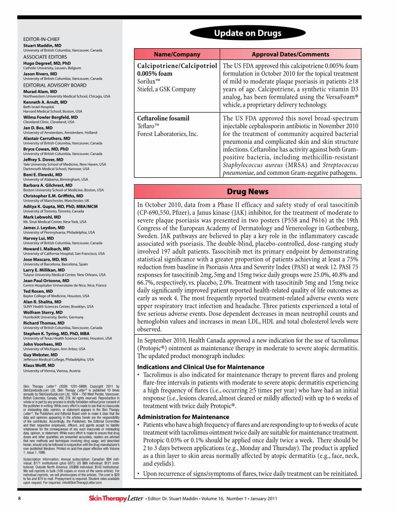
Name/Company Approval Dates/Comments
Calcipotriene/Calcipotriol 0.005% foamSorilux™Stiefel, a GSK Company
The US FDA approved this calcipotriene 0.005% foam formulation in October 2010 for the topical treatment of mild to moderate plaque psoriasis in patients ≥18 years of age. Calcipotriene, a synthetic vitamin D3 analog, has been formulated using the VersaFoam® vehicle, a proprietary delivery technology.
Ceftaroline fosamilTeflaro™ Forest Laboratories, Inc.
The US FDA approved this novel broad-spectrum injectable cephalosporin antibiotic in November 2010 for the treatment of community acquired bacterial pneumonia and complicated skin and skin structure infections. Ceftaroline has activity against both Gram-positive bacteria, including methicillin-resistant Staphylococcus aureus (MRSA) and Streptococcus pneumoniae, and common Gram-negative pathogens.
Drug News
In October 2010, data from a Phase II efficacy and safety study of oral tasocitinib (CP-690,550, Pfizer), a Janus kinase (JAK) inhibitor, for the treatment of moderate to severe plaque psoriasis was presented in two posters (P558 and P616) at the 19th Congress of the European Academy of Dermatology and Venereology in Gothenburg, Sweden. JAK pathways are believed to play a key role in the inflammatory cascade associated with psoriasis. The double-blind, placebo-controlled, dose-ranging study involved 197 adult patients. Tasocitinib met its primary endpoint by demonstrating statistical significance with a greater proportion of patients achieving at least a 75% reduction from baseline in Psoriasis Area and Severity Index (PASI) at week 12. PASI 75 responses for tasocitinib 2mg, 5mg and 15mg twice daily groups were 25.0%, 40.8% and 66.7%, respectively, vs. placebo, 2.0%. Treatment with tasocitinib 5mg and 15mg twice daily significantly improved patient reported health-related quality of life outcomes as early as week 4. The most frequently reported treatment-related adverse events were upper respiratory tract infection and headache. Three patients experienced a total of five serious adverse events. Dose dependent decreases in mean neutrophil counts and hemoglobin values and increases in mean LDL, HDL and total cholesterol levels were observed.
In September 2010, Health Canada approved a new indication for the use of tacrolimus (Protopic®) ointment as maintenance therapy in moderate to severe atopic dermatitis. The updated product monograph includes:
Indications and Clinical Use for Maintenance• Tacrolimus is also indicated for maintenance therapy to prevent flares and prolong
flare-free intervals in patients with moderate to severe atopic dermatitis experiencing a high frequency of flares (i.e., occurring ≥5 times per year) who have had an initial response (i.e., lesions cleared, almost cleared or mildly affected) with up to 6 weeks of treatment with twice daily Protopic®.
Administration for Maintenance• Patients who have a high frequency of flares and are responding to up to 6 weeks of acute
treatment with tacrolimus ointment twice daily are suitable for maintenance treatment. Protopic 0.03% or 0.1% should be applied once daily twice a week. There should be 2 to 3 days between applications (e.g., Monday and Thursday). The product is applied as a thin layer to skin areas normally affected by atopic dermatitis (e.g., face, neck, and eyelids).
• Upon recurrence of signs/symptoms of flares, twice daily treatment can be reinitiated.
EDITOR-IN-CHIEFStuart Maddin, MDUniversity of British Columbia, Vancouver, Canada
ASSOCIATE EDITORSHugo Degreef, MD, PhDCatholic University, Leuven, Belgium
Jason Rivers, MDUniversity of British Columbia, Vancouver, Canada
EDITORIAL ADVISORY BOARDMurad Alam, MDNorthwestern University Medical School, Chicago, USA
Kenneth A. Arndt, MDBeth Israel HospitalHarvard Medical School, Boston, USA
Wilma Fowler Bergfeld, MDCleveland Clinic, Cleveland, USA
Jan D. Bos, MDUniversity of Amsterdam, Amsterdam, Holland
Alastair Carruthers, MDUniversity of British Columbia, Vancouver, Canada
Bryce Cowan, MD, PhDUniversity of British Columbia, Vancouver, Canada
Jeffrey S. Dover, MDYale University School of Medicine, New Haven, USADartmouth Medical School, Hanover, USA
Boni E. Elewski, MDUniversity of Alabama, Birmingham, USA
Barbara A. Gilchrest, MDBoston University School of Medicine, Boston, USA
Christopher E.M. Griffiths, MDUniversity of Manchester, Manchester, UK
Aditya K. Gupta, MD, PhD, MBA/MCMUniversity of Toronto, Toronto, Canada
Mark Lebwohl, MDMt. Sinai Medical Center, New York, USA
James J. Leydon, MDUniversity of Pennsylvania, Philadelphia, USA
Harvey Lui, MDUniversity of British Columbia, Vancouver, Canada
Howard I. Maibach, MDUniversity of California Hospital, San Francisco, USA
Jose Mascaro, MD, MSUniversity of Barcelona, Barcelona, Spain
Larry E. Millikan, MDTulane University Medical Center, New Orleans, USA
Jean Paul Ortonne, MDCentre Hospitalier Universitaire de Nice, Nice, France
Ted Rosen, MDBaylor College of Medicine, Houston, USA
Alan R. Shalita, MDSUNY Health Sciences Center, Brooklyn, USA
Wolfram Sterry, MDHumboldt University, Berlin, Germany
Richard Thomas, MDUniversity of British Columbia, Vancouver, Canada
Stephen K. Tyring, MD, PhD, MBAUniversity of Texas Health Science Center, Houston, USA
John Voorhees, MDUniversity of Michigan, Ann Arbor, USA
Guy Webster, MDJefferson Medical College, Philadelphia, USA
Klaus Wolff, MDUniversity of Vienna, Vienna, Austria
Skin Therapy Letter © (ISSN 1201–5989) Copyright 2011 by SkinCareGuide.com Ltd. Skin Therapy Letter © is published 10 times annually by SkinCareGuide.com Ltd, 1004 – 750 West Pender, Vancouver, British Columbia, Canada, V6C 2T8. All rights reserved. Reproduction in whole or in part by any process is strictly forbidden without prior consent of the publisher in writing. While every effort is made to see that no inaccurate or misleading data, opinion, or statement appears in the Skin Therapy Letter ©, the Publishers and Editorial Board wish to make it clear that the data and opinions appearing in the articles herein are the responsibility of the contributor. Accordingly, the Publishers, the Editorial Committee and their respective employees, officers, and agents accept no liability whatsoever for the consequences of any such inaccurate or misleading data, opinion, or statement. While every effort is made to ensure that drug doses and other quantities are presented accurately, readers are advised that new methods and techniques involving drug usage, and described herein, should only be followed in conjunction with the drug manufacturer’s own published literature. Printed on acid-free paper effective with Volume 1, Issue 1, 1995.
Subscription Information. Annual subscription: Canadian $94 indi-vidual; $171 institutional (plus GST); US $66 individual; $121 insti-tutional. Outside North America: US$88 individual; $143 institutional. We sell reprints in bulk (100 copies or more of the same article). For individual reprints, we sell photocopies of the articles. The cost is $20 to fax and $15 to mail. Prepayment is required. Student rates available upon request. For inquiries: [email protected]
Update on Drugs

AAbobotulinumtoxinA 2:6Accutane™ 4:6Acne 3:1-2,5;8:10;10:1-4; 10:5-7;10:8Actinickeratoses 5:5-7;10:8Acyclovir+hydrocortisone 2:6Adalimumab 4:6Adapalene 7:8;8:10Agalsidasealfa 4:6Agingskin 7:5-9Alefacept 4:1-3Alitretinoin 1:8;2:7Alopecia 7:5-7Alphamelanocyte-stimulatinghormonereceptor 1:5-7Altabax® 1:1-4Altargo® 1:1-4Ambilify® 9:2Amevive® 4:1-3Anesthetics(local) 8:6Anti-androgens 10:2-3Antibacterial 1:1-4;10:5-7;10:8Antibioticresistance 1:1-4;10:5-7Antidepressants 8:7Antifungals 5:1-4;6:8Antihistamines 8:7Antioxidants 4:5Anxietydisorder 9:1-3Aripiprazole 9:2Asclera® 5:8Atopicdermatitis 1:1-4;8:10Azzalure® 2:6
BBacterialdecolonization 1:3Basalcellcarcinoma 1:5-7;7:1-4BCC 1:5-7;7:1-4Belimumab 7:8Benlysta® 7:8Benzoylperoxide(BP) 10:5-7Benzylalcohollotion 2:7Beyaz™ 10:8Biologics 4:1-3Biologics+traditionalagents 4:1-3Botulinumtoxin-typeA 2:6Butorphanol 2:2-3
CCalcineurininhibitors(topical) 2:1-3;5:2;8:6Calcitriol 2:6Cancer 1:8Cannabinoids 8:7Capsaicin 1:8;2:7;8:6Ceftarolinefasamil 10:8Cervarix® 2:7;9:8
Chemoprevention 7:1-4Chemotherapy 7:5-7Chronickidneydisease 2:1-5CKD 2:1-5Clindamycin 8:10;10:5-7Coaltar 5:2Collagenaseclostridiumhistolyticum 4:6Collagen-baseddermalfiller 2:6Corticosteroids 5:1-4;8:6Cryotherapy 5:5-6Curettage 5:5-6Cyclobutanepyrimidinedimmers 1:5
DDairyproducts 3:1-2,5Darunavir 2:7Delusionalparasitosis 9:1-3Deoxyribonucleicacid 1:5-7;2:8;9:4Dermatitisartefacta 9:1-3Dialysis 2:1-5Diclofenacsodiumgel 3:6;5:6Diet 3:1-2,5DNA 1:5-7;2:8;9:4Drospirenone+ethinylestradiol 6:8;10:8Drugdelivery 8:1-4;9:4-7Dysport™ 2:6
EEczema 1:1-4Eflornithinehydrochloride 7:8Enbrel® 2:6EpiCeram® 8:10Erythromycin 10:5-7Estradioltablet 2:7Etanercept 2:6Etravirine 2:7Eumelanin 1:5Evolence® 2:6
FFabrydisease 4:6Fieldtherapy 5:6-7Finasteride 10:3Flutamide 10:3
GGabapentin 2:3-4Gadolinium 9:6;10:8Gammalinolenicacid 2:2-3Gardasil® 2:7;4:6;8:10;9:8Gaucherdisease 4:6;9:8Genitalwarts 10:8Genomictesting 1:5-7Glycemicindex 3:1-2,5Glycemicload 3:1-2,5Golimumab 2:7
HHaircare 7:7Hairloss 7:5-7Herpeslabialis 2:6Hidradenitissuppurativa 5:6HIV/AIDS 2:7;9:8HPVvaccine 1:8;2:7;4:6;8:10;9:8Humanpapillomavirus 1:8;2:7;4:6;6:6;8:10;9:8Humira® 4:6Hyaluronicaciddermalfiller 2:6
IIL-12/IL-23 2:6IMGN901 6:8Imiquimod 2:6;5:5-7;5:8;10:8Impetigo 1:2Infrared(IRA) 4:4-5Insulinsensitizingagents 10:3Intelence® 2:7Intensepulsedlightsource 3:3-5Interleukin-12/interleukin-23 2:6Ipilimumab 7:8IPL 3:3-5IRA 4:4-5Isopropylmyristate 9:8Isotretinoin 4:6Istodax® 2:6Itch 2:1-5;7:5-9
JJuvéderm®XC 3:6
KK101 6:8K301 7:8Kaprolac® 6:8;7:8
LLasers 3:3-5Latanoprost 3:6Leukemia 1:8Levocetirizinedihydrochloride 2:6Levomefolatecalcium 10:8Lice 2:7;9:8Lipodissolve/lipotherapy 5:8Lipsovir® 2:6Lithiumsalts 5:2Lupuserythematosus 6:3;6:5;7:8Lymphoma 1:8
MMC1R 1:5-7Melanin 1:5Melanocortin1receptor 1:5-7Melanoma 1:5-7;7:8Menthol 8:6
Articles are indexed by drug names, trade names and disease terms. Bold entries refer to major references.
Key Word / Drug Name Issue #: Page # Key Word / Drug Name Issue #: Page # Key Word / Drug Name Issue #: Page #
Index for Volume 15
• Editor: Dr. Stuart Maddin • Index for Volume 15

Merkelcellcarcinoma 6:8Methicillin-resistantStaphylococcus aureus 1:2-3;10:8Metronidazole 5:2Microcyn® 2:7;6:8Minocyclinehydrochloride 8:10Minoxidil 7:5-7Moisturizers 8:6;8:10MRSA 1:2-3;10:8
NNalfurafine 2:2-3Naltrexone 2:2-3Nanodermatology 8:1-4;9:4-7Nanotechnology 8:1-4;9:4-7Nephrogenicsystemicfibrosis 9:6;10:8Neuroleptics 8:7-8NMSC 1:5-7;7:1-4Nonmelanomaskincancer 1:5-7;7:1-4
OOpioidagonists 2:1-5;8:7-8Opioidantagonists 2:1-5;8:7-8Oralcontraceptivepills 10:2-3;10:8Orap® 9:2Organtransplantation 7:1-4Osmolytes 4:5Oxychlorinecompound 2:7;6:8
PPCOS 10:1-4Pentoxyfilline 2:2-3Perlane®-L 3:6Photoaging 1:5-7;4:4-5Photodermatoses 6:1-3Photodynamictherapy 5:7Photopheresis 2:6Photoprotection 4:4-5;6:1-3Photosensitivitydisorders 6:1-3Phototherapy 8:8Pigmentation 1:5-7Pimecrolimus 5:2;8:6Pimozide 9:2PMLE 6:1-3Polidocanolinjection 5:8Polycysticovarysyndrome 10:1-4Poly-L-lacticacid 2:7Polymorphiclighteruption 6:1-3Prezista™ 2:7Propionibacteriumacnes 10:5-7Protopic® 2:1-3Pruritus 2:1-5;8:5-9Psoralen+UVA 1:5,7Psoriasis 1:8;4:1-3;6:5;8:10Psoriaticarthritis 2:7Psychocutaneousdisease 9:1-3Psychophysiologicdisorder 9:1-3PUVA 1:5,7;5:3
QQ-switchedlasers 3:3-5Quality-switched 3:3-5Qutenza™ 1:8;2:7
RReplagal® 4:6Restylane®-L 3:6Resultz® 9:8Retapamulin 1:1-4Retinoids(systemic) 7:1-4;7:8Risperdal® 9:2Risperidone 9:2Romidepsin 2:6
SSafety 9:4-7Salicylicacid 8:6Scalpcooling 7:5-7SCC 1:5-7;7:1-4SculptraAesthetic® 2:7Seborrheicdermatitis 5:1-4;7:8Secondarilyinfectedtraumaticlesions 1:2Seleniumsulphide 5:2Silkis™ 8:10Simponi™ 2:7Skincancer 1:5-7;5:5-7;6:5;7:1-4Smoking 5:4-7Solarkeratosis 1:6Solarurticaria 6:3Spironolactone 10:2-3Squamouscellcarcinoma 1:5-7;5:5-7;7:1-4Staphylococcus aureus 1:1-4;6:8Staphylococcus epidermidis 10:6Stelara™ 2:6Sunscreens 4:4-5;6:1-3;8:1-4;9:4-7
TTabaccosmoking 5:4-7Tacrolimus 2:1-3;5:2;8:6Tattoo 3:3-5Tattooremoval 3:3-5Televancin 2:6Tenofovirdisoproxilfumarate 9:8Thalidomide 2:3Therakos™ 2:6Titaniumdioxide 2:8;9:4-5Toctino® 1:8Toxicity 9:4-7Tretinoin 8:10Triclosan 6:8
UUlesfia™ 2:7Ultravioletphototherapy 2:3-4;5:2Ultravioletradiation 1:5-7;4:4-5;6:1-3Ustekinumab 2:6UVA 1:5-7;4:4-5;6:1-3;8:8UVB 1:5-7;4:4-5;5:2;6:1-3;8:8UVR 1:5-7
VVagifem® 2:7Valomaciclovir 2:8Vaniqa® 7:8Varicellazoster 2:8Vectical™ 2:6;8:10
Velaglucerasealfa 4:6;9:8Veltin™ 8:10Vericoseveins 5:8Vibativ™ 2:6Viread® 9:8VitaminD3 2:6;8:10Voltaren®gel 3:6Vpriv™ 4:6;9:8
XXerclear® 2:6Xerosis 7:5-9Xiaflex™ 4:6XYZAL® 2:6
YYasmin® 6:8Yaz® 6:8;10:8
ZZincpyrithione 5:2Zyclara™ 2:6;5:5-7;5:8
Key Word / Drug Name Issue #: Page # Key Word / Drug Name Issue #: Page # Key Word / Drug Name Issue #: Page #
• Editor: Dr. Stuart Maddin • Index for Volume 15