CROSSFIRE: Controversies in Neuromuscular and ... · CROSSFIRE: Controversies in Neuromuscular and...
64
2006 COURSE F AANEM 53 rd Annual Meeting Washington, DC Copyright © October 2006 American Association of Neuromuscular & Electrodiagnostic Medicine 2621 Superior Drive NW Rochester, MN 55901 PRINTED BY JOHNSON PRINTING COMPANY, INC. Lawrence R. Robinson, MD Ernest W. Johnson, MD Jun Kimura, MD Morris A Fisher, MD Steve R. Geiringer, MD Francis P. Lagattuta, MD CROSSFIRE: Controversies in Neuromuscular and Electrodiagnostic Medicine
Transcript of CROSSFIRE: Controversies in Neuromuscular and ... · CROSSFIRE: Controversies in Neuromuscular and...

2006 COURSE F AANEM 53rd Annual Meeting
Washington, DC
Copyright © October 2006 American Association of Neuromuscular & Electrodiagnostic Medicine
2621 Superior Drive NW Rochester, MN 55901
Printed by Johnson Printing ComPany, inC.
Lawrence R. Robinson, MD
Ernest W. Johnson, MD
Jun Kimura, MD
Morris A Fisher, MD
Steve R. Geiringer, MD
Francis P. Lagattuta, MD
CROSSFIRE: Controversies in Neuromuscular and Electrodiagnostic Medicine

CROSSFIRE: Controversies in Neuromuscular and Electrodiagnostic Medicine
Faculty
Lawrence R. Robinson, MDProfessorDepartment of Rehabilitation MedicineVice Dean, Clinical AffairsUniversity of WashingtonSeattle, WashingtonDr. Robinson attended Baylor College of Medicine and completed his resi-dency training in rehabilitation medicine at the Rehabilitation Institute of Chicago. He now serves as professor in the Department of Rehabilitation Medicine at the University of Washington and is the Director of the Harborview Medical Center Electrodiagnostic Laboratory. He is also currently Vice Dean for Clinical Affairs at the University of Washington. His current clinical interests include the statistical interpretation of elec-trophysiologic data, laryngeal electromyography, and the study of trau-matic neuropathies. He recently received the Distinguished Academician Award from the Association of Academic Physiatrists and the AANEM Distinguished Researcher Award.
Ernest W. Johnson, MDEmeritus ProfessorDepartment of Physical Medicine and RehabilitationOhio State UniversityColumbus, OhioDr. Johnson received his medical degree from The Ohio State University in Columbus, Ohio, interned at Philadelphia General Hospital, and completed his residency in physical medicine and rehabilitation at Ohio State University under the sponsorship of the National Foundation of Infantile Paralysis. He has edited the textbook Practical EMG, and au-thored over 143 peer-reviewed articles. He established the Super EMG continuing medical education course in 1978, and is still involved in planning and teaching this course. Currently, Dr. Johnson is an emeritus professor at Ohio State University. He has conducted research on electro-diagnostic medicine in recurrent carpal tunnel syndrome and the use of H waves in upper limb radiculopathies. Dr. Johnson is a past-president of the AANEM, AAPM&R, AAP, former chair of the American Board of Electrodiagnostic Medicine, and has been editor of the American Journal of Physical Medicine and Rehabilitation.
Jun Kimura, MDProfessor Department of NeurologyUniversity of Iowa Hospitals and ClinicsIowa City, IowaDr. Kimura received his Bachelor of Technology in 1957 and MD in 1967 from Kyoto University in Japan. He came to the United States as a Fulbright scholar in 1962 for his neurology residency and electrophysiol-ogy fellowships at the University of Iowa. He taught at the University of Manitoba in Canada, the University of Iowa, and Kyoto University in Japan. He has served as the AAEM’s secretary-treasurer, president of the AAEM, and editor of Muscle & Nerve. Dr. Kimura received the AAEM’s Distinguished Researcher Award in 1995 and Lifetime Achievement Award in 1999. He published the 3rd edition of Electrodiagnosis in Diseases of Nerve and Muscle in 2001. He holds an honorary membership in ap-proximately 20 national societies of neurology and neurophysiology. His current professional titles include Professor Emeritus at Kyoto University, and Professor of Neurology at the University of Iowa.
Morris A. Fisher, MDProfessorDepartment of NeurologyLoyola University Stritch School of MedicineMaywood, IllinoisDr. Fisher is a graduate of Harvard Medical School. He completed his neurology training as well as a fellowship in clinical neurophysiology at Massachusetts General Hospital in Boston. For the past 14 years, he has been a professor of neurology at Loyola University Chicago Stritch School of Medicine in Mayowood, Illinois, and an attending neurologist at the Hines VA Hospital in Hines, Illinois. His current positions include Director of the Neuromuscular Program at Loyola University Medical Center as well as Director of the Clinical Neurophysiology Laboratories at the Hines VA Hospital. Dr. Fisher has been a member of the AANEM since 1975 and has served as a member of the AANEM Board of Directors. He has also been on the Board of Directors and served as President of the American Academy of Clinical Neurophsiology. Dr. Fisher has an ongoing interest in F waves. He also funded research in neuropathies including the effect of exercise in diabetic neuropathies and investigations into the pathogenesis of immunologically mediated neuropathies.
Course Chair: Jeffrey A. Strakowski, MD
The ideas and opinions expressed in this publication are solely those of the specific authors and do not necessarily represent those of the AANEM.
Dr. Fisher is a consultant for NeuroMetrix. All other authors had nothing to disclose.
F-ii
Steve R. Geiringer, MDProfessorDepartment of Physical Medicine and RehabilitationWayne State UniversityDetroit, MichiganDr. Geiringer completed undergraduate, medical school, and residency training at the University of Michigan in Ann Arbor, Michigan. He then spent over 8 years on the faculty at the same facility in the department of Physical Medicine and Rehabilitation. In 1991, Dr. Geiringer joined the faculty at Wayne State University in Detroit, Michigan, gaining the rank of Professor while there. Dr. Geiringer remains on Wayne State’s faculty as Professor, although currently is in a solo private practice of PM&R. His scholarly contributions include over 20 articles in peer-reviewed journals, and nearly 20 books and book chapters. Dr. Geiringer’s handbook on anatomy relevant to EMG is now the standard in the field, and has been widely translated internationally. He is currently an associate editor for the American Journal of PM&R, and in 2000 he was elected a director of the American Board of PM&R.
Francis P. Lagattuta, MDDirectorLAGS Spine and SportscareSanta Maria, CaliforniaDr. Francis Lagattuta is one of the pioneer pain interventionalists. He is board-certified in pain medicine, physical medicine and rehabilitation, and electrodiagnostic medicine. He originally had a practice in the western suburbs of Chicago. While in Chicago, he was involved in sports medicine and was the team doctor for the Naperville and Wheaton high schools, and the Chicago Bulls. In 1998, Dr. Lagatutta moved to the Central Coast of California. He now has offices in Santa Barbara, Santa Maria, Atascadero, and Lompoc with a surgical center in Santa Maria. He is the Current Procedural Terminology (CPT) representative for the American Academy of Physical Medicine and Rehabilitation (AAPMR) from 2004 to present. He was the CPT representative for the North American Spine Society (NASS) from 2000-2004. Dr. Lagattuta has been a board member for the American Academy of Neurodiagnostic Medicine and the Physiatrist Association of Spine, Sports Medicine, and Occupational Medicine in addition to serving on multiple committees for AANEM, AAPMR, and NASS. He has written numerous articles and given many national presentations.
F-iii
Please be aware that some of the medical devices or pharmaceuticals discussed in this handout may not be cleared by the FDA or cleared by the FDA for the specific use described by the authors and are “off-label” (i.e., a use not described on the product’s label). “Off-label” devices or pharmaceuticals may be used if, in the judgement of the treating physician, such use is medically indicated to treat a patient’s condition. Information regarding the FDA clearance status of a particular device or pharmaceutical may be obtained by reading the product’s package labeling, by contacting a sales representative or legal counsel of the manufacturer of the device or pharmaceutical, or by contacting the FDA at 1-800-638-2041.

F-iv AANEMCourse

CROSSFIRE: Controversies in Neuromuscular and Electrodiagnostic Medicine
Contents
Faculty ii
Objectives iii
CourseCommittee iv
Controversies in Electrodiagnosis of Ulnar Neuropathy 1LawrenceR.Robinson,MD
Controversies in Electrodiagnosis of Ulnar Neuropathy 7ErnestW.Johnson,MD
F Waves are Overutilized in Radiculopathy 11JunKimura,MD
F Waves are Underutilized in Radiculopathy 15MorrisA.Fisher,MD
Epidural Steroid Injections are Overutilized 21SteveR.Geiringer,MD
Epidural Steroid Injections are Underutilized 27FrancisP.Lagattuta,MD
CMESelf-AssessmentTest 35
Evaluation 39
Ob j e c t i v e s—Attending this course will provide participants the opportunity to discuss (1) controversies surrounding electrodiagnosis of ulnar neuropathies, (2) whether F waves are over- or underutilized in radiculopathy, and (3) whether epidural steroid injections are over- or underutilized.
Pr e r e q u i s i t e—This course is designed as an educational opportunity for residents, fellows, and practicing clinical EDX physicians at an early point in their career, or for more senior EDX practitioners who are seeking a pragmatic review of basic clinical and EDX principles. It is open only to persons with an MD, DO, DVM, DDS, or foreign equivalent degree.
Ac c r e d i tAt i O n stAt e m e n t—The AANEM is accredited by the Accreditation Council for Continuing Medical Education to provide continuing medical education (CME) for physicians.
cme cr e d i t—The AANEM designates this activity for a maximum of 3.25 hours in AMA PRA Category 1 Credit(s)TM. This educational event is approved as an Accredited Group Learning Activity under Section 1 of the Framework of Continuing Professional Development (CPD) options for the Maintenance of Certification Program of the Royal College of Physicians and Surgeons of Canada. Each physician should claim only those hours of credit he or she actually spent in the educational activity. CME for this course is avail-able 10/06 - 10/09.
F-v
F-vi
ThomasHyattBrannagan,III,MDNew York, New York
HopeS.Hacker,MDSan Antonio, Texas
KimberlyS.Kenton,MDMaywood, Illinois
DaleJ.Lange,MD New York, New York
SubhadraNori,MDBronx, New York
JeremyM.Shefner,MD,PhDSyracuse, New York
T.DarrellThomas,MDKnoxville, Tennessee
BryanTsao,MDShaker Heights, Ohio
2005-2006 AANEM PRESIDENT
JaniceM.Massey,MDDurham, North Carolina
2005-2006 AANEM COURSE COMMITTEE
KathleenD.Kennelly,MD,PhDJacksonville, Florida

Controversies in Electrodiagnosis of Ulnar Neuropathy
Lawrence R. Robinson, MDProfessor
DepartmentofRehabilitationMedicineViceDean,ClinicalAffairs
UniversityofWashingtonSchoolofMedicineSeattle,Washington
INTRODUCTION
Ulnar neuropathy is the second most common entrapment neu-ropathy in the upper limbs. Assessment of the ulnar nerve can, however, be problematic from many perspectives. This manuscript will review (1) the best way to perform motor nerve conduction studies (NCSs); (2) when to consider Martin-Gruber anastomo-sis (MGA) and how to test for it; (3) when and how to perform inching studies; (4) how to perform and interpret sensory con-duction studies; and (5) whether or not needle electromyography (EMG) should be performed, and how the results should be inter-preted.
THE BEST WAY TO PERFORM MOTOR CONDUCTION STUDIES
What is the best way to perform motor conduction studies? Motor NCSs are a good way to begin assessment for possible ulnar neuropathy at the elbow (UNE). This is usually the most useful technique for localizing the site of UNE and determining the pathophysiology of the lesion.
BEST RECORDINg SITES
While the abductor digiti minimi (ADM) is the most common re-cording site, it is preferable to record from two muscles. Recording from the first dorsal interosseus (FDI) muscle, the most distal muscle supplied by the ulnar nerve can reveal abnormalities missed
by recording from only theADM.5,12,14 While the overall sensitivity of the ADM and FDI are comparable, there is incomplete overlap in cases identified by either recording technique—i.e., some cases are identified only by ADM recording and others only by FDI recording.
The physician can use a two-channel technique to record from both muscles simultaneously so that extra stimulations are not required. The recording site for the FDI is usually described as the active electrode over the bulk of the muscle, with the reference distally over the metacarpal joint of the index finger. Such a recording arrangement often produces an initial positive deflection, which is difficult to interpret. An initial negative deflection is more com-monly seen when the reference is placed over the carpo-metacarpal joint of the thumb, therefore this is the preferred technique.
Stimulation Sites
Stimulation is usually performed at the wrist, below the elbow, above the elbow, and at the axilla. While some electrodiagnostic (EDX) physicians do not routinely stimulate at the axilla, the advantage of this technique is that it offers a conduction velocity (CV) across one more segment (the arm), which can be compared with the across-elbow CV. Study of the across-elbow segment re-quires great care in technique and interpretation. The position of the elbow greatly influences the measured CV. When the elbow is extended, it is thought that the ulnar nerve may become loose or redoubled in the ulnar groove, and that surface measurements do not reflect the true distance of the underlying nerve. Flexing the elbow stretches the nerve to its full length and measurement of the

F-� Controversies in Electrodiagnosis of Ulnar Neuropathy AANEMCourse
distance over the ulnar groove more closely reflects the distance along the nerve.1,3
The distance between above- and below-elbow stimulation sites may also influence the accuracy of the CV measurement. Since surface measurements can be in error by many millimeters, use of short distances between stimulation sites means that there will be a relatively large percentage of error in the distance and hence CV measurements. Many EDX physicians recommend using at least a 10-cm across elbow distance to reduce this measurement error.10 The rationale has been that the addition of errors in dis-tance and latency measures produces unacceptable margins of error when shorter distances are used. However, the original study that produced the 10-cm estimate used older technology for latency measurements (oscilliscopes in the predigital era). Recent data indicates that only 6 cm might be needed with the improved ac-curacy of today’s EDX instruments.9 The shorter distance has the advantage of not diluting focal slowing by long distances of normal conduction.
How to Interpret Conduction Velocities
How much slowing in the across-elbow segment is sufficient to diagnose UNE? Some EDX physicians compare the across-elbow velocity to the forearm velocity, allowing up to 11-15 ms differ-ence between the across-elbow and forearm segments before calling the finding abnormal.8 However, comparison with the forearm segment assumes that the forearm segment remains normal in UNE. But, as axon loss progresses and faster conducting fibers are lost, the distal velocity often slows, making the comparison to the forearm segment less useful.
This author prefers to use the absolute CV rather than a compari-son between segments.12,15 A recent study has suggested that absolute velocities of less than 48 m/s are suggestive of UNE and that this is superior to comparison with the forearm velocity13 (Table 1). As axon loss progresses and compound muscle action potential (CMAP) is reduced in size, the utility of comparing the across elbow with the forearm segments is diminished13 (Table 2).
Slowed CV is not the only finding that should be considered diag-nostic of UNE. Such patients may also have a drop in amplitude in the across-elbow segment or increased temporal dispersion. Some authors state that an amplitude reduction of more than 10% in the across-elbow 10-cm segment may be abnormal,8 but this is more convincing if accompanied by focal slowing or temporal disper-sion.
Most of the abnormalities seen on NCSs require the presence of demyelination for localization. However, in many traumatic ulnar
Table 1 MotorConductionVelocity:Sensitivityandspecificityatvariedlimitsofnormal.
Reference Specificity Sensitivity* Area under ROC Value Curve+ADMCV 48 95% 80%(76%-84%) 0.94(0.89-0.99)FDICV 49 95% 77%(7�%-8�%) 0.90(0.85-0.94)ADMCVdiff 10 95% 51%(46%-56%) 0.81(0.75-0.87)FDICVdiff 1� 95% 38%(33%-43%) 0.76(0.69-0.8�)*The95%confidenceintervalswerecalculatedforallsensitivitiesandpresentedinparenthesis.+Theareaunderthecurverepresentsoveralltestaccuracy,ortheratioofthenumberofsubjectscorrectlyclassifiedbythetestasnormalorabnormaloverthetotalnumberofsubjects.
ADMCV–conductionvelocity(m/s)betweenaboveandbelowelbowsites(ADM)FDICV–conductionvelocity(m/s)betweenaboveandbelowelbowsites(FDI)ADMCVdiff–difference(m/s)betweenacrosselbowandforearmsegments(ADM)FDICVdiff–difference(m/s)betweenacrosselbowandforearmsegments(FDI)
ADM=abductordigitiminimi;CV=conductionvelocity;FDI=firstdorsalinteros-seus.
Table 2 The Influence of compound muscle action potentialamplitudeonsensitivityofusingconductionvelocity(CV)acrosstheelbowandCVdifferencebetweenacrosselbowandforearmsegments(CV-DIFF).
Recording CMAP CV CV DIFF Site Amplitude Sensitivity* Sensitivity*ADM <4mV 85%(77%-93%) 30%(�0%-40%)ADM 4-10mV 78%(7�%-84%) 61%(53%-69%)ADM >10mV 71%(6�%-80%) 50%(40%-60%)FDI <3mV 83%(75%-91%) 4�%(3�%-5�%)FDI 3-11mV 77%(71%-83%) 37%(30%-44%)FDI >11mV 67%(56%-78%) 33%(��%-44%)
*Thesensitivitiespresentedareforlimitsofnormalthatgiveaspecificityof95%.The95%confidenceintervalsareshowninparenthesis.
ADM=abductordigitiminimi;CMAP=compoundmuscleactionpotential;CV=conductionvelocity;CV-DIFF=conductionvelocitydifference;FDI=firstdorsalinterosseus.

AANEMCourse CROSSFIRE:ControversiesinNeuromuscularandElectrodiagnosticMedicine F-3
neuropathies in which there is only axon loss without demyelin-ation, localization of ulnar neuropathy is far more difficult. In such cases, there will be diffuse mild slowing of CV (affecting all segments) without focal slowing, conduction block, or temporal dispersion; thus there are no focal nerve conduction changes across the lesion.
WHEN TO CONSIDER MARTIN-gRUBER ANASTOMOSIS AND HOW TO LOOK FOR IT
Martin-Gruber anastomosis is often discussed in the context of median NCSs, yet it is far more important to recognize this anomaly when performing ulnar NCSs. It is here that a false-posi-tive diagnosis can be made. When present, this anomaly will result in a much lower amplitude response with below-elbow stimulation compared to amplitude obtained with wrist stimulation, simulating conduction block. The FDI is the muscle most commonly affected by MGA, and it is not uncommon to see a marked reduction in CMAP amplitude in the FDI, but not the ADM (Figure 1).
When faced with a drop in CMAP amplitude between wrist and elbow, the inexperienced EDX physician may suspect a focal ulnar neuropathy in the proximal forearm which could even be “con-firmed” by inching studies along the ulnar nerve and across the anastomosis. However, in all such cases, the presence of an MGA can and should be ruled out very simply by stimulating the median nerve at the elbow and recording over the ADM and FDI muscles. Presence of any significant response with an initial negative takeoff indicates the presence of the anomaly.
PERFORMINg INCHINg STUDIES
Although previously stated that a 10 cm minimum distance across the elbow is recommended for CV measurements, study of very short segments yields a higher sensitivity for very focal lesions. With short-segment studies, the injured segment with demyelin-ation occupies a higher percentage of the distance studied when compared to longer segments in which normal nerve dilutes the measurement. Inching studies (or perhaps more appropriately called “centimetering” studies) can be performed by stimulating the nerve at 2-cm increments across the elbow.6,11 Landmarks are best established by drawing a line between the medial epicondyle and the olecranon,2 and measuring 2-cm increments distal and proxi-mal to this line. Stimulation must be carefully performed at just barely supramaximal since overstimulation may cause nerve activa-tion distal to the cathode and potentially distal to a lesion. Using this technique, a conduction delay of more than 0.7 ms across 2-cm segments is probably abnormal.6 Focal changes in amplitude or waveform morphology across a segment are more impressive than a latency change alone (Figure 2).
PERFORMINg AND INTERPRETINg SENSORY CONDUCTION STUDIESSensory NCSs are often of less localizing value than motor NCSs. There are several technical problems that make the responses seen with sensory NCSs more difficult to interpret. First, with stimula-tion of the ulnar nerve and antidromic recording over the little finger, there is often a large-amplitude hypothenar motor response volume conducted to the recording electrodes which precludes accurate identification and measurement of the sensory nerve action potential (SNAP). Second, due to phase cancellation,7 the amplitudes of the sensory responses fall dramatically over distance, and reductions of 50% or more are not unusual or abnormal in the wrist-to-elbow segment. Third, it is much harder to record sensory responses, particularly with proximal stimulation, when their amplitudes are reduced by significant ulnar neuropathy with temporal dispersion.
Nevertheless, sensory responses are often helpful for measuring the degree of sensory axon loss. Reduction in the amplitude of the ulnar SNAP after distal stimulation is probably one of the more sensitive indicators of the UNE.4 Of course, a low amplitude sensory response in the wrist to little finger segment is not local-izing and simply means that there has been sensory axon loss at or distal to the dorsal root ganglion at C8.
Measurement of SNAPs may be helpful to exclude lesions other than UNE. When attempting to distinguish UNE from ulnar neu-ropathy at the wrist, measurement of the dorsal ulnar cutaneous sensory response can be helpful. This nerve is involved with lesions at the elbow, but not at the wrist (where it bypasses Guyon’s canal). When this response it normal and symmetrical it is more indicative of ulnar neuropathy at the wrist. When the lesion is at the elbow, the dorsal ulnar cutaneous response is typically reduced in ampli-tude or absent. Similarly, the medial antebrachial cutaneous nerve (MABCN) can be studied to rule out more proximal lesions such as a lower brachial plexus lesion. Lower plexus lesions would be expected to have a small amplitude or absent MABCN response, whereas in ulnar neuropathies this nerve should be spared.
PERFORMINg NEEDLE ELECTROMYOgRAPHY AND INTERPRETINg RESULTS
Needle EMG of ulnar-innervated muscles is important, both to determine whether or not any axon loss has occurred, and to help localize lesions that are purely axonal in nature. Thus, even if NCSs are entirely normal, when ulnar neuropathy is clinically suspected, needle EMG should still be performed. The most helpful hand muscles to assess are the ADM and FDI, two muscles commonly involved in UNE.5 Study of the flexor carpi ulnaris (FCU) and ulnar half of the flexor digitorum profundus (FDP) is marginally helpful. Although the branch to these muscles usually comes off

Figure 1 UlnarmotorconductionstudiesrecordingfromasubjectwithMartin-Gruberanastamosis.ThetopfourtracesarerecordedfromADM,andthebottomfourfromFDI.Notethelargedropinamplitudebetweenwristandbelow-elbowwhenrecordingatFDI.
ADM=abductordigitiminimi;FDI=firstdorsalinterosseus.
F-4 Controversies in Electrodiagnosis of Ulnar Neuropathy AANEMCourse

distal to most entrapment sites at the elbow, the fascicles supplying these muscles are in a relatively protected position within the nerve and these muscles are consequently often spared.
Needle EMG of non-ulnar-innervated muscles is often useful to rule out other lesions that may mimic ulnar neuropathy. Examination of thenar muscles or the extensor indicis proprius offers the opportu-nity to compare C8 - T1 muscles not innervated by the ulnar nerve. This can be useful to rule out lower cervical radiculopathies as well as lower brachial plexopathies. When interpreting abnormalities in the FDI muscle, it should be remembered that this is the muscle most commonly innervated by the MGA. Moreover, in many cases of MGA, the anomalous branch is derived from the anterior inter-
osseus nerve.16 Thus, median neuropathy or anterior interosseus nerve syndrome should be considered when evidence of axon loss is found in the FDI and the clinical presentation is not typical of ulnar neuropathy.
SUMMARY
The diagnosis of UNE is potentially complex. The EDX consultant will want to thoughtfully plan out the approach before seeing the patient. There are many technical details to be mindful of and in-terpretation of results can be challenging. Despite these challenges,
Figure 2 Inchingstudiesinapatientwithfocalulnarneuropathy.Notethefocalchangeinlatencyandamplitude.
AbdDigMin=abductordigitiminimi
AANEMCourse CROSSFIRE:ControversiesinNeuromuscularandElectrodiagnosticMedicine F-5

in many cases, one can accurately diagnose and localize ulnar neu-ropathy in the elbow region.
REFERENCES
1. Bielawski M, Hallett M. Position of the elbow in determination of abnormal motor conduction of the ulnar nerve across the elbow. Muscle Nerve 1989;12:803-809.
2. Campbell WW, Pridgeon RM, Sahni KS. Short segment incremental studies in the evaluation of ulnar neuropathy at the elbow. Muscle Nerve 1992;15:1050-1054.
3. Checkles NS, Russakov AD, Piero DL. Ulnar nerve conduction velocity--effect of elbow position on measurement. Arch Phys Med Rehabil 1971;52:362-365.
4. Eisen A. Early diagnosis of ulnar nerve palsy. An electrophysiologic study. Neurology 1974;24:256-262.
5. Jabre JF, Wilbourn AJ. The EMG findings in 100 consecutive ulnar neuropathies. Acta Neurol Scand 1979;60:S73-S91.
6. Kanakamedala RV, Simons DG, Porter RW, Zucker RS. Ulnar nerve entrapment at the elbow localized by short segment stimulation. Arch Phys Med Rehabil 1988;69:959-963.
7. Kimura J, Machida M, Ishida T, Yamada T, Rodnitzky RL, Kudo Y, Suzuki S. Relation between size of compound sensory or
muscle action potentials, and length of nerve segment. Neurology 1986;36:647-652.
8. Kincaid JC. AAEE Minimonograph #31: The electrodiagnosis of ulnar neuropathy at the elbow. Muscle Nerve 1988;11:1005-1015.
9. Landau ME, Barner KC, Campbell WW. Optimal screening distance for ulnar neuropathy at the elbow. Muscle Nerve 2003;27:570-574.
10. Maynard FM, Stolov WC. Experimental error in determination of nerve conduction velocity. Arch Phys Med Rehabil 1972;53:362-372.
11. Miller RG. The cubital tunnel syndrome: diagnosis and precise local-ization. Ann Neurol 1979;6:56-59.
12. Payan J. Electrophysiological localization of ulnar nerve lesions. J Neurol Neurosurg Psychiatry 1969;32:208-220.
13. Shakir A, Micklesen PJ, Robinson LR. Which motor nerve conduc-tion study is best in ulnar neuropathy at the elbow? Muscle Nerve 2004;29:585-590.
14. Stewart JD. The variable clinical manifestations of ulnar neuropathies at the elbow. J Neurol Neurosurg Psychiatry 1987;50:252-258.
15. Tackmann W, Vogel P, Kaeser HE, Ettlin T. Sensitivity and localizing significance of motor and sensory electroneurographic parameters in the diagnosis of ulnar nerve lesions at the elbow. A reappraisal. J Neurol 1984;231:204-211.
16. Wertsch JJ. AAEM Case Report #25: Anterior interosseous nerve syndrome. Muscle Nerve 1992;15:977-983.
F-6 Controversies in Electrodiagnosis of Ulnar Neuropathy AANEMCourse

INTRODUCTION
Entrapment at the wrist has been reported and well-studied.5,12
Controversy remains, however, about the vulnerability around the ulnar nerve at the elbow. There are many studies to evaluate pos-sible compromise of the ulnar nerve at the elbow (UNE), but pos-sibilities are manifold for mistakes and misinterpretation.1,7,8,11,17
ANATOMIC AND OTHER CONSIDERATIONS
When discussing the UNE, it is important to understand the ana-tomical considerations and other considerations. Potential sites of compromise, subluxation or dislocation of the ulnar nerve, and the differences between men and women are important to consider. The potential sites of compromise include pressure on the ulnar nerve as it courses near the medial humeral epicondyle, distal as it enters the cubital tunnel, and proximal to the medial condyle in a fascial loop often referred to as the “arcade of Struthers” (Figures 1 and 2).18 There has been controversy about the arcade of Struthers. Some argue that it is historically incorrect as named, yet it is present 3-10 cm proximal to the medial humeral epicondyle in 13% of individuals. Anatomic dissection clearly shows the potential vulner-ability of the ulnar nerve as it lies in the medial femoral epicondyle and then proceeds through the cubital tunnel into the forearm.6
Further vulnerability of the ulnar nerve occurs in approximately 20% of individuals who have subluxation or dislocation of the ulnar nerve when the elbow is flexed,8 which makes the nerve more subject to pressure and injury. Tardy ulnar nerve compromise
occurs 10-25 years following fractures at the elbow—usually in the supracondylar area. This is best diagnosed with nerve conduction studies (NCSs) proximal and distal to the fracture site.
There are significant differences between men and women regard-ing the presentation of UNE. Contreras and associates noted that pressure on the nerve is caused by the tubercle of the coronoid process. This is 1.5 times larger in men than in women. Also, the subcutaneous fat overlying the tubercle is thicker in women. Both of these factors contribute to a higher incidence of UNE in men.6
ELECTRODIAgNOSTIC TECHNIQUES
Nerve conduction studies are used to study UNE. The importance of elbow position during the performance of NCSs across the elbow was first demonstrated over 30 years ago3 (Figure 3). Standard elec-trodiagnosis of ulnar nerve entrapment at the elbow has tradition-ally consisted of measuring motor conduction velocity (CV) across the elbow compared with motor CV in the forearm. More recently, short segment stimulation of the ulnar nerve across the elbow has been shown to identify conduction block and slowing at a specific site.7,9,10 Short-segment stimulation is performed by measuring 1 or 2 cm marks across the elbow (or by using a pre-formed set of stimulating electrodes spaced in a holder) and then recording the compound muscle action potential (CMAP) of the abductor digiti quinti. The CMAP amplitude can be noted as well as the CV calculated. The CV is expected to be faster as one proceeds proxi-mally. If the ulnar nerve is compromised at the elbow, there will be
Controversies in Electrodiagnosis of Ulnar Neuropathy
Ernest W. Johnson, MDProfessorEmeritus
DepartmentofPhysicalMedicineandRehabilitationOhioStateUniversity
Columbus,Ohio
F-7

slowing across that segment and a reduction in CMAP amplitude (conduction block).7
Some have suggested that short segment evaluation of ulnar nerve at the elbow can be compromised by the lower temperature of the nerve in its superficial course.11 In a novel study by Merlevede and associates,14 it was suggested that the latency of the compound nerve action potential (CNAP) could bypass the exact position of the nerve and avoid the complications of movement as the elbow was flexed. This variation in measurements introduced errors, something re-emphasized by Kim and colleagues in 2005.8
To identify UNE, the ulnar and median nerves are stimulated at the wrist and percutaneously. The CNAP of each nerve is then re-corded 10 cm above the elbow and compared. Differences in laten-cies in normal subjects is 1.4 ms. In classic instances of UNE, there is a difference greater than 1.4 ms or the absence of ulnar CNAP. One explanation could be the proximity of the ulnar and median nerves 10 cm above the medial epicondyle, thus causing confusion about which nerve is being recorded. Unfortunately, the location of the compromise can not be determined, either.
F-8 Controversies in Electrodiagnosis of Ulnar Neuropathy AANEMCourse
Figure 1 Superficialdissectionofanteriorforearm(Netter).
Figure 2 Deepdissectionofanteriorforearm(Netter).
Figure 3 Withelbowextendedconduction velocity (CV) is 40m/swhilewithelbowflexedtheCVis58m/s.

Rarely have investigators used the SNAP amplitude to determine UNE. The difficulties with phase cancellation may be an obstacle to using SNAP amplitude. In 2001, Hermann and colleagues7 used the SNAP to indicate conduction block at the elbow, but did not use the amplitude variation diagnostically. In their 1-cm segments of stimulation they indicated a 20% reduction in CMAP amplitude or a .5 ms latency shift over 1 cm distance as conduction block. They also considered a SNAP amplitude (antidromic of digit 5) of below 10 µV or absent as an indication of conduction block. This study found that the CMAP at distal stimulation was reduced in only 15% of the patients, but the SNAP was reduced in 38%, pre-sumably suggesting greater vulnerability of the sensory fibers.
Wongsam and colleagues20 studied the SNAP amplitude and laten-cies of the median nerve in carpal tunnel syndrome at 14 cm and 7 cm. Although the nerve is in its distal distribution, the results would suggest that .2 ms per centimeter and 4-5 µV per centimeter could be used as changes reflecting phase cancellation. This author and colleagues performed NCSs on patients with ulnar nerve vul-nerability and normal subjects to validate this observation (Figures 4-6). The stimulation technique was modified to use a monopolar needle as the cathode and a ground electrode as the anode, then the antidromic SNAP was recorded over digit 5. This provided a more exact localization of the stimulation. The SNAP was recorded with surface electrodes on the little finger separated by 4 cm.
TREATMENT
Some courses of treatment for UNE include modifying the place-ment of a flexed elbow and avoiding pressure across the ulnar nerve. The patient could be fitted for an elbow pad, or in the case of a basketball player, an elbow brace. Surgical approaches are possible
but should be used with caution. Relocation of the ulnar nerve has been the most common operative approach, but the many studies to assess the outcome have been varied and report uneven results.
Paternostro-Sluga and colleagues16 investigated post operative, subcutaneously transferred ulnar nerve at the elbow with inching technique and reported that a focal increase in latency did not show a lack of improvement, but was “a clinically irrelevant deficit.” In contrast, only a sharply localized amplitude reduction was signifi-cant in ongoing nerve compromise. This reinforced the importance of amplitude in diagnosing UNE.
AANEMCourse CROSSFIRE:ControversiesinNeuromuscularandElectrodiagnosticMedicine F-9
Figure 4 Noteulnarnervedislocationwhenelbowisflexed.
Figure 5 Note theulnarsensorynerveactionpotentialaboveandbelow elbow (6 cm) drop from 18 µV to 6 µV (motor artifact is largepotential).
Figure 6 Ulnarsensorynerveactionpotentialaboveandbelowelbow.(10cm;�ms;10µV).(Shotatangletoremoveflash.)

Nathan and associates15 reported NCSs on 102 patients with simple decompression of cubital tunnel syndrome and noted that the relief of symptoms did not correlate with conduction across the elbow. Thus, 74 of these postoperative patients, although reporting excellent results, still had slowing at the elbow.
This author’s own clinical experience over several years dictates caution about operative intervention in UNE, especially in diabetic patients.
SUMMARY
Ulnar neuropathy is a common problem facing the electrodiagnos-tic physician. The more exact techniques of needle electromyog-raphy (EMG) and SNAP amplitudes should be used rather than CMAPs and slowing at the elbow. Most of the literature does not support the accuracy of needle EMG for estimating the prognosis and severity of the nerve entrapment. It is therefore important to evaluate UNE using the needle stimulating and SNAP recording techniques described above.
REFERENCES
1. Campbell W. Ulnar neuropathy at the elbow. Muscle Nerve 2000;23:450-452.
2. Campbell WW, Pridgeon RM, Riaz G, Astruc J, Leahy M, Crostic EG. Sparing of flexor carpi ulnaris in ulnar neuropathy at elbow. Muscle Nerve 1980;12:965-967.
3. Checkles N, Russakov AD, Piero DL. Ulnar nerve conduction ve-locity: effect of elbow position on measurement. Arch Phys Med Rehabil 1971;52:362-365.
4. Checkles NS, Balmaseda M. Standardization of ulnar sensory fiber conduction velocity. Presented at: The 38th Annual Assembly of the American Academy of Physical Medicine and Rehabilitation, 1976.
5. Capitani D, Beer S. Handlebar palsy. A compression syndrome of the deep terminal (motor) branch of the ulnar nerve in biking. J Neurol 2002;249:1441-1445.
6. Contreras MG, Warner M, Charboneau WJ, Cahill DR. Anatomy of the ulnar nerve at the elbow: potential relationship of acute ulnar neuropathy to gender differences. Clin Anat 1998;11:372-378.
7. Herrmann DN, Preston DC, McIntosh KA, Logigian EL. Localization of ulnar neuropathy with conduction block across elbow. Muscle Nerve 2001;24:698-700.
8. Kim BJ, Date ES, Lee SH, Yoon JS, Hur SY, Kim SJ. Distance measure error induced by displacement of the ulnar nerve when the elbow is flexed. Arch Phys Med Rehabil 2005;86:809-812.
9. Kim D, Kang Y, Hwang M, Jo HS, Kim KH. Localization of ulnar neuropathy at the elbow using new simulator for the inching test. Clin Neurophysiol 2004;115:1021-1026.
10. Kostera-Pruszczyk A, Stälberg E, Falck B. Short segment studies of the motor ulnar nerve at the elbow in healthy subjects. Electroencephalogr Clin Neurophysiol 1997;103:140.
11. Landau ME, Barner KC, Murray ED, Campbell WW. Cold elbow syndrome: spurious slowing of ulnar nerve conduction velocity. Muscle Nerve 2005;32:815-817.
12. Laroy V, Spaans F. No exclusive ulnar or median sensory innerva-tion of the ring finger. Electroencephalogr Clin Neurophysiol 1997;103:140.
13. Matel C, Logigian E, Shefner J. Evaluation of patients with recur-rent symptoms after ulnar nerve transposition. Muscle Nerve 2004;30:493-496.
14. Merlevede K, Theys P, van Hees J. Diagnosis of ulnar neuropathy: a new approach. Muscle Nerve 2000;23:478-481.
15. Nathan PA, Istvan JA, Meadows KD. Intermediate and long-term outcomes following simple decompression of the ulnar nerve at the elbow. Chir Main 2005;24:29-34.
16. Paternostro-Sluga T, Ciovica R, Turkof E, Zauner-Dungl A. The diagnostic value of the “inching technique” of ulnar motor con-duction for postoperative electrophysiological evaluation of ulnar compression at the elbow. Electroencephalogr Clin Neurophysiol 1996;99:304.
17. Schuhfried O, Angst M, Herceg M, Paternostro-Sluga T. Interexaminer repeatability of antidromic ulnar sensory conduction velocity mea-surements. Arch Phys Med Rehabil 2005;86:2047-2050.
18. Siqueira MG, Martins RS. The controversial arcade of Struthers. Surg Neurol 2005;64:S10-S16.
19. Visser LH, Beekman R, Franssen MD. Short segment nerve con-duction studies in ulnar neuropathy at the the elbow. Muscle Nerve 2005;31:331-338.
20. Wongsam P, Johnson EW, Weinerman J. Carpal tunnel syndrome: use of palmar stimulation of sensory fibers. Arch Phys Med Rehabil 1983;64:19-19.
F-10 Controversies in Electrodiagnosis of Ulnar Neuropathy AANEMCourse
INTRODUCTION
Nerve conduction studies (NCSs) supplement clinical observation by characterizing the conduction abnormalities and delineating the extent and distribution of a neural lesion.13 The type of lesion dictates the choice of techniques used to quantitate the degree of involvement. Thus, physiological studies become a reliable means of testing peripheral nerve function if conducted as an extension of the clinical examination. The topic of this debate is whether or not F waves should be used in the evaluation of a radiculopathy. This author believes the use of F waves is unjustified empirically, as well as theoretically.
This discussion will focus on the unique characteristics of nerve conduction measurements over a short segment of nerve as com-pared to a long segment of nerve. Short segments best identify focal pathology (e.g., entrapment neuropathies and radiculopathies), while long segments best demonstrate a diffuse or multisegmental process (i.e., polyneuropathies). This manuscript will show that F-wave latencies do not serve as a useful measure of a radiculopathy because of their focal nature. It will also discuss other theoretical and evidence-based objections for testing F waves when studying radiculopathies. Finally, this manuscript will cover the proper appli-cation of F waves in the study of diffuse neuropathic processes. This will further illustrate the importance of selecting the technique that is best suited to evaluate the lesion in question.
NERVE CONDUCTION STUDIES: THE LONg AND SHORT OF IT
A question often posed, but rarely tested, relates to the length of the nerve segment being studied and the yield of NCSs:9 or, restated, other factors being equal, to achieve the best results, should the shorter or longer segment be studied?
Conventional NCSs help document the site of a focal lesion within a peripheral nerve segment using successive stimuli, usually 10-20 cm apart. The inching technique in which the stimulus is applied in shorter increments in the range of 1 to several centimeters allows for more precise localization to isolate the exact site of involvement within the affected segment.9
A focal lesion tends to escape detection if evaluated along a longer course of the nerve because the inclusion of the unaffected seg-ments dilutes the effect of restricted slowing, lowering the sensitiv-ity. Studying a shorter segment reveals the slowing, and helps isolate a localized abnormality with better resolution of focal pathology that may otherwise remain undetected. A nerve impulse normally conducts at a rate of 50 m/s or 0.2 ms/cm. For example, assume a 1-cm segment, with localized demyelination, has a doubled con-duction time of 0.4 ms/cm. A study of a 10-cm segment would reveal an increase from 2.0 ms to 2.2 ms or a 10% change. This amounts to one standard deviation, well within the normal range
F Waves are Overutilized in Radiculopathy
Jun Kimura, MD Professor
DepartmentofNeurologyUniversityofIowaHospitalsandClinics
IowaCity,Iowa
F-11

of variability. The same 0.2-ms increase in latency measured over the affected 1 cm segment shows an increase from 0.2 ms to 0.4 ms or a 100% change in latency, signaling a clear abnormality. A large percentage increase in latency associated with an abrupt change in waveform morphology signifies a focal lesion despite inherent measurement error of short incremental stimulation.
In inching studies, inaccurate advances of the stimulating electrodes may result in an excessive latency increase. Also, inadvertent spread of stimulus current may activate a less affected and consequently more excitable, neighboring segment of nerve. In practice, however, these theoretical concerns seem to affect incremental measurements very little. An abrupt change in waveform of the recorded response nearly always accompanies a latency increase across the site of compression. In fact, waveform analysis provides an additional and perhaps more convincing sign for a focal lesion even in the absence of abnormal latency prolongation. The inching technique, origi-nally described in determining the precise site of involvement in carpal tunnel syndrome,8,10,20 also has value in assessing ulnar neu-ropathy at the elbow2 and peroneal nerve entrapment at the knee.6 It also helps characterize the focal nature of some widespread ab-normalities such as multifocal motor neuropathies.5 Unfortunately, a radiculopathy, because of its proximal site of involvement, does not avail itself to this type of approach.
LIMITATION OF F-WAVE STUDIES IN RADICULOPATHIES
For technical reasons, short incremental stimulation cannot pass through a proximal lesion in radiculopathy as previously described. The latency of an F wave elicited after a proximal stimulation close to the lesion can isolate a relatively short central loop that contains the site of involvement. However, the F wave elicited in this manner overlaps with the M response, unless combined with a collision method, which separates the two components for latency determination.11 Even when it is assessed along a relatively short central segment, a focal radicular lesion escapes detection because normally conducting unaffected portions of this loop will dilute the abnormality.
There are other reasons for the failure of F waves to provide clini-cally useful information in radiculopathy. First, as only a small pool of neurons normally generate F waves, a surviving fast conducting neuron may give rise to a normal F-wave latency in an incomplete lesion. Second, the F waves recorded from the intrinsic hand and foot muscles target mostly C8 - T1 and S1 - S2 roots, excluding more commonly affected C7 and L5 levels from evaluation. Thus, normal F waves derived from an unaffected root have no clinical relevance in the evaluation of radiculopathy. Finally, F-wave ab-normalities, if seen in a patient with suspected radiculopathy, indicate slowing somewhere along the length of the axon distally or
proximally. Therefore, if a study is abnormal, the lesion cannot be precisely localized to confirm the diagnosis of a radicular process.
SENSITIVITY OF F-WAVE STUDIES IN RADICULOPATHY
The theoretical considerations discussed earlier clearly imply the limitation of F-wave studies in the electrodiagnosis of radiculopa-thies. This not withstanding, the F wave is commonly used in the evaluation of suspected radiculopathies, and most reported studies show disappointingly low yields. In one well-controlled study19 of cervical radiculopathy, sensitivity of the F wave ranged from 10%-20%. More specifically, 10% of 2093 patients with clinical symptoms of cervical radiculopathy showed F-wave abnormalities compared with 3% of 1005 patients with normal needle electro-myography (EMG). In the same series, only 7% of patients with clinical and needle EMG evidence of radiculopathy had increased F-wave latencies. The F wave showed abnormalities twice as often in patients with clinical symptoms consistent with a radiculopathy as compared to those with normal examination. The likelihood of finding an abnormal F wave approached 20% in patients with an abnormal needle EMG examination, indicating a C8 radiculopa-thy. These findings indicate that F-wave studies add little if needle EMG examination shows changes consistent with a radiculopathy.
F-wave abnormalities, if found in a patient with normal needle EMG studies, have limited clinical value in diagnosing radiculopa-thy because of the lack of localizing value. Finally, an F-wave study may show statistically significant changes in patients with radicu-lopathy compared to control subjects. A group statistical difference, however, does not suffice because an electrophysiologic technique is used to confirm the diagnosis in individual patients within the clinical context.
USEFULNESS OF F-WAVE LATENCIES FOR DIFFUSE NEUROPATHIC CONDITIONS
Evaluation of a longer nerve segment by means of F wave mea-surement, though of limited value for a focal lesion, provides an excellent measure for assessing a diffuse or multi-segmental process such as polyneuropathies.4 F-wave studies also aid in the assessment of neurogenic intermittent claudication in lumbar spinal stenosis, especially if they are combined with a walking stress test.1 A longer nerve path tends to accumulate segmental abnormalities, which collectively might show a clear deviation from the normal range. Assuming a nerve impulse conducting at 50 m/s or at a rate of 0.2 ms/cm, a 20% delay for a 10-cm segment amounts to only 0.4 ms. The same change, if calculated for a 100 cm segment (20 ms), becomes 4.0 ms, an obvious increase making it easily detectable. Thus, for a diffuse process, a longer segment gives rise to a greater
F-1� F Waves are Overutilized in Radiculopathy AANEMCourse
conduction delay. In addition, evaluating a longer segment also improves overall accuracy in measuring the distance and latency because the same absolute error constitutes a smaller percentage change when compared to a shorter segment.
Sequential studies depend on high reproducibility of measured values. In this author’s studies, those measurements showing the range of relative intertrial variation (RIV) within 10% included F-wave latency and F-wave conduction velocity of both median and tibial nerves, and sensory conduction velocity of the median nerve in healthy subjects, as well as patients with diabetes.12,14 In contrast, amplitudes showed a much greater RIV than latencies or nerve conduction velocities. Intra-class correlation coefficient (ICC), another test of reproducibility, exceeded 0.9 for F-wave latency of the median and tibial nerves. A large among-subject vari-ance of the amplitudes also led to a high ICC for amplitude of the median nerve sensory potential and median and tibial nerve com-pound muscle action potentials. These measures, however, showed a considerably large RIV, indicating that a high ICC value does not necessarily provide proof for good reproducibility.
To further characterize various aspects of F waves in a healthy pop-ulation17,18 and establish normative data for future clinical use, 100 healthy volunteers were selected and studied.15 Based on this study, it was concluded that the use of a height nomogram served as an acceptable means to adjust F-wave latencies for the limb length. In addition to the commonly used minimal latency, maximal F-wave conduction velocity and persistence, other clinically relevant mea-sures with a narrow variability include mean and maximal latencies, chronodispersion, and mean duration. In particular, mean latency obtained with 10 stimuli gives accurate results either for group or individual analysis. A new, reliable automated analysis of F waves may prove meaningful as a test in clinical neurophysiology.3
Additionally, the F wave may provide a means to clarify the role of central drive on the excitability of the anterior horn cells. This author and colleagues studied the effect of sustained rest lasting from 1-12 hours on F waves and transcranial motor evoked po-tentials (MEPs).16 F waves and MEPs recorded from the abductor pollicis brevis in 10 and 6 healthy subjects respectively, showed a progressive suppression after volitional muscle relaxation respec-tively and showed a quick recovery upon a brief, standardized voluntary muscle contraction. F-wave persistence also showed a very similar time course from control to suppression and recovery. These findings indicate that MEP amplitude, commonly used as a measure of cortical excitability, reflects in part a reversible change at the level of the anterior horn cell, and the absence of F waves, usually taken as a sign of conduction block of the peripheral motor axons, may also result from inexcitability of spinal motor neurons after volitional immobilization.7
CONCLUSION
In NCSs, a shorter or longer length of nerve segment may be chosen to increase sensitivity of measurement and improve accu-racy. Either approach poses technical merits and limitations, but the pattern of the conduction abnormalities dictates the selection of technique. A short segmental study inching across the affected site best identifies a focal lesion involving a restricted zone, which might be obscured with evaluation of a longer nerve segment. In contrast, studies of a longer segment using F waves detect diffuse or multisegmental motor abnormalities better than a short segment study, increasing sensitivity with summation of conduction delay along the length of the affected nerve. Measurement errors also diminish in proportion to the overall latency and surface distance under consideration. Increased accuracy of measured values, in turn, improves the reproducibility of the results. Nerve conduction studies of radiculopathies, a sharply focal lesion, theoretically call for short segmental stimulation, which unfortunately is not feasible for its proximal location. No currently available conduction studies provide sensitive, specific useful information in the assessment of radiculopathy. Specifically F-wave measurements raise theoretical concerns about their utility in suspected radiculopathy, which has been borne out in the study of patient and control populations.
In NCSs, short segment studies magnify focal conduction abnor-malities despite increased measurement error. Long segment studies such as F waves, though insensitive to focal lesions, provide better yield and reliability for diffuse or multisegmental processes. These findings also underscore the importance of choosing nerve stimu-lation techniques appropriate for the clinically suspected lesion. Thus, electrophysiologic studies are more reliable when conducted as an extension of the history and physical examination, which provide the overall orientation for the subsequent physiologic evaluation.
REFERENCES
1. Bal S, Celiker R, Palaoglu S, Cila A. F wave studies of neuorgenic intermittent claudication in lumbar spinal stenosis. Am J Phys Med Rehabil 2006;85:135-140.
2. Campbell WW, Pridgeon RM, Sahni KS. Short segment incremental studies in the evaluation of ulnar neuropathy at the elbow. Muscle Nerve 1992;15:1050-1054.
3. Fisher MA. Comparison of automated and manual F-wave latency measurements. Clin Neurophysiol 2005;116:264-269.
4. Islam MR, Bhowmik NB, Haque A, Haque S, Haque A, Rahman HR. F wave latency a frequent and early involved nerve conduction parameter in young diabetic subjects. Mymensingh Med J 2005;14:46-49.
AANEMCourse CROSSFIRE:ControversiesinNeuromuscularandElectrodiagnosticMedicine F-13

5. Kaji R, Oka N, Tsuji T, Mezaki T, Nishio T, Akiguchi I, Kimura J. Pathological findings at the site of conduction block in multifocal motor neuropathy. Ann Neurol 1993;33:152-158.
6. Kanakamedala RV, Hong CZ. Peroneal nerve entrapment at the knee localized by short segment stimulation. Am J Phys Med Rehabil 1989;68:116-122.
7. Kimura J. Long and short of nerve conduction measures: repro-ducibility for sequential assessments. J Neurol Neurosurg Psychiatr 2001;71:427-430.
8. Kimura J. Current understanding of F-wave physiology in the clini-cal domain. In: Barber C, Tsuji S, Uozumi T, Akamatsu N, Eisen A, editors. Functional neuroscience: evoked potentials and related tech-niques. Amsterdam: Elsevier; 2006.
9. Kimura J. Facts, fallacies, and fancies of nerve conduction studies: twenty-first annual Edward H. Lambert Lecture. Muscle Nerve 1997;20:777-787.
10. Kimura J. The carpal tunnel syndrome: localization of conduction abnormalities within the distal segment of the median nerve. Brain 1979;102:619-635.
11. Kimura J, Butzer JF. F-wave conduction velocity in Guillain-Barré syndrome. Assessment of nerve segment between axilla and spinal cord. Arch Neurol 1975;32:524-429.
12. Kohara N, Kimura J, Kaji R, Goto Y, Ischii J. Multicenter analysis on intertrial variability of nerve conduction studies: healthy subjects
and patients with diabetic polyneuropathy. In Kimura J, Shibasaki H, editors. Recent advances in clinical neurophysiology. Oxford: Elsevier; 1996. p 809-815.
13. Lambert EH. Diagnostic value of electrical stimulation of motor nerves. Electroencephalogr Clin Neurophysiol 1962;S22:9-16.
14. Nodera H, Kaji R. F-wave latency is the most reproducible NCS parameter in repeated studies performed at short intervals. Muscle Nerve 2005;31:407-408.
15. Nobrega JA, Pinheiro DS, Manzano GM, Kimura J. Various aspects of F-wave values in a healthy population. Clin Neurophysiol 2004;115:2336-2342.
16. Okada F, Kimura J, Yamada T, Shinohara M, Ueno H. Effect of sustained volitional muscle relaxation on the excitability of the ante-rior horn cells: comparison between F wave and transcranial motor evoked potential (MEP). Japan J Clin Neurophysiol 2004;32:213-219.
17. Puksa L, Stälberg E, Falck B. Occurence of A-waves in F-wave studies of healthy nerve. Muscle Nerve 2003;28:626-629.
18. Puksa L, Stälberg E, Falck B. Reference values of F wave parameters in healthy subjects. Clin Neurophysiol 2003;114:1079-1090.
19. Rivner MH. The contemporary role of F-wave studies. F-wave studies: limitation. Muscle Nerve 1998;21:1101-1104.
20. Seror P. Orthodromic inching test in mild carpal tunnel syndrome. Muscle Nerve 1998;21:1206-1208.
F-14 F Waves are Overutilized in Radiculopathy AANEMCourse

INTRODUCTION
F waves are intriguing motor artifacts that have an established role in clinical neurophysiology.14 This manuscript will focus on the specific aspects of F waves relevant to understanding their role in the evaluation of radiculopathies. This is an area of immediate relevance and controversy.1
F waves result from antidromic activation (“backfiring”) of motor neurons and consist of discharges of one to several motor units.13 As such, they are low in amplitude and are inherently variable in latency, amplitude, and configuration. They may not appear after each stimulus (Figure 1). As such, meaningful evaluation of F waves requires recording a series of F waves and analyzing a number of different parameters. These requirements may vary from muscle to muscle and may differ depending on the parameter of interest. The parameters of interest may vary depending on the questions being asked and the clinical context, and the mode of analyses may need to vary depending on the particular parameter being used. Valid judgments about F waves cannot be made by simply recording a minimal F-wave latency (FWL) following 10 stimuli, except in a limited context. Although this may sound complicated, the issues are clear when there is an understanding of the physiology of F-waves.
A reasonable number of F waves need to be recorded in order to obtain meaningful F-wave data, and this will vary depending on the recording muscle. Consistent with increased resting excitability, the number of discernible (>20 µV) F waves in antigravity muscles (about 80%-90%) will characteristically be considerably greater
(about 30%-40%) than their antigravity antagonists.8,9,16,19 In bipeds such as man, the antigravity muscles are the flexors in the arms and the extensors in the legs.
F-wave latencies are the most frequently reported F-wave param-eter and are most frequently reported as minimal latencies. F-wave latencies are directly related to height, limb length, and, to a lesser degree, age. It is important to consider these variables when estab-lishing normal FWL values. Regression equations and tables are available.27 Individual FWLs may be difficult to define for techni-cal reasons and may overlap with A waves.7 Recording FWLs as mean values minimizes errors and, in a review of multiple studies over more than 20 years, has now been reported to be more reli-able and sensitive than minimal latencies.10,12,15,24,25,28,32,40 By using mean values, the distribution of a particular set of F-wave param-eters becomes muted for statistical analysis since the central limit theorem states that the distribution of mean values is normally distributed, even if the underlying distribution is not.
Analysis of F-wave parameters other than FWL has clinical utility and may at times be more important than latency measurements. There has been a longstanding interest in these parameters.26 The difference between minimal and maximal latencies in a series of F waves (chronodispersion) provides a measure of the range of conduction velocities in the axons contributing to the recorded F waves. F-wave duration and amplitude are related to both the size and the number of motor units in a particular F-wave. The ratio of F-wave amplitudes to that of the associated M waves (i.e., mean F/M ratios) is a measure of the proportion of a motor neuron pool ac-tivated by antidromic stimulation. F-wave persistence refers to the
F Waves are Underutilized in Radiculopathy
Morris A. Fisher, MDAttendingNeurologistandDirector
ClinicalNeurophysiologyLaboratoriesHines,Illinois
ProfessorofNeurologyandDirectorNeuromuscularProgram
LoyolaUniversityChicagoStritchSchoolofMedicine
Chicago,Illinois
F-15

percentage of measurable F responses (>20 µV) that follow a series of stimuli and is related to the antidromic excitability of a particular motor neuron pool. The recurrence of individual motor units in a series of F waves (repeater waves) measures the selectivity of F wave discharge. This measurement can be further refined by consider-ing the number (percentage) of repeater waves in contrast to the number of individual repeater waves. For example, if there are four repeater waves, this could be due to one repeater wave present four times versus two repeater waves that each “repeat” twice.
When using these different F-wave parameters, the number of stimuli, and ultimately the number of F waves, needs to be consid-ered in order to obtain meaningful data. As previously discussed, this may vary depending on the recording muscles and the inten-
sity of stimulation used. F waves are most prominent—increased amplitudes and persistence—at supramaximal stimulation, which is most commonly used. At submaximal stimuli, data from this author’s laboratory would indicate more stimuli are required for effective evaluation of F-wave parameter. Except for latency and duration, normative values differ from those obtained with supra-maximal stimulation. Using supramaximal stimulation, a recent set of reference values by Puksa and colleagues has been based on 20 “artifact free” F waves greater than 20 µV in amplitude.27 Accordingly, accurate mean latency values in healthy subjects might be obtained recording from the abductor digiti minimi and abduc-tor hallucis muscles following 10 stimuli.24 Other reports have recommended 20 stimuli.15,25,28 Up to 40, however, have been rec-ommended when recording from the antigravity antagonist exten-sor digitorum brevis.10 When recording from small hand muscles and antigravity muscles in the legs (i.e., abductor hallucis and calf muscles), 20 stimuli are also adequate for measures of persistence, duration, and the percentage of repeater waves. More stimuli (i.e., 40-60) may be required for accurate measurements of chronodis-persion.10,15,28 At the same time, only two F waves may be needed for determining abnormal chronodispersion if these two values are above the accepted normal. Even more stimuli—up to 100—may be required for accurate measurement of the absolute number of repeater waves.15 Although two studies suggest these number of stimuli for F-wave parameters would also be adequate in patients with neuropathies,10,23 in reality allowance would have to be made in subjects with pathologically low persistence. Modern computer databases are allowing for accumulation of meaningful information to evaluate these questions for F-wave parameters.
F waves are ubiquitous and therefore can be recorded from any muscle. At the same time, they are low in amplitude, generally less than 5% of the associated maximum evoked motor response amplitude. As such, in the clinical setting, they are commonly re-corded only from muscles in the feet or legs or in the hands. With more proximal stimulation, F waves will be obscured by the much larger M wave.
F WAVES AND RADICIULOPATHIES
Theoretical Issues
Much of the criticism of the use of F waves in radiculopathies has been based on theoretical considerations.
A few comments about the use F waves in general are indicated. F waves are the most sensitive study for determining abnormalities in patients with axonal polyneuropathies—significantly more so than motor conduction studies.17,37 F-wave latencies are also the most sensitive nerve conduction parameter in patients with dia-betes mellitus5 as well as the most stable and reliable conduction study for monitoring patients with neuropathies during sequential examinations.21 This may be true because F waves are affected by dysfunction along the entire course of a nerve. In addition,
F-16 F Waves are Underutilized in Radiculopathy AANEMCourse
Figure 1 F waves (right) with associated M waves (left) recordedfromtheabductorpollicisbrevismusclefollowingsupramaximalstimula-tion.The inherentvariabilityofFwaves isemphasized in thesuperim-posed recordings (B).The chronodispersion is the difference betweentheshortestandlongestFwavelatencies.Thepersistenceinthisseriesof10 recordings is90percent,since inoneanFwave isabsent.Thetwolargestresponsesarerepeaterwaves(calibrationperdivision,5mVfor M waves, 500 μV; 5 ms).

however, F-wave abnormalities have been long described in entrap-ment neuropathies with focal nerve injury. Abnormal F waves have a high sensitivity in acquired demyelinating neuropathies17,20 in which focal proximal demyelination may be the main pathological feature. In addition to abnormal FWLs, increased chronodispersion and decreased persistence may occur in up to 50% of the nerves in these patients and may be the only abnormality in those nerves.20 Experimentally, root injury is due to compression and inflamma-tion. This would produce demyelination, and slowing of nerve conduction can be demonstrated.29
The use of F waves in radiculopathies has been criticized because the injury may not involve all of the motor axons in a particular nerve root. This argument might be reasonable if the minimal latency F-wave parameter was the only parameter that could be analyzed. F waves are, in fact, uniquely qualified to analyze data where there may be a range of normal and abnormal latency values. Relevant F-wave parameters include mean, median, and maximum latencies as well as chronodispersion. The same argument applies to those who argue that F waves cannot be used because the recording muscles may have multiple root innervation. These types of argu-ment are less relevant today than they were in the past. Given the current diagnostic quality of radiographic studies, most patients now having electrodiagnostic (EDX) examinations for lumboscral root injury may in fact have spinal stenosis. In the case of spinal stenosis, multiple root injury might be expected.
Another theoretical criticism of the use of F waves in radiculopa-thies has been based on the concept of “dilution”—namely, the relatively small latency delay associated with nerve root compres-sion is obscured by the much longer FWL. This argument ignores the reliability and reproducibility of FWLs previously discussed and, if analyzed appropriately, the ability to compare differences between sides (i.e., 2 ms in the hands, 3 ms in the legs, and 4 ms in the feet). The argument also ignores the additional information that may be obtained by analyzing F-wave parameters other than latency. Modeling FWL changes in radiculopathies using the signal detection theory18 indicates that absolute FWL does not influence the accuracy of detecting focal lesions. This negates the theoreti-cal rational for the “dilution” hypothesis. The important variable appears in fact to be variance. This emphasizes the importance of using techniques that decrease the variance of FWL measurements such as use of mean rather than minimal latency values.
Finally, the use of F waves in radiculopathies has been criticized because the data essentially overlaps with that obtained with needle electromyography (EMG). This is not necessarily supported by available reports, and it is not what one might expect based on the pathophysiology of root injury. Needle EMG requires axonal injury while F-wave abnormalities could occur with demyelination. At a more fundamental level, the argument debatably misses the point. The important question is whether F-waves are helpful in diagnos-ing patients with radiculopathies. If this is true, as noted in an early
study on this issue,33 F-wave studies are indicated where the infor-mation could be meaningful for the diagnosis of a radiculopathy.
Studies
There are a number of studies that have considered the use of F waves in radiculopathies. Although direct comparisons between studies may be difficult due to differing methodologies, there is enough information at this time to draw meaningful conclusions. This author studies only the evaluation of the role of F waves in lumbosacral radiculopathies. Up to 90% of radiculopathies occur at this level and up to 80% involve the L5 and/or S1 roots.39 These roots innervate muscles commonly used for F-wave recordings. By contrast, approximately 90% of cervical radiculopathies involve the C5, C6, or C7 root.22 These roots do not supply the C8, T1 innervated muscles commonly used for F-wave recordings and are therefore not readily subject to F-wave analysis in patients with cervical radiculopathies.
Based on prolonged latencies or abnormal side-to-side differences, sensitivities of approximately 50%-80% were reported for F waves in the evaluation of lumbosacral radiculopathies.11,16 Sensitivities were particularly high for S1 radiculopathies when recording from calf muscles, and there were patients with normal needle EMG studies and abnormal F-waves. These studies were based on analysis of minimal F-wave latencies following a limited number of stimuli (i.e., 10). Subsequently, the value of F waves in the evaluation of radiculopathies was questioned in well-recognized reviews.39
The most commonly cited article criticizing the use of F waves in radiculopathies is by Aminoff and colleagues.4 This study evaluated 28 patients with clinically unequivocal lumbosacral radiculopathy (L5 and/or S1); 4 did not have confirmatory radiographic studies. The authors state that the diagnostic yield of the F waves was dis-appointing, (5/28 patients) and all of patients had needle EMG abnormalities. These conclusions were based on F waves recorded from the extensor digitorum brevis muscle (EDB) only, following 10 stimuli, and based on normative values that did not include corrections for height, limb length, or age. Any abnormality was therefore based on absolute latency values using minimal laten-cies predictably from 3-4 F waves in the antigravity antagonist EDB. This methodology of F-wave analysis by current standards would be considered inadequate and the conclusions meaningless. Furthermore, the predominant innervation of the EDB is L5, and yet more than 75% of these patients had S1 lesions. This is the only study cited by Wilbourn and Aminoff 39 relating to lumbo-sacral root injury in their critical review of F waves in patients with radiculopathies.
Aiello and colleagues2 published a methodologically reasonable study evaluating 24 patients with clinical and radiological L5 root injury, which had been surgically confirmed. The authors evaluated FWLs and persistences. The authors conclude that EDB F waves
AANEMCourse CROSSFIRE:ControversiesinNeuromuscularandElectrodiagnosticMedicine F-17

did not provide meaningful additional information in comparison to needle EMG. Analysis of their data, however, indicates that there was a meaningful decrease in the mean persistences on the affected side in comparison to the unaffected side (p<0.02).
Albeck and colleagues3 examined the diagnostic value of various blinded electrophysiological studies evaluated in 25 patients with monoradicular sciatica (16 L5, 9 S1). Studies included F-wave recordings stimulating the peroneal nerve for L5 patients and the tibial nerve for S1 patients. The methodology of the F-wave recordings was not otherwise defined, and abnormality was based on latency and inter-side differences. Analyses included receiver operating curves. The only electrophysiological modality found to have a high predictive value was H reflexes. This was not true for F waves, but also not true for other modalities including needle EMG.
More recent studies using F-wave parameters, in addition to minimal F-wave latencies, have reported a sensitivity in L5/S1 radiculopathies comparable to that for needle electrode examina-tion. In 96 patients with L5/S1 radiculopathies, over 40% had clinically relevant, absent, or prolonged latency F waves, and 76% had abnormal chronodispersion.6 In a similar series of patients with L5/S1 injury, using similar F-wave parameters, needle EMG studies were abnormal in 70% while F-wave abnormalities were found in 69%.30 F-wave abnormalities were found in 13 of the 23 patients where the only needle EMG denervation was in the paraspinal muscles, thereby providing unique evidence for injury to the anterior rami. In 95 patients with L5, S1, or L5 and S1 root lesions confirmed by surgery (78) or myelography, F waves were abnormal in 70% of patients and needle EMG was abnormal in 77%.35 The F-wave parameters evaluated included chronodisper-sion and mean F-wave duration. Using similar criteria for normal versus abnormal F waves, improvement in F-wave parameters has been correlated at a statistically significant level with recovery in strength following surgery.34 In 20 patients with surgically verified L4, L5, or S1 radiculopathies, needle EMG was abnormal in 12 and F-wave abnormalities were noted in 8; in 3 of the 8 patients, needle EMG was unrevealing. F-wave abnormalities in this study were based on prolonged FWLs, abnormal latency differences between sides, and/or abnormal persistence.36 Wells and colleagues used a multiparameter, computer analyzed composite measurement to evaluate lumbosacral root compression.38 This measurement included five F-wave latency parameters. The study was blinded, prospective, and consisted of a control group. Using this composite approach, the authors reported a diagnostic specificity of 84.3% and a sensitivity of 83.3%.
Using increased minimal latencies and/or chronodispersion, 69% of the tibial or peroneal nerves studied in patients with spinal stenosis had abnormal F waves, while only 24% of the nerves in patients
with L5/S1 root compression syndromes had abnormal F waves. In both sets of patients, however, 3 minutes of standing produced an abnormal increase in F-wave chronodispersion.31 In some patients, this increase in chrondispersion with standing was as much as 8 ms. This study is consistent with F waves having meaningful diagnostic utility in spinal stenosis, and shows that focal radicular injury can produce discernible changes in F waves.
CONCLUSION
There are no convincing theoretical arguments and no convincing studies indicating that F waves cannot be helpful in the diagnosis of lumbosacral radiculopathies. Theoretical considerations along with the weight of several clinical studies indicate that F waves can be abnormal in L5/S1 radiculopathies and may have a sensitivity comparable to needle EMG. For this to be true, F waves need to be analyzed appropriately. Minimum F-wave data alone is not adequate, and multiple F-wave parameters need to be evaluated. Abnormality of F waves can result from injury along the length of a nerve. As with any EDX study (or studies),3 F waves cannot be used as the sole evidence for a radiculopathy. The current evidence, however, would support the usefulness of F waves in the EDX evaluation of radiculopathies where evidence of injury to the ante-rior rami could be helpful.
REFERENCES
1. AANEM Position Statement: Proper performance and interpreta-tion of electrodiagnostic studies. Muscle Nerve 2006;33:436-439.
2. Aiello I, Patraskakis S, Sau GF, Zirattu G, Bissakou M, Patta G, Traccis S. Diagnostic value of extensor digitorum brevis F-wave in L5 root compression. Electromyogr Clin Neurophysiol 1990;30:73-76.
3. Albeck MJ, Taher G, Lauritzen M, Trojaborg W. Diagnsotic value of electorphysiological tests in patients with sciatica. Acta Neurol Scand 2000:101:249-254.
4. Aminoff MJ, Goodin DS, Parry GJ, Barbaro NM, Weinstein PR, Rosenblum ML. Electrophysiologic evaluation of lumbosacral radi-caulopthies: electromyography, late responses, and somatosensory evoked potentials. Neurology 1985;35:1514-1518.
5. Andersen H, Stälberg E, Falck B. F-wave latency sensitive nerve con-duction parameter in patients with diabetes mellitus. Muscle Nerve 1997;20:1296-1302.
6. Berger AR, Sharma K, Lipton RB. Comparison of motor conduc-tion abnormalities in lumbosacral radiculopathy and axonal polyneu-ropathy. Muscle Nerve 1999;22:1053-1057.
7. Bischoff C, Stälberg E, Falck B, Puska L. Significance of A-waves recorded in routine nerve conduction studies. Electroenceph clin Neurophysiol 1996;101:528-533.
8. Buschbacher R. Tibial nerve F-wave latencies recorded from the abductor hallucis. Am J Phys Med Rehabil 1999;78:S43-S47.
F-18 F Waves are Underutilized in Radiculopathy AANEMCourse

9. Buschbacher RM. Peroneal nerve F-wave latencies recorded from the extensor digitorum brevis. Am J Phys Med Rehabil 1999;78:S48-S52.
10. Chroni E, Taub N, Panayiotopoulos CP. The importance of sample size for the estimation of F wave latency parameters in the peroneal nerve. Electroencephalogr Clin Neurophysiol 1996;101:375-378.
11. Eisen A, Schomer D, Melmed C. An electrohysiological method for examining lumbosacral root compression. Can J Neurol Sci 1977;4:117-123.
12. Fisher M. F response latency determination. Muscle Nerve 1982;5:730-734.
13. Fisher MA. Electrophysiology of radiculopathies. Clin Neurophysiol 2002;113:317-335.
14. Fisher MA. H-reflex and F-response studies. In: Aminoff M, editor. Electrodiagnosis in clinical neurology, 5th edition. New York: Churchill-Livingstone; 2005. p 357-369.
15. Fisher MA, Hoffen B, Hultman C. Normative F wave values and the number of recorded F waves. Muscle Nerve 1994;17:1185-1189.
16. Fisher MA, Shivde AJ, Teixera C, Grainer LS. Clinical and electro-physiological appraisal of the significance of radicular injury in back pain. J Neurol Neurosurg Psychiatry 1978;41:303-306.
17. Fraser JL, Olney RK. The relative diagnostic sensitivity of differ-ent F-wave parameters in various polyneuropathies. Muscle Nerve 1992;15:912-918.
18. Gozani SN, Kong X, Fisher MA. Factors influencing F-wave latency detection of lumbosacral root lesions using a detection theory based model. Clin Neurophysiol 2006;117:1449-1457.
19. JušićA,BarabaR,BogunovićA.H-reflexandF-wavepotentialsinleg and arm muscles. Electromyogr Clin Neurophysiol 1995;35:471-478.
20. Kiers L, Clouston P, Zunigz G, Cros D. Quantitative studies of F responses in Guillain-Barré syndrome and chronic inflammatory demyelinating polyneuropathy. Electroenceph Clin Neurophysiol 1994;93:255-264.
21. Kohara N, Kimura J, Kaji R, Ishii J. Multicenter analysis on intertrial variability of nerve conduction studies: healthy subjects and patients with diabetic neuropathy. In Kimura J, Shibasaki H, editors: Recent advances in clinical neurophysiology. Amsterdam: Elsevier; 1996. p. 809.
22. Levin KH, Maggiano HJ, Wilbourn AJ. Cervical radiculopathies: comparison of surgical and EMG localization of single-root lesions. Neurology 1996;46:1022-1025.
23. Nobrega JA, Manzano GM, Novo NF, Monteagudo PT. Sample size and the study of F waves. Muscle Nerve 1999;22:1275-1278.
24. Nobrega JA, Pinheiro DS, Manzano GM, Kimura J. Various aspects of F-wave values in a healthy population. Clin Neurophysiol 2004;115:2336-2442.
25. Panayiotopoulos CP, Chroni E. F-waves in clinical neurophysiology: a review, methodological issues and overall value in peripheral neu-ropathies. Electroencephalogr Clin Neurophysiol 1996;101:365-374.
26. Peioglou-Harmoussi S, Fawcett PR, Howel D, Barwick DD. F-response behavior in a control population. J Neurol Neurosurg Psychiatry 1985;48:1152-1158.
27. Puksa L, Stälberg E, Falck B. Reference values of F-wave parameters in healthy subjects. Clin Neurophysiol 2003;114:1079-1090.
28. Raudino F. F-wave: sample size and normative values. Electromyogr Clin Neurophysiol 1997;37:107-109.
29. Sakamoto Y, Nakamura T, Tagaki K. Functional and morphological changes of lumbar nerve roots induced by mechanical compression of the nucleus pulposus in contact with the root: anlysis of fiber-size dependent vulnerability in rabbits. J Orthop Sci 2004:9:598-604.
30. Scelsa SN, Herskovitz S, Berger AR. The diagnostic utility of F waves in L5/S1 radiculopathy. Muscle Nerve 1995;18:1496-1497.
31. Tang LM, Schwartz MS, Swash M. Postural effects on F wave pa-rameters in lumbosacral root compression and canal stenosis. Brain 1988;207:207-213.
32. Taniguchi MH, Hayes J, Rodriquez AA. Reliability determination of F mean response latency. Arch Phys Med Rehabil 1993;74:1139-1143.
33. Tonzola RF, Ackil AA, Shahani BT, Young RR. Usefulness of elec-trophysiological studies in the diagnosis of lumbosacral root disease. Ann Neurol 1981;9:305-308.
34. Toyokura M, Ishida A, Murakami K. Follow-up study on F-wave in patients with lumbosacral radiculopathy. Comparison between before and after surgery. Electromyogr Clin Neurophysiol 1996;36:207-214.
35. Toyokura M, Murakami K. F-wave study in patients with lumbosacral radiculopathies. Electromyogr Clin Neurophysiol 1997;37:19-26.
36. Tullberg T, Svanborg E, Isaccsson J, Grane P. A preoperative and postoperative study of the accuracy and value of electrodiagnosis in patients with lumbosacral disc herniation. Spine 1993;7:837-842.
37. Weber F. The diagnostic sensitivity of different F wave parameters. J Neurol Neurosurg Psychiatry 1998;65;535-540.
38. Wells MD, Meyer AP, Emley M, Kong X, Sanchez R, Gozani SN. Detection of lumbosacral nerve root compression with a novel com-posite nerve conduction measurement. Spine 2002;27:2811-2819.
39. Wilbourn AJ, Aminoff MJ. AANEM minimonograph #32: The elec-trodiagnostic examination in patients with radiculopathies. Muscle Nerve 1998;21:1621-1631.
40. Zappia M, Valentino P, Marchello LP, Paniccia M, Montagna P. F-wave normative values in different nerves of healthy subjects. Electorencephalogr Clin Neurophysiol 1993;89:67-72.
AANEMCourse CROSSFIRE:ControversiesinNeuromuscularandElectrodiagnosticMedicine F-19

F-�0 AANEMCourse

INTRODUCTION
Neck and back pain are extremely common ailments seen by physicians. For this reason, physicians who manage patients with cervical and lumbar pain welcome effective additions to the treat-ment armamentarium. For those with radiculopathy, the properly performed epidural steroid injections (ESI) can sometimes be a useful adjunct in the overall treatment regimen, especially if used in the acute or subacute stages.
Even for patients with clear-cut radiculopathy, ESIs are overuti-lized. The most common scenario is one where the patient receives three injections, regardless of the efficacy of the first or second procedure. It clearly makes no sense to perform the third injection, and sometimes not even the second. In some patients, though, ESIs seem to be useful. Having said that, based on anecdotal and practi-cal experience, the evidence basis for ESI—even in the setting of radiculopathy—is at best flimsy.
Of much greater concern is the injudicious use of ESIs for non-radicular spine pain. It has become virtually routine to interview patients with muscular or disc-related back or neck pain who were subjected to another treating physician’s recommended regimen of three ESI. It should come as no great surprise that this course of treatment appears to be no more successful than a placebo, but ESI carries a considerably greater risk of complications (usually not serious) than placebo. This lack of ESI efficacy is apparent on an anecdotal basis (outpatient musculoskeletal practice with >1000 acute/subacute spine patients yearly). The literature is vir-
tually silent on the use of ESI for axial spine pain. Investigators are perhaps wary of formally studying what should be intuitively obvious; epidural placement of drugs has no documentable benefit for subdural structural problems.
ANATOMY REVIEW
The prefix “epi” is from the Greek meaning for upon, on, or over.18 Therefore, the word epidural means situated on or over the dura mater.18 The majority of pain-sensitive structures in the spine lie under the dura, e.g., the disc, annular fibers, posterior longitudinal ligament, as well as the origins of the spinal nerve roots, and the anterior and posterior primary rami. The roots emerge from their individual sheaths through the dura and into the epidural space.
RATIONALE FOR EPIDURAL STEROID INJECTIONS FOR NONRADICULAR PAIN
Given the unswerving consistency of spinal anatomy, the use of ESI for radiculopathy is more than defensible, but objectively not effective, as will be reviewed. The use of ESI for axial pain, though, requires belief in one of two theories. The first of these is that the material placed in the epidural space somehow finds its way through the nerve root sheath into the cerebrospinal fluid (CSF). Picture the conical-shaped perturbation of the dura as each root exits. The dura has to end somewhere, and perhaps the fluid injected can squeeze between the dura and the root to happily swim subdurally. Don’t
Epidural Steroid Injections are Overutilized
Steve R. geiringer, MDProfessor
DepartmentofPhysicalMedicineandRehabilitationWayneStateUniversity
Detroit,Michigan
F-�1

bet your mortgage on it. Scant few references address this point. One3 makes the specific point that the nerve root sleeve is NOT a route for the spread of drugs from epidural to subdural spaces. The only other direct reference found on this point15 found that con-tinuous infusion, rather than bolus, might lead to some diffusion of material through the root sleeve.
The second possible theory, and certainly more plausible, is that the injected medicine diffuses through the dura. A literature search on studies of drug diffusion into plasma or CSF after various routes of administration produced 228 references. Presumably, all studies with epidural placement would have been captured. None of these pertained to corticosteroids, however; the majority looked at mor-phine, other opioids, anesthetics, meperidine, and/or fentanyl. Any other search using “steroid” or its derivations, along with cues to CSF, diffusion, etc., yielded no relevant results. From these results, it seems that physicians must rely on faith that epidural placement of steroids indeed ends up under the dura. It does appear that the lipid solubility of a substance is not the basis for its permeability through the dura.3
EVIDENCE BASIS FOR EPIDURAL STEROID INJECTIONS FOR NONRADICULAR PAIN
From this point on, the literature search results are separated based on whether they met at least three of these four widely accepted design criteria: prospective, control group, blinded, and random-ized. In “good” studies, blinding was most commonly missing, but the other three criterion were usually met. In “bad” studies, usually only prospective analysis criterion was met, and in some cases, even that one was not met. Design criteria limits were based on two parameters: English language speaking and adult subjects. Also, only a few cervical studies were reported in the literature; therefore, the remainder of this manuscript will only address ESI for lumbar conditions.
In most of these studies, outcome measures included pain scales and, sometimes, functional scales. Other specifics are detailed if pertinent.
Studies Not Well-controlled
In the literature search, four references were found that did not meet the criteria for a good investigation meeting three criteria and looked at ESI for nonradicular symptoms. Buttermann7 studied 100 people with herniated lumbar discs who had no benefit 6 or more weeks after treatment. Epidural steroid injections were found to be approximately 50% effective versus a greater than 90% benefit from discectomy. Rivest20 studied 212 patients with pain from either herniated nucleus pulposis (HNP) or lumbar spinal stenosis. Only one ESI was provided. Of those with stenosis, 38% found benefit, while 61% with HNP had some reduced symptoms.
However, there was no blinding, randomization, or control group, and follow-up was only 2 weeks.
A 1987 study1 of 16 patients included some with and some without leg pain. Once again, only one injection was performed. Sixty-two percent of patients stated they had 50% or greater pain relief 1 day after injection; 43% after 1 month; and only 1 patient had 50% or greater pain relief after 6 months. As in earlier studies, no control, blinding, or randomization occurred. Finally, in 2004 Delport10 interviewed 140 people by telephone 6-36 months after ESI for lumbar stenosis. Of those interviewed, 32% found more than 2 months of relief; 39% found less than 2 months of relief; and 29% found no relief. Despite the remarkable resemblance of these data to the known placebo effect, this retrospective review concluded that ESIs are “reasonable” for lumbar spinal stenosis.
Well-controlled Study
Only one investigation was found that looked at nonradicular pain treated with ESI, and was prospective, randomized, and placebo-controlled. In 2004, Khot11 gave steroid or saline epidural injec-tions to 120 patients with lumbar discogenic pain. The pattern of pain was confirmed by discography. Follow-up was for 12 months; outcome was determined with pain and disability scales. Neither of these measures showed any statistically significant benefit of the steroid injection over the placebo.
EVIDENCE BASIS FOR EPIDURAL STEROID INJECTIONS FOR RADICULAR PAIN
Studies Not Well-controlled
Six studies on ESIs for radicular pain were found using the limits mentioned earlier. In 2001, Papagelopoulos16 provided ESI to 50 patients who had previously failed nonoperative treatment for ste-nosis- or disc-related lumbar radiculopathy. Average follow up was at 24 months, at which time 68% of patients were asymptomatic. There was no control group, randomization, or blinding.
Vad24 claims to have performed a randomized study of steroid versus saline epidural injections (EIs), but in fact the randomization was by patient choice! Outcomes were determined by question-naires given to 48 patients with lumbosacral (LS) radiculopathy from HNP. After an average of 1.4 years, 84% of the steroid group compared to 48% of the saline group, had greater than 50% pain relief.
In another study,21 40 patients were retrospectively asked about pain relief after ESI. All but one reported radicular symptoms by the end of an 8-month follow-up. The results were deemed “poor” due to the following: 60% had some immediate relief, but by the last follow-up appointment, 40% reported no effect from the
F-�� Epidural Steroid Injections are Overutilized AANEMCourse

injection(s), 35% had inconsistent results, and 24% were asymp-tomatic.
A prospective study in 199813 included no desirable design factors. Sixty-nine patients with radicular symptoms underwent selective injections under fluoroscopy. Serial questionnaires up to an average of 80 weeks later showed that 75% had 50% or greater reduction in pain scores and 78% were “satisfied” with their final outcomes. Quite similar methodology and results were recorded by Botwin.4 In this study, 75% of 34 patients had 50% or greater pain relief after 1 year while 64% had improved walking tolerance.
The last of these less-than optimally designed investigations in-cluded 18 patients with radiculopathy thought to be from facet joint cysts.22 This was a slightly different population, and treatment combined ESI with facet injection under fluoroscopy. The outcome measures, other than general patient satisfaction, included avoid-ance of surgical treatment. Follow-up averaged 9.9 months, and 50% avoided an operation. This was a retrospective series from the office of a single provider.
Five of the six studies in this category show good or very good results. Unfortunately, the following section will show that careful control of similar research questions yields very different results.
Well-controlled Studies
An encouraging total of 12 papers were found on a literature search on ESIs for radicular pain. These incorporated at least three studies showing prospective design, randomization, blinding, and a control group. Only one favorable study was found describing the use of ESIs to avoid operation. Fifty-five patients were thought to be op-erative candidates and had requested an operation. They received EIs with or without steroid. By final follow-up (up to 28 months later), a much greater proportion (p< .004) avoided an operation.
Bush6 found the EI treatment group had better results than placebo at 1 month. However, at 1 year the only significant difference in the groups was better straight leg raising (SLR) in the treatment group; other subjective and objective measures improved in both groups.
Another carefully-designed investigation by Arden and colleagues2 included 228 subjects with LS radiculopathy. The ESI treatment group had a better outcome at the 3-week follow-up time frame, but at every follow-up point thereafter—up to the final 1-year evaluation—there was no benefit from ESI over placebo in any outcome measure. Some of the same authors,17 using the same patient group, concluded ESIs in this population had no cost benefit as well.
A recent study by Ng and colleagues14 provided one ESI or placebo injection in 86 patients with chronic radicular pain. There was no benefit in either pain scales or walking distances. Snoek and col-leagues23 also provided only one ESI to 55 patients with herniated
lumbar discs and found no benefit. Cuckler and colleagues9 gave up to two ESIs in 73 patients with radiculopathy and radiographic evidence of nerve root compression. No benefit was demonstrated at up to 20 months follow-up.
Carette and colleagues8 found that a steroid treatment group had limited and temporary benefit over a placebo group. In 158 patients with sciatica from HNP, the only differences at 3 weeks were in finger-to-floor distances and amount of sensory deficit. By 6 weeks, those measures equalized, and the only difference was in lower-limb pain. However, by 3 months, all measures showed no benefit from treatment. At 1 year, the operative rate was virtually identical in the treatment/placebo groups. The authors concluded that short-term relief of leg pain might result from ESI without any lasting effect on function or the need for surgical treatment.
Buchner and colleagues5 used 3 injections in 36 patients with radiculopathy. At 2 weeks, SLR was better in the treatment group, but there were no other demonstrable benefits. At 6-week and 6-month evaluations, however, the groups did not differ in any measure. Wilson-Macdonald25 used intramuscular injections (steroid + anesthetic) as a control in a study of 93 patients with nerve root compression. Pain scales were better early on in the ESI group; long-term follow up—for at least 2 years—showed no group differences.
Finally, a 1992 study by Lavyne and colleagues12 looked at 84 patients using intraoperative EI with or without steroids included, at the end of microsurgical lumbar discectomy. Naturally, this is different than typical ESI, but interesting nonetheless. No relevant differences were detected in hospital lengths-of-stay, pain, function, or return-to-work measures.
COMPLICATIONS AND SIDE-EFFECTS
A Pub Med search for ESI plus “side-effects” or “complications” yields hundreds of papers, case reports, and letters, all of which could not be included in this manuscript. The reported problems range anywhere from headache to abscess, meningitis, and paraple-gia (after inadvertent injection into the spinal cord with attempted epidural placement). In general, lumbar ESI have a low complica-tion rate, and the problems that arise do not tend to be serious or catastrophic.
SUMMARY
Physicians probably have treated patients who appeared to be cured by a series of ESIs. This author does not administer ESIs, but advises patients about their use, and there is a small handful of physicians to whom patients are referred (typically in the acute to subacute settings only). It is this author’s belief that ESIs are more likely useful if a burst-and-taper of oral prednisone (360 mg total
AANEMCourse CROSSFIRE:ControversiesinNeuromuscularandElectrodiagnosticMedicine F-�3

dose over 8 days) is moderately successful (used only for frank ra-diculopathy). The next clinic appointment is made 7-10 days after the second injection. If no benefit accrues from these two injec-tions, a third injection is not offered.
Based on this author’s involvement with thousands of back pain patients, it is clear that ESI (along with other injections) are over-utilized. This is particularly true when the physician advising the patient about ESIs is also the one performing them. Epidural steroid injections are routinely used for vague, nonradicular pain, and are often repeated when it makes no medical or general common sense to do so, i.e., when they are ineffective. Too many charts have been
read by this author stating, “We’ll try a series of ESI; if that doesn’t help, we’ll go on to facet injections, perhaps selective blocks, and consider RF.”
Needless to say, the injudicious use of ESI or any related procedure cannot be considered to be in the best interest of the patient. This author does not see justification for uses beyond radiculopathy; however, it was surprising to see the consistency of poor results among the well-controlled studies previously cited (Figures 1 and 2). The data makes the compelling argument that even this author’s pattern of infrequent referral for ESI to experienced practitioners could represent overuse.
Figure 1 Percentofstudiesshowingbenefit fromESI for radicularpain
ESI=epiduralsteroidinjection
Figure 2 Percent of studies showing Benefit from ESI for nonra-dicularpain
ESI=epiduralsteroidinjection
F-�4 Epidural Steroid Injections are Overutilized AANEMCourse

REFERENCES
1. Andersen KH, Mosdal C. Epidural application of corticosteroids in low back pain and sciatica. Acta Neurochir (Wien) 1987;87:52-53.
2. Arden NK, Price C, Reading I, Stubbing J, Hazelgrove J, Dunne C, Michel M, Rogers P, Cooper C. A multicentre randomized controlled trial of epidural corticosteroid injections for sciatica: the WEST study. Rheumatology 2005;44:1399-1406.
3. Bernards CM, Hill HF. The spinal nerve root sleeve is not a preferred route for redistribution of drugs from the epidural space to the spinal cord. Anesthesiology 1991;77:827-832.
4. Botwin KP, Gruber RD, Bouchlas CG, Torres-Ramos FM, Sanelli JT, Freeman ED, Slaten WK, Rao S. Fluoroscopically guided lumbar transforaminal epidural steroid injections in degenerative lumbar ste-nosis: an outcome study. Am J Phys Med Rehabil 2002;81:898-905.
5. Buchner M, Zeifang F, Brocai DR, Schiltenwolf M: Epidural corti-costeroid injection in the conservative management of sciatica. Clin Orthop Relat Res 2000;375:149-156.
6. Bush K, Hillier S. A controlled study of caudal epidural injections of triamcinolone plus procaine for the management of intractable sciatica. Spine 1991;16:572-575.
7. Butterman GR. Treatment of lumbar disc herniation: epidural steroid injection compared with discectomy: A prospective, random-ized study. J Bone Joint Surg Am 2005;86:670-679.
8. Carette S, Leclaire R, Marcoux S, Morin F, Blaise GA, St-Pierre A, Truchon R, Parent F, Levesque J, Bergeron V, Montminy P, Blanchette C. Epidural corticosteroid injections for sciatica dir to herniated nucleus pulposus. N Engl J Med 1997;336:1634-1640.
9. Cuckler JM, Bernini PA, Wiesel SW, Booth RE Jr, Rothman RH, Pickens GT. The use of epidural steroids in the treatment of lumbar radicular pain. A prospective, randomized, double-blind study. J Bone Joint Surg Am 1985;67:63-66.
10. Delport EG, Cucuzzella AR, Marley JK, Pruitt CM, Fisher JR. Treatment of lumbar spinal stenosis with epidural steroid injections; a retrospective outcome study. Arch Phys Med Rehabil 2004;85:479-484.
11. Khot A, Bowditch M, Powell J, Sharp D. The use of intradiscal steroid therapy for lumbar spinal discogenic pain: a randomized controlled trial. Spine 2004;29:833-836.
12. Lavyne MH, Bilsky MH. Epidural steroids, postoperative morbidity, and recovery in patients undergoing microsurgical lumbar discec-tomy. J Neurosurg 1992;77:90-95.
13. Lutz GE, Vad VB, Wisneski RJ. Fluoroscopic transforaminal lumbar epidural steroids: an outcome study. Arch Phys Med Rehabil 1998;79:1362-1366.
14. Ng L, Chaudhary N, Sell P. The efficacy of corticosteroids in perira-dicular infiltration for chronic radicular pain: a randomized, double-blind, controlled trial. Spine 2005;30:857-862.
15. Nishimura N, Fujimaki T, Oshibuchi M, Yoshikawa I, Aida M. The distribution of solutions in the epidural space. Masui 1991;40:350-360.
16. Papagelopoulos PJ, Petrou HG, Triantafyllidis PG, Vlamis JA, Psomas-Pasalis M, Korres DS, Stamos KG. Treatment of lumbo-sacral radicular pain with epidural steroid injections. Orthopedics 2001;24:145-149.
17. Price C, Arden N, Coglan L, Rogers P. Cost-effectiveness and safety of epidural steroids in the management of sciatica. Health Technol Assess 2005;9:1-58.
18. Random House Dictionary of the English Language, 2nd edition. unabridged. New York: Random House; 1987.
19. Riew KD, Yin Y, Gilula L, Bridwell KH, Lenke LG, Lauryssen C, Goette K. The effect of nerve-root injections on the need for opera-tive treatment of lumbar radicular pain. A prospective, randomized, controlled, double-bline study. J Bone Joint Surg Am 2000;82:1589-1593.
20. Rivest C, Katz JN, Ferrante FM, Jamison RN. Effects of epidural steroid injection on pain due to lumbar spinal stenosis or herniated disks: a prospective study. Arthritis Care Res 1998;11:291-297.
21. Rosen CD, Kahanovitz N, Bernstein R, Viola K. A retrospective analysis of the efficacy of epidural steroid injections. Clin Orthop Relat Res 1988;228:270-272.
22. Sabers SR, Ross SR, Grogg BE, Lauder TD. Procedure-based non-surgical management of lumbar zygapophyseal joint cyst-induced radicular pain. Arch Phys Med Rehabil 2005;86:1767-1771.
23. Snoek W, Weber H, Jorgensen B. Double blind evaluation of extra-dural methylprednisolone for herniated lumbar discs. Acta Orthop Scand 1977;48:635-641.
24. Vad VB, Bhat AL, Lutz GE, Cammisa F. Transforaminal epidural steroid injections in lumbosacral radiculopathy: a prospective ran-domized study. Spine 2002;27:11-16.
25. Wilson-Macdonald J, Burt G, Griffin D, Glynn C: Epidural steroid injection for nerve root compression. A randomised, controlled trial. J Bone Joint Surg Br 2005;87:352-355.
AANEMCourse CROSSFIRE:ControversiesinNeuromuscularandElectrodiagnosticMedicine F-�5

F-�6 AANEMCourse

INTRODUCTION
Do epidural steroid injections (ESIs) play a role in pain control? And what is their role? To answer these questions, the physician should first identify the disease or pathology that needs to be treated. The most frequent indication for ESIs is degenerative disc disease in the spine.
DEgENERATIVE DISC DISEASE
As children, the discs in the spine are fully hydrated shock absorb-ers. The nucleus pulposus (with proteoglyoglycans), located in the center of the disc, is contained by the annulus fibrosis. The annulus is made of material that is similar to cartilage in the knee, having a limited blood supply and little chance for regeneration after injury. Therefore, the first time back pain is experienced for more than a day, chances are the annulus has been torn and the degenerative disc cascade has begun. The limited blood supply to the disc has been disrupted and a chronic, degenerative process has begun. This will continue until the organism dies. The literature supports this as an inflammatory process, and studies by Saal and colleagues have shown that phospholipase A2 is the enzyme responsible for the inflammation.18 The phospholipase A2 leaks from the cell membranes, which causes excessive prostaglandin and leukotriene production. The phospholipase A2 activity is 100,000 times greater than normal activity. This causes edema in the nerve by local mi-gration of leukocytes, increased phagocytic activity, and increased
fibroblastic proliferation, all of which cause significant chemical damage to the nerve.
There can also be direct damage from disc material compressing the nerve. In addition, vascular abnormalities can be seen in spinal stenosis when there is a failure of the arteries to vasodilate, or when there is increased activity (i.e., walking). It is not clear why some spinal stenosis patients dilate and are not symptomatic despite structural abnormalities.
Unfortunately, back pain becomes a central nervous system problem as well as a peripheral nerve problem. Pain is an unpleasant sensory and emotional experience associated with actual or potential tissue damage. People with low back pain enter an augmented pain state with severe persistent pain. The pain is disproportionate to tissue damage with atypical sensory features. There is emotional distress, behavioral dysfunction, fatigue with sleep disturbance, diminished libido, memory complaints, generalized stress, and hyperarous-ability to environmental stimuli. Functional magnetic resonance images show an additional 10 years of brain atrophy in patients with a 10-year history of back pain. The atrophy occurs in the bilateral prefrontal cortex and right thalamus.
In chronic degenerative disc disease, the spine is not the only structure involved. There is also generalized deconditioning which affects the cardiovascular system. The medications themselves cause a variety of problems. Opiates are among the most commonly prescribed treatments for low back pain and are probably the
Epidural Steroid Injections Are Underutilized
Francis P. Lagattuta, MDLAGSSpineandSportscare
SantaMaria,California
F-�7

safest medication, if used sparingly (the way ESIs are being used). However, since opiates have no ceiling, the side effects of constipa-tion, stupor, lack of libido, and liver dysfunction are common. This class of drugs is more of a liability in chronic back pain than an asset. Of course, there are also the social problems associated with opiates such as drug addiction and diversion. Data from chronic opiate addiction, show that there are also changes of blood flow and atrophy in the brain.
Muscle relaxants have no role in the treatment of chronic back pain, yet carisoprodol is a frequently prescribed drug for the condi-tion. California has tried to ban this drug due to its metabolite, meprobamate. Other non-cox II nonsteriodal anti-inflammatory drugs (NSAIDs) have had reported safety issues such as gastroin-testinal bleeds, and kidney and liver issues. The list of problems with these medications is endless, yet at this time there are few alternatives. With these problems, what is the solution and what is the safest and most cost-effective way to treat chronic degenerative disc disease?
The answer is simple. Exercise is the most important treatment modality. It has not been proven in clinical trials, but it is still the best available treatment. Exercise can be broken down into three types: (1) aerobic conditioning, (2) flexibility/stretching, and (3) strengthening. Aerobic conditioning is the most important. It has been shown that discs leak inflammatory mediators that injure the nerve. If aerobic exercise is performed in the morning, the inflam-matory mediators will be carried away and highly oxygenated blood with nerve repairing enzymes will be transported to the injured areas. The beneficial effects on the cardiovascular system will keep the entire body more healthy and allow for less postural dysfunc-tion. Aerobic exercise also improves sleep and raises endorphin levels causing much less need of artificial opiates.
Flexibility/stretching exercises should concentrate on the areas of dysfunction and should be performed after aerobic conditioning. This will allow for proper biomechanics which will prevent self-injury to the important structures in the body,12 as well as improve joint function, and reduce stress to the discs and joints in the body.
Finally, strengthening exercises are needed as part of chronic de-generative disc disease. Muscles are one of the few structures in the body that do not degenerate. Muscle mass is lost at less than 1% per year as humans age, but the muscles never lose the ability to become stronger through hypertrophy and central mechanisms. The muscles need to be strengthened to protect the aging struc-tures. Stronger hip, back, and abdominal muscles lessen thoracic and lumbar pain. Stronger scapular, shoulder, and neck muscles lessen cervical pain. Thus, core strengthening is key to the treat-ment plan for chronic degenerative disc disease. If everyone started a core exercise program in high school and continued it until death, there would be very few cases of surgery and other treatments for degenerative disc disease.
Flare ups can occur when large amounts of inflammatory mediators are released secondary to a mechanical and chemical injury to the disc. This can be more than the body can handle, and should be treated quickly with anti-inflammatories. If there is nerve involve-ment on examination, then corticosteroids should be used. If there is no nerve involvement, standard NSAIDs can be used; appropri-ate short-term analgesics can be used as well. If the inflammation cannot be suppressed orally, an ESI should be performed, prefer-ably sooner rather than later in order to prevent nerve damage. If the pain is radicular, transformational ESIs should be performed (Figure 1). If the pain is central with positive bilateral straight leg raising and listing, a caudal or interlaminar ESI should be per-formed (Figures 2 and 3).6,7 This will decrease the inflammation and stop damage to the nerves. If pain persists, a series of 3 epidur-als 2 weeks apart should be performed to maximize the effects of the long-acting steroids. Other physical therapy modalities should be performed during this time, with aerobic exercise being the most important. By following these treatment modalities, the in-flammatory reaction and pain will subside 98% of the time.19 This treatment modality strategy should be performed whenever there is a flare up. The only rate-limiting factor is the systemic effects of the corticosteroids. Using 3-month intervals, no systemic effects should occur. This process should be repeated until the patient is free of pain.
LONg-TERM TREATMENT
If a patient with a long history of back and leg pain is seen by a physician for the first time, an electrodiagnostic (EDX) test should be performed. If the test is positive for nerve root disease, a series of ESIs along with exercise and cognitive behavioral treatment needs to be started. If the EDX test is negative, treatment with medial branch blocks and neurolysis may be necessary. If pain persists after the posterior element work-up is complete and radicular pain exists, a series of three epidurals should be started as long as it is used in context with exercises and cognitive behavioral training.
WHAT IS THE MECHANISM OF ACTION FOR EPIDURAL STEROID INJECTIONS?
Pain from the herniated disc or nerve root compression from the disc causes irritation and inflammation of the nerve roots. If there is sustained neuronal activity due to the nerve injury there can be central changes. During spinal formed flexion in the anterior-pos-terior plane and extension, the nerve root normally will lengthen approximately 7 cm. The dura is firmly anchored at its cranial end to the base of the skull. It is fixed with fibrous tissue between the root sleeves and the intervertebral canal. Therefore, the neural tissues move poorly with changes in passive stretch. After the spinal cord ends at the first lumbar vertebra, the spinal nerves continue in the cauda equina. The nerve roots have ample room to move except through the foramina where the majority of the nerves have prob-
F-�8 Epidural Steroid Injections are Underutilized AANEMCourse
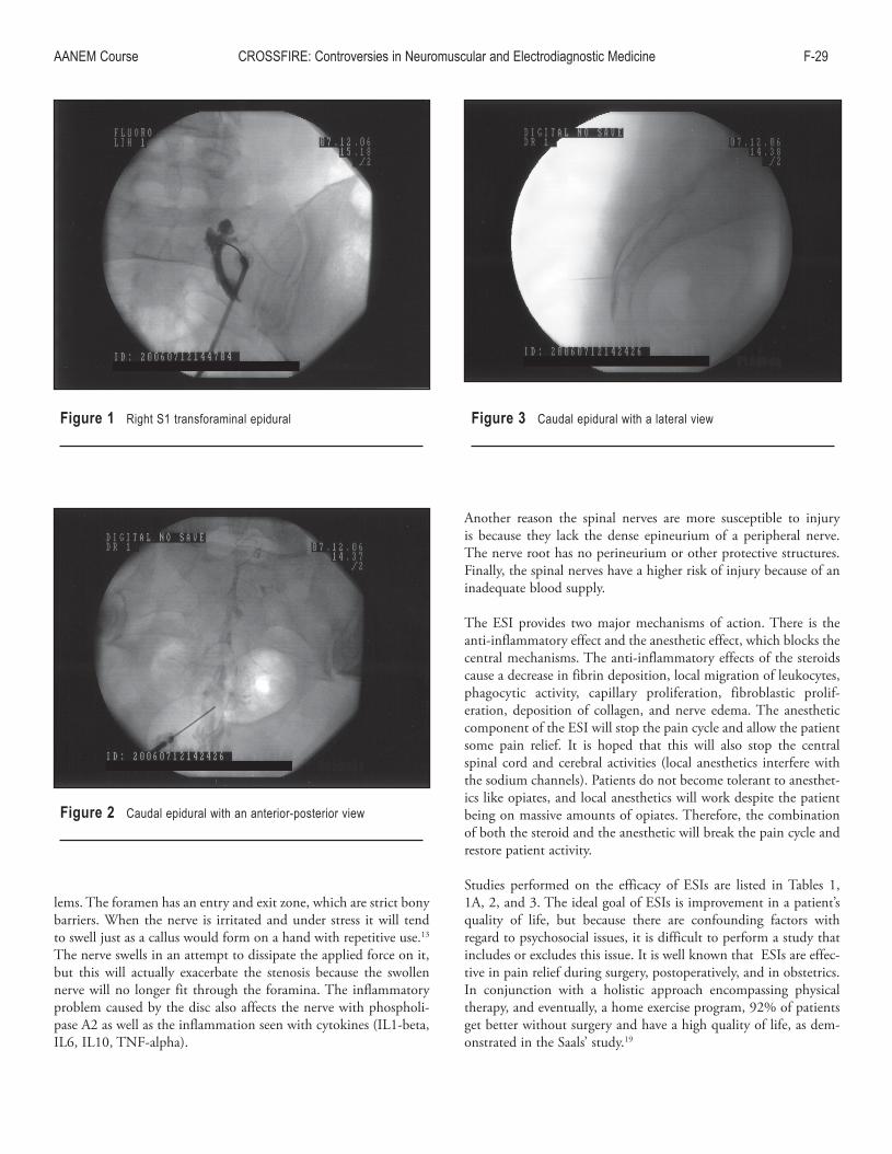
lems. The foramen has an entry and exit zone, which are strict bony barriers. When the nerve is irritated and under stress it will tend to swell just as a callus would form on a hand with repetitive use.13 The nerve swells in an attempt to dissipate the applied force on it, but this will actually exacerbate the stenosis because the swollen nerve will no longer fit through the foramina. The inflammatory problem caused by the disc also affects the nerve with phospholi-pase A2 as well as the inflammation seen with cytokines (IL1-beta, IL6, IL10, TNF-alpha).
Another reason the spinal nerves are more susceptible to injury is because they lack the dense epineurium of a peripheral nerve. The nerve root has no perineurium or other protective structures. Finally, the spinal nerves have a higher risk of injury because of an inadequate blood supply.
The ESI provides two major mechanisms of action. There is the anti-inflammatory effect and the anesthetic effect, which blocks the central mechanisms. The anti-inflammatory effects of the steroids cause a decrease in fibrin deposition, local migration of leukocytes, phagocytic activity, capillary proliferation, fibroblastic prolif-eration, deposition of collagen, and nerve edema. The anesthetic component of the ESI will stop the pain cycle and allow the patient some pain relief. It is hoped that this will also stop the central spinal cord and cerebral activities (local anesthetics interfere with the sodium channels). Patients do not become tolerant to anesthet-ics like opiates, and local anesthetics will work despite the patient being on massive amounts of opiates. Therefore, the combination of both the steroid and the anesthetic will break the pain cycle and restore patient activity.
Studies performed on the efficacy of ESIs are listed in Tables 1, 1A, 2, and 3. The ideal goal of ESIs is improvement in a patient’s quality of life, but because there are confounding factors with regard to psychosocial issues, it is difficult to perform a study that includes or excludes this issue. It is well known that ESIs are effec-tive in pain relief during surgery, postoperatively, and in obstetrics. In conjunction with a holistic approach encompassing physical therapy, and eventually, a home exercise program, 92% of patients get better without surgery and have a high quality of life, as dem-onstrated in the Saals’ study.19
Figure 1 RightS1transforaminalepidural
Figure 2 Caudalepiduralwithananterior-posteriorview
Figure 3 Caudalepiduralwithalateralview
AANEMCourse CROSSFIRE:ControversiesinNeuromuscularandElectrodiagnosticMedicine F-�9

Table 1 Reviewsoftheepiduralsteroidliteratureandauthor’scomments
Primary author Type of review Original author’s commentsKepes8 Literaturereview,1985 Rationalefortheirusehasnotbeenscientificallyproven.Benzon� Literaturereview,1985 Initialsuccessrateof81%-90%inpatientswithdisksyndromesand0.0- 1.5%inpatientswithspinalstenosis,spondylolysis,and spondylolisthesis.Spitzer�4 QuebecTaskForceonSpinalDisorders,1987 Epiduralinfiltrationshavebeenthetopicofanumberofclinicaltrials,with variableresultsandtheirusefulnessremainscontroversial.Koes10 Reviewedmethodologyof1�controlled One-halfoftrialsreportedpositiveoutcomesandone-halfreportednegative clinicalstudies,1995 outcomes.Thecriticalassessmentofthemethodsusedinthesetrials revealedflawsinthedesignofmoststudies.Bogduk3 Literaturereview,1995 Literatureprovidesendorsementbutofferslittlecompellingdataonrational andefficacy.Watts�5 Metaanalysis,11studies,1995 Short-term(upto60days)increasedoddsratioofpainrelief(>75% improvementto�.61whencomparedtoplacebo.)Longterm(1�mo)odds ratioof1.87whencomparedtoplacebo.Weinstein�6 Literaturereview,1995 Doshowasignificantlypositivepainreducingbenefit.Spaccarelli�3 Literaturereview,1996 65%ofpatientsreceivereliefinnoncontrolledstudies. Tabulationof�6noncontrolledstudies Reviewofnoncontrolledstudies Atlong-termfollowup,nodifferencebetweencontrolgroupAtherapeutic effectseemstooccurinlowerextremityradicularsymptomsat intermediate-termfollowup(�wkto3mo).Rydevik17 Literaturereviewonepiduralsteroids Moststudiesdonotsupporttheiruse. Iradicularpainfromspinalstenosis,1997Ringsdal15 Literaturereviewofprospectivecontrolledstudies,1977 Resultsareconflicting.Correctlydesignedstudiesarenecessary.
F-30 Epidural Steroid Injections are Underutilized AANEMCourse

Table 1A
Primaryauthor EfficacyofepiduralsteroidsSnoek�� NegativeMatthews11 SlightlypositiveBreivik4 PositiveBush5 PositiveSerrao�1 NegativeKlenerman9 NegativeRocco16 PositiveRidley14 ShorttermonlyBeliveau1 NegativeYates�7 PositiveReviewedbyKoes,theirresultsandmethodologicscoreoutofapossible100points.ModifiedfromKoesBW,andcollegues.Efficacyofepiduralsteroidinjectionsforlowbackpainandsciatica:asystematicreviewofrandomizedclinicaltrials,Pain1995;63:�79-�88,withpermission.
Table 2 Complicationsofepiduralsteroidinjections
EpiduralhematomaInfectionEpiduralabscessMeningitisOsteomyelitisSpinalHeadachePseudomyeloceleNerveandcordinjurySpinalSteroidsHypertensionFluidretentionCongestiveheartfailureEpidurallipomatosisHypothalamic-pituitary-adrenalaxissuppressionCushingsyndromeHyperglycemiaSteroidmyopathyIrregularmensesLocalAnestheticMotorblockCardiacarrhythmiaSeizureRetinalhemorrhage/blindnessIdiopathicFacialflushingandgeneralizederythemaTransientParalysisComplexregionalpainsyndromeAsepticmeningitis
AANEMCourse CROSSFIRE:ControversiesinNeuromuscularandElectrodiagnosticMedicine F-31

COMPARISONS AND TREATMENT OF CHRONIC ILLNESSES Patients with chronic rheumatoid arthritis have chronic illnesses similar to degenerative disc disease where inflammatory mediators destroy the skeletal structures, causing significant problems. In this situation, patients are given intravenous (IV) or subcutaneous treat-ments in addition to oral medications even though the IV injection will only last 1 month, and the subcutaneous injection will only last 1 week. This treatment is still given because it will preserve the joints and keep the disease under control. With ESIs, inflammation and damage to the nerve roots is contained by reducing signifi-cant scarring that can develop post-operatively. Scarring can also result from a lack of treatment. Monthly or weekly ESIs are also performed in inflammatory diseases of the bowel where treatment provides relief for the patient and improves their quality of life. This same situation occurs in patients with chronic back pain with radicular features (chronic nerve irritation). If patients receives ap-propriate ESIs to allow them to do the appropriate exercises, their pain can be controlled. If a flare-up arises from physical trauma like traveling or a slip and fall, an ESI will stop the inflammation at the nerve root level and allow patients to return to normal activity.
Epidural steroid injections can also be useful in patients with complex regional pain syndrome with severe extremity pain. Treatment includes performing a nerve root block at the nerve root level of the involved structure in addition to a sympathetic block. This will stop the central mechanism at the spinal cord and create pain relief and allow patients to go into a physical therapy or a home exercise program.
SUMMARY
In conclusion, ESIs are the most effective treatment for radicular symptoms after oral prednisone and NSAIDS have been tried. Treatment includes up to 3 injections every 3 months, but this is rarely needed. Most patients may need only one or two ESIs throughout the year to maintain their level of decreased inflamma-tion. Every 3 or 4 years a significant flare-up may occur and a series of three ESIs may need to be repeated. By no means should there ever be a situation where ESIs are not used because they are not long-acting enough. It has been shown with rheumatoid inflamma-tory disease and from the chronic inflammatory bowel disease that short-term relief is necessary and beneficial. Epideral steroid injec-tions are cost-effective, safe, and if used appropriately with a home exercise program, the most effective way to treat a radiculopathy.
Table 3
Oxycodone 1 tab #60 #90 #120�0mg 75.00 101.00 1�7.00 153.0040mg 9�.00 134.00 177.00 �19.0080mg 1�4.00 198.00 �71.00 345.00Hydrocodone7.5/750 �1.50 34.00 46.00 55.00Oxycodone/ACAP5/3�5mg 35.00 5�.00 58.00 61.00Tramadol 45.00 8�.00 1�0.00 158.00Celexoxib 11�.00 �15.00 �98.00 380.00Naprosyn generic500mg 46.00 55.00 57.00 60.00Zolpidem10mg 1�5.00 �35.00Zolpidem cr 119.00 �18.001�.5mgCarisoprodol �3.00 39.00 55.00 57.00Methocarbamol 19.00 300.00 41.00 5�.00500mgMethacaramol generic750mg�1.00 35.00 50.00 58.00Oxaprozin 54.00 58.00 6�.00 66.00gabapentin100mg �3.00 49.00 455.00300mg 47.00 66.00 74.00 8�.00400mg 55.00 64.00 70.00 78.00600mg 63.00 76.00 90.00 103.00Pregabalin�5mg 71.00 133.0050mg 99.00 133.0075mg 71.00 98.00100mg 71.00 148.00150mg 71.00 148.00Avinza30mg 118.00 �01.90 �78.00 354.0060mg 195.00 340.00 485.00 630.0090mg �70.00 490.00 710.00 930.001�0mg 307.00 564.00 8�5.00 1080.00Kadian�0mg 110.55 198.00 �7�.00 346.0030mg 118.00 �03.00 3��.00 356.0050mg 163.00 �75.00 387.00 500.0060mg 163.00 �75.00 387.00 500.00100mg 318.00 58.00 855.00 11�3.00
F-3� Epidural Steroid Injections are Underutilized AANEMCourse

REFERENCES
1. Beliveau P. A comparison between epidural anesthesia and without corticosteroid in the treatment of sciatica. Rheumatol Phys Med 1971;11:40-43.
2. Benzon HT. Epidural steroid injections for low back pain and lum-bosacral radiculopathy. Pain 1986;24:277-295.
3. Bogduk N. Epidural steroids. Spine 1995;20:845-888.4. Brevik HT, Hesla PE, Molnar I, et al. Treatment of chronic low
back pain and sciatica: comparison of caudal epidural injections of Bupivacaine and methylprednisolone with Bupivacaine followed by saline. In: Bonica JJ, Albe Fessard DG, editors. Advances in pain research and therapy, volume 3. New York: Raven Press; 1976. p 927-932.
5. Bush K, Hillier S. A controlled study of caudal epidural injections of triamcinolone plus procaine for the management of intractable sciatica. Spine 1991;16:572-575.
6. Dilke TF, Burry HC, Grahame R. Extradural corticosteroid injection in management of lumbar nerve root compression. BMJ 1973:2:635-637.
7. Epidural use of steroids in the management of back pain. Canberra, Australia: National Health and Medical Research Council of Australia; 1994. p 13.
8. Kepes ER, Duncalf D. Treatment of backache with spinal injec-tions of local anesthetics, spinal and systemic steroids. A review. Pain 1985;22:33-47.
9. Klenerman L, Greenwood R, Davenport HT, White DC, Peskett S. Lumbar epidural injections in the treatment of sciatica. Br J Rheumatol 1984;23:35-38.
10. Koes BW, Scholten RJ, Mens JM, Bouter LM. Efficacy of epidural steroid injections for low back pain and sciatica: a systematic review of randomized clinical trials. Pain 1995;63:279-288.
11. Mathews JA, Mills SB, Jenkins VM, Grimes SM, Morkel MJ, Mathews W, Scott CM, Sittampalam Y. Back pain and sciatica: controlled trials of manipulation, traction, sclerosant and epidural injections. Br J Rheumatol 1987;26:416-423.
12. McCarron RF, Wimpee MW, Hudkins PG, Laros GS. The inflamma-tory effect of nucleus pulposus. A possible element in the pathogen-esis of low back pain. Spine 1987;12:760-764.
13. Murphy RW. Nerve roots and spinal nerves in degenerative disk disease. Clin Orthop 1977:129:46-60.
14. Ridley MG, Kingsley GH, Gibson T, Grahame R. Outpatient lumbar epidural corticosteroid injection in the management of sciatica. Br J Rheumatol 1988;27:295-299.
15. Ringsdal VS, Nielsen NA, Slot O, Kryger P. Epidural glucocorticoid injection in lumbago sciatica. Ugeskr Laeger 1997;159:5653-5657.
16. Rocco AG, Frank E, Kaul AF, Lipson SJ, Gallo JP. Epidural steroids, epidural morphine and steroids combined with morphine in the treatment of post laminectomy syndrome. Pain 1989;36:297-303.
17. Rydevik BL, Cohen DB, Kostuik JP. Spine epidural steroids for pa-tients with lumbar spinal stenosis. Spine 1997;22:2313-2317.
18. Saal JS, Franson RC, Dobrow R, Saal JA, White AH, Goldthwaite N. High levels of inflammatory phospholipase A2 activity in lumbar disc herniations. Spine 1990;15:674-678.
19. Saal JA, Saal JS. Nonoperative treatment of herniated lumbar intervertebral disc with radiculopathy: An outcome study. Spine 1989;14:431-437.
20. Schlumpf U, Johr M. Acute lumbar disk displacement with nerve root compression. Indications for peridural steroid injection. Schweiz Rundsch Medication Prax 1997;86:292-295.
21. Serrao JM, Marks RL, Morley SJ, Goodchild CS. Intrathecal mid-azolam for the treatment of chronic mechanical low back pain: a controlled comparison with epidural steroid in a pilot study. Pain 1992;48:5-12.
22. Snoek W, Weber H, Jorgensen B. Double blind evaluation of extra-dural methylprednisolone for herniated lumbar discs. Acta Orthop Scand 1977;635-641.
23. Spaccarelli KC. Lumbar and caudal epidural corticosteroid injections. Mayo Clin Proc 1996;71:169-179.
24. Spitzer WO, LeBlanc FE, Dupuis M. Scientific approach to the as-sessment and management of activity-related spine disorders. Spine 1987;7:S1-S59.
25. Watts RW, Silagy CA. A meta-analysis on the efficacy of epidural corticosteroids in the treatment of sciatica Anesth Intensive Care 1995;23:564 569.
26. Weinstein SM, Herring SA, Derby R. Contemporary concepts in spine care. Epidural steroid injections. Spine 1995;20:1842-1846.
27. Yates DW. A comparison of the types of epidural injection com-monly used in the treatment of low back pain and sciatica. Rheumatol Rhehabil 1978;17:181-186.
AANEMCourse CROSSFIRE:ControversiesinNeuromuscularandElectrodiagnosticMedicine F-33

F-34 AANEMCourse

1. Which of the following is the best criterion for slowing of ulnar motor conduction across the elbow?A. Across elbow conduction velocity (CV) is slower than
forearm by > 5 m/s.B. Across elbow CV is slower than forearm by > 11m/s.C. Across elbow CV is slower than 53 m/s.D. Across elbow CV is slower than 48 m/s.E. Across elbow CV is slower than 43 m/s.
2. A large drop in ulnar compound muscle action potential (CMAP) amplitude is seen between wrist and below-elbow stimulation sites. Which of the following is most likely?A. Guyon’s canal neuropathy.B Cubital tunnel syndrome.C. Entrapment at the intermuscular septum of flexor digito-
rum profundus.D. Arcade of Struthers neuropathy.E. Martin-Gruber anastamosis.
CROSSFIRE: Controversies in Neuromuscular and Electrodiagnostic Medicine
CME SELF-ASSESSMENT TEST
Select the ONE best answer for each question.
AANEMCourse F-35
Fill
in an
swer
s here Last 4 digits of
your phone number
1 2 3 4
Instructions for filling out your parSCORE sheet
Ontheright-handsideoftheparSCOREsheet, you will need to fill in the following:
Under ID number, please write out and fill inthelast4digitsofyourphonenumber.Be sure to start in the first box on the left (asshown).
Under Test Form, please fill in “A”.
Leavethecompletedformatthetableoutsideyoursession.

3. The minimal criteria for prolonged latency across 2 cm on inching studies of the ulnar nerve is:A. 0.4 ms.B. 0.8 ms.C. 1.2 ms.D. 1.7 ms.E. 2.3 ms.
4. An absent ulnar sensory response is seen with stimulation at the wrist and recording over the small finger. Regarding local-ization, this suggests:A. Ulnar neuropathy at the wrist.B. Ulnar neuropathy at the elbow (UNE).C. No localization.D. C8 radiculopathy.E. Deep ulnar neuropathy.
5. Which of the following muscles is most likely to be normal in UNE?A. Flexor carpi ulnaris.B. First dorsal interosseous (FDI).C. Abductor digiti minimi (ADM).D. Palmaris brevis.E. Adductor pollicis.
6. Where is the “arcade of Struthers” located?A. Just distal to medial humeral condyle.B. 6-8 cm proximal to medial humeral condyle.C. Anterio-medial to medial humeral condyle.D. 8 cm distal to condyle.E. There is no such anatomic site.
7. Subluxing or dislocation of ulnar nerve from medial humeral condylar groove occurs in what percent of the general popula-tion?A. 10%.B. 20%.C. 35%.D. 40%.E. It is extremely rare.
8. Which would be most sensitive and specific test for ulnar neu-ropathy at the elbow?A. Needle electromyography (EMG).B. Motor conduction velocity (CV) slowing across the
elbow.C. Sensory CV slowing across the elbow.D. CMAP amplitude of abductor digiti quinti (ADQ) digit
5 above the elbow and wrist stimulation.E. Sensory nerve action potential (SNAP) digit 5 above and
below elbow (across elbow).
9. Which would suggest a compromise of the ulnar nerve proxi-mal to wrist?A. Reduced CMAP of first dorsal interosseus muscle.B. Reduced CMAP of adq digit 5.C. Reduced SNAP digit 5.D. Needle EMG abnormalities in first dorsal interosseous.E. Reduced SNAP of dorsal ulnar cutaneous nerve.
10. What anatomic variations make ulnar UNE more common in men than women?A. The humeral coronoid tubercle is larger.B. The subcutaneous fat pad is thicker.C. The carrying angle of humerus is greater.D. The cubital tunnel is smaller.E. There is no difference.
11. Which is the most likely abnormal electrodiagnostic (EDX) finding in UNE?A. Fibrillation and positive waves in flexor carpi ulnaris.B. Reduced CMAP ADQ digit 5.C. Slowing of motor CV across elbow.D. Fibrillation and positive waves in first dorsal interro-
seous.E. Reduced recruitment in flexor carpi ulnaris.
12. The best motor nerve conduction study (NCS) for diabetic polyneuropathy is: A. Median motor latency to lumbrical. B. Radial motor latency to extensor indicis. C. Median/ulnar motor latency to first dorsal interosseous. D. Tibial F latency to abductor hallucis. E. Peroneal F latency to tibialis anterior.
13. Which is the most sensitive EDX technique for radiculopa-thy? A. Needle EMG. B. F-wave latency (FWL). C. H-reflex latency. D. Motor evoked potential. E. High voltage electric stimulation over spine.
14. In order to counter individual differences in FWL, which of the following should be used? A. Use a height normogram. B. Measure the side-to-side difference. C. Compare one nerve to another in the same limb. D. All of above. E. None of above.
F-36 CME Self-Assessment Test AANEMCourse

15. What happens to F waves if the nerve is stimulated more proximally? A. Latency shortens. B. Latency lengthens. C. Amplitude increases. D. Amplitude decreases. E. Nothing happens.
16. What happens if one intentionally relaxes the target muscle for a few hours? A. Decreased CMAP amplitude. B. Decreased CMAP latency. C. Decreased F-wave latency. D. Decreased F-wave persistence. E. Nothing happens.
17. Which nerve conduction result has the highest reproducibil-ity? A. Motor nerve conduction velocity (NCV). B. Sensory NCV. C. Minimal latency of F wave. D. Amplitude of CMAP. E. Amplitude of sensory nerve action potential.
18. Which nerve conduction measurement will be most affected by patient’s height? A. Sensory amplitude. B. Sensory CV C. Motor amplitude. D. Motor CV. E. F-wave latency.
19. Which of the following is true about F waves?A. They are large in amplitude.B. They result from antidromic activation of motor
neurons.C. They are enhanced by submaximal stimulation.D. They always occur after each stimulus.E. They are restricted in distribution.
20. All are true about F waves EXCEPT which of the following?A. Adequate analysis requires evaluating a series of F waves.B. The difference between the minimum and maximum
FWL in a series of F waves has been called chronodisper-sion.
C. The number of stimuli needed may depend on the record-ing muscle.
D. F waves are inherently variable in lateancy, amplitude, and configuration.
E. Minimal FWLs are the most reliable FWL measurement.
21. F waves have been reported as abnormal in which of the fol-lowing?
1. Peripheral neuropathies.2. Entrapment neuropathies.3. Plexopathies.4. Lumbosacral radiculopathies.
A. Only 1, 2, and 3 are correct.B. Only 1 and 3 are correct.C. Only 1 and 4 are correct.D. Only 1 is correct.E. All are correct.
22. Which of the following is true about F waves and radiculopa-thies?A. Analysis of multiple F-wave parameters can be helpful.B. All reports indicate F waves are less sensitive than needle
EMG.C. F waves are usually recorded from muscles innervated by
cervical roots commonly injured in cervical radiculopa-thies.
D. Criticism of F waves based on “dilution” has been sup-ported by theoretical modeling.
E. The use of minimal FWLs following 10 stimuli as the sole F-wave parameter required has been found to be adequate in published reports.
23. All are true about F waves and radiculopathies EXCEPT which of the following?A. The only F-wave parameter reported abnormal in lumbo-
sacral radiculopathies has been latency.B. F waves may be abnormal even with unremarkable needle
EMG studies.C. F-wave data should be interpreted with other EDX and
clinical information.D. F-wave persistence and/or chronodispersion have been
reported as the only F-wave abnormality present. E. F waves should be used where the information could be
helpful.
24. The great majority of research investigations regarding the use of epidural steroid injections (ESIs) pertain to what type of pain syndrome?A. Myofascial.B. Radicular cervical.C. Axial cervical.D. Radicular lumbar.E. Axial lumbar.
AANEMCourse CMESelf-AssessmentTest F-37

25. Material injected into the epidural space is most likely to reach the subdural region by:A. Diffusion through the dura.B. Migration through the nerve root sleeves.C. Inadvertent subdural needle placement.D. Systemic absorption and redistribution.E. Dissolving in lipids.
26. Well-controlled research studies have found that ESIs have a proven benefit for:A. Cervical radiculopathy.B. Thoracic radiculopathy.C. Lumbar radiuclopahty.D. Lumbar spinal stenosis.E. None of the above.
27. A possible beneficial outcome of treatment with ESIs in the population affected with lumbar radiculopathy is:A. More rapid return to work.B. Long-term reduction in leg pain.C. Avoidance of a surgical procedure.D. Higher overall functional level.E. Ability to walk longer distances.
28. Some well-designed research studies conclude that ESIs lead to reduced pain levels, judged subjectively. The duration of this beneficial effect is typically:A. 0-6 weeks.B. 6 weeks to 6 months.C. 6-12 months.D. 1-2 years.E. Over 2 years.
29. The most important factor causing an injury to the nerves from a herniated disc is:A. Direct compression of the nerve from the disc material.B. Inflammation from a reaction to the disc material. C. Lack of oxygen to the nerves.D. Lack of nutrients to the nerves.E. All of the above.
30. Surgery is not indicated in most cases of a herniated disc because:A. 98% of the cases resolve with nonoperative treatment.B. Surgery restores the normal biomechanics. C. Surgery will not cause scar tissue.D. Once surgery is performed, it will never be needed again.E. None of the above.
31. This medication has no role in the treatment of a radiculopa-thy:A. Naproxen.B. Hydrocodone.C. Nortriptyline. D. Soma.E. None of the above.
32. Which statement is true about corticosteroids?A. If you take more than 3 doses a year, you will die.B. It is a synthetic substance.C. Used properly, it can be used up to 12 doses per year.D. Given as an epidural will cause osteoporosis.E. All statements are true.
33. Which of the following is incorrect in the treatment of chronic lumbar radiculopathy?A. A cognitive behavioral program.B. A home exercise program.C. Anti-inflammatory medications by mouth or epidural
routes.D. Multiple surgeries.E. Salsa dancing.
F-38 AANEMCourse

34. How would you rate the quality of instruction received during Dr. Robinson’s presentation?A. Best possible.B. Good.C. Average.D. Poor.E. Worst possible.
35. Select any item(s), that, if changed, would have appreciably improved Dr. Robinson’s presentation:A. Quality of slides.B. Quality of handout.C. Amount of clinically relevant information in the presenta-
tion.D. Amount of scientific content in the presentation.E. Other: please explain on the comment form at the back of
this handout.
36. Did you perceive any commercial bias in Dr. Robinson’s pre-sentation?A. Yes.B. No.
37. How would you rate the quality of instruction received during Dr. Johnson’s presentation?A. Best possible.B. Good.C. Average.D. Poor.E. Worst possible.
38. Select any item(s), that, if changed, would have appreciably improved Dr. Johnson’s presentation:A. Quality of slides.B. Quality of handout.C. Amount of clinically relevant information in the presenta-
tion.D. Amount of scientific content in the presentation.E. Other: please explain on the comment page at the back of
this handout.
39. Did you perceive any commercial bias in Dr. Johnson’s presen-tation?A. Yes.B. No.
40. How would you rate the quality of instruction received during Dr. Kimura’s presentation?A. Best possible.B. Good.C. Average.D. Poor.E. Worst possible.
41. Select any item(s), that, if changed, would have appreciably improved Dr. Kimura’s presentation:A. Quality of slides.B. Quality of handout.C. Amount of clinically relevant information in the presenta-
tion.D. Amount of scientific content in the presentation.E. Other: please explain on the comment page at the back of
this handout.
42. Did you perceive any commercial bias in Dr. Kimura’s presen-tation?A. Yes.B. No.
43. How would you rate the quality of instruction received during Dr. Fisher’s presentation?A. Best possible.B. Good.C. Average.D. Poor.E. Worst possible.
CROSSFIRE: Controversies in Neuromuscular and Electrodiagnostic Medicine
EVALUATION
Select ANY of the answers that indicate your opinions.
Your input is needed to critique our courses and to ensure that we use the best faculty instructors and provide the best course options in future years. Make additional comments or list suggested topics or faculty for future courses on the comment form provided at the end of this handout.
AANEMCourse F-39

44. Select any item(s), that, if changed, would have appreciably improved Dr. Fisher’s presentation:A. Quality of slides.B. Quality of handout.C. Amount of clinically relevant information in the presenta-
tion.D. Amount of scientific content in the presentation.E. Other: please explain on the comment page at the back of
this handout.
45. Did you perceive any commercial bias in Dr. Fisher’s presenta-tion?A. Yes.B. No.
46. How would you rate the quality of instruction received during Dr. Geiringer’s presentation?A. Best possible.B. Good.C. Average.D. Poor.E. Worst possible.
47. Select any item(s), that, if changed, would have appreciably improved Dr. Geiringer’s presentation:A. Quality of slides.B. Quality of handout.C. Amount of clinically relevant information in the presenta-
tion.D. Amount of scientific content in the presentation.E. Other: please explain on the comment page at the back of
this handout.
48. Did you perceive any commercial bias in Dr. Geiringer’s pre-sentation?A. Yes.B. No.
49. How would you rate the quality of instruction received during Dr. Lagattuta’s presentation?A. Best possible.B. Good.C. Average.D. Poor.E. Worst possible.
50. Select any item(s), that, if changed, would have appreciably improved Dr. Lagattuta’s presentation:A. Quality of slides.B. Quality of handout.C. Amount of clinically relevant information in the presenta-
tion.D. Amount of scientific content in the presentation.E. Other: please explain on the comment page at the back of
this handout.
51. Did you perceive any commercial bias in Dr. Lagattuta’s pre-sentation?A. Yes.B. No.
52. As a result of your attendance at this educational session, did you learn anything that will improve the care of your pa-tients?A. Yes, substantially.B. Yes, somewhat.C. Not sure.D. Probably not.E. This session was not applicable to my patients.
53. Do you feel that the information presented in this session was based on the best evidence available?A. Yes.B. No: Please explain on the comment page at the back of
this handout.
54. Select ALL items where improvement was needed.A. The accuracy of advance descriptions of this course.B. The specific topics selected for presentation.C. The number of speakers in this course.D. The amount of time allotted for discussion in this
course.E. Other: please add other areas and outline specific rec-
ommendations for areas needing improvement on the comment page at the back of this handout.
55. I plan to attend the 2007 AANEM Annual Meeting in Phoenix, AZ October 17-20.A. Yes, definitely.B. No, definitely.C. Will wait to see the program content.D. Will wait to see if budget allows my attendance.
F-40 Evaluation AANEMCourse
AANEMCourse CROSSFIRE:ControversiesinNeuromuscularandElectrodiagnosticMedicine F-41
56. We would like the AANEM Annual Meeting to be one of your “must attend” meetings each year. In order to do this, we would have to do what to make it happen? Please explain on the comment page at the back of this handout.
57. If you are a member of the AANEM, what would you rate as the most valuable benefit of your membership?A. My subscription to the journal Muscle & Nerve.B. Receiving free educational materials such as the Muscle &
Nerve Invited Reviews and the AANEM Resource CD.C. The availability of CME opportunities.D Member discounts on AANEM CME products and ser-
vices.E. AANEM’s advocacy work on issues that impact my pro-
fession.
58. In 2006, the AANEM distributed 6 bound copies of the Muscle & Nerve Invited Reviews. How would you rate this new member benefit?A. Very valuable.B. Somewhat valuable.C. Not very valuable.D. I do not receive this since I am not an AANEM
member.E. Other: please explain on the comment page at the back of
this handout.
59. When you receive the bound copies of the Muscle & Nerve Invited Reviews, you can visit the AANEM website and complete CME questions to receive a certificate at no charge. Have you utilized this new service?A. Yes, I have completed CME for the Invited Reviews
online and found the system easy to utilize.B. Yes, I have completed CME for the Invited Reviews
online, but found the system difficult to utilize.C. No, I have not utilized this service because I was unaware
it was available. D. No, I have not utilized it although I was aware of its avail-
ability: please explain on the comment page at the back of this handout.
E. Other: please explain on the comment page at the back of this handout.
60. In 2006, the AANEM added Online Case Studies to the website which are also available for CME credit. How would you rate this new member benefit?A. Very valuable.B. Somewhat valuable.C. Not very valuable.D. I was not aware that this was available on the AANEM
website.E. Other: please explain on the comment page at the back of
this handout.
61. The AANEM has recently launched new Marketing Slides on the website that can assist EDX physicians in marketing to referral sources. How would you rate this member benefit?A. Very valuable.B. Somewhat valuable.C. Not very valuable.D. I have not reviewed the marketing slides yet.E. Other: please explain on the comment page at the back of
this handout.

2006 COURSE G AANEM 53rd Annual Meeting
Washington, DC
Copyright © October 2006 American Association of Neuromuscular & Electrodiagnostic Medicine
2621 Superior Drive NW Rochester, MN 55901
Printed by Johnson Printing ComPany, inC.
Mark A. Ferrante, MD
Bryan E. Tsao, MD
Lisa S. Krivickas, MD
Asa J. Wilbourn, MD
Brachial Plexus Assessment

Brachial Plexus Assessment
Faculty
Mark A. Ferrante, MDClinical Associate ProfessorDepartment of NeurologyTulane University School of MedicineNew Orleans, LouisianaDr. Ferrante received his medical degree at the University of South Florida-Tampa and performed his EMG/neuromuscular fellowship at the Cleveland Clinic Foundation in Cleveland, Ohio. Dr. Ferrante was formerly Chief in the Department of Neurology and Director of the EMG Laboratory at Keesler Medical Center at Keesler Air Force Base, where he also received the United States Air Force Meritorious Service Award. In addition to the AANEM, Dr. Ferrante is a member of the American Academy of Neurology. He has previously received the AANEM Junior Member Recognition Award and has been a Board Examiner for the American Board of Electrodiagnostic Medicine. His current research interests include the application of sensory nerve conduction studies to di-agnose focal brachial plexopathies, neuralgic amyotrophy, and the brachial plexus in general.
Bryan E. Tsao, MDStaffDepartment of NeurologyCleveland ClinicAssistant ProfessorLerner College of MedicineCleveland ClinicCleveland, OhioDr. Tsao completed his neurology residency at Loma Linda University, fol-lowed by a fellowship in clinical neurophysiology in EMG and neuromus-cular disease at Cleveland Clinic. He is an assistant professor of medicine at Cleveland Clinic Lerner College of Medicine and a staff neuromuscular neurologist at Cleveland Clinic. His clinical and research interests include the diagnosis and management of peripheral nerve injury, neuralgic amy-otrophy, and the electrodiagnosis of the brachial plexus. He is a fellow of the AANEM and serves on the Course Committee.
Lisa S. Krivickas, MDAssociate ProfessorDepartment of Physical Medicine and RehabilitationHarvard Medical SchoolBoston, MassachusettsDr. Krivickas is Associate Professor of Physical Medicine and Rehabilitation (PM&R) at Harvard Medical School, Associate Chief of PM&R at Massachusetts General Hospital, and Director of EMG at Spaulding Rehabilitation Hospital. She received a B.S. in mechanical engineering from Cornell University and her medical degree from Harvard Medical School. She has over 50 publications including peer-reviewed scientific papers, book chapters, and review articles. Dr. Krivickas’ current research involves the study of muscle physiology in neuromuscular disorders and aging, investigation of new electrophysiologic techniques for the assess-ment of neuromuscular disorders, and clinical trials for amyotrophic lateral sclerosis and other neuromuscular diseases. She has been elected to the Board of Trustees of the American College of Sports Medicine and is a director for the American Board of Electrodiagnostic Medicine. She is a member of the Neuromuscular Medicine Examination Committee of the American Board of Psychiatry and Neurology. She received the 2006 Young Academician Award from the Association of Academic Physiatrists.
Asa J. Wilbourn, MDDirector, EMG LaboratoryCleveland ClinicAssociate ProfessorDepartment of NeurologyCase Western Reserve UniversityCleveland, OhioDr. Wilbourn received his neurology training at Yale University and his electrophysiology training at Mayo Clinic, Rochester, Minnesota. He is the Director of the EMG Laboratory at Cleveland Clinic, and is Associate Professor of Neurology at the Case Western Reserve University School of Medicine. Dr. Wilbourn has extensively published on all aspects of the electrodiagnostic evaluation of plexopathies, radiculopathies, entrapment neuropathies, and iatrogenic nerve injuries. He has served as a member of the AANEM Education and Training Program Committees, and has been the chair of both the Membership and Progam Committees. Dr. Wilbourn has also served on the AANEM Board of Directors.
Course Chair: Asa J. Wilbourn, MD
The ideas and opinions expressed in this publication are solely those of the specific authors and do not necessarily represent those of the AANEM.
Authors had nothing to disclose.
G-ii

Brachial Plexus Assessment
Contents
Faculty ii
Objectives iii
CourseCommittee iv
Electrodiagnostic Assessment of the Brachial Plexus 1MarkA.Ferrante,MD
Traumatic Brachial Plexopathies 13BryanE.Tsao,MD
Nontraumatic Brachial Plexopathies 21LisaS.Krivickas,MD
Iatrogenic Brachial Plexopathies 27AsaJ.Wilbourn,MD
CMESelf-AssessmentTest 33
Evaluation 37
Ob j e c t i v e s—This session will (1) review the anatomy and pathophysiology of the brachial plexus, (2) discuss how each major compo-nent of the brachial plexus is assessed by nerve conduction studies and needle EMG, and (3) describe the clinical and electrodiagnostic features of the more common brachial plexus lesions encountered.
Pr e r e q u i s i t e—This course is designed as an educational opportunity for residents, fellows, and practicing clinical EDX physicians at an early point in their career, or for more senior EDX practitioners who are seeking a pragmatic review of basic clinical and EDX principles. It is open only to persons with an MD, DO, DVM, DDS, or foreign equivalent degree.
Ac c r e d i tAt i O n stAt e m e n t—The AANEM is accredited by the Accreditation Council for Continuing Medical Education to provide continuing medical education (CME) for physicians.
cme cr e d i t—The AANEM designates this activity for a maximum of 3.25 hours in AMA PRA Category 1 Credit(s)TM. This educational event is approved as an Accredited Group Learning Activity under Section 1 of the Framework of Continuing Professional Development (CPD) options for the Maintenance of Certification Program of the Royal College of Physicians and Surgeons of Canada. Each physician should claim only those hours of credit he or she actually spent in the educational activity. CME for this course is avail-able 10/06 - 10/09.
Please be aware that some of the medical devices or pharmaceuticals discussed in this handout may not be cleared by the FDA or cleared by the FDA for the specific use described by the authors and are “off-label” (i.e., a use not described on the product’s label). “Off-label” devices or pharmaceuticals may be used if, in the judgement of the treating physician, such use is medically indicated to treat a patient’s condition. Information regarding the FDA clearance status of a particular device or pharmaceutical may be obtained by reading the product’s package labeling, by contacting a sales representative or legal counsel of the manufacturer of the device or pharmaceutical, or by contacting the FDA at 1-800-638-2041.
G-iii

G-iv
ThomasHyattBrannagan,III,MDNew York, New York
HopeS.Hacker,MDSan Antonio, Texas
KimberlyS.Kenton,MDMaywood, Illinois
DaleJ.Lange,MD New York, New York
SubhadraNori,MDBronx, New York
JeremyM.Shefner,MD,PhDSyracuse, New York
T.DarrellThomas,MDKnoxville, Tennessee
BryanE.Tsao,MDShaker Heights, Ohio
2005-2006 AANEM PRESIDENT
JaniceM.Massey,MDDurham, North Carolina
2005-2006 AANEM COURSE COMMITTEE
KathleenD.Kennelly,MD,PhDJacksonville, Florida

Electrodiagnostic Assessment of the Brachial Plexus
Mark A. Ferrante, MDClinicalAssociateProfessor
DepartmentofNeurologyTulaneUniversitySchoolofMedicine
NewOrleans,Louisiana
INTRODUCTION
The brachial plexus (BP) is an immense structure that is composed of over 100,000 nerve fibers that extend from the neck to the axilla.9 The sensory fibers are derived from sensory neurons located in the dorsal root ganglia (DRG) and the motor fibers are derived from neurons located in the anterior horns of the spinal cord, re-spectively. These fibers enter the BP at multiple levels (generally, C5 through T1), travel through it, and exit from various sites as named nerves. As these fibers traverse the BP, they reorganize, sequentially giving rise to multiple elements of five different types. In sequence, from proximal to distal, they are termed roots, trunks, divisions, cords, and terminal nerves. Lesions of the BP are not uncommon. This reflects its inherent susceptibility to trauma, which is related to its superficial location and to its position between highly mobile structures (i.e., the head and neck, proximally, and the upper ex-tremity and shoulder, distally), and the secondary acquisition of pathology from neighboring diseased structures (e.g., major blood vessels, lymph nodes, the lung). Thus, physicians frequently refer patients with brachial plexopathies for electrodiagnostic (EDX) as-sessment. At a minimum, EDX physicians require a strong grasp of BP anatomy, an awareness of the various disorders affecting the BP, an understanding of the underlying pathophysiologies and EDX manifestations of the latter, and knowledge of the BP elements as-sessed by each nerve conduction study (NCS) performed and each muscle sampled by needle electrode examination (NEE). Because of its great expanse, no single NCS or muscle assessed by NEE could possibly evaluate it in its entirety. Consequently, an organized approach is required. In addition, based on historical and physical examination information, as well as information gleaned as the
study proceeds, the EDX physician must decide which additional nerves and muscles require study. Since the entry and exit sites of the sensory and motor fibers composing each BP-derived nerve are known, the course traversed through the BP by these fibers can be deduced. Consequently, for all BP-derived nerves amenable to NCS or NEE assessment, the BP elements assessed are known. Most importantly, since the fibers composing the individual BP-derived nerves traverse the BP via different routes, focal BP lesions produce different EDX patterns, the recognition of which yields localizing information.2,3,7 Once the lesion is localized, the un-derlying pathophysiology and severity are determined, ultimately yielding diagnostic and prognostic information of importance to the referring physician.
ANATOMY OF THE BRACHIAL PLEXUS
The BP elements, sequentially, from proximal to distal, include: (1) five roots: the C5 through T1 anterior primary rami (APR); (2) three trunks: upper, middle, and lower; (3) six divisions: three anterior and three posterior; (4) three cords: lateral, posterior, and medial; and (5) several terminal nerves (Figure 1).4,8
Anterior Primary Rami
Anatomically, the dorsal and ventral rootlets emanating from each spinal cord segment fuse to form single dorsal and ventral roots. The latter enter the intervertebral foramen and, just distal to the DRG, fuse to form a mixed spinal nerve (MSN). The MSN exits the foramen and immediately gives off a posteriorly directed branch,
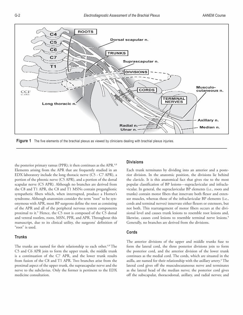
G-� Electrodiagnostic Assessment of the Brachial Plexus AANEMCourse
the posterior primary ramus (PPR); it then continues as the APR.4,8 Elements arising from the APR that are frequently studied in an EDX laboratory include the long thoracic nerve (C5 - C7 APR), a portion of the phrenic nerve (C5 APR), and a portion of the dorsal scapular nerve (C5 APR). Although no branches are derived from the C8 and T1 APR, the C8 and T1 MSNs contain preganglionic sympathetic fibers which, when interrupted, produce a Horner’s syndrome. Although anatomists consider the term “root” to be syn-onymous with APR, most BP surgeons define the root as consisting of the APR and all of the peripheral nervous system components proximal to it.9 Hence, the C5 root is composed of the C5 dorsal and ventral rootlets, roots, MSN, PPR, and APR. Throughout this manuscript, due to its clinical utility, the surgeons’ definition of “root” is used.
Trunks
The trunks are named for their relationship to each other.4,8 The C5 and C6 APR join to form the upper trunk, the middle trunk is a continuation of the C7 APR, and the lower trunk results from fusion of the C8 and T1 APR. Two branches arise from the proximal aspect of the upper trunk, the suprascapular nerve and the nerve to the subclavius. Only the former is pertinent to the EDX medicine consultation.
Divisions
Each trunk terminates by dividing into an anterior and a poste-rior division. In the anatomic position, the divisions lie behind the clavicle. It is this anatomical fact that gives rise to the most popular classification of BP lesions—supraclavicular and infracla-vicular. In general, the supraclavicular BP elements (i.e., roots and trunks) contain motor fibers that innervate both flexor and exten-sor muscles, whereas those of the infraclavicular BP elements (i.e., cords and terminal nerves) innervate either flexors or extensors, but not both. This rearrangement of motor fibers occurs at the divi-sional level and causes trunk lesions to resemble root lesions and, likewise, causes cord lesions to resemble terminal nerve lesions.9 Generally, no branches are derived from the divisions.
Cords
The anterior divisions of the upper and middle trunks fuse to form the lateral cord, the three posterior divisions join to form the posterior cord, and the anterior division of the lower trunk continues as the medial cord. The cords, which are situated in the axilla, are named for their relationship with the axillary artery.4 The lateral cord gives off the musculocutaneous nerve and terminates as the lateral head of the median nerve; the posterior cord gives off the subscapular, thoracodorsal, axillary, and radial nerves; and
Figure 1Thefiveelementsofthebrachialplexusasviewedbycliniciansdealingwithbrachialplexusinjuries.

AANEMCourse BrachialPlexusAssessment G-3
the medial cord gives off the medial brachial, medial antebrachial, and ulnar nerves and terminates as the medial head of the median nerve. The lateral and medial pectoral nerves emanate from the lateral and medial cords, respectively, just after their formation.
Terminal Nerves
Depending on the publication, the terminal nerves number from three (median, ulnar, and radial nerves) to five (when the musculo-cutaneous and axillary nerves are included).4,9
CLASSIFICATION OF BRACHIAL PLEXUS LESIONS
Since divisional injuries are infrequent, and since root lesions re-semble trunk lesions while cord lesions resemble terminal nerve lesions (regarding contiguous elements only), BP injuries have been classified into supraclavicular and infraclavicular plexopathies. Importantly, this anatomical division has clinical significance since these two categories differ in their incidence, severity, and progno-sis.9 Supraclavicular plexopathies are more common, more severe, and tend to have a worse prognosis (a reflection of the injuries producing them). The supraclavicular plexus is further divided into the: (1) upper plexus (upper trunk; C5 and C6 roots); (2) middle plexus (middle trunk; C7 root); and (3) lower plexus (lower trunk; C8 and T1 roots). These three categories also have clinical relevance in regard to severity, prognosis, and incidence.9 Upper plexopathies, which represent the most frequent site of BP involve-ment, tend to be less severe than those involving the lower plexus because: (1) the pathophysiology of an upper plexus lesion is more frequently demyelinating conduction block (DCB), (2) the muscles innervated by upper plexus motor fibers are more proximate to the lesion (i.e., more likely to be reinnervated via axon regrowth), and (3) the location of an upper plexus lesion is more likely to be extraforaminal (i.e., more amenable to surgical repair). Conversely, lower plexus lesions are less frequently associated with DCB, result in muscle denervation further from the lesion site, and tend to be preganglionic.
ELECTRODIAGNOSTIC MANIFESTATIONS
Although nerve fibers can be damaged in innumerable ways, the pathologic and pathophysiologic responses to these forces are limited. With more severe lesions, axon disruption (termed “axon loss”) occurs with ensuing Wallerian degeneration (pathologically) and conduction failure (pathophysiologically). Prior to Wallerian degeneration (up to 7 days for motor axons and 10-11 days for sensory axons), the distal stump of the severed axon remains capable of conducting impulses and, thus, during that period conduction block is observed. With lesions of lesser severity, the resultant pathology is limited to demyelination and the resultant pathophysiology is either DCB (when action potentials cannot
traverse the lesion site) or demyelinating conduction slowing (when action potentials traverse the lesion site at a slower rate). Each of these pathophysiologies has unique EDX features.
Electrodiagnostic Features of Axon Loss Lesions
Most BP lesions are axon loss in nature (e.g., avulsions, neoplastic processes), although concomitant demyelination may occur (e.g., early traumatic lesions). Since axon loss produces conduction failure, affected sensory and motor fibers can no longer contribute to sensory nerve action potential (SNAP) and compound muscle action potential (CMAP) formation. Consequently, the latter are reduced in size or absent and their latencies and conduction ve-locities (CVs) are normal or mildly reduced. Unlike the response amplitude which reflects the total number of conducting fibers, the latency and CV reflect only the fastest conducting fibers (i.e., the majority of the conducting fibers are not reflected). As a result, whenever a few of the larger fibers remain intact, the latency and CV values are normal, despite the fact that most of the nerve fibers are disrupted. For that reason, amplitude measurements are a much more sensitive indicator of axon loss. Once the distal stump can no longer conduct, the response amplitude is the same regardless of whether the stimulus is applied above, at, or below the lesion. On NEE, depending on the timing of the EDX medicine consultation and the severity of the insult, fibrillation potentials and a neuro-genic pattern of motor unit action potential (MUAP) recruitment are observed. Although MUAP drop out occurs immediately, it may not be appreciable when the sampled muscles are mildly or moderately denervated. Reinnervation occurs in two ways: (1) proximodistal regeneration of the affected fibers, and (2) collateral sprouting of unaffected ones. The degree of reinnervation is de-termined by several factors, including the grade of the injury, the distance between the lesion site and the innervated structures, and the completeness of the lesion. The grade of an axon loss lesion re-flects the amount of damage sustained by the supporting structures of the nerve (i.e., the endoneurium, perineurium, and epineurium). When all of these structures remain intact, reinnervation by proxi-modistal regeneration can occur. Unfortunately, motor axon ad-vancement is limited by the “time-distance factor.” Since regrowth occurs at a rate of about 1 inch per month and denervated muscle fibers undergo degeneration after 18-24 months in the denervated state, denervated muscle fibers located more than 2 feet from the injury site cannot be reinnervated by this mechanism. The sensory end organs are not affected by the time-distance factor. Since collat-eral sprouting requires intact axons, the completeness of the lesion determines whether there are enough motor axons for functional recovery (i.e., complete or nearly complete axon disruption is not amenable to reinnervation via collateral sprouting).
Electrodiagnostic Features of Demyelinating Lesions
Since demyelinating conduction slowing typically is not associated with weakness or sensory loss, patients with BP lesions limited to

G-� Electrodiagnostic Assessment of the Brachial Plexus AANEMCourse
this form of pathophysiology tend to be asymptomatic. Thus, they are infrequently referred to EDX laboratories. Conversely, lesions producing DCB, like axon loss lesions, produce weakness and large fiber sensory modality dysfunction. Unlike axon loss lesions, they seldom occur in isolation and do not induce pathologic changes beyond the lesion site. Consequently, comparison of the responses recorded with stimulation above and below the lesion reveals am-plitude discrepancies that have localizing value (i.e., the CMAP amplitude obtained with distal stimulation is significantly larger than that obtained with proximal stimulation). Although demy-elinating lesions located proximal to both the proximal and distal stimulation sites do not produce such an amplitude discrepancy, their presence is indicated whenever the CMAP amplitude is “too good” for the severity of neurogenic recruitment. This applies to BP lesions located proximal to the midtrunk level (i.e., the most proximal level of the BP capable of being stimulated by a hand-held surface stimulator). Although focal demyelinating lesions typically are associated with an excellent prognosis, there are two chronic BP lesions in which the predominant pathophysiology is demyelin-ation and for which resolution does not occur—the early stages of radiation-induced plexopathy (these lesions later convert to axon loss and never resolve) and multifocal motor neuropathy with DCB (when untreated, these lesions may convert to axon loss).
Severity Determination
The degree of CMAP amplitude (or negative area under the curve) decrement correlates well with the number of motor axons dis-rupted and, hence, with the degree of clinical weakness. Prior to reinnervation, whenever the CMAP recorded from the affected
muscle shows a decrement of 50%—in comparison to the homolo-gous response recorded from the asymptomatic contralateral limb or in comparison to the more distally recorded CMAP (regarding focal DCB lesions)—then approximately half of the motor axons composing the nerve innervating that muscle are nonconducting. Similarly, unelicitable responses typically are associated with para-lyzed muscles.
ELECTRODIAGNOSTIC ASSESSMENT OF THE BRACHIAL PLEXUS
Due to its large size and complexity, there is no single NCS or muscle assessable by NEE that is capable of evaluating the BP in its entirety. Instead, its regions are studied individually. Since each element of the BP has its own SNAP domain (i.e., the SNAPs assessing the sensory fibers contained in that element), CMAP domain (i.e., the CMAPs assessing the motor fibers contained in that element), and muscle domain (i.e., the muscles innervated by the motor fibers contained in that element), focal lesions of the BP typically produce unique patterns of EDX involvement which, when recognized, contain localizing information (Tables 1, 2, and 3). First, each component of the EDX medicine consultation (i.e., sensory NCS, motor NCS, NEE) is performed. The pattern of sensory NCS abnormalities localizes the lesion to a particular BP region or element. The motor NCSs corroborate the lesion’s local-ization and provide an assessment of its severity. The NEE may further localize the lesion to a particular portion of a region or of an element. Although the NEE is the most sensitive component for identifying motor axon loss, it has several limitations, including:
Table 1TheSNAPdomainsofthebrachialplexuselements*
Sensory NCSs Trunk Elements Cord Elements UT MT LT LC PC MCLABC 100% - - 100% - -Med-D1 100% - - 100% - -Med-D� �0% 80% - 100% - -Med-D3 10% 70% �0% 80% - �0%Radial 60% �0% - - 100% -Uln-D5(andDUC) - - 100% - - 100%MABC - - 100% - - 100%
DUC=recordingdorsumofthehand;LABC=lateralantebrachialcutaneious;LC=lateralcord;LT=lowertrunk;MABC=medialantebrachialcutaneous;MC=medialcord;MT=middletrunk;NCS=nerveconductionstudy;PC=posteriorcord;SNAP=sensorynerveactionpotential;UT=uppertrunk
*ThepercentagesshownindicatetheapproximatefrequenciesthatthesensorynervefiberssubservingthelistedsensoryNCStraversetheindividualbrachialplexusele-ments.

AANEMCourse BrachialPlexusAssessment G-5
(1) distal mislocalization (e.g., a lower trunk lesion sparing motor fibers destined for the radial nerve may be mislocalized to the medial cord); (2) impaired recognition of mild to moderate lesions after reinnervation occurs; and (3) inability to discriminate between axon loss and DCB when isolated MUAP dropout is observed.
As previously stated, the accurate localization of BP lesions requires an understanding of the BP elements traversed by its sensory and motor fibers. For BP motor fibers, knowing the segmental in-nervation of the studied muscles identifies the BP elements being assessed. For example, since the biceps muscle is a C5, C6 muscu-locutaneous nerve-innervated muscle, the motor fibers innervating it traverse the C5 and C6 roots, the upper trunk, the lateral cord, and the musculocutaneous nerve. Thus, performance of a muscu-locutaneous motor NCS (or NEE assessment of the biceps muscle) assesses these BP elements. The pathways taken through the BP by the sensory fibers distally composing its named nerves vary among individuals and, thus, are assigned percentages (Table 1). Those appropriate to the EDX medicine consultation are discussed in the following section.
THE BRACHIAL PLEXUS ELEMENTS ASSESSED BY ELECTRODIAGNOSTIC STUDIES OF BRACHIAL PLEXUS-DERIVED NERVES
The dorsal scapular, long thoracic, and suprascapular nerves all derive from the supraclavicular aspect of the BP and all are “pure”
motor nerves. Thus, there are no sensory NCSs available for their assessment. There are also no reliable motor NCSs available, which use surface recording electrodes to assess the dorsal scapular and long thoracic nerves. Although motor NCSs can be performed using needle electrodes, the collected responses only reflect those muscle fibers located adjacent to the tip of the recording electrode. Thus, unlike surface electrode recording, which reflects the total number of muscle fibers responding to the stimulus, these record-ings only reflect the latency values of the fastest conducting fibers located near the recording electrode. As previously stated, latency values are of little value in the EDX assessment of axon loss brachial
Table 2TheCMAPdomainsofthebrachialplexuselements
BP Element Motor NCS assessing itUpperTrunk* Axillary,recordingdeltoid;Musculocutaneous, recordingbicepsMiddleTrunk Radial,recordinganconeusLowerTrunk Ulnar,recordingabductordigitiminimiorfirstdorsal interosseous;Median,recordingabductorpollicis brevis;Radial,recordingextensorindicispropriusLateralCord Musculocutaneous,recordingbicepsPosteriorCord Axillary,recordingdeltoid;Radial,recordingextensor digitorumcommunisorextensorindicispropriusMedialCord Ulnar,recordingabductordigitiminimiorfirstdorsal interosseous;Median,recordingabductorpollicis brevis
BP=brachialplexus;CMAP=compoundmuscleactionpotential;NCS=nerveconductionstudy
*Becausethesuprascapularnerveexitsfromtheuppertrunkjustafteritsforma-tion,thesuprascapularmotorNCS,recordinginfraspinatus,typicallyisnotusefulforassessingthiselement.Itisusefulforassessingthemoreproximallylocatedelementsoftheupperplexus(i.e.,theC5andC6roots).
Table 3 The muscle domains of the brachial plexuselements*
Upper plexus Middle plexus Lower plexusSerratusanterior Pronatorteres FlexorpollicislongusRhomboids Flexorcarpiradialis AbductorpollicisbrevisSupraspinatus Triceps ExtensorcarpiulnarisInfraspinatus Anconeus Extensordigitorum communisBiceps Extensorcarpiradialis ExtensorpollicisbrevisDeltoid ExtensorindicispropriusTeresminor Extensordigitorum Flexorcarpiulnaris communisTriceps Flexordigitorum profundus-3,�Pronatorteres AbductordigitiminimiFlexorcarpiradialis FirstdorsalinterosseousBrachioradialisExtensorcarpiradialis
Lateral cord Posterior cord Medial cordBiceps Deltoid FlexorpollicislongusPronatorteres Teresminor AbductorpollicisbrevisFlexorcarpiradialis Triceps Flexorcarpiulnaris Anconeus Flexordigitorum profundus-3,� Brachioradialis Abductordigitiminimi Extensorpollicisbrevis Firstdorsalinterosseous Extensorcarpiradialis Extensorindicisproprius Extensordigitorumcommunis Extensorcarpiulnaris
*Onlythosemuscleseasilyassessedbyelectrodiagnosticmedicinephysiciansareshown.
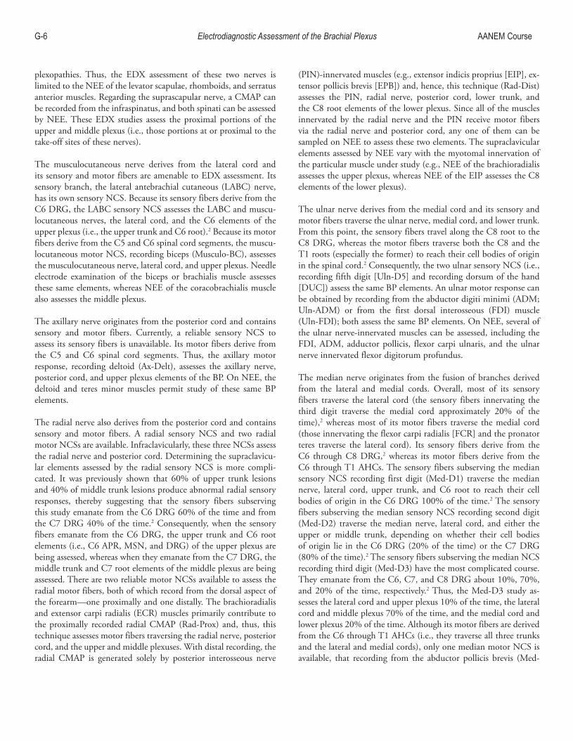
G-6 Electrodiagnostic Assessment of the Brachial Plexus AANEMCourse
plexopathies. Thus, the EDX assessment of these two nerves is limited to the NEE of the levator scapulae, rhomboids, and serratus anterior muscles. Regarding the suprascapular nerve, a CMAP can be recorded from the infraspinatus, and both spinati can be assessed by NEE. These EDX studies assess the proximal portions of the upper and middle plexus (i.e., those portions at or proximal to the take-off sites of these nerves).
The musculocutaneous nerve derives from the lateral cord and its sensory and motor fibers are amenable to EDX assessment. Its sensory branch, the lateral antebrachial cutaneous (LABC) nerve, has its own sensory NCS. Because its sensory fibers derive from the C6 DRG, the LABC sensory NCS assesses the LABC and muscu-locutaneous nerves, the lateral cord, and the C6 elements of the upper plexus (i.e., the upper trunk and C6 root).2 Because its motor fibers derive from the C5 and C6 spinal cord segments, the muscu-locutaneous motor NCS, recording biceps (Musculo-BC), assesses the musculocutaneous nerve, lateral cord, and upper plexus. Needle electrode examination of the biceps or brachialis muscle assesses these same elements, whereas NEE of the coracobrachialis muscle also assesses the middle plexus.
The axillary nerve originates from the posterior cord and contains sensory and motor fibers. Currently, a reliable sensory NCS to assess its sensory fibers is unavailable. Its motor fibers derive from the C5 and C6 spinal cord segments. Thus, the axillary motor response, recording deltoid (Ax-Delt), assesses the axillary nerve, posterior cord, and upper plexus elements of the BP. On NEE, the deltoid and teres minor muscles permit study of these same BP elements.
The radial nerve also derives from the posterior cord and contains sensory and motor fibers. A radial sensory NCS and two radial motor NCSs are available. Infraclavicularly, these three NCSs assess the radial nerve and posterior cord. Determining the supraclavicu-lar elements assessed by the radial sensory NCS is more compli-cated. It was previously shown that 60% of upper trunk lesions and 40% of middle trunk lesions produce abnormal radial sensory responses, thereby suggesting that the sensory fibers subserving this study emanate from the C6 DRG 60% of the time and from the C7 DRG 40% of the time.2 Consequently, when the sensory fibers emanate from the C6 DRG, the upper trunk and C6 root elements (i.e., C6 APR, MSN, and DRG) of the upper plexus are being assessed, whereas when they emanate from the C7 DRG, the middle trunk and C7 root elements of the middle plexus are being assessed. There are two reliable motor NCSs available to assess the radial motor fibers, both of which record from the dorsal aspect of the forearm—one proximally and one distally. The brachioradialis and extensor carpi radialis (ECR) muscles primarily contribute to the proximally recorded radial CMAP (Rad-Prox) and, thus, this technique assesses motor fibers traversing the radial nerve, posterior cord, and the upper and middle plexuses. With distal recording, the radial CMAP is generated solely by posterior interosseous nerve
(PIN)-innervated muscles (e.g., extensor indicis proprius [EIP], ex-tensor pollicis brevis [EPB]) and, hence, this technique (Rad-Dist) assesses the PIN, radial nerve, posterior cord, lower trunk, and the C8 root elements of the lower plexus. Since all of the muscles innervated by the radial nerve and the PIN receive motor fibers via the radial nerve and posterior cord, any one of them can be sampled on NEE to assess these two elements. The supraclavicular elements assessed by NEE vary with the myotomal innervation of the particular muscle under study (e.g., NEE of the brachioradialis assesses the upper plexus, whereas NEE of the EIP assesses the C8 elements of the lower plexus).
The ulnar nerve derives from the medial cord and its sensory and motor fibers traverse the ulnar nerve, medial cord, and lower trunk. From this point, the sensory fibers travel along the C8 root to the C8 DRG, whereas the motor fibers traverse both the C8 and the T1 roots (especially the former) to reach their cell bodies of origin in the spinal cord.2 Consequently, the two ulnar sensory NCS (i.e., recording fifth digit [Uln-D5] and recording dorsum of the hand [DUC]) assess the same BP elements. An ulnar motor response can be obtained by recording from the abductor digiti minimi (ADM; Uln-ADM) or from the first dorsal interosseous (FDI) muscle (Uln-FDI); both assess the same BP elements. On NEE, several of the ulnar nerve-innervated muscles can be assessed, including the FDI, ADM, adductor pollicis, flexor carpi ulnaris, and the ulnar nerve innervated flexor digitorum profundus.
The median nerve originates from the fusion of branches derived from the lateral and medial cords. Overall, most of its sensory fibers traverse the lateral cord (the sensory fibers innervating the third digit traverse the medial cord approximately 20% of the time),2 whereas most of its motor fibers traverse the medial cord (those innervating the flexor carpi radialis [FCR] and the pronator teres traverse the lateral cord). Its sensory fibers derive from the C6 through C8 DRG,2 whereas its motor fibers derive from the C6 through T1 AHCs. The sensory fibers subserving the median sensory NCS recording first digit (Med-D1) traverse the median nerve, lateral cord, upper trunk, and C6 root to reach their cell bodies of origin in the C6 DRG 100% of the time.2 The sensory fibers subserving the median sensory NCS recording second digit (Med-D2) traverse the median nerve, lateral cord, and either the upper or middle trunk, depending on whether their cell bodies of origin lie in the C6 DRG (20% of the time) or the C7 DRG (80% of the time).2 The sensory fibers subserving the median NCS recording third digit (Med-D3) have the most complicated course. They emanate from the C6, C7, and C8 DRG about 10%, 70%, and 20% of the time, respectively.2 Thus, the Med-D3 study as-sesses the lateral cord and upper plexus 10% of the time, the lateral cord and middle plexus 70% of the time, and the medial cord and lower plexus 20% of the time. Although its motor fibers are derived from the C6 through T1 AHCs (i.e., they traverse all three trunks and the lateral and medial cords), only one median motor NCS is available, that recording from the abductor pollicis brevis (Med-

AANEMCourse BrachialPlexusAssessment G-7
Figure 2 The pathway traversed through the brachial plexus bythe sensory nerve fibers subserving the lateral antebrachial cutaneoussensory nerve conduction study and, thus, the brachial plexus elementsassessedbythisstudy.
Figure 3 Thepathwaytraversedthroughthebrachialplexusbythesensorynervefiberssubservingthemediannerve(recordingfirstdigit)sensorynerveconductionstudyand,thus,thebrachialplexuselementsassessedbythisstudy.
Figure 5 The different pathways traversed through the brachialplexus by the sensory nerve fibers subserving the median nerve(recording third digit) sensory nerve conduction study and, thus, thepotential brachial plexus elements assessed by this study (the solidline indicates themorecommonpathway;10%toC6,70%toC7,and�0%toC8).
Figure 4 The different pathways traversed through the brachialplexusbythesensorynervefiberssubservingthemediannerve(record-ingseconddigit)sensorynerveconductionstudyand,thus,thepotentialbrachialplexuselementsassessedbythisstudy(thesolidlineindicatesthemorecommonpathway;�0%toC6and80%toC7).
APB). Since the APB is a C8,T1-innervated muscle (T1 > C8), this study assesses the median nerve, medial cord, and lower plexus elements of the brachial plexus.5 On NEE, the pronator teres and FCR, both innervated by motor fibers traversing the lateral cord, and the APB, innervated by fibers traversing the medial cord, are easily assessed. The flexor pollicis longus (an anterior interosseous nerve-innervated muscle) is also helpful.
The medial antebrachial cutaneous (MABC) nerve is composed of sensory fibers that traverse the proximal portion of the medial cord, the lower trunk, and the T1 root to reach the T1 DRG. The MABC sensory NCS is useful for assessing these BP elements. The
pathways traversed by the sensory nerve fibers subserving these seven sensory NCS are shown in Figures 2 through 8.2
ELECTRODIAGNOSTIC PATTERNS ASSOCIATED WITH REGIONAL/ELEMENTAL BRACHIAL PLEXOPATHIES
Although an isolated low-amplitude NCS response, stimulated and recorded distally, identifies an axon loss process, it does not localize the lesion. The lesion could be situated anywhere along the nerve. In addition, it could involve BP nerve fibers distally compos-ing the studied nerve. Thus, other NCS responses traversing the

G-8 Electrodiagnostic Assessment of the Brachial Plexus AANEMCourse
same elements must be assessed. Based on the pattern of normal and abnormal responses, a particular region or individual element may become highly probable or highly unlikely. For example, when median sensory and motor NCS response abnormalities are coupled with normal ulnar sensory and motor NCS responses, the lesion lies between the axilla (the site where the median nerve proper is formed) and the palm (the site where the median sensory and motor fibers diverge). However, when ulnar sensory, ulnar motor, and median motor response abnormalities are coupled with a normal Med-D2 response, the lesion must lie proximal to the median nerve and involve either the medial cord or the lower plexus. Knowing which studies assess which BP elements provides
the basis for this approach. Since the pathways that the sensory and motor fibers composing the various BP-derived nerves take through the plexus are known, the SNAP, CMAP, and muscle domains of each BP element can be derived (Tables 1, 2, and 3).1,2 This infor-mation is mandatory for BP lesion localization. Since the sensory fibers subserving the individual sensory NCS do not always traverse the BP elements in the same manner, their approximate reliability is shown in parenthesis (from Table 1).
C5 APR. Currently, the C5 APR does not have a SNAP domain since a reliable sensory NCS has not yet been devised for its as-sessment. Its CMAP domain includes the Musculo-BC and the Ax-Delt motor NCSs. Its muscle domain is equivalent to the C5 myotome, less the paraspinal muscles.
C6 APR. The SNAP domain of the C6 APR includes the LABC (100%), Med-D1 (100%), superficial radial (60%), Med-D2 (20%), and Med-D3 (10%) sensory NCSs.2 Its CMAP domain includes the Musculo-BC and the Ax-Delt motor NCSs. Its muscle domain is equivalent to the C6 myotome, less the paraspinal muscles.
C7 APR. The SNAP domain of the C7 APR includes the Med-D2 (80%), Med-D3 (70%), and superficial radial (40%) sensory NCSs.2 Its CMAP domain includes the radial motor NCS, record-ing anconeus. Its muscle domain is equivalent to the C7 myotome, less the paraspinal muscles.
C8 APR. The SNAP domain of the C8 APR includes the Uln-D5 (100%), DUC (100%), and Med-D3 (20%) sensory NCSs.2 Its CMAP domain includes the Rad-Dist, Uln-ADM, Uln-FDI, and Med-APB motor NCSs. Its muscle domain is equivalent to the C8
Figure 8Thepathwaytraversedthroughthebrachialplexusbythesensory nerve fibers subserving the medial antebrachial cutaneoussensorynerveconductionstudyand,thus,thebrachialplexuselementsassessedbythisstudy.
Figure 7Thepathwaytraversedthroughthebrachialplexusbythesensorynervefiberssubservingtheulnarnervesensorynerveconduc-tion study and, thus, the brachial plexus elements assessed by thisstudy.
Figure 6 The different pathways traversed through the brachialplexus by the sensory nerve fibers subserving the superficial radialnervesensorynerveconductionstudyand, thus, thepotentialbrachialplexus elements assessed by this study (the dotted lines indicate thetwooptions;60%toC6and�0%toC7).
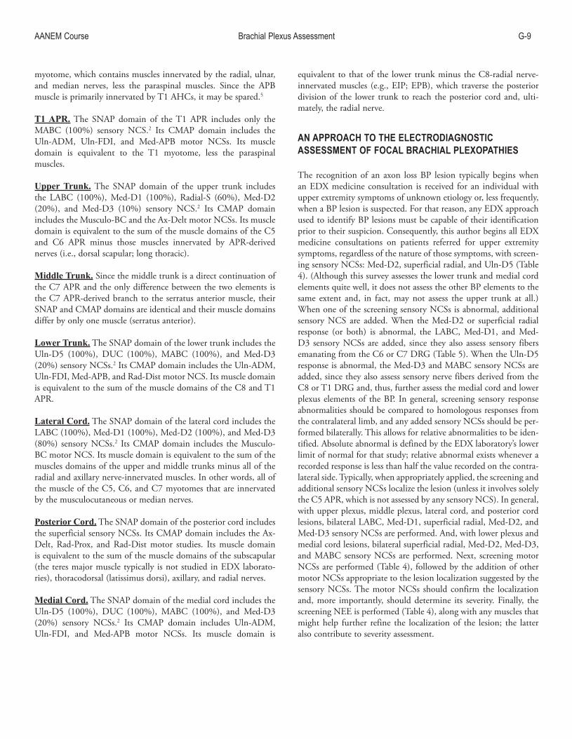
AANEMCourse BrachialPlexusAssessment G-�
myotome, which contains muscles innervated by the radial, ulnar, and median nerves, less the paraspinal muscles. Since the APB muscle is primarily innervated by T1 AHCs, it may be spared.5
T1 APR. The SNAP domain of the T1 APR includes only the MABC (100%) sensory NCS.2 Its CMAP domain includes the Uln-ADM, Uln-FDI, and Med-APB motor NCSs. Its muscle domain is equivalent to the T1 myotome, less the paraspinal muscles.
Upper Trunk. The SNAP domain of the upper trunk includes the LABC (100%), Med-D1 (100%), Radial-S (60%), Med-D2 (20%), and Med-D3 (10%) sensory NCS.2 Its CMAP domain includes the Musculo-BC and the Ax-Delt motor NCSs. Its muscle domain is equivalent to the sum of the muscle domains of the C5 and C6 APR minus those muscles innervated by APR-derived nerves (i.e., dorsal scapular; long thoracic).
Middle Trunk. Since the middle trunk is a direct continuation of the C7 APR and the only difference between the two elements is the C7 APR-derived branch to the serratus anterior muscle, their SNAP and CMAP domains are identical and their muscle domains differ by only one muscle (serratus anterior).
Lower Trunk. The SNAP domain of the lower trunk includes the Uln-D5 (100%), DUC (100%), MABC (100%), and Med-D3 (20%) sensory NCSs.2 Its CMAP domain includes the Uln-ADM, Uln-FDI, Med-APB, and Rad-Dist motor NCS. Its muscle domain is equivalent to the sum of the muscle domains of the C8 and T1 APR.
Lateral Cord. The SNAP domain of the lateral cord includes the LABC (100%), Med-D1 (100%), Med-D2 (100%), and Med-D3 (80%) sensory NCSs.2 Its CMAP domain includes the Musculo-BC motor NCS. Its muscle domain is equivalent to the sum of the muscles domains of the upper and middle trunks minus all of the radial and axillary nerve-innervated muscles. In other words, all of the muscle of the C5, C6, and C7 myotomes that are innervated by the musculocutaneous or median nerves.
Posterior Cord. The SNAP domain of the posterior cord includes the superficial sensory NCSs. Its CMAP domain includes the Ax-Delt, Rad-Prox, and Rad-Dist motor studies. Its muscle domain is equivalent to the sum of the muscle domains of the subscapular (the teres major muscle typically is not studied in EDX laborato-ries), thoracodorsal (latissimus dorsi), axillary, and radial nerves.
Medial Cord. The SNAP domain of the medial cord includes the Uln-D5 (100%), DUC (100%), MABC (100%), and Med-D3 (20%) sensory NCSs.2 Its CMAP domain includes Uln-ADM, Uln-FDI, and Med-APB motor NCSs. Its muscle domain is
equivalent to that of the lower trunk minus the C8-radial nerve-innervated muscles (e.g., EIP; EPB), which traverse the posterior division of the lower trunk to reach the posterior cord and, ulti-mately, the radial nerve.
AN APPROACH TO THE ELECTRODIAGNOSTIC ASSESSMENT OF FOCAL BRACHIAL PLEXOPATHIES
The recognition of an axon loss BP lesion typically begins when an EDX medicine consultation is received for an individual with upper extremity symptoms of unknown etiology or, less frequently, when a BP lesion is suspected. For that reason, any EDX approach used to identify BP lesions must be capable of their identification prior to their suspicion. Consequently, this author begins all EDX medicine consultations on patients referred for upper extremity symptoms, regardless of the nature of those symptoms, with screen-ing sensory NCSs: Med-D2, superficial radial, and Uln-D5 (Table 4). (Although this survey assesses the lower trunk and medial cord elements quite well, it does not assess the other BP elements to the same extent and, in fact, may not assess the upper trunk at all.) When one of the screening sensory NCSs is abnormal, additional sensory NCS are added. When the Med-D2 or superficial radial response (or both) is abnormal, the LABC, Med-D1, and Med-D3 sensory NCSs are added, since they also assess sensory fibers emanating from the C6 or C7 DRG (Table 5). When the Uln-D5 response is abnormal, the Med-D3 and MABC sensory NCSs are added, since they also assess sensory nerve fibers derived from the C8 or T1 DRG and, thus, further assess the medial cord and lower plexus elements of the BP. In general, screening sensory response abnormalities should be compared to homologous responses from the contralateral limb, and any added sensory NCSs should be per-formed bilaterally. This allows for relative abnormalities to be iden-tified. Absolute abnormal is defined by the EDX laboratory’s lower limit of normal for that study; relative abnormal exists whenever a recorded response is less than half the value recorded on the contra-lateral side. Typically, when appropriately applied, the screening and additional sensory NCSs localize the lesion (unless it involves solely the C5 APR, which is not assessed by any sensory NCS). In general, with upper plexus, middle plexus, lateral cord, and posterior cord lesions, bilateral LABC, Med-D1, superficial radial, Med-D2, and Med-D3 sensory NCSs are performed. And, with lower plexus and medial cord lesions, bilateral superficial radial, Med-D2, Med-D3, and MABC sensory NCSs are performed. Next, screening motor NCSs are performed (Table 4), followed by the addition of other motor NCSs appropriate to the lesion localization suggested by the sensory NCSs. The motor NCSs should confirm the localization and, more importantly, should determine its severity. Finally, the screening NEE is performed (Table 4), along with any muscles that might help further refine the localization of the lesion; the latter also contribute to severity assessment.

G-10 Electrodiagnostic Assessment of the Brachial Plexus AANEMCourse
DIFFERENTIATING BETWEEN VARIOUS BRACHIAL PLEXUS LOCALIZATIONS
Differentiating Between Upper Plexus and Lateral Cord Lesions
With upper plexus lesions, the LABC and Med-D1 responses usually are abnormal (since the sensory fibers being assessed emanate from the C6 DRG) and the Med-D2 and Med-D3 re-sponses tend to be spared (or affected to a lesser extent, since the sensory nerve fibers being assessed infrequently derive from the C6 DRG); the superficial radial response is affected when the superfi-cial radial fibers derive from the C6 DRG (60%).2 With lateral cord lesions, the LABC, Med-D1, Med-D2, and Med-D3 responses tend to be homogeneously affected (although the Med-D3 may be spared in the 20% of cases in which its fibers derive from the C8
Table 5TheDRGderivationsofthesensoryfiberssubservingvarioussensoryNCS
Sensory NCS Assessing C6 DRG-Derived Sensory FibersLateralantebrachialcutaneousMediansensory,recordingdigit1Radialsensory
Sensory NCS Assessing C7 DRG-Derived Sensory FibersRadialsensoryMediansensory,recordingdigit�Mediansensory,recordingdigit3
Sensory NCS Assessing C8 DRG- or T1 DRG-Derived Sensory FibersUlnarsensory,recordingdigit5DorsalulnarcutaneousMedialantebrachialcutaneous
DRG=dorsalrootganglion;NCS=nerveconductionstudy
Table 4UsefulEDXstudiesoftheupperextremity
Sensory Nerve Conduction StudiesMediansensorynerve,recordingdigit�*Ulnarsensorynerve,recordingdigit5*Radialsensorynerve,recordingdorsumofthehand*Lateralantebrachialcutaneousnerve,recordinglateralforearmDorsalulnarcutaneousnerve,recordingdorsumofhandMediansensorynerve,recordingdigit1Mediansensorynerve,recordingdigit3Medialantebrachialcutaneousnerve,recordingmedialforearm
Motor Nerve Conduction StudiesMedianmotornerve,recordingabductorpollicisbrevis*Ulnarmotornerve,recordingabductordigitiminimi*Musculocutaneousnerve,recordingbicepsAxillarynerve,recordingdeltoidRadialnerve,recordingdorsalaspectofproximalforearmRadialnerve,recordingdorsalaspectofdistalforearmUlnarmotornerve,recordingfirstdorsalinterosseous
Screening Needle Electrode ExaminationFirstdorsalinterosseousExtensorindicispropriusFlexorpollicislongusPronatorteresBicepsTricepsDeltoidParaspinalmuscles
*ScreeningnerveconductionstudiesthatareperformedonallindividualsreferredforupperextremityevaluationsEDX=electrodiagnostic

AANEMCourse BrachialPlexusAssessment G-11
DRG) and the superficial radial response is always spared (not a lateral cord assessor).2 On motor NCS, the Musculo-BC and Ax-Delt responses may be affected (depending on severity), whereas only the former can be affected with lateral cord lesions. On NEE, upper plexus lesions may be associated with dorsal scapular, long thoracic, suprascapular, axillary, and C5, C6-radial nerve inner-vated muscle abnormalities, whereas these muscles are unaffected by lateral cord lesions. When the dorsal scapular, long thoracic, and suprascapular nerve-innervated muscles are spared and the axillary and C5, C6-radial nerve innervated muscles are affected, the lesion likely involves the upper trunk element of the upper plexus, since the suprascapular nerve exits the upper trunk just after its forma-tion (i.e., it is usually spared with upper trunk lesions because it exits the latter so proximally).
Differentiating Between Middle Plexus and Posterior Cord Lesions
With middle plexus lesions, the Med-D2 (80%) and Med-D3 (70%) responses tend to be abnormal, whereas with posterior cord lesions they are always spared (since these studies are lateral cord assessors).2 On motor NCS, Rad-Dist and Ax-Delt response abnormalities indicate posterior cord involvement, since they do not assess the middle plexus. On NEE, C6, C7-median nerve in-nervated muscles (e.g., pronator teres; FCR) are affected by middle plexus, but not posterior cord, lesions. C5, C6-radial and axillary nerve innervated muscle abnormalities may be observed with poste-rior cord lesions, but not with middle plexus involvement.
Differentiating Between Lower Plexus and Medial Cord Lesions
With both lower plexus and medial cord lesions, the Uln-D5 and MABC sensory responses may be abnormal. The Med-D3 response is less frequently abnormal (20%).2 On motor NCS, ulnar and median motor response abnormalities may be associated with either. When the Rad-Dist response is abnormal, however, a medial cord lesion is excluded, since the C8-radial nerve innervated muscles receive their innervation via the lower plexus, posterior division of the lower trunk, and posterior cord (i.e., these fibers are not medial cord assessors). Likewise, on NEE, C8-radial nerve innervated muscle abnormalities indicate lower plexus involve-ment. Unfortunately, sparing of these muscles cannot be taken as
proof of a medial cord lesion because partial lower trunk lesions may spare the C8-radial motor fibers. Regarding the lower plexus, whenever the lesion involves predominantly the C8 APR or the T1 APR, differential EDX patterns frequently are observed.2,5,6 These differences reflect the fiber composition differences between these two elements. The C8 APR contains predominantly ulnar sensory and motor fibers, radial motor fibers (e.g., to EIP and EPB), as well as median motor fibers to those muscles receiving C8 input (e.g., flexor pollicis longus). The T1 APR contains predominantly MABC sensory and median motor fibers to those muscles receiv-ing T1 input (especially the APB muscle). For that reason, with T1 APR lesions (e.g., true neurogenic thoracic outlet syndrome), the MABC response is more affected than are the ulnar sensory responses, the Med-APB response is more affected than the ulnar motor responses, and the NEE shows the most pronounced ab-normalities in the APB muscle as compared to other muscles of the lower plexus muscle domain. Conversely, with C8 APR lesions (e.g., post-median sternotomy brachial plexopathy), the ulnar sensory responses are affected out of proportion to the MABC response (the latter is frequently spared), the ulnar motor responses are affected out of proportion to the Med-APB response (the latter is frequently spared), and NEE shows the APB muscle to be the least affected muscle of the lower plexus muscle domain.
SUMMARY
Despite its large size and complicated anatomical arrangement, the BP is very amenable to EDX evaluation. Utilizing the sensory NCSs as screening tools, the region of involvement can be deter-mined, permitting the motor NCS and NEE to further localize the lesion within the identified region and to determine its severity.
REFERENCES
1. Ferrante MA, Wilbourn AJ. Plexopathies. In: Levin KH, Luders HO, editors. Comprehensive clinical neurophysiology. Philadelphia: WB Saunders; 2000. p 201-214.
2. Ferrante MA, Wilbourn AJ. The utility of various sensory conduc-tion responses in assessing brachial plexopathies. Muscle Nerve 1995;18:879-889.

3. Gilliatt RW, Sears TA. Sensory nerve action potentials in patients with peripheral nerve lesions. J Neurol Neurosurg Psychiatry 1958;21:109-118.
4. Goss CM. Gray’s anatomy, 29th American edition. Philadelphia: Lea and Febiger; 1973.
5. Maggiano H, Levin KH, Wilbourn AJ. Relationship between the medial antebrachial cutaneous sensory and median motor responses in brachial plexopathies. Muscle Nerve 1993;16:1113-1114.
6. Nishida T, Price SJ, Minieka MM. Medial antebrachial cutaneous nerve conduction in true neurogenic thoracic outlet syndrome. Electromyogr Clin Neurophysiol 1993;33:285-288.
7. Stewart JD. Focal peripheral neuropathies. New York: Elsevier; 1987. p 97.
8. Sunderland S. Nerves and nerve injuries, 2nd edition. New York: Churchill Livingstone; 1978.
9. Wilbourn AJ. Brachial plexus lesions. In: Dyck PJ, Thomas PK, editors. Peripheral neuropathy, volume 2, 4th edition. Philadelphia: WB Saunders; 2005. p 1339-1373.
G-1� AANEMCourse

2006 AANEM Course F: SELF-ASSESSMENT EXAMINATION ANSWER SHEET
1. D
2. E
3. B
4. C
5. A
6. C
7. A
8. E
9. E
10. A
11. C
12. D
13. A
14. D
15. A
16. D
17. C
18. E
19. B
20. E
21. E
22. A
23. A
24. D
25. A
26. E
27. C
28. A
29. B
30. A
31. D
32. C
33. D
2006 AANEM Course G: SELF-ASSESSMENT EXAMINATION ANSWER SHEET
1. B
2. A
3. C
4. A
5. A
6. A
7. A
8. A
9. C
10. A
11. D
12. B
13. C
14. A
15. E
16. B
17. C
18. A
19. E
20. E
21. A
22. D
23. C
24. C
2006 AANEM Course H: SELF-ASSESSMENT EXAMINATION ANSWER SHEET
1. B
2. C
3. D
4. A
5. E
6. C
7. E
8. A
9. D
10. E
11. E
12. E
13. A
14. B
15. A
16. D
17. A
18. C
19. A
20. B
21. E
22. A
23. C
24. B