Cross-species approaches to cognitive neuroplasticity research
9
Cross-species approaches to cognitive neuroplasticity research J. Mishra a,b, ⁎, A. Gazzaley a,b,c, ⁎ a Department of Neurology, University of California, San Francisco, San Francisco, CA, USA b Department of Psychiatry, University of California, San Francisco, San Francisco, CA, USA c Department of Physiology, University of California, San Francisco, San Francisco, CA, USA abstract article info Article history: Accepted 1 September 2015 Available online xxxx Keywords: Neuroplasticity Cognition Cross-species Translational Intervention Animal model Neuroplasticity studies investigate the neural mechanisms that support learning-induced changes in cognition and behavior. These studies are performed in both experimental animals and humans across development from childhood to aging. Here, we review select recent studies that have sought to combine both animal and human neuroplasticity research within the same study. In investigating the same cognitive/behavioral functions in parallel in animals and humans, these studies take advantage of complementary neuroscience research methods that have been established for each species. In animals, these methods include investigations of genetic and molecular biomarker expression and micro-scale electrophysiology in single neurons in vivo or in brain slices. In humans, these studies assess macro-scale neural network dynamics using neuroimaging methods in- cluding EEG (electroencephalography) and functional and structural MRI (magnetic resonance imaging). Thus, by combining these diverse and complementary methodologies cross-species studies have the unique ability to bridge molecular, systems and cognitive neuroscience research. Additionally, they serve a vital role in transla- tional neuroscience, providing a direct bridge between animal models and human neuropsychiatric disorders. Comprehensive cross-species understanding of neural mechanisms at multiple scales of resolution and how these neural dynamics relate to behavioral outcomes, then serve to inform development and optimization of treatment strategies. © 2015 Elsevier Inc. All rights reserved. Introduction How do cross-species studies enrich neuroplasticity research, inform translational neuroscience and contribute to the development of novel interventions? In this review, we attempt to answer these questions by highlighting select recent studies that have performed cross-species experiments within the same study (Soliman et al., 2010; Pattwell et al., 2012; Sagi et al., 2012; Malter Cohen et al., 2013; Narayanan et al., 2013; Mishra et al., 2014). For each of these studies we describe: (1) the rationale for the cross-species investigation, (2) the experiments performed in animals and humans, and (3) how these experiments provide complementary insight into the cognitive/ behavioral phenomenon under investigation. In performing cross- species research, these studies are able to unite a diverse array of genet- ic, molecular, systems and cognitive neuroscience methods—invasive in animals and non-invasive in humans, and direct them at a specific neuroscientific question. Additionally, we discuss how the study outcomes contribute to translational research, especially toward the design and optimization of novel interventions. Next generation neuro- therapeutics will be powered by the rich diversity of individual informa- tion ranging from the genetic scale to the dynamics of macro-scale neural network interactions; cross-species studies are providing these insights in terms of how to integrate diverse neurobiological in- formation and thereby inform personalized interventions that are tai- lored to the biological state of the developing brain, by genotype as well as cognitive/behavioral phenotype (Lee et al., 2014; Casey et al., 2015). Indeed, such personalized interventions promise greater efficacy, which is aligned with the goals of precision medicine and N-of-1 trial in- vestigations (Schork, 2015). The studies that we review here span various aspects of learning from fear-conditioning (Soliman et al., 2010; Pattwell et al., 2012) and responding in a novel, threatening environment (Malter Cohen et al., 2013) to spatial learning (Sagi et al., 2012), learning control of motor errors (Narayanan et al., 2013), and maintaining cognitive performance in the presence of sensory distractions (Mishra et al., 2014). Interesting- ly, while they may appear disparate, what many of these studies have in common is the interaction between the frontal cortex, which enables pro-active top-down control of information processing, and more bottom-up sensory-motor and emotion processing brain regions. For in- stance, fear conditioning (Soliman et al., 2010; Pattwell et al., 2012) and responding in a novel, potentially threatening, environment (Malter NeuroImage xxx (2015) xxx–xxx ⁎ Corresponding authors at: University of California, San Francisco-Mission Bay, Sandler Neurosciences Center Room 502 and 511C, MC 0444 675 Nelson Rising Lane, San Francisco, CA 94158. E-mail addresses: [email protected] (J. Mishra), [email protected] (A. Gazzaley). YNIMG-12557; No. of pages: 9; 4C: 3, 4, 5, 6, 7, 8 http://dx.doi.org/10.1016/j.neuroimage.2015.09.002 1053-8119/© 2015 Elsevier Inc. All rights reserved. Contents lists available at ScienceDirect NeuroImage journal homepage: www.elsevier.com/locate/ynimg Please cite this article as: Mishra, J., Gazzaley, A., Cross-species approaches to cognitive neuroplasticity research, NeuroImage (2015), http:// dx.doi.org/10.1016/j.neuroimage.2015.09.002
Transcript of Cross-species approaches to cognitive neuroplasticity research

NeuroImage xxx (2015) xxx–xxx
YNIMG-12557; No. of pages: 9; 4C: 3, 4, 5, 6, 7, 8
Contents lists available at ScienceDirect
NeuroImage
j ourna l homepage: www.e lsev ie r .com/ locate /yn img
Cross-species approaches to cognitive neuroplasticity research
J. Mishra a,b,⁎, A. Gazzaley a,b,c,⁎a Department of Neurology, University of California, San Francisco, San Francisco, CA, USAb Department of Psychiatry, University of California, San Francisco, San Francisco, CA, USAc Department of Physiology, University of California, San Francisco, San Francisco, CA, USA
⁎ Corresponding authors at: University of California, SanNeurosciences Center Room 502 and 511C, MC 0444Francisco, CA 94158.
E-mail addresses: [email protected] (J. Mishra), ad(A. Gazzaley).
http://dx.doi.org/10.1016/j.neuroimage.2015.09.0021053-8119/© 2015 Elsevier Inc. All rights reserved.
Please cite this article as: Mishra, J., Gazzaledx.doi.org/10.1016/j.neuroimage.2015.09.00
a b s t r a c t
a r t i c l e i n f oArticle history:Accepted 1 September 2015Available online xxxx
Keywords:NeuroplasticityCognitionCross-speciesTranslationalInterventionAnimal model
Neuroplasticity studies investigate the neural mechanisms that support learning-induced changes in cognitionand behavior. These studies are performed in both experimental animals and humans across developmentfrom childhood to aging. Here, we review select recent studies that have sought to combine both animal andhuman neuroplasticity research within the same study. In investigating the same cognitive/behavioral functionsin parallel in animals and humans, these studies take advantage of complementary neuroscience researchmethods that have been established for each species. In animals, thesemethods include investigations of geneticand molecular biomarker expression and micro-scale electrophysiology in single neurons in vivo or in brainslices. In humans, these studies assess macro-scale neural network dynamics using neuroimaging methods in-cluding EEG (electroencephalography) and functional and structural MRI (magnetic resonance imaging). Thus,by combining these diverse and complementary methodologies cross-species studies have the unique abilityto bridge molecular, systems and cognitive neuroscience research. Additionally, they serve a vital role in transla-tional neuroscience, providing a direct bridge between animal models and human neuropsychiatric disorders.Comprehensive cross-species understanding of neural mechanisms at multiple scales of resolution and howthese neural dynamics relate to behavioral outcomes, then serve to inform development and optimization oftreatment strategies.
© 2015 Elsevier Inc. All rights reserved.
Introduction
How do cross-species studies enrich neuroplasticity research,inform translational neuroscience and contribute to the developmentof novel interventions? In this review, we attempt to answer thesequestions by highlighting select recent studies that have performedcross-species experiments within the same study (Soliman et al.,2010; Pattwell et al., 2012; Sagi et al., 2012; Malter Cohen et al., 2013;Narayanan et al., 2013; Mishra et al., 2014). For each of these studieswe describe: (1) the rationale for the cross-species investigation,(2) the experiments performed in animals and humans, and (3) howthese experiments provide complementary insight into the cognitive/behavioral phenomenon under investigation. In performing cross-species research, these studies are able to unite a diverse array of genet-ic, molecular, systems and cognitive neurosciencemethods—invasive inanimals and non-invasive in humans, and direct them at a specificneuroscientific question. Additionally, we discuss how the study
Francisco-Mission Bay, Sandler675 Nelson Rising Lane, San
y, A., Cross-species approach2
outcomes contribute to translational research, especially toward thedesign and optimization of novel interventions. Next generation neuro-therapeuticswill be poweredby the richdiversity of individual informa-tion ranging from the genetic scale to the dynamics of macro-scaleneural network interactions; cross-species studies are providingthese insights in terms of how to integrate diverse neurobiological in-formation and thereby inform personalized interventions that are tai-lored to the biological state of the developing brain, by genotype aswell as cognitive/behavioral phenotype (Lee et al., 2014; Casey et al.,2015). Indeed, such personalized interventions promise greater efficacy,which is alignedwith the goals of precisionmedicine andN-of-1 trial in-vestigations (Schork, 2015).
The studies that we review here span various aspects of learningfrom fear-conditioning (Soliman et al., 2010; Pattwell et al., 2012) andresponding in a novel, threatening environment (Malter Cohen et al.,2013) to spatial learning (Sagi et al., 2012), learning control of motorerrors (Narayanan et al., 2013), andmaintaining cognitive performancein the presence of sensory distractions (Mishra et al., 2014). Interesting-ly, while theymay appear disparate, whatmany of these studies have incommon is the interaction between the frontal cortex, which enablespro-active top-down control of information processing, and morebottom-up sensory-motor and emotion processing brain regions. For in-stance, fear conditioning (Soliman et al., 2010; Pattwell et al., 2012) andresponding in a novel, potentially threatening, environment (Malter
es to cognitive neuroplasticity research, NeuroImage (2015), http://
2 J. Mishra, A. Gazzaley / NeuroImage xxx (2015) xxx–xxx
Cohen et al., 2013) invoke interactions between the prefrontal–amygdalar networks. Prediction error-driven learning involves inter-action between medial frontal cortex and motor cortex (Narayananet al., 2013). And learning to suppress sensory distractions involvesprefrontal-sensory cortical dynamics (Mishra et al., 2014). Cross-species investigations in these different learning domains are enabledby the preservation and homology of neural network function across ro-dents and humans (Buzsáki et al., 2013). Additionally, neurotrophicfactors, especially the brain derived neurotrophic factor (BDNF) playsa key role inmodulating synaptic plasticity in these networks (reviewedin Rattiner et al., 2005; Casey et al., 2015). BDNF promotes neuronalsurvival and differentiation, andmediates long-term potentiation espe-cially in the hippocampus, which facilitates learning and memory con-solidation. Thus, genetic variation in BDNF expression is of interest tomany of these cross-species studies. Overall, common findings acrossanimals and humans confirm the translatability of animal experimentresults toward understanding the human brain. Here, we provide anoverview of these recent cross-species studies with a common empha-sis in each on the rationale for cross-species research, the experimentalmethods and species-convergent outcomes.
Cross-species understanding of fear learning
Learning to respond to dangerous threats in our environment is anevolutionary necessity. In fear conditioning, an initially non-fear-inducing cue is predictably associated with a threat until the individuallearns the cue–threat association and starts to physiologically respondto the cue as if it were itself threatening. Rodents demonstrate a freezingresponse to the conditioning cue and humans show a pronounced stressresponse as measured by elevated galvanic skin conductance responses(SCR). The learned fear responses can also be extinguished by repeated-ly presenting the cue dissociated from the threat. This is referred to asfear extinction and is the foundation for exposure therapy used in thetreatment of anxiety disorders, phobias and post-traumatic stress disor-der. Exposure therapy is needed in these neuropsychiatric conditions, asfear extinction is abnormal, i.e. individuals continue to have an abnor-mal aversive response to neutral cues.
Soliman et al. (2010) applied a cross-species approach to investi-gate the role of the BDNF gene in fear extinction. The BDNF gene is sus-ceptible to a common mutation at codon 66 where valine (Val) getssubstituted for methionine (Met). The Val/Val genotype is thus thetypical form of BDNF, while theMet allele (Val/Met andMet/Met) is as-sociated with treatment resistant anxiety-like behaviors (Chen et al.,2006). The rationale for the Soliman et al. (2010) study was to test ifBDNF Met allele carriers indeed show differences in fear extinctionfrom non-carriers, in both mice and humans. If this were shown to bethe case then there would be compelling evidence for a genetic mousemodel for anxiety disorders and a standard fear extinction paradigmcould be used to test novel targeted anxiolytic therapeutics developedin mice or humans. The study indeed showed that both mice andhuman who were Met allele carriers showed impaired fear-extinction.Wildtype mice and typical humans progressively reduced their fear re-sponse over early vs. late extinction trials, as measured by percent timefreezing in mice and SCR in humans, but Met allele carriers did not(Fig. 1A, B). The authors additionally performed a functional MRI(fMRI) study in humans and showed enhanced activity in ventromedialprefrontal cortex (vmPFC) and reduced responses in the amygdaladuring extinction in typical humans. Met carriers showed opposingresults, i.e. significantly reduced vmPFC activation and significantlyelevated amygdala activity (Fig. 1C, D). Thus, Met carriers had alteredfronto-amygdalar circuitry. Many studies show that vmPFC signalsinhibit the amygdala and thus regulate fear responses (reviewedin Milad and Quirk, 2012). So in the absence of vmPFC activation,Met allele carriers continue to have heightened fear responses support-ed by the elevated amygdalar activity. The fMRI findings thus extendthe validity of the cross-species translation beyond behavior to
Please cite this article as: Mishra, J., Gazzaley, A., Cross-species approachdx.doi.org/10.1016/j.neuroimage.2015.09.002
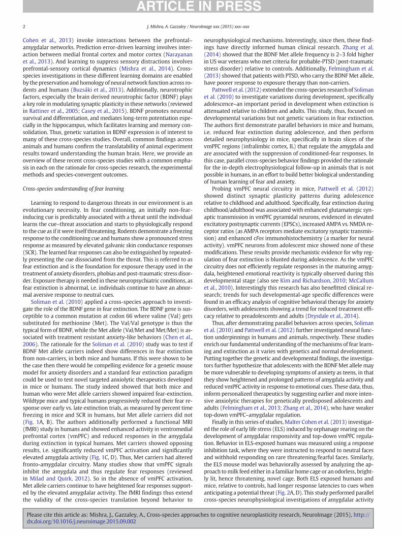
neurophysiological mechanisms. Interestingly, since then, these find-ings have directly informed human clinical research. Zhang et al.(2014) showed that the BDNF Met allele frequency is 2–3 fold higherin USwar veterans whomet criteria for probable-PTSD (post-traumaticstress disorder) relative to controls. Additionally, Felmingham et al.(2013) showed that patients with PTSD, who carry the BDNFMet allele,have poorer response to exposure therapy than non-carriers.
Pattwell et al. (2012) extended the cross-species research of Solimanet al. (2010) to investigate variations during development, specificallyadolescence–an important period in development when extinction isattenuated relative to children and adults. This study, thus, focused ondevelopmental variations but not genetic variations in fear extinction.The authors first demonstrate parallel behaviors in mice and humans,i.e. reduced fear extinction during adolescence, and then performdetailed neurophysiology in mice, specifically in brain slices of thevmPFC regions (infralimbic cortex, IL) that regulate the amygdala andare associated with the suppression of conditioned-fear responses. Inthis case, parallel cross-species behavior findings provided the rationalefor the in-depth electrophysiological follow-up in animals that is notpossible in humans, in an effort to build better biological understandingof human learning of fear and anxiety.
Probing vmPFC neural circuitry in mice, Pattwell et al. (2012)showed distinct synaptic plasticity patterns during adolescencerelative to childhood and adulthood. Specifically, fear extinction duringchildhood/adulthoodwas associated with enhanced glutamatergic syn-aptic transmission in vmPFC pyramidal neurons, evidenced in elevatedexcitatory postsynaptic currents (EPSCs), increased AMPA vs. NMDA re-ceptor ratios (as AMPA receptors mediate excitatory synaptic transmis-sion) and enhanced cFos immunohistochemistry (a marker for neuralactivity). vmPFC neurons from adolescent mice showed none of thesemodifications. These results provide mechanistic evidence for why reg-ulation of fear extinction is blunted during adolescence. As the vmPFCcircuitry does not efficiently regulate responses in the maturing amyg-dala, heightened emotional reactivity is typically observed during thisdevelopmental stage (also see Kim and Richardson, 2010; McCallumet al., 2010). Interestingly this research has also benefitted clinical re-search; trends for such developmental-age specific differences werefound in an efficacy analysis of cognitive behavioral therapy for anxietydisorders, with adolescents showing a trend for reduced treatment effi-cacy relative to preadolescents and adults (Drysdale et al., 2014).
Thus, after demonstrating parallel behaviors across species, Solimanet al. (2010) and Pattwell et al. (2012) further investigated neural func-tion underpinnings in humans and animals, respectively. These studiesenrich our fundamental understanding of themechanisms of fear learn-ing and extinction as it varies with genetics and normal development.Putting together the genetic and developmental findings, the investiga-tors further hypothesize that adolescents with the BDNFMet allele maybe more vulnerable to developing symptoms of anxiety as teens, in thatthey show heightened and prolonged patterns of amygdala activity andreduced vmPFC activity in response to emotional cues. These data, thus,inform personalized therapeutics by suggesting earlier and more inten-sive anxiolytic therapies for genetically predisposed adolescents andadults (Felmingham et al., 2013; Zhang et al., 2014), who have weakertop-down vmPFC–amygdalar regulation.
Finally in this series of studies,Malter Cohen et al. (2013) investigat-ed the role of early life stress (ELS) induced by orphanage rearing on thedevelopment of amygdalar responsivity and top-down vmPFC regula-tion. Behavior in ELS-exposed humans was measured using a responseinhibition task, where they were instructed to respond to neutral facesand withhold responding on rare threatening/fearful faces. Similarly,the ELS mouse model was behaviorally assessed by analyzing the ap-proach tomilk feed either in a familiar home cage or an odorless, bright-ly lit, hence threatening, novel cage. Both ELS exposed humans andmice, relative to controls, had longer response latencies to cues whenanticipating a potential threat (Fig. 2A, D). This study performed parallelcross-species neurophysiological investigations of amygdalar activity
es to cognitive neuroplasticity research, NeuroImage (2015), http://
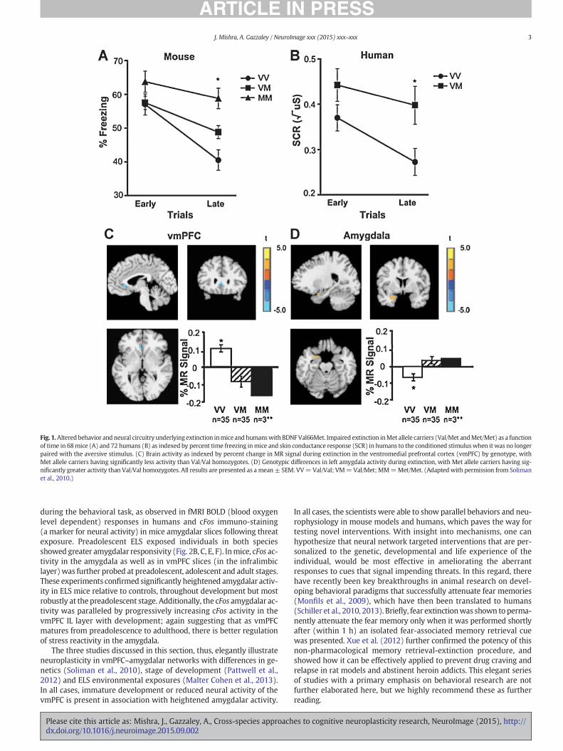
Fig. 1.Altered behavior and neural circuitry underlying extinction inmice and humanswith BDNFVal66Met. Impaired extinction inMet allele carriers (Val/Met andMet/Met) as a functionof time in 68mice (A) and 72 humans (B) as indexed by percent time freezing inmice and skin conductance response (SCR) in humans to the conditioned stimuluswhen it was no longerpaired with the aversive stimulus. (C) Brain activity as indexed by percent change in MR signal during extinction in the ventromedial prefrontal cortex (vmPFC) by genotype, withMet allele carriers having significantly less activity than Val/Val homozygotes. (D) Genotypic differences in left amygdala activity during extinction, with Met allele carriers having sig-nificantly greater activity than Val/Val homozygotes. All results are presented as a mean± SEM. VV=Val/Val; VM=Val/Met; MM=Met/Met. (Adaptedwith permission from Solimanet al., 2010.)
3J. Mishra, A. Gazzaley / NeuroImage xxx (2015) xxx–xxx
during the behavioral task, as observed in fMRI BOLD (blood oxygenlevel dependent) responses in humans and cFos immuno-staining(a marker for neural activity) in mice amygdalar slices following threatexposure. Preadolescent ELS exposed individuals in both speciesshowed greater amygdalar responsivity (Fig. 2B, C, E, F). Inmice, cFos ac-tivity in the amygdala as well as in vmPFC slices (in the infralimbiclayer)was further probed at preadolescent, adolescent and adult stages.These experiments confirmed significantly heightened amygdalar activ-ity in ELS mice relative to controls, throughout development but mostrobustly at the preadolescent stage. Additionally, the cFos amygdalar ac-tivity was paralleled by progressively increasing cFos activity in thevmPFC IL layer with development; again suggesting that as vmPFCmatures from preadolescence to adulthood, there is better regulationof stress reactivity in the amygdala.
The three studies discussed in this section, thus, elegantly illustrateneuroplasticity in vmPFC–amygdalar networks with differences in ge-netics (Soliman et al., 2010), stage of development (Pattwell et al.,2012) and ELS environmental exposures (Malter Cohen et al., 2013).In all cases, immature development or reduced neural activity of thevmPFC is present in association with heightened amygdalar activity.
Please cite this article as: Mishra, J., Gazzaley, A., Cross-species approachdx.doi.org/10.1016/j.neuroimage.2015.09.002
In all cases, the scientists were able to show parallel behaviors and neu-rophysiology in mouse models and humans, which paves the way fortesting novel interventions. With insight into mechanisms, one canhypothesize that neural network targeted interventions that are per-sonalized to the genetic, developmental and life experience of theindividual, would be most effective in ameliorating the aberrantresponses to cues that signal impending threats. In this regard, therehave recently been key breakthroughs in animal research on devel-oping behavioral paradigms that successfully attenuate fear memories(Monfils et al., 2009), which have then been translated to humans(Schiller et al., 2010, 2013). Briefly, fear extinctionwas shown to perma-nently attenuate the fear memory only when it was performed shortlyafter (within 1 h) an isolated fear-associated memory retrieval cuewas presented. Xue et al. (2012) further confirmed the potency of thisnon-pharmacological memory retrieval-extinction procedure, andshowed how it can be effectively applied to prevent drug craving andrelapse in rat models and abstinent heroin addicts. This elegant seriesof studies with a primary emphasis on behavioral research are notfurther elaborated here, but we highly recommend these as furtherreading.
es to cognitive neuroplasticity research, NeuroImage (2015), http://

Fig. 2. Greater amygdala activity in humans and mice following ELS (early life stress). (A) Stressed preadolescent humans take longer than their standard reared counterparts to detectfrequently presented neutral targets embedded among rare threat nontarget cues that they were instructed to ignore. (B) Parameter estimates of amygdala activity in response to thethreat cue (i.e., fearful face) were greater in stressed preadolescent humans than their standard-reared counterparts. (C) Bilateral regions of the amygdala identified as more reactiveto threat (i.e., fear face stimuli) in stressed preadolescent humans than their standard reared counterparts. (D) The difference in time that control and stressed preadolescent mice taketo approach a cue in a novel cage compared with their home cage. (E) The density of c-Fos protein in the amygdala following exposure to the threatening context (i.e., novel cage) wasgreater in stressed preadolescent mice than their standard-reared counterparts. (F) An individual slice cut through the amygdala taken from each mouse was stained for c-Fos (red)and PVA (parvalbumin, green) and used for quantification of c-Fos following exposure to the threatening context, clustered by experimental group and at 10× magnification. All dataare z-scored and expressed as means ± SEM. (Adapted with permission from Malter Cohen et al., 2013.)
4 J. Mishra, A. Gazzaley / NeuroImage xxx (2015) xxx–xxx
Cross-species plasticity underlying rapid learning
While in the previous section, we described a set of neuroplasticitystudies at the prolonged timescales over the course of neurode-velopment, early life experiences and hereditary genetics, here wedescribe a cross-species neuroplasticity study at the very rapid time-scale of two hours. Sagi et al. (2012) tested spatial learning in humansusing a car racing video game and asked whether changes in brainstructure can be observed after two hours of learning. For this, theyemployed diffusion tensor imaging (DTI) in humans before and aftertraining, which is sensitive to self-diffusion of water molecules andserves as a marker of tissue architecture. Mean diffusivity (MD) is anoutcomemeasure, which is high if there is ample space between tissue(such as neurons, glia and blood vessels) for water to move freely, butMD is lower if tissue spacing is reduced, possibly driven by growthand proliferation of new neurons, glia or blood vessels (Johansen-Berg et al., 2012). In MD maps generated two hours apart, Sagi et al.(2012) showed significant MD decreases in the hippocampus andparahippocampus—brain regions that are particularly important forspatial learning and memory (Fig. 3A). The study also showed thatthis structural brain plasticity was behaviorally relevant in that fasterlearners showed greater decreases inMD. Additionally, an active controlgroup, which also practiced game-based car driving, but without anyrepetitive learning on a specific spatial track, and a non-training controlgroup did not show these structural changes (Fig. 3B).
To gain a deeper understanding of the rapid structural changes asso-ciated with short-term spatial learning, Sagi et al. (2012) performed a
Please cite this article as: Mishra, J., Gazzaley, A., Cross-species approachdx.doi.org/10.1016/j.neuroimage.2015.09.002
parallel experiment in rats learning awatermaze task. Sowhile humanslearned the spatial organization of a car-racing track in a video game,rats learned to memorize the location of a hidden platform in a waterpool using spatial cues. So even though short-term spatial learningwas implemented in both species, apparent differences in task para-digms should also be kept in mind when evaluating the convergenceof results between rats and humans.
Like humans, rats showed a decrease in MD in the posterior hippo-campus after twohours of spatial learning (Fig. 3C, D). Follow-up immu-nohistochemistry in the hippocampus of learners vs. controls showedenhanced synaptophysin (a marker of synaptic vesicles), glial fibrillaryacidic protein (GFAP; amarker of astrocyte activation andproliferation),and BDNF. These markers suggest that regions of MD decrease maybe undergoing rapid structural proliferation of synaptic vesicles andastrocytes with learning. Specifically, activation, proliferation and re-modeling of astrocyte and glial processes, which support the cellularlearning network, possibly make the most prominent contribution tothe MD signal at the rapid hourly learning timescales (Sagi et al.,2012; Johansen-Berg et al., 2012). This is because DTI does not havesufficient resolution to capture changes at the level of synapses resultingfrom synaptogenesis, and evidence in the literature suggests thatneurogenesis and angiogenesis occur on much longer timescales ofdays to weeks, but not hours.
Overall, the cross-species investigation in this study enabledSagi et al. (2012) to draw links between microscale cellular dynamicsand macroscale measures of structural change. Many open questionsremain—how do these structural dynamics evolve with time, are they
es to cognitive neuroplasticity research, NeuroImage (2015), http://

Fig. 3. Structural remodeling of brain tissue measured by DTI as changes inMD (mean diffusivity) after 2 h of training on a spatial learning and memory task. Panels A and B show humandata and C andD show rat data. (A) Significant decreases inMDwith learning are seen in the humanhippocampus and (B) only in the learning group (LG) but not two control groups (CG1and 2). (C) The posterior hippocampus in rats shows decreases in MD after learning, parallel to findings in humans, and again only in the learning group (L) but not the active control orpassive untrained control (C and P). (Adapted with permission from Sagi et al., 2012.)
5J. Mishra, A. Gazzaley / NeuroImage xxx (2015) xxx–xxx
ephemeral or lead to stable and sustained structural changes, andwhat learning parameters govern long-term structural stability? Futurecross-species studies are needed to comprehensively answer thesequestions and further our understanding of associations between themicro- and macro-scale neural dynamics.
Cross-species plasticity underlying error-related adaptive learning
Adaptive learning is essential for rapid learning; it allows individualsto appropriately change behavior in response to errors. It is also im-portant to study as it is compromised in several neuropsychiatric con-ditions (Ridderinkhof et al., 2004; Velligan et al., 2002; Fitzgeraldet al., 2005; van Meel et al., 2007), and understanding of its neuralmechanisms will lead to better targeted diagnostics and treatments.Narayanan et al. (2013) conducted a cross-species investigation ofadaptive learning focusing on themedial frontal cortex (MFC), especial-ly the anterior cingulate, as it has been shown to be involved in adaptiveerror control in both animals and humans. The authors employed asimple time estimation task in both humans and rats in which a re-sponse was required after an estimated time interval (human, 1.4 s;rat, 1 s). EEG and intracortical field potentials were recorded simulta-neous with task engagement, at mid-frontal electrodes in humans andmedial frontal sites in rodents, respectively. Event related potentials(ERPs) at these sites confirmed conserved processing across species,i.e. enhanced signaling post-error vs. post-correct trials (Fig. 4A, B).Spectral decomposition showed a significant increase in low frequencypower (4–8 Hz theta oscillations in humans) exclusively on post-errortrials, which was significantly correlated with response latencies(slower responses post-error vs. post-correct trials). These results inhumans were paralleled in rats by the intracortical MFC recordings in
Please cite this article as: Mishra, J., Gazzaley, A., Cross-species approachdx.doi.org/10.1016/j.neuroimage.2015.09.002
the 4–25 Hz frequency range. These data demonstrate that humansand rodents share features of error-driven adaptive control throughlow-frequency oscillations in the MFC (Fig. 4C, D).
The authors then conducted a detailed electrophysiological inves-tigation of MFC activity and MFC-motor cortex interactions in therat. Neurophysiological finding were consistent with the role of MFCin behavioral monitoring: (1) Phase coherence of local field poten-tials (LFP) across MFC and motor cortex sites was enhanced post-errortrials. (2) MFC single neuron spikes were coupled to the enhanced lowfrequency local-field oscillations only on post-error trials. (3) A largefraction of the nearly 100 investigated MFC neurons encoded priorbehavioral outcome, while current response latency was encodedbymotor cortex neurons. (4) Pharmacological inactivation ofMFC elim-inated both post-error adaptive control behaviors and underlyingneural mechanisms. The inactivated-MFC rats showed greater propor-tion of errors, shorter response latencies and no post-error slowing.Mechanistically, the selective expression of low-frequency oscillationsin motor cortex post-errors, as well as spike-field coherence of motorcortex neurons exclusively post-errors, was eliminated in inactivated-MFC rats.
These results demonstrate the causal role of MFC in adaptive controlof action, as well as how it is achieved via selective post-error couplingof motor neuron spike activity to the low frequency oscillations gener-ated by theMFC. The study suggests that individualswith a dysfunction-al MFC may function in a mode that is less cognitively flexible and maynot benefit from information about previous behavioral outcomes. Theconserved neurobehavioral signatures of adaptive control across speciesfurther suggest that novel interventions, including targeted neuro-pharmacology, neurostimulation as well as closed-loop neurofeedbackapproaches (Mishra and Gazzaley, 2014) that enhance MFC function
es to cognitive neuroplasticity research, NeuroImage (2015), http://

Fig. 4. Commonmechanisms of medial frontal cortical oscillations during adaptive control in rats and humans. (A) Average event-related potentials over themidfrontal cortex (electrodeCz) in humans aligned to the target time. Amplitudes were significantly increased in post-error (red) as compared to post-correct (black) trials. (B) Rodent medial frontal field potentialswere also significantly increased in post-error (red) as compared to post-correct (black) trials; the results were highly similar to those in humans. (C) Time-frequency analysis revealingenhanced low-frequency power after errors trials relative to correct trial in humans and (D) in rodents. (Adapted with permission from Narayanan et al., 2013.)
6 J. Mishra, A. Gazzaley / NeuroImage xxx (2015) xxx–xxx
can now be tested at multiple levels of neural resolution in animals andhumans.
Cross-species validation of behavioral closed loop cognitive training toresolve sensory distractions
We recently conducted a cross-species intervention study to evalu-ate a targeted and personalized training to ameliorate distractibility inaging (Mishra et al., 2014). The study, conducted in aged rodents andolder humans, wasmotivated by previous demonstrations of a selectivedeficit in suppressing distractions in both older animals and humans(de Villers-Sidani et al., 2010; Hasher et al., 1999; Gazzaley et al.,2005; Gazzaley, 2013); a deficit that had not been addressed by anyother neurotherapeutic approach. In this study, we developed a novelbehavioral closed loop distractor training program implemented inthe auditory domain. Trainees discriminated specific target tone fre-quencies from other distracting tones. The training was closed loopand adaptive on each trial, such that correct behavioral performanceled to increased distractor challenge (distractor tone frequenciesbecame more similar to the target frequency), while incorrect behaviorreduced distractor challenge (distractor tone frequencies became dis-similar to the target frequency). Both older rats and humans performed36 training sessions, each with a unique set of targets and distractors,over a one-month period. Behavioral and neural outcomes of distractortraining were compared to repeatedmeasurements (one-month apart)in untrained control groups. In humans, we also compared results ofadaptive distractor training to amechanistic control, which implement-ed adaptive target training. The latter training appeared exactly thesame as the distractor training with the goal to discriminate targetsand distractors; but in this case, training challenge adapted targets(target frequency became more similar to that of distractors on correctresponses or becamemore dissimilar to distractor frequencies on incor-rect responses).
In both species, behavioral and neural outcome measures revealedthat training resulted in selective improvements in the suppressionof distractions; and that these results were specific to the adaptivedistractor training intervention and were not observed in controlgroups. Behaviorally, both trained rats and humans made fewerdistractor-related false positive errors (Fig. 5A, B). Neural outcomes
Please cite this article as: Mishra, J., Gazzaley, A., Cross-species approachdx.doi.org/10.1016/j.neuroimage.2015.09.002
were investigated in the two species using complementary electrophys-iological methods—single neuron recordings and auditory tonotopicmaps were probed in anesthetized animals and whole-brain EEG wasrecorded in humans on a target vs. distractor discrimination assess-ment. Notably, anesthetized recordings in animals allowed us to discernthe extent of neuroplasticity in auditory sensory cortex in the absence oftop-down regulation, which is not possible in humans.
In rats, single neuron electrophysiology showed selective suppres-sion of distractor processing; in contrast the response to oddball targetswas unchanged (Fig. 5C). Tonotopic maps of auditory cortex showedenhanced spatial and spectral resolution of sound frequencies. Inhumans, early sensory event-related responses to distractors, whichlocalized to auditory cortex, were selectively diminished post-training(Fig. 5D). Similar to findings in rats, responses to targets remained unal-tered, and neither of the control groups showed this outcome. Thisevent-related distractor suppression was behaviorally relevant as itcorrelated to the training gain in sensory resolution. The neural changefurther correlated with several transfer measures (i.e. assessments ofcognitive benefits beyond the trained task), specifically improvementsin working memory span and sustained auditory attention. BeyondERPs, spectral analyses of the human data showed neuroplasticity infrontal low frequency responses (in the theta band). Frontal oscillationslocalized to themiddle frontal cortex near the inferior frontal junction, aregion known to play a critical role in interference resolution (Brasset al., 2005; Zanto et al., 2011). Frontal theta was selectively restrainedin response to distractors post-training, while the theta response to tar-gets was enhanced. Finally, measures of frontal-sensory theta phase co-herence showed selective suppression of this network during post-training distractor processing.
In summary, we found converging evidence in rats and humansthat closed loop adaptive distractor training can alleviate deficits indistractor processing. The novel deficit-targeted training procedurewas able to harness plasticity at multiple neural scales. (1) At themicro-scale, it achieved selective distractor suppression in single neu-rons of rat auditory cortex, and thereby improved the signal-to-noiseresolution of tonotopic maps. (2) At the macro-scale, it selectivelyreduced early sensory processing of distractor ERPs in humans. (3) Inhumans, low frequency oscillations in frontal cortex were also selec-tively restrained to distractors and frontal-sensory communication
es to cognitive neuroplasticity research, NeuroImage (2015), http://

Untrained older human Trained older human
A B
C D
300 ms
Fig. 5. Evidence for behavioral and neural distractor suppression selectively achieved after adaptive distractor training. (A) Training progressively improved the ability to discriminate tar-get tones amidst varied distractor tones in both rodents and humans (lower octave values imply better discrimination resolution) (B) Underlying the gains in sensory resolution was aselective reduction in false positive errors on distractor trials at the end of training ‘T2’ assessment relative to baseline, ‘T1’. Bar graphs on the right show no significant change in targethits in both species. Data aremeans±SEM. (C) Single neurons in auditory cortex of the trained rat show suppression of distractor processingwhen a sequence of distractor tones is playedto the anesthetized rat ear with infrequent oddball tone stimuli. The green horizontal lines represent the response asymptote of the sample neuron to the oddball and repeating distractortones; only the response asymptote to the distractors, but not targets, is significantly reduced in the trained rat. (D) ERP recordings to distractor stimuli in humans show a selective re-duction in distractor processing at 150–160 ms post-stimulus onset at the T2 vs. T1 assessment, while responses in the control group remain unaltered. Positive microvolt deflectionsare plotted below the horizontal axis. (Adapted with permission from Mishra et al., 2014.)
7J. Mishra, A. Gazzaley / NeuroImage xxx (2015) xxx–xxx
(coherence) was suppressed for distractors post-training. Additionally,we showed that this neuroplasticity is directly associated with behav-ioral gains on-task as well as benefits in untrained transfer measuresof sustained attention and working memory span. Overall, our resultsclearly demonstrated the utility of using a cross-species neuroscientificapproach to validate a novel intervention. The study further highlightedhow adaptive trainingmechanics can be specifically focused on a cogni-tive deficit, heightened distractibility in this case, to generate selectiveimprovements within that neuro-cognitive domain. Notably, theseinsights inform future closed loop neurotherapeutic research; theyprovide proof-of-principle that it is possible to engineer closed looptraining programs that are selectively targeted to specific behavioral,cognitive and even neural network deficits (Mishra and Gazzaley,2014).
Discussion
Here, we provide an overview of recent advances in cross-speciesneuroplasticity research. Each of these studies has significantly con-tributed toward a deeper scientific understanding of how the brain sup-ports learning. Diverse aspects of learning have been investigated, fromnegative emotion/fear learning, to spatial learning, to error-relatedlearning, to learning to resolve sensory interference. In performingcross-species research, all of these studies have utilized complementarymethodologies in animals and humans within the same study, whichconverge to provide insights that would be impossible to glean fromresearch in a single species alone. In these studies, expression of par-allel behaviors in both species is most commonly the rationale forperformingmore in-depth neurobiological investigations, using physio-logical or structural assessment methods appropriate for each species.The mechanistic research integrates findings from genetics, immu-nohistochemistry and micro-scale electrophysiology in animals with
Please cite this article as: Mishra, J., Gazzaley, A., Cross-species approachdx.doi.org/10.1016/j.neuroimage.2015.09.002
measures of macro-scale network dynamics probed using EEG/ MRIneuroimaging in humans (Fig. 6).
These cross-species studies substantially contribute to translationalresearch and they generate more credibility for the use of animalmodels that parallel human behavior (Fig. 6). The detailed neuro-mechanistic knowledge gained from all of these studies has the poten-tial to inform the development of novel interventions, as is already thecase for our recent study (Mishra et al., 2014). Of note, the breakthroughstudies byMonfils et al. (2009), Schiller et al. (2010, 2013) andXue et al.(2012) that performed a series of animal and human behavioral exper-iments across multiple studies to show robust unlearning of fearfulmemories and addictions, are also very relevant here. Together, thesestudies inform new interventions, which can now be developed totarget an individual's traits such as genetics, life experience anddevelopmental stage as well as their dynamic neural and cognitivestates. For instance, the cross-species translational research on fearlearning has already directly informed clinical findings of greater riskof PTSD and poor response to exposure therapy in BDNF Met allelecarriers (Felmingham et al., 2013; Zhang et al., 2014), and the trendfor poor treatment efficacy in adolescents relative to other age groups(Drysdale et al., 2014). Yet, much research still remains to systematical-ly investigate parallels in neuroplasticity across species as a function ofage, genetics and life experience.
Mechanistically informed intervention development is extremelyimportant if we are to deliver therapeutics that provide comprehensiveand long-lasting benefit to the individual—not just at the level of ob-servable behavior, but also underlying brain function. Furthermore,the task paradigms and assessments in these studies can now be usedas biomarkers and outcome measures to evaluate the effectiveness offuture intervention research. If an intervention significantly benefitsneuro-cognitive status it can be readied for validation in large samplesize randomized controlled human trials, which are then followedby studies of practical implementation in the field. In contrast, if the
es to cognitive neuroplasticity research, NeuroImage (2015), http://

Animal Model
Genetics
Human Study
Cognition Behavior
Development
micro-scale
cellular and
network plasticity
macro-scale
network
plasticity
Plasticity Mechanisms
Biomarker Identification
Translational Research
Fig. 6. The diagram encapsulates how cross-species studies inform neuroscience. Animal models and human studies are best integrated in research when they demonstrate conservedgenetics, developmental profiles, and outcomes on specific cognitive and behavioral assessments (top row). In-depth investigations in animal models provide insights onmicro-scale cel-lular and network plasticity (bottom left). Cellular activity profiles of both neurons and glia are measured using immunohistochemistry and slice and in-vivo electrophysiology (synapticcurrents,firing rates, spike-field and LFP coherence). Human research incorporates neuroimaging, EEG andMRI, which informmacro-scalewhole brain neural network dynamics (bottomright). Comprehensive and converging findings provided by thesemethods contribute to discovery of neuroplasticity mechanisms, identification of biomarkers that can be used to assessefficacy of targeted interventions and to the development of mechanistic interventions in translational research (center).
8 J. Mishra, A. Gazzaley / NeuroImage xxx (2015) xxx–xxx
neuro-cognitive assessments developed in these cross-species studiesas biomarkers, are only mildly impacted by a new intervention, then iturges informed iterative improvement of the intervention. In everyway, building comprehensive knowledge of underlying neural mecha-nisms using cross-species research models is a win–win for interven-tional research. At the same time, we must not only take advantagesof the converging findings across species, but also be aware of the dis-similarities in network complexity and in experimental protocols andnot over-interpret outcomes.
Conclusion
Here, we have presented a case for the usefulness of cross-speciesneuroscience research in uncovering mechanisms of neuroplasticityand informing novel targeted interventions. In our opinion, such re-search is especially critical for the future of translational neurosciences.Engaging in cross-species research is challenging, as it requires collabo-rations between neuroscientists with diverse expertise. Yet, it is this di-versity of knowledge that spurs innovations, and we urge the initiationof more studies using the cross-species research model.
Acknowledgments
This work was supported by a grant from the NIH R01AG040333 iscofounder and chief science advisor of Akili Interactive Labs. J.M. andA.G. are co-inventors on a patent for “Methods of Suppressing IrrelevantStimuli”; our research associated with this patent is discussed here.
References
Brass, M., Derrfuss, J., Forstmann, B., von Cramon, D.Y., 2005. The role of the inferior fron-tal junction area in cognitive control. Trends Cogn. Sci. 9, 314–316.
Buzsáki, G., Logothetis, N., Singer, W., 2013. Scaling brain size, keeping timing: evolution-ary preservation of brain rhythms. Neuron 80, 751–764.
Casey, B.J., Glatt, C.E., Lee, F.S., 2015. Treating the developing versus developed brain:translating preclinical mouse and human studies. Neuron 86, 1358–1368.
Chen, Z.-Y., Jing, D., Bath, K.G., Ieraci, A., Khan, T., Siao, C.-J., Herrera, D.G., Toth,M., Yang, C.,McEwen, B.S., Hempstead, B.L., Lee, F.S., 2006. Genetic variant BDNF (Val66Met) poly-morphism alters anxiety-related behavior. Science 314, 140–143.
De Villers-Sidani, E., Alzghoul, L., Zhou, X., Simpson, K.L., Lin, R.C.S., Merzenich, M.M.,2010. Recovery of functional and structural age-related changes in the rat primaryauditory cortex with operant training. Proc. Natl. Acad. Sci. U. S. A. 107, 13900–13905.
Please cite this article as: Mishra, J., Gazzaley, A., Cross-species approachdx.doi.org/10.1016/j.neuroimage.2015.09.002
Drysdale, A.T., Hartley, C.A., Pattwell, S.S., Ruberry, E.J., Somerville, L.H., Compton, S.N., Lee,F.S., Casey, B.J., Walkup, J.T., 2014. Fear and anxiety from principle to practice: impli-cations for when to treat youth with anxiety disorders. Biol. Psychiatry 75, e19–e20.
Felmingham, K.L., Dobson-Stone, C., Schofield, P.R., Quirk, G.J., Bryant, R.A., 2013. Thebrain-derived neurotrophic factor Val66Met polymorphism predicts response to ex-posure therapy in posttraumatic stress disorder. Biol. Psychiatry 73, 1059–1063.
Fitzgerald, K.D., Welsh, R.C., Gehring, W.J., Abelson, J.L., Himle, J.A., Liberzon, I., Taylor, S.F.,2005. Error-related hyperactivity of the anterior cingulate cortex in obsessive-compulsive disorder. Biol. Psychiatry 57, 287–294.
Gazzaley, A., 2013. Top-down modulation deficit in the aging brain: an emerging theoryof cognitive aging. In: Knight, R.T., Stuss, D.T. (Eds.), Principles of Frontal Lobe Func-tion. Oxford University Press, USA, pp. 593–608.
Gazzaley, A., Cooney, J.W., Rissman, J., D'Esposito, M., 2005. Top-down suppression deficitunderliesworkingmemory impairment in normal aging. Nat. Neurosci. 8, 1298–1300.
Hasher, L., Zacks, R., May, C., 1999. Inhibitory control, circadian arousal, and age. In:Gopher, D., Koriat, A. (Eds.), Attention and Performance, XVII, Cognitive Regulationof Performance: Interaction of Theory and Application. MIT Press, Cambridge, MA,pp. 653–675.
Johansen-Berg, H., Baptista, C.S., Thomas, A.G., 2012. Human structural plasticity at recordspeed. Neuron 73, 1058–1060.
Kim, J.H., Richardson, R., 2010. New findings on extinction of conditioned fear early in de-velopment: theoretical and clinical implications. Biol. Psychiatry 67, 297–303.
Lee, F.S., Heimer, H., Giedd, J.N., Lein, E.S., Šestan, N., Weinberger, D.R., Casey, B.J., 2014.Mental health. Adolescent mental health—opportunity and obligation. Science 346,547–549.
Malter Cohen, M., Jing, D., Yang, R.R., Tottenham, N., Lee, F.S., Casey, B.J., 2013. Early-lifestress has persistent effects on amygdala function and development in mice andhumans. Proc. Natl. Acad. Sci. 110, 18274–18278.
McCallum, J., Kim, J.H., Richardson, R., 2010. Impaired extinction retention in adolescentrats: effects of D-cycloserine. Neuropsychopharmacology 35, 2134–2142.
Milad, M.R., Quirk, G.J., 2012. Fear extinction as a model for translational neuroscience:ten years of progress. Annu. Rev. Psychol. 63, 129–151.
Mishra, J., Gazzaley, A., 2014. Closed-loop rehabilitation of age-related cognitive disorders.Semin. Neurol. 34, 584–590.
Mishra, J., de Villers-Sidani, E., Merzenich, M., Gazzaley, A., 2014. Adaptive training dimin-ishes distractibility in aging across species. Neuron 84, 1091–1103.
Monfils, M.-H., Cowansage, K.K., Klann, E., LeDoux, J.E., 2009. Extinction–reconsolidationboundaries: key to persistent attenuation of fear memories. Science 324, 951–955.
Narayanan, N.S., Cavanagh, J.F., Frank, M.J., Laubach, M., 2013. Common medial frontalmechanisms of adaptive control in humans and rodents. Nat. Neurosci. 16, 1888–1895.
Pattwell, S.S., Duhoux, S., Hartley, C.A., Johnson, D.C., Jing, D., Elliott, M.D., Ruberry, E.J.,Powers, A., Mehta, N., Yang, R.R., Soliman, F., Glatt, C.E., Casey, B.J., Ninan, I., Lee,F.S., 2012. Altered fear learning across development in both mouse and human.Proc. Natl. Acad. Sci. U. S. A. 109, 16318–16323.
Rattiner, L.M., Davis, M., Ressler, K.J., 2005. Brain-derived neurotrophic factor inamygdala-dependent learning. Neuroscientist 11, 323–333.
Ridderinkhof, K.R., Ullsperger, M., Crone, E.A., Nieuwenhuis, S., 2004. The role of the me-dial frontal cortex in cognitive control. Science 306, 443–447.
Sagi, Y., Tavor, I., Hofstetter, S., Tzur-Moryosef, S., Blumenfeld-Katzir, T., Assaf, Y., 2012.Learning in the fast lane: new insights into neuroplasticity. Neuron 73, 1195–1203.
Schiller, D., Monfils, M.-H., Raio, C.M., Johnson, D.C., Ledoux, J.E., Phelps, E.A., 2010.Preventing the return of fear in humans using reconsolidation update mechanisms.Nature 463, 49–53.
es to cognitive neuroplasticity research, NeuroImage (2015), http://

9J. Mishra, A. Gazzaley / NeuroImage xxx (2015) xxx–xxx
Schiller, D., Kanen, J.W., LeDoux, J.E., Monfils, M.-H., Phelps, E.A., 2013. Extinction duringreconsolidation of threat memory diminishes prefrontal cortex involvement. Proc.Natl. Acad. Sci. U. S. A. 110, 20040–20045.
Schork, N.J., 2015. Personalizedmedicine: time for one-person trials. Nature 520, 609–611.Soliman, F., Glatt, C.E., Bath, K.G., Levita, L., Jones, R.M., Pattwell, S.S., Jing, D., Tottenham,
N., Amso, D., Somerville, L.H., Voss, H.U., Glover, G., Ballon, D.J., Liston, C., Teslovich,T., Van Kempen, T., Lee, F.S., Casey, B.J., 2010. A genetic variant BDNF polymorphismalters extinction learning in both mouse and human. Science 327, 863–866.
VanMeel, C.S., Heslenfeld, D.J., Oosterlaan, J., Sergeant, J.A., 2007. Adaptive control deficitsin attention-deficit/hyperactivity disorder (ADHD): the role of error processing.Psychiatry Res. 151, 211–220.
Velligan, D.I., Ritch, J.L., Sui, D., DiCocco, M., Huntzinger, C.D., 2002. Frontal systems behav-ior scale in schizophrenia: relationships with psychiatric symptomatology, cognitionand adaptive function. Psychiatry Res. 113, 227–236.
Please cite this article as: Mishra, J., Gazzaley, A., Cross-species approachdx.doi.org/10.1016/j.neuroimage.2015.09.002
Xue, Y.-X., Luo, Y.-X., Wu, P., Shi, H.-S., Xue, L.-F., Chen, C., Zhu, W.-L., Ding, Z.-B., Bao, Y.,Shi, J., Epstein, D.H., Shaham, Y., Lu, L., 2012. Amemory retrieval-extinction procedureto prevent drug craving and relapse. Science 336, 241–245.
Zanto, T.P., Rubens, M.T., Thangavel, A., Gazzaley, A., 2011. Causal role of the prefrontalcortex in top-down modulation of visual processing and working memory. Nat.Neurosci. 14, 656–661.
Zhang, L., Benedek, D.M., Fullerton, C.S., Forsten, R.D., Naifeh, J.A., Li, X.X., Hu, X.Z., Li, H., Jia,M., Xing, G.Q., Benevides, K.N., Ursano, R.J., 2014. PTSD risk is associated with BDNFVal66Met and BDNF overexpression. Mol. Psychiatry 19, 8–10.
es to cognitive neuroplasticity research, NeuroImage (2015), http://