Critical Care Suite 2 - GE Healthcare
8
gehealthcare.com Critical Care Suite 2.0 ON-DEVICE ARTIFICIAL INTELLIGENCE. HELPING YOU WHEN EVERY MINUTE COUNTS.
Transcript of Critical Care Suite 2 - GE Healthcare

gehealthcare.com
Critical Care Suite 2.0 ON-DEVICE ARTIFICIAL INTELLIGENCE. HELPING YOU WHEN EVERY MINUTE COUNTS.

Critical Care Suite is a collection of AI algorithms embedded on X-ray systems for automated measurements, detection and triage of critical conditions and quality control
In these challenging times, radiographers, radiologists, and physicians are all under tremendous pressure to manage an ever-increasing number of cases. Every minute counts when dealing with high-risk procedures such as tracheal intubation1 and critical conditions including pneumothorax. When X-ray exams alone contribute to 60 percent of imaging,2 it’s crucial to highlight critical information and help clinicians respond fast without compromising diagnostic precision.
C R I T I C A L C A R E S U I T E 2 . 0
2
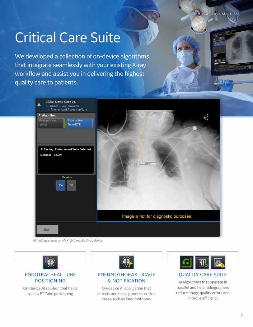
Critical Care SuiteWe developed a collection of on-device algorithms that integrate seamlessly with your existing X-ray workflow and assist you in delivering the highest quality care to patients.
ENDOTRACHEAL TUBE POSITIONING
On-device AI solution that helps assess ET Tube positioning
PNEUMOTHORAX TRIAGE & NOTIFICATION
On-device AI application that detects and helps prioritize critical
cases such as Pneumothorax
QUALITY CARE SUITEAI algorithms that operate in
parallel and help radiographers reduce image quality errors and
improve efficiency
C R I T I C A L C A R E S U I T E 2 . 0
AI findings shown on AMX™ 240 mobile X-ray device
3

Endotracheal Tube Positioning One of the high-risk procedures carried out in the ICU and ER is tracheal intubation. Improper positioning of the endotracheal tube during intubation poses a serious health risk to patients. A major complication is accidental migration of the tube tip into a mainstem bronchus. If unrecognized, this can lead to hypoxemia and collapse of the contralateral lung, hyperinflation of the intubated lung with resultant tension pneumothorax, and cardiac arrest.
Confidence at Point of Care• Provides an accurate and automated measurement of
ET Tube position on the mobile X-ray device within secondsof image acquisition
• On-device algorithm automatically detects the presence ofET tube in AP chest X-ray images on mobile X-ray
• In 94% of cases the ET Tube tip-to-Carina distance calculationis accurate to within 1.0 cm7
• The vertical distance between the tube tip and the carina isautomatically calculated and displayed on device
C R I T I C A L C A R E S U I T E 2 . 0
Up to 25% of patients intubated outside of the operating room have mispositioned ETTs on chest X-rays3-6
ET Tube Cases at a Glance• Enables immediate access to AI-derived
measurements in PACS worklist viaconfigurable DICOM® tags
• Displays AI-generated measurementswith an image overlay in PACS
Critical Care Suite’s Endotracheal Tube Positioning algorithm is available on AMX 240 mobile X-ray systems.
THE FIRST ON-DEVICE AI SOLUTION THAT HELPS ASSESS ET TUBE POSITIONINGGE Healthcare’s Critical Care Suite 2.0 now includes an AI algorithm that helps assess endotracheal tube positioning.
4

Pneumothorax Detection and Triage Given the high number of chest X-rays ordered as “STAT,” i.e. with immediate priority, the triaging of true STATs has become challenging for radiographers and radiologists. Turnaround times can be as long as eight hours, even when chest X-rays are marked as urgent for patients with potentially life-threatening conditions such as Pneumothorax, or collapsed lung.8,9
ON-DEVICE AI APPLICATION THAT HELPS PRIORITIZE PNEUMOTHORAXCritical Care Suite includes the world’s first, on-device AI solution that can detect and help triage critical conditions, such as pneumothorax, alleviating the overwhelming demand that urgent X-rays are placing on radiology teams.
High Accuracy10
• Detects nearly all large pneumothoraces(96% sensitivity)
• Identifies 3 out of 4 small pneumothoraces(75% sensitivity)
• Limits false alerts (94% specificity)
• An Area Under Curve (AUC) of 0.96
Triage Notifications• Sends a secondary capture DICOM image to PACS
and presents the AI results to the radiologist
• Image flags help enable worklist prioritization andhave the potential to expedite review of criticalfindings
Pneumothorax detection and triage is included in Critical Care Suite 2.0 and is available on AMX 240 mobile X-ray systems as well as Definium™ 656 HD and Definium 646 HD fixed X-ray systems.
C R I T I C A L C A R E S U I T E 2 . 0
5

Quality Care SuiteMinutes matter when intubating a critically ill patient or dealing with a collapsed lung. Every minute spent imaging the patient matters; every moment processing the medical image matters; every hour that passes before an image is reviewed matters – and it’s vital the process is as fast as possible.
Intelligent Auto RotateSaves technologists 3–4 user
interface clicks on more than 80% of mobile chest X-ray exams, saving
up to 70,000 “clicks” a year7
Intelligent Protocol CheckConducts an automated quality
check to detect errors on the acquisition system, such as improper
protocol used (AUC >0.99)10
Intelligent Field of ViewDetects when a lung field is clipped in a frontal chest X-ray (AUC >0.99) and allows technologists to determine if a repeat is required before sending the
image to PACS10
REAL-TIME QUALITY ALERTSQuality Care Suite includes AI algorithms that operate in parallel and help technologists reduce image quality errors and improve efficiency.
Quality Care Suite is included in Critical Care Suite 2.0 and is available on AMX 240 mobile X-ray systems as well as Definium 656 HD and Definium 646 HD fixed X-ray systems.
C R I T I C A L C A R E S U I T E 2 . 0
6

Seamless integration with your X-ray imaging workflowCritical Care Suite’s on-device AI algorithms automatically analyze X-rays without routing images to a server. The AI output is sent directly to PACS via a secondary capture DICOM image.
Built to improve the standard of care you’re delivering to patientsCritical Care Suite is the world’s first collection of on-device AI algorithms for automated measurements, detection and triage of critical conditions and quality control.
AI embedded on device, at point of care without need for additional IT infrastructure
Critical Care Suite embedded on X-ray device
DICOM PACS
Robust AI algorithm trained on a large, global,diverse data set>30K images, multiple countries and institutions
Seamless integration with PACS for rapid review by the radiologistAI-derived measurement is sent to PACS within configurable public DICOM tags
On-device AI notifications to radiographer:
• Intelligent Auto Rotate
• Intelligent Protocol Check
• Intelligent Field of View
• Pneumothorax Detection & Triage
• Endotracheal Tube Positioning
Original DICOM image
+
Secondary capture DICOM image for AI output
+
Public DICOM tags
PTX triage flag on PACS worklist
Immediate access to AI-derived
measurements in worklist
AI-generated measurements
displayed on PACS with an image
overlay
C R I T I C A L C A R E S U I T E 2 . 0
7

References:
1. Stephen E. Lapinsky. Endotracheal intubation in the ICU. Crit Care. 2015; 19(1): 258.
2. World Health Organization Report -Communicating Radiation Risks in Pediatric Imaging.
3. Jemmett ME, Kendal KM, Fourre MW, Burton JH. Unrecognized misplacement of endotracheal tubes in a mixed urban to rural emergency medical services setting. Acad Emerg Med 2003;10:961–5.
4. Katz SH, Falk JL. Misplaced endotracheal tubes by paramedics in an urban emergency medical services system. Ann Emerg Med 2001;37:32–7.
5. Lotano R, Gerber D, Aseron C, Santarelli R, Pratter M. Utility of postintubation chest radiographs in the intensive care unit. Crit Care 2000;4:50–3.
6. McGillicuddy DC et al. Is a postintubation chest radiograph necessary in the emergency department? Rachh, Pratik, et al. “Reducing STAT Portable Chest Radiograph Turnaround Times: Int J Emerg Med 2009;2:247–9.
7. GE Healthcare Data on File.
8. A Pilot Study.” Current problems in diagnostic radiology (2017).
9. Lorenz, Jonathan, and Matthew Blum. “Complications of percutaneous chest biopsy.” Seminars in interventional radiology. Vol. 23. No. 2. Thieme Medical Publishers, 2006.
10. FDA 510(k) K183182.
© 2020 General Electric Company – All rights reserved.
GE Healthcare reserves the right to make changes in specifications and features shown herein, or discontinue the product described at any time without notice or obligation. Contact your GE Healthcare representative for the most current information. GE, the GE Monogram, AMX and Definium are trademarks of General Electric Company. DICOM is a trademark of National Electrical Manufacturers Association. GE Healthcare, a division of General Electric Company. GE Medical Systems, Inc., doing business as GE Healthcare. All other third party trademarks are the property of their respective owners.
November 2020JBxxxxxxx