Could Licorice prevent Bisphenol A-Induced Biochemical ...
15
73 Ain Shams Journal of Forensic Medicine and Clinical Toxicology Jan 2018, 30:73-87 Could Licorice prevent Bisphenol A-Induced Biochemical, Histopathological and Genetic Effects in the Adult Male Albino Rats? Walaa Yehia Abdelzaher 1 , Dalia Mohamed Ali 2 and Wagdy K. B. Khalil 3 1 Department of Pharmacology, Faculty of Medicine, Minia University, Minia, Egypt. 2 Department of Forensic Medicine and Clinical Toxicology, Faculty of Medicine, Minia University, Minia, Egypt. 3 Department of Cell Biology, National Research Centre, Egypt. All right received. Introduction isphenol A (BPA) is an ecological estrogenic endocrine disruptors with similar chemical structure to estradiol and diethylstilbestrol. It is one of the most common worldwide chemicals (Ritter, 2011) that have been commonly used in polycarbonate plastics as drinking bottles, toys and dental sealant and in epoxy resins lining containers for food and beverage (Fenichel et al., 2013). Exposure of polycarbonate products to high temperature, acidic or basic compounds and repeated washing hastens hydrolysis of the ester bond linking BPA molecules and degradation of the polymers leading to leakage of BPA from food and beverage containers (Lim et al., 2009). Nowadays, BPA are everywhere in the environment. Human at different ages become in contact with BPA continuously through packaged food, drinking water, dermal exposure, and inhalation of household dusts (Lakind and Naiman, 2011). BPA and their metabolites have been detected in about 92.6 % of the human population bodies (Kitraki et al., 2015). European Food Safety Authority set 0.05 mg/kg body weight as the standard tolerable daily dose of BPA. Recently, the safe daily human exposure limit to BPA has been lowered to 4 μg/kg/day (EFSA, 2015). As a result of increasing evidence for the hazards of exposure to BPA, more and more studies have been conducted to prove the side effects of BPA on different body organs either in vivo or in vitro (Ahbab et al., 2017; Fang et al., 2015). The adverse effects of BPA are mostly due to its estrogenic activity (Kurosawa et al., 2002). Their actions are mediated by endocrine signaling pathways that resulting in large changes in the cell functions even at very low concentrations (Welshons et al., 2003). These changes have deleterious effects on health including prostate and breast cancer (Prins et al., 2008; Pupo et al., 2012). It has been reported that BPA induces oxidative stress in different body organs (Kabuto et al., 2004; Mourad and Khadrawy, 2012). Furthermore, other effects of BPA have been recorded such as mitochondrial mediated apoptosis in the hepatic tissue (Xia et al., 2014) and inflammatory cytokine dysregulation (Ben-Jonathan et al., 2009). Abstract Bisphenol A (BPA) is an ecological estrogenic endocrine disruptor used commonly in polycarbonate plastics. This study aimed to investigate the biochemical, histopathological and genetic effects of BPA at different doses and to evaluate the protective role of licorice against such effects. Thirty Wistar male albino rats were divided into five groups administered BPA daily at 2.4 μg/kg and 500 mg/kg orally with or without licorice (150 mg/kg) for 4 weeks. The results revealed that the high toxic dose decreased GSH, SOD and catalase levels and increased MDA level significantly. Serum TNF-α, testosterone and testicular cholesterol levels were significantly decreased while serum alkaline phosphatase was significantly increased. Histopathological changes were observed in testes, lungs and stomach. Alteration in the expression of NF-κB1 gene in lung occurred. These results suggested that BPA induced oxidative stress; resulted in its complication in the examined rats and treatment with licorice alleviated the toxicity induced by BPA. Keywords Bisphenol A, licorice, oxidative stress, genetic B
Transcript of Could Licorice prevent Bisphenol A-Induced Biochemical ...

73
Ain Shams Journal of Forensic Medicine and Clinical Toxicology
Jan 2018, 30:73-87
Could Licorice prevent Bisphenol A-Induced Biochemical, Histopathological and Genetic Effects in the Adult Male Albino Rats?
Walaa Yehia Abdelzaher1, Dalia Mohamed Ali2 and Wagdy K. B. Khalil3
1 Department of Pharmacology, Faculty of Medicine, Minia University, Minia, Egypt. 2 Department of Forensic Medicine and Clinical Toxicology, Faculty of Medicine, Minia University, Minia, Egypt. 3 Department of Cell Biology, National Research Centre, Egypt.
All right received.
Introduction isphenol A (BPA) is an ecological estrogenic
endocrine disruptors with similar chemical
structure to estradiol and diethylstilbestrol. It is
one of the most common worldwide chemicals (Ritter,
2011) that have been commonly used in polycarbonate
plastics as drinking bottles, toys and dental sealant and in
epoxy resins lining containers for food and beverage
(Fenichel et al., 2013).
Exposure of polycarbonate products to high
temperature, acidic or basic compounds and repeated
washing hastens hydrolysis of the ester bond linking
BPA molecules and degradation of the polymers leading
to leakage of BPA from food and beverage containers
(Lim et al., 2009). Nowadays, BPA are everywhere in the
environment. Human at different ages become in contact
with BPA continuously through packaged food, drinking
water, dermal exposure, and inhalation of household
dusts (Lakind and Naiman, 2011).
BPA and their metabolites have been detected in
about 92.6 % of the human population bodies (Kitraki et
al., 2015). European Food Safety Authority set 0.05
mg/kg body weight as the standard tolerable daily dose
of BPA. Recently, the safe daily human exposure limit to
BPA has been lowered to 4 μg/kg/day (EFSA, 2015).
As a result of increasing evidence for the
hazards of exposure to BPA, more and more studies have
been conducted to prove the side effects of BPA on
different body organs either in vivo or in vitro (Ahbab et
al., 2017; Fang et al., 2015).
The adverse effects of BPA are mostly due to its
estrogenic activity (Kurosawa et al., 2002). Their actions
are mediated by endocrine signaling pathways that
resulting in large changes in the cell functions even at
very low concentrations (Welshons et al., 2003). These
changes have deleterious effects on health including
prostate and breast cancer (Prins et al., 2008; Pupo et al.,
2012). It has been reported that BPA induces oxidative
stress in different body organs (Kabuto et al., 2004;
Mourad and Khadrawy, 2012). Furthermore, other effects
of BPA have been recorded such as mitochondrial
mediated apoptosis in the hepatic tissue (Xia et al., 2014)
and inflammatory cytokine dysregulation (Ben-Jonathan
et al., 2009).
Abstract Bisphenol A (BPA) is an ecological estrogenic endocrine disruptor used commonly in polycarbonate
plastics. This study aimed to investigate the biochemical, histopathological and genetic effects of BPA at
different doses and to evaluate the protective role of licorice against such effects. Thirty Wistar male
albino rats were divided into five groups administered BPA daily at 2.4 µg/kg and 500 mg/kg orally with
or without licorice (150 mg/kg) for 4 weeks. The results revealed that the high toxic dose decreased GSH,
SOD and catalase levels and increased MDA level significantly. Serum TNF-α, testosterone and testicular
cholesterol levels were significantly decreased while serum alkaline phosphatase was significantly
increased. Histopathological changes were observed in testes, lungs and stomach. Alteration in the
expression of NF-κB1 gene in lung occurred. These results suggested that BPA induced oxidative stress;
resulted in its complication in the examined rats and treatment with licorice alleviated the toxicity induced
by BPA.
Keywords Bisphenol A, licorice, oxidative stress, genetic
B
74 Abdelzaher et al., / Ain Shams J Forensic Med Clin Toxicol, Jan 2018 (30): 73-87
Herbal medicine has been used for treatment of
several diseases even after the evolution of modern
medicines. Licorice, from the dried roots of Glycyrrhiza
glabra L.(Fabaceae), is one of the common herbal
medicines that are grown and used broadly in different
regions of the world (Karkanisa et al., 2016). Several
countries have used extracted licorice as a sweetening
additive in candies, tobacco, and beverages (Hesham et
al., 2012).
Licorice has many components, including
chalcones, flavonoids, isoflavonoids, glycyrrhizic acid
and glycyrrhizin which is considered the major
biologically active one (Huo et al., 2011). Previous
studies have reported that licorice root has many medical
activities such as antimicrobial, anti-atherosclerotic, anti-
ulcer, anticancer, anti-viral, anti-inflammatory,
antioxidant effects (Gafner et al., 2011; Liao et al., 2012;
Fu et al., 2013) and free-radical scavenging activities (Di
Mambro and Fonseca, 2005). It has been used as a
medical raw material to reduce weight gain because of its
diuretic effect (Wang et al., 2013), to increase white
blood cell count, and as an antidote, a relaxant (Jung et
al., 2016) and an expectorant (Takhshid et al., 2012).
Rahnama et al. (2013) proved that licorice is
effective in the detoxification and protection of the liver.
Moreover, licorice root contain liquiritin apioside which
is a potent antitussive compound (Kamei et al., 2003).
The aim of the present study to investigate the
biochemical, histopathological and genetic effects of
BPA at different doses and to evaluate the protective role
of licorice against such effects.
Materials and Methods Animals:
Thirty Wistar male albino rats weighing 200-
220 gm, were obtained from the National Research
Center, Cairo, Egypt. Rats were housed and left in their
cages for one week to allow appropriate acclimatization
to the animal house conditions (45± 5% humidity, 25±2 °C temperature and 12 h lighting cycle) and had free
access to tap water and standard rodent chow (El-Nile
Company, Egypt).
Chemicals
Bisphenol A (BPA) was purchased from Sigma
Aldrich (Saint Louis, MO, USA) in the form of
crystalline white powder (CAS number: 80-05-7, purity
of 99%). Licorice (Glycyrrhizic acid ammonium salt
(C42H62O16.NH3) from glycyrrhiza root) was purchased
from Sigma Aldrich (CAS Number: 53956-04-0, purity
of ≥ 95.0%).
Animal treatment schedule:
The rats were randomly divided into 5 equal
groups:
Group I (control group): was received the
standard diet with corn oil (0.5 ml/day).
Group II (Licorice group): was received the
standard diet with licorice at a dose of 150 mg/kg b.w.
(body weight) dissolved in water, according to Takhshid
et al., (2012).
Group III (BPA-low dose group) & Group IV
(BPA-high dose group) were received the standard diet
with BPA at doses of 2.4 µg/kg/day (Tiwari et al., 2012)
and 500 mg/kg/day (Kattaia and Abdel Baset, 2014)
respectively dissolved in corn oil.
Group V (treated group): was received the
standard diet with BPA at a dose of 500 mg/kg b.w./day
and licorice at the same previous dose.
The treatments were given orally by gastric tube
for 4 weeks. Corn oil was given to the control group as it
is the vehicle of BPA.
The selected low dose of BPA in our study was
based on Howdeshell et al., (1999) who decided that the
environmental level of BPA at 2.4 µg/kg b.w. Previous
researches have reported that 2.4 µg/kg BPA has
reproductive toxicity and same biological effects of its
environmental amount which gives attention to the
hazard of human exposure to BPA (Vomsaal and
Hughes, 2005; Salian et al., 2009a). The high dose of
BPA was about 10% of the LD50 value of BPA (LD50
oral for rats was 5000 mg/kg body weight) according to
Chapin et al. (2008).
Animals, at the end of the experiment, were
anesthetized with ether and sacrificed. Blood samples
were suitably collected from each rat and centrifuged
(centrifuge Jantezki, T30, Germany), at 5000 rpm for 10
minutes for serum collection. Then sera were separated
and kept at -80°c until estimation of MDA
(malondialdhyde), SOD (superoxide dismutase), CAT
(catalase), GSH (reduced glutathione), TNF-α, ALP
(alkaline phosphatase) and testosterone hormone. The
whole lungs, stomach and testes were dissected out.
Preparation of Tissue Homogenates
Specimens from testes, lung and stomach were
homogenized separately in phosphate buffer solution
(prepared by dissolving 8.01g NaCl, 0.20g KCl, 1.78g
Na2HPO4, 2H2O and 0.27g KH2PO4 in 1 liter of distilled
water and pH was adjusted at 7.4) using homogenizer
(Tri-R Stir-R homogenizer, Tri-R Instruments, Inc.,
Rockville Centre, NY). The ratio of tissue weight to
homogenization buffer was 1:5. Centrifugation of the
homogenates was done at 5000 rpm for 10 min at 4°C.
The resulted supernatant was kept at -80°c until
assessment of MDA level in lung, stomach and testes and
cholesterol level in testes.
Biochemical Analysis:
At the end of the experiment, the concentrations
of Alkaline phosphatase and cholesterol (BioMed,
Egypt), catalase and reduced glutathione (Biodiagnostic,
Egypt), TNF-α [ELISA kit (IDlabsTM inc. Biotechnology,
Canada)] and testosterone [ELISA kits (Enzo Biochem
Inc., USA)] were estimated by using diagnostic kits. The
activities were detected according to the manufacturers’
guidelines using commercially available kits.
The index of lipid peroxidation
[malondialdehyde (MDA)] was detected by using
1,1,3,3-tetramethoxypropane as standard (Buege and
Aust, 1978). Superoxide dismutase (SOD) activity was
determined according to the previously described
75 Abdelzaher et al., / Ain Shams J Forensic Med Clin Toxicol, Jan 2018 (30): 73-87
method (Marklund and Marklund, 1974) based on the
fact that the autoxidation of pyrogallol is inhibited by
SOD. Gastric mucosal malondialdehyde (MDA) level
was measured by the method of Mihara and Uchiyama,
(1978).
Analysis of the gastric juice: Gastric juice from each animal was collected
and centrifuged at 1000 rpm for 10 min to get rid of any
solid debris and the volume of the supernatant was
determined. Next, the supernatant was assayed for total
acid concentration and pepsin concentration.
Determination of total acidity of the gastric
juice:
The total acidity was determined according to
the method of Hara et al., (1991).
Determination of the proteolytic activity:
This was determined by a modified
spectrophotometric method (Sanyal et al., 1971). The
pepsin activity is the main factor concerned with the
proteolytic activity of gastric secretion. The measured
pepsin activity represent the amount of liberated tyrosine
in micromole per 1 ml of gastric juice per minute using
1:100 diluted gastric juice and 2% bovine serum albumin
in 0.01 NHCL as substrate.
Determination of the mucin concentration:
This was determined by Winzler, (1955).
Histological Examination: Samples from testes, lung and stomach were
fixed in 10% buffered formalin, set in paraffin wax,
sectioned and stained with hematoxylin and eosin. Slides
were examined under Olympus (U.TV0.5XC-3) light
microscopy and photographed by Olympus digital
camera. Adobe Photoshop was used to process the
images.
Gene Expression Analysis
Reverse transcription (RT) followed by
polymerase chain reaction (PCR) is considered the ideal
method for the detection and quantification of mRNA.
Quantitative real time PCR (RT-qPCR) is progressively
more used due to its high sensitivity, accuracy, excellent
reproducibility, and broad dynamic quantification range
(Cikos and Koppel, 2009).
Extraction of total RNA and cDNA synthesis
Lung tissues of male rats were used for
isolation of the total RNA by TRIzol® Reagent
(Invitrogen, Germany) Kit according to the
manufacturer’s guidlines of the above Kit. Aliquots of
RNA were ready after isolation to be reverse transcribed
into complementary DNA (cDNA). The reaction volume
was done in 20 µl and prepared according to the protocol
of the RevertAid First Strand cDNA Synthesis Kit (MBI
Fermentas, USA). The reverse transcription (RT)
reaction was done for 10 min at 25°C. Then, the tubes of
the reaction were kept in thermo-cycler machine for 60
min at 42°C. Finally, the reaction was terminated for 5
min at 99°C. The PCR products containing the cDNA
were preserved at -20°C till DNA amplification.
Quantitative real time-PCR:
Evaluation of the copy of the cDNA of male rats
was done by a StepOne Real-Time PCR System (Applied
Biosystem, USA) to identify the expression values of the
tested genes. The PCR reaction was described previously
by El-Baz et al., (2016).
Briefly, 25 L of reaction mixtures was
prepared containing 12.5 l of SYBR® green (TaKaRa,
Biotech. Co. Ltd.), 0.5 l of 0.2 M forward and reverse
primers, 6.5 L DNA-RNA free water, and 2.5 l of the
synthesized cDNA. Propagation of the cDNA was done
by reaction program in three steps. Firstly: incubation of
the PCR tubes at 95°C for 3 min. Then: the reaction
program had 50 cycles. Each cycle had 3 sub-steps: (a)
15 sec at 95°C; (b) 30 sec at 60°C; and (c) 30 sec at
72°C. Finally: the reaction program contained 71 cycles,
with temperature ranged from 60 °C to 95 °C that
increased in steps of 0.5°C/10 s. A melting curve analysis
of the reaction followed these cycles was done for each
qRT-PCR termination at 95.0°C to detect the quality of
the primers. The qRT-PCR reaction without any
contamination was confirmed by using PCR tubes
containing non template control. The 2−ΔΔCT method
was used to determine the relative quantification of the
target genes to the reference (β-Actin).
Statistical analysis The collected data from all groups were analyzed using
SPSS 16 (Statistical Package for Social Sciences, SPSS
Inc., Chicago, IL, USA). The data was expressed as
means ± standard deviation. One-way ANOVA was
applied to compare the means of the quantitative
parameters among the examined groups. Post hoc
Tukey’s test was applied to identify which groups’ means
differed.
Results Biochemical analysis:
Serum GSH, SOD and catalase were
significantly decreased while level of TNF-α showed a
significant increase following BPA (500 mg/kg/day) for
4 weeks as compared to the control group. Meanwhile,
treatment of rats with licorice in combination with BPA
(500 mg/kg/day) caused significant increase in serum
levels of GSH, SOD and catalase and a significant
decrease in level of TNF-α as compared to high dose
BPA group (Table I).
The level of MDA in serum, lung, testis and
stomach in BPA- high dose group increased significantly
in comparison to the control group. On the other hand,
administration of licorice in combination with BPA (500
mg/kg/day) showed a significant decrease in level of
MDA in serum, testis and stomach as compared to BPA
(500 mg/kg/day) group (Table II).
Serum testosterone and testicular cholesterol
were significantly decreased while serum alkaline
phosphatase was significantly increased in BPA (500
mg/kg/day) group as compared to the control group.
Licorice in group V caused a significant increase in both
serum testosterone and testicular cholesterol and a
76 Abdelzaher et al., / Ain Shams J Forensic Med Clin Toxicol, Jan 2018 (30): 73-87
significant decrease in serum alkaline phosphatase as
compared to BPA (500 mg/kg/day) group (Table III).
Bisphenol A in a dose of 500 mg/kg/day caused
a significant increase in the levels of total acidity and
pepsin, however, the level of mucin was significantly
reduced as compared to the control group. While,
significant decrease in the levels of total acidity and
pepsin with significant increase in mucin was recorded in
rats treated with licorice in combination with BPA (500
mg/kg/day) as compared to BPA (500 mg/kg/day) group
(Table IV).
Histological examination
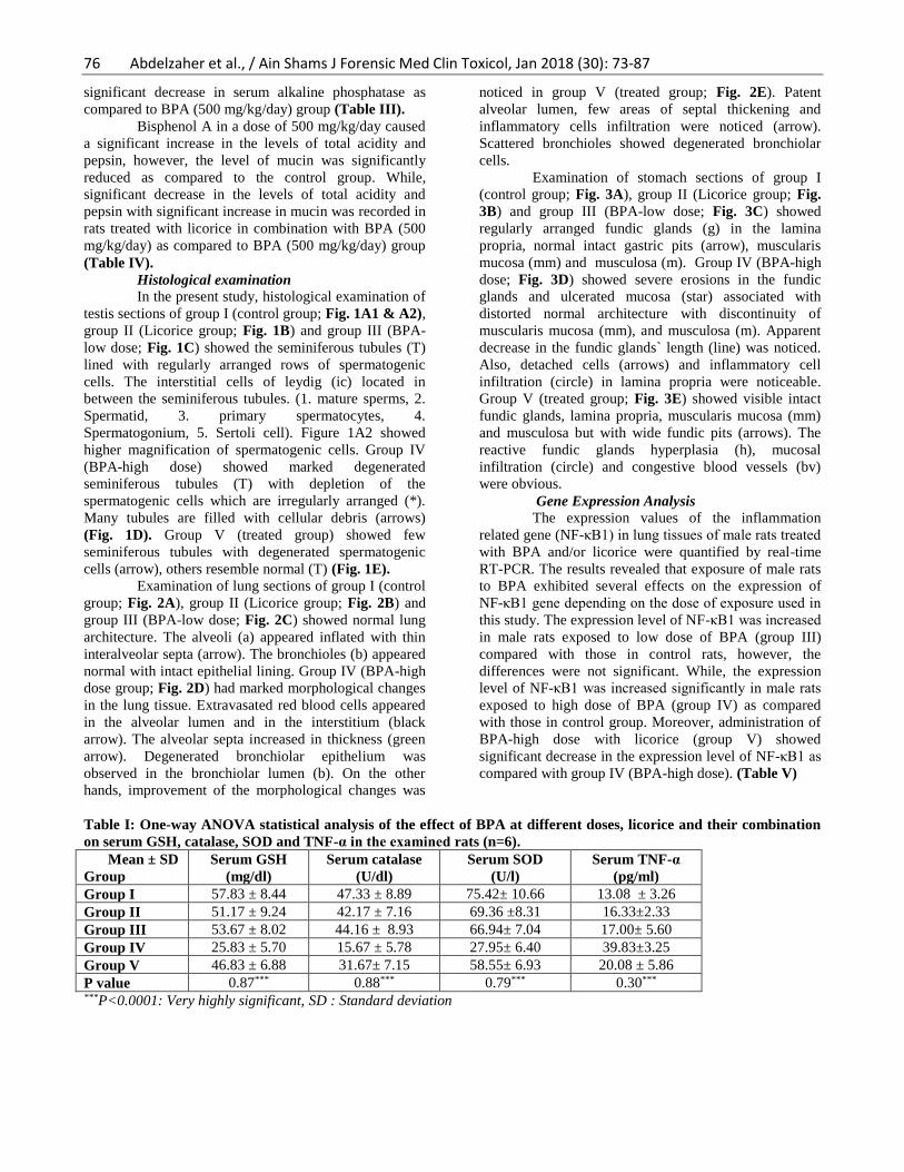
In the present study, histological examination of
testis sections of group I (control group; Fig. 1A1 & A2),
group II (Licorice group; Fig. 1B) and group III (BPA-
low dose; Fig. 1C) showed the seminiferous tubules (T)
lined with regularly arranged rows of spermatogenic
cells. The interstitial cells of leydig (ic) located in
between the seminiferous tubules. (1. mature sperms, 2.
Spermatid, 3. primary spermatocytes, 4.
Spermatogonium, 5. Sertoli cell). Figure 1A2 showed
higher magnification of spermatogenic cells. Group IV
(BPA-high dose) showed marked degenerated
seminiferous tubules (T) with depletion of the
spermatogenic cells which are irregularly arranged (*).
Many tubules are filled with cellular debris (arrows)
(Fig. 1D). Group V (treated group) showed few
seminiferous tubules with degenerated spermatogenic
cells (arrow), others resemble normal (T) (Fig. 1E).
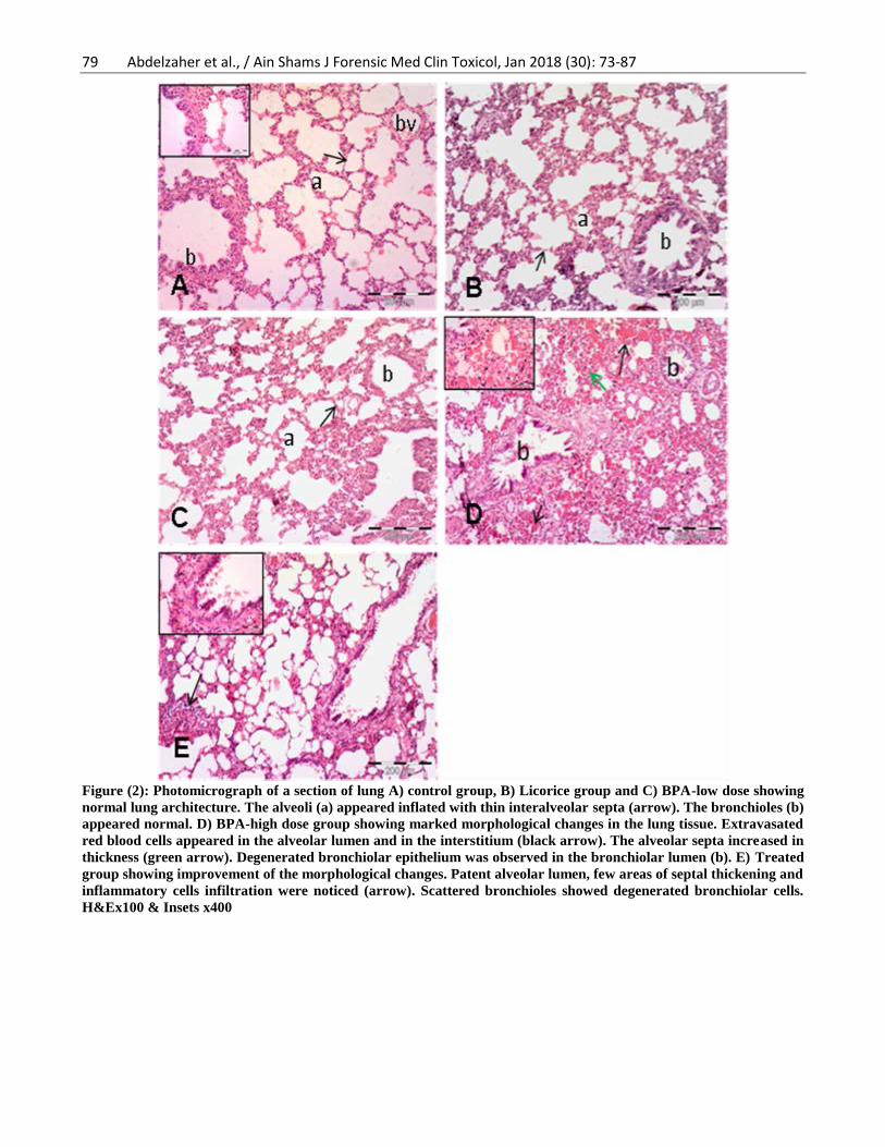
Examination of lung sections of group I (control
group; Fig. 2A), group II (Licorice group; Fig. 2B) and
group III (BPA-low dose; Fig. 2C) showed normal lung
architecture. The alveoli (a) appeared inflated with thin
interalveolar septa (arrow). The bronchioles (b) appeared
normal with intact epithelial lining. Group IV (BPA-high
dose group; Fig. 2D) had marked morphological changes
in the lung tissue. Extravasated red blood cells appeared
in the alveolar lumen and in the interstitium (black
arrow). The alveolar septa increased in thickness (green
arrow). Degenerated bronchiolar epithelium was
observed in the bronchiolar lumen (b). On the other
hands, improvement of the morphological changes was
noticed in group V (treated group; Fig. 2E). Patent
alveolar lumen, few areas of septal thickening and
inflammatory cells infiltration were noticed (arrow).
Scattered bronchioles showed degenerated bronchiolar
cells.
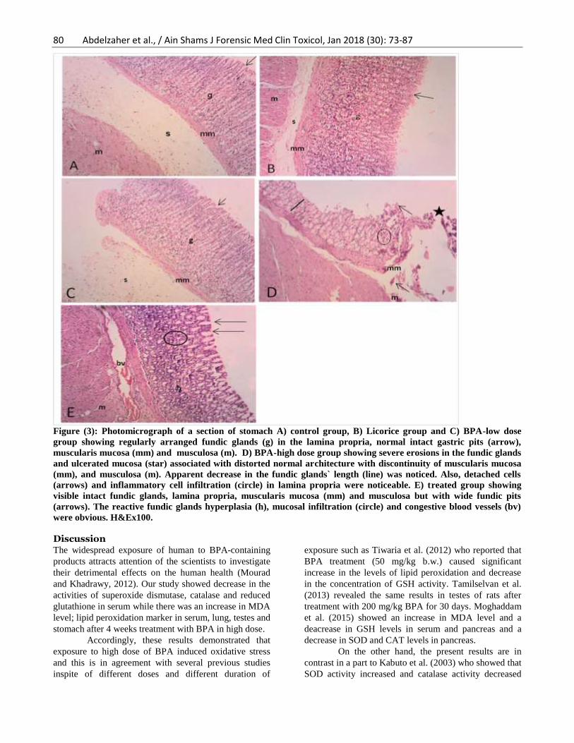
Examination of stomach sections of group I
(control group; Fig. 3A), group II (Licorice group; Fig.
3B) and group III (BPA-low dose; Fig. 3C) showed
regularly arranged fundic glands (g) in the lamina
propria, normal intact gastric pits (arrow), muscularis
mucosa (mm) and musculosa (m). Group IV (BPA-high
dose; Fig. 3D) showed severe erosions in the fundic
glands and ulcerated mucosa (star) associated with
distorted normal architecture with discontinuity of
muscularis mucosa (mm), and musculosa (m). Apparent
decrease in the fundic glands` length (line) was noticed.
Also, detached cells (arrows) and inflammatory cell
infiltration (circle) in lamina propria were noticeable.
Group V (treated group; Fig. 3E) showed visible intact
fundic glands, lamina propria, muscularis mucosa (mm)
and musculosa but with wide fundic pits (arrows). The
reactive fundic glands hyperplasia (h), mucosal
infiltration (circle) and congestive blood vessels (bv)
were obvious.
Gene Expression Analysis
The expression values of the inflammation
related gene (NF-κB1) in lung tissues of male rats treated
with BPA and/or licorice were quantified by real-time
RT-PCR. The results revealed that exposure of male rats
to BPA exhibited several effects on the expression of
NF-κB1 gene depending on the dose of exposure used in
this study. The expression level of NF-κB1 was increased
in male rats exposed to low dose of BPA (group III)
compared with those in control rats, however, the
differences were not significant. While, the expression
level of NF-κB1 was increased significantly in male rats
exposed to high dose of BPA (group IV) as compared
with those in control group. Moreover, administration of
BPA-high dose with licorice (group V) showed
significant decrease in the expression level of NF-κB1 as
compared with group IV (BPA-high dose). (Table V)
Table I: One-way ANOVA statistical analysis of the effect of BPA at different doses, licorice and their combination
on serum GSH, catalase, SOD and TNF-α in the examined rats (n=6).
Mean ± SD
Group
Serum GSH
(mg/dl)
Serum catalase
(U/dl)
Serum SOD
(U/l)
Serum TNF-α
(pg/ml)
Group I 57.83 ± 8.44 47.33 ± 8.89 75.42± 10.66 13.08 ± 3.26
Group II 51.17 ± 9.24 42.17 ± 7.16 69.36 ±8.31 16.33±2.33
Group III 53.67 ± 8.02 44.16 ± 8.93 66.94± 7.04 17.00± 5.60
Group IV 25.83 ± 5.70 15.67 ± 5.78 27.95± 6.40 39.83±3.25
Group V 46.83 ± 6.88 31.67± 7.15 58.55± 6.93 20.08 ± 5.86
P value 0.87*** 0.88*** 0.79*** 0.30***
***P<0.0001: Very highly significant, SD : Standard deviation
77 Abdelzaher et al., / Ain Shams J Forensic Med Clin Toxicol, Jan 2018 (30): 73-87
Table II: One-way ANOVA statistical analysis of the effect of BPA at different doses, licorice and their combination
on serum, lung, testis and gastric MDA in the examined rats (n=6).
Mean ± SD
Group
Serum MDA
(nmol/ dL)
Lung MDA
(nmol/gm tissue)
Testis MDA
(nmol/gm tissue)
Gastric MDA
(nmol/gm tissue)
Group I 182.71± 9.72 38.39±6.46 53.90±4.80 24.55±8.25
Group II 196.02 ± 15.11 38.95±4.92 51.46±6.32 26.11±7.24
Group III 200.31±13.36 39.66±4.69 54.29±8.09 25.51±9.84
Group IV 384.13 ± 11.83 80.41±7.97 81.16±3.57 72.72±9.11
Group V 203.92± 12.25 47.15±7.77 60.22±4.24 35.49 ± 9.20
P value 0.92*** 0.70*** 0.41*** 0.97***
***P<0.0001: Very highly significant , SD: Standard deviation
Table III: One-way ANOVA statistical analysis of the effect of BPA at different doses, licorice and their combination
on serum testosterone, alkaline phosphatase and testicular cholesterol in the examined rats (n=6).
Mean ± SD
Group
Serum testosterone
(pg/ml)
Serum alkaline
phosphates (U/L)
Testicular cholesterol
(mg/gm tissue)
Group I 4.93± 1.24 125.95± 12.42 34.27±7.98
Group II 5.25±1.16 129.36 ± 18.79 35.62±7.82
Group III 4.79±1.39 132.43 ± 17.90 33.11± 9.35
Group IV 1.84±0.96 186.87± 10.46 15.49± 3.73
Group V 4.24 ± 1.65 156.36±8,.66 26.02± 6.41
P value 0.82*** 0.41*** 0.44***
***P<0.0001: Very highly significant, SD: Standard deviation
Table IV: One-way ANOVA statistical analysis of the effect of BPA at different doses, licorice and their combination
on total acidity, pepsin and mucin in the examined rats (n=6).
Mean ± SD
Group
Total acidity
(mEq/3h)
Pepsin
(μg/ml tyrosine)
Mucin (mg %
hexose)
Group I 47.50± 9.33 116.50±11.09 104.83± 7.83
Group II 48.66± 9.07 119.83±11.80 102.60±7.63
Group III 47.76± 7.97 118.50±11.05 106.33± 8.29
Group IV 96.00± 8.25 183.83±6.05 65.50 ± 17.98
Group V 57.33± 11.54 135.16±6.55 99.33±11.18
P value 0.93*** 0.49*** 0.25***
***P<0.0001: Very highly significant, SD : Standard deviation
Table V: One-way ANOVA statistical analysis of the effect of BPA at different doses, licorice and their combination
on the expression of NF-κB1 gene in the lung tissue of the examined rats (n=6).
Group Mean ± SD
Group I 0.63 ± 0.01c
Group II 0.59 ± 0.05c
Group III 0.93 ± 0.02bc
Group IV 1.93 ± 0.02a
Group V 1.13 ± 0.047b
P value 0.13***
***P<0.0001: Very highly significant, SD : Standard deviation
78 Abdelzaher et al., / Ain Shams J Forensic Med Clin Toxicol, Jan 2018 (30): 73-87
Figure (1): Photomicrograph of a section of testis A) Control group, B) Licorice group and C) BPA-low dose group
showing the seminiferous tubules (T) lined with regularly arranged rows of spermatogenic cells. The interstitial cells
of leydig (ic) located in between the seminiferous tubules. Inset key (1. mature sperms, 2. Spermatid, 3. primary
spermatocytes, 4. Spermatogonium, 5. Sertoli cell). A2) showing higher magnification of spermatogenic cells. D)BPA-
high dose showing marked degenerated seminiferous tubules (T) with depletion of the spermatogenic cells which are
irregularly arranged (*). Many tubules are filled with cellular debris (arrows). E) Treated group showing few
seminiferous tubules with degenerated spermatogenic cells (arrow), others resemble normal (T). H&Ex100 & Insets
x400
79 Abdelzaher et al., / Ain Shams J Forensic Med Clin Toxicol, Jan 2018 (30): 73-87
Figure (2): Photomicrograph of a section of lung A) control group, B) Licorice group and C) BPA-low dose showing
normal lung architecture. The alveoli (a) appeared inflated with thin interalveolar septa (arrow). The bronchioles (b)
appeared normal. D) BPA-high dose group showing marked morphological changes in the lung tissue. Extravasated
red blood cells appeared in the alveolar lumen and in the interstitium (black arrow). The alveolar septa increased in
thickness (green arrow). Degenerated bronchiolar epithelium was observed in the bronchiolar lumen (b). E) Treated
group showing improvement of the morphological changes. Patent alveolar lumen, few areas of septal thickening and
inflammatory cells infiltration were noticed (arrow). Scattered bronchioles showed degenerated bronchiolar cells.
H&Ex100 & Insets x400
80 Abdelzaher et al., / Ain Shams J Forensic Med Clin Toxicol, Jan 2018 (30): 73-87
Figure (3): Photomicrograph of a section of stomach A) control group, B) Licorice group and C) BPA-low dose
group showing regularly arranged fundic glands (g) in the lamina propria, normal intact gastric pits (arrow),
muscularis mucosa (mm) and musculosa (m). D) BPA-high dose group showing severe erosions in the fundic glands
and ulcerated mucosa (star) associated with distorted normal architecture with discontinuity of muscularis mucosa
(mm), and musculosa (m). Apparent decrease in the fundic glands` length (line) was noticed. Also, detached cells
(arrows) and inflammatory cell infiltration (circle) in lamina propria were noticeable. E) treated group showing
visible intact fundic glands, lamina propria, muscularis mucosa (mm) and musculosa but with wide fundic pits
(arrows). The reactive fundic glands hyperplasia (h), mucosal infiltration (circle) and congestive blood vessels (bv)
were obvious. H&Ex100.
Discussion The widespread exposure of human to BPA-containing
products attracts attention of the scientists to investigate
their detrimental effects on the human health (Mourad
and Khadrawy, 2012). Our study showed decrease in the
activities of superoxide dismutase, catalase and reduced
glutathione in serum while there was an increase in MDA
level; lipid peroxidation marker in serum, lung, testes and
stomach after 4 weeks treatment with BPA in high dose.
Accordingly, these results demonstrated that
exposure to high dose of BPA induced oxidative stress
and this is in agreement with several previous studies
inspite of different doses and different duration of
exposure such as Tiwaria et al. (2012) who reported that
BPA treatment (50 mg/kg b.w.) caused significant
increase in the levels of lipid peroxidation and decrease
in the concentration of GSH activity. Tamilselvan et al.
(2013) revealed the same results in testes of rats after
treatment with 200 mg/kg BPA for 30 days. Moghaddam
et al. (2015) showed an increase in MDA level and a
deacrease in GSH levels in serum and pancreas and a
decrease in SOD and CAT levels in pancreas.
On the other hand, the present results are in
contrast in a part to Kabuto et al. (2003) who showed that
SOD activity increased and catalase activity decreased
81 Abdelzaher et al., / Ain Shams J Forensic Med Clin Toxicol, Jan 2018 (30): 73-87
significantly in the liver after intraperitoneal injection of
25 mg/kg and 50 mg/kg BPA for 5 days but SOD activity
showed no significant change in brain, lung, kidney,
liver, fat body, testes, and blood. This difference may be
due to different doses and different duration of treatment.
It has been suggested that oxidative stress
induced cytotoxicity through direct membrane damage
and generally increases the membrane permeability. This
leads to rising in the intracellular calcium which disturbs
mitochondrial function and ultimately stimulates reactive
oxygen species (ROS) production (Demuro et al., 2005;
Gleichmann and Mattson, 2011).
The ROS are formed in profuse in pro-oxidant
states and play an essential role against pathological
conditions; however overproduction of ROS may harm
the body tissues. The antioxidant enzymes act against
ROS toxicity (Birben et al., 2012). Superoxide anion
radical has been dismutased by Superoxide dismutase
(SOD) to hydrogen peroxide, which is further degraded
by catalase through reduced glutathione (Hayyan et al.,
2016). The decrease in antioxidant enzymes activities
explained the malfunction of antioxidant system to
overcome the free radicals formed after exposure to BPA
(Lobo et al., 2010). Sajiki (2001) suggested that
bisphenol A is metabolized to a reactive metabolite 4,5-
bisphenol O-quinone and bisphenol radical by oxygen
radical reactions.
Alkaline phosphatases (ALP) are a group of
enzymes that found in several body tissues such as liver,
kidney, intestine, bone and white blood cells. The main
function of alkaline phosphatase is transporting across
the cell membranes (Celik et al., 2009). Hence,
destruction of these tissues results in leakage of ALP into
the blood. Our study showed a significant increase in
ALP serum level which may be attributed to oxidative
stress induced by BPA treatment that cause tissue
damage and release of the enzymes from tissues in
serum. This result is in agreement with Eshak and Osman
(2014). Sangai and Verma (2012) found that BPA may
serve as a plasma membrane labilizer. The raise in ALP
activities can comprise a danger to the cell life which is
reliant on phosphate esters for its vital processes.
The current study showed a significant increase
in the serum level of TNF-α after exposure of rats to high
dose of BPA for 4 weeks. This result revealed that
exposure to high dose of BPA induced inflammatory
reaction which is in accordance with Zhu et al. (2015).
High TNF-α level may affect the apoptotic pathways in
various organs and increase the susceptibility to
autoimmunity (Zhang and An, 2007).
In the present study, significant decrease in
testicular cholesterol level was demonstrated after
exposure of examined rats to 500 mg/kg BPA. This result
had been explained by Negri-Cesi (2015) who reported
that BPA can disturb the hypothalamic neuroendocrine
functions which is partly involved in regulation of fat
metabolism (Dieguez et al., 2011).
Decrease in testicular cholesterol level
accompanied by reduction in serum testosterone level.
Similar to these results, Eshak and Osman (2014)
revealed that BPA induced endocrine and reproductive
toxicity. Cholesterol is a precursor of all sex hormones.
Testosterone synthesis process is a series of events
through which conversion of cholesterol into testosterone
occurs when luteinizing hormone (LH) triggers the
testicular leydig cells (Miller and Auchus, 2011). The
reduction in plasma testosterone level may be due to
reduction in cholesterol level and/or degeneration of
Leydig cells in testes (Nanjappa et al., 2012) that is
revealed by histopathological examination of exposed
rats in our study. Moreover, it has been reported that
BPA acts as estrogen agonist and androgen antagonist
activity (Acconcia et al., 2015).
The testosterone is the major androgen hormone
which is important for maintenance of spermatogenesis
and inhibition of germ cell apoptosis (Dohle et al., 2003).
Akingbemi et al. (2004), Nakamura et al. (2010) and
Gurmeet et al. (2014) are in agreement with our study,
they showed reduction in serum testosterone level after
treatment with BPA. While Jahan el al. (2016) are party
in contrast, they revealed that BPA (50 mg/kg) induced
decrease in serum testosterone and increase in serum
cholesterol levels. This difference may be due to different
doses of exposure to BPA.
These results were supported by marked
histological changes of testes after administration of 500
mg/kg BPA. These changes have been clarified by
Gurmeet et al. (2014) who supposed that the decreased
testosterone level may be the cause of alteration of
spermatogenesis and disturbance of the seminiferous
epithelium. Additionally, the disturbances in
hypothalamic-pituitary-gonadal axis and decrease in
Sertoli cell phagocytic function that is induced by BPA
administration could affect the histological structure of
the testes (Vandenberg et al., 2009). The cytotoxic agents
can affect the spermatogenic cells due to their high
mitotic activity (Endo et al., 2003). Our results are in
accordance with Li et al. (2009), Jahan et al. (2016) and
Tian et al. (2017). They showed morphological damages
in testis after administration of BPA and provide the
evidence for their toxicity on the male reproductive
system.
The current microscopic examination of the
lung showed severe morphological disruption in the lung
after chronic administration of 500 mg/kg BPA. This
result is in agreement with Amaravathi et al. (2012) and
Kattaia and Abdel Baset (2014). Previous researchers
suggested that BPA induced inflammation by production
of different cytokines which are secreted by macrophages
(Wetherill et al., 2007). These cytokines resulted in
endothelial cell adhesion molecules that provoked the
82 Abdelzaher et al., / Ain Shams J Forensic Med Clin Toxicol, Jan 2018 (30): 73-87
adhesion and transport of neutrophils from the vascular
space into parenchyma. This lead to production of
proteases and oxidants that damaged the vascular
endothelial cells and clarified the extravasated RBCs of
the lung tissue and degenerated bronchial epithelium
found in the present study (Wetherill et al., 2007). The
thickened alveolar septa may be due to marked
deposition of interstitial collagen fiber and severe cellular
infiltration with eosinophils, neutrophils, lymphocytes
and macrophages (Zidan, 2011).
The current study also revealed that the high
dose of BPA resulted in gastric ulcer formation,
inflammatory cell infiltration and distortion of the normal
architecture of the gastric tissue after 4 weeks. It has been
reported that a disturbance between aggravating and
protective factors leads to induction of gastric ulcer
formation. Gastric acid, pepsin and oxidant agents are
considered as aggravating factors, while mucous, and
antioxidant agents are protective factors (Suleyman,
2012). To our knowledge, there are no studies have been
reported about the effect of BPA on the stomach.
Moreover, our study reported that high dose of
BPA increased the gastric juice total acidity and pepsin
activity significantly. This effect may be attributed to
several processes; generation of ROS, enhancement of
lipid peroxidation, infiltration of leukocytes and initiation
of apoptosis (Bech et al., 2000). Also, a significant
reduction in mucin was recorded in group IV. These
results were evidently supported our findings in the
histological examination of the stomach. Mucin is
essential for gastric mucosa against ulcerogens and
promotes the repair of the damaged gastric epithelium by
decrease in the stomach wall friction during peristalsis
and serving as an important barrier to back diffusion of
hydrogen ions (Sevak et al., 2002). Furthermore, mucus
is able to act as an antioxidant as it can minimize
mucosal hurt enhanced by oxygen free radicals (Repetto
and Llesuy, 2002).
Nuclear factor kappa-light-chain-enhancer of
activated B cells (NF-κB) is a protein complex that is
present in approximately all cell types. NF-κB is
involved in DNA transcription and cell survival
(Gilmore, 2006). It is concerned with cellular responses
to different stimuli for example; cytokines, viral or
bacterial antigens, oxidative stress and free radicals
(Perkins, 2007). Additionally, NF-κB has an important
role in controlling the immune response to infection (κ
light chains are essential structures of antibodies). The
mammalian NF-κB family has five proteins; NF-κB1,
NF-κB2, RelA, RelB and c-Rel (Nabel and Verma,
1993). False regulation of NF-κB may be due to viral
infection, inflammation, septic shock, cancer,
and autoimmune diseases (Meffert et al., 2003). Hence,
we aimed to study NF-κB transcription and activation.
NF-κB is remained in the cytoplasm in
unstimulated cells. Once the cells are activated, NF-κB
leads to nuclear translocation, which is responsible for
regulation of certain cytokines genes, like TNF-alpha
(Oeckinghaus et al., 2011). Our study revealed a
significant increase in the expression level of NF-κB
after exposure to high dose of BPA as compared to the
control group. Li et al. (2012) determined that BPA can
modify different transcriptional factors, such as Sp1
(specificity protein 1) that affect the combining site of
NF-κB and hence change the transcriptional capacity of
NF-κB. Moreover, Jung et al. (2016) reported that
oxidant generation enhanced activation of NF-κB which
resulted in raising the TNF-α level that lead to tissue
injury and this is in concomitant with the results in the
present study. Previous studies supposed that of NF-κB
activation could stimulate invasion of cancer cells
(Palumbo et al., 2007; Wu et al., 2009).
The current study revealed that low dose of
BPA (2.4 µg/kg) had no significant effect on the exposed
rats and this is in agreement with Tiwari et al. (2012) and
Kattaia and Abdel Baset (2014). The International Food
Safety Authorities Network (INFOSAN) reported that 5
mg/kg/day showed no-observed adverse- effect level
(NOAEL) for BPA. NOAEL is the maximum dose that
did not stimulate any adverse effect in the examined
animals (INFOSAN, 2009). But, this result disagrees
with Salian et al. (2009a, 2009b) and Akingbemi et al.
(2004) who reported that the environmental dose of BPA
induced adverse effects on testicular functions of
experimental animals.
The current study revealed that treatment with
licorice improved the oxidative stress induced by high
dose of BPA, raised the levels of cholesterol and
testosterone to normal and decreased the level of TNF-α
significantly. Also, it improved the histopathological
changes induced by high dose of BPA in testis, lung and
stomach in group V in comparison to group IV. These
results indicated that antioxidant and anti-inflammatory
effect of licorice. This is in agreement with Yousaf et al.
(2016) and Rashwan and Anfenan (2012) who
contributed the antioxidant properties of licorice extract
to metabolism of glycyrrhizic acid to glycyrrhetinic acid
that has an antioxidant activity (Marí et al., 2009). It also
may be due to their content of glabridin (Asl and
Hosseinzadeh, 2008). Glabridin inhibits oxidation of
LDL by binding to it, minimizes NADPH oxidase
activation and increases cellular glutathione that results
in reduction of cellular oxidative stress (Rosenblat et al.,
2002).
It has been reported that the anti-inflammatory
effect of licorice may be as a result of its effect on the
adrenal gland which is responsible for production of
cortisol (Konovalova et al., 2000). Previous studies
suggested that licorice extract has different bioactive
compounds which inhibit NO production and
inflammatory cytokine (Chung et al., 2014; Yu et al.,
2015). Our results are in accordance with Takhshid et al.
83 Abdelzaher et al., / Ain Shams J Forensic Med Clin Toxicol, Jan 2018 (30): 73-87
(2012) and Jung et al. (2016) who clarified that licorice-
derived compounds enhance prostaglandins
concentration in the digestive system and this increases
secretion of mucus from the stomach which has healing
effects.
Furthermore, licorice treatment led to a
significant reduction in the expression level of NF-κB1
as compared with group IV (BPA-high dose). Previous
research demonstrated the efficiency of Licorice extract
in vitro to prevent the signaling pathways leading to NF-
κB activation (Schröfelbauer et al., 2009).
According to the authors` knowledge, there is
no previous studies have investigated the protective
effect of licorice for inhibition and/or reduction of
toxicity induced by BPA.
Conclusion In conclusion, 4 weeks exposure to high level of
bisphenol a (BPA) induced oxidative stress,
inflammatory reaction, histopathological changes in
different body organs, and genetic effect. The licorice
can reduce and/or prevent the toxic effects induced by
BPA due to its antioxidant, anti-inflammatory, anti-
atherosclerotic, anti-ulcer effects and free-radical
scavenging activities. Hence, we can use the licorice as a
prophylaxis against BPA toxicity, taking into
consideration the dose and duration of treatment.
Funding This research received no specific grant from any
funding agency in the public, commercial, or not-for-
profit sectors.
Conflict of interest The authors declared no potential conflicts of interest
with respect to the research, authorship, and/or
publication of this article.
Ethical approval Animals and their care were approved according to the
animal care Committee of Faculty of Medicine - Minia
University, Egypt. All applicable international, national,
and/or institutional guidelines for the care and use of
animals were followed.
This article does not contain any studies with human
participants performed by any of the authors.
References Acconcia F, Pallottini V and Marino M (2015):
Molecular Mechanisms of Action of BPA. Dose
Response13:1-9.
Ahbab MA, Barlas N and Karabulut G (2017): The
toxicological effects of bisphenol A and
octylphenol on the reproductive system of
prepubertal male rats. Toxicol. Ind. Health
33:133-46. (Epub ahead of print).
Akingbemi BT, Sottas CM, Koulova AI et al., (2004):
Inhibition of testicular steroidogenesis by the
xenoestrogen bisphenol a is associated with
reduced pituitary luteinizing hormone secretion
and decreased steroidogenic enzyme gene
expression in rat Leydig cells. Endocrinology
145:592-603.
Amaravathi P, Srilatha CH, Ramadevi V et al., (2012):
Pulmonary and genotoxicity of bisphenol-A in
Wistar albino rats. Curr. Biotica. 6:53-60.
Asl MN and Hosseinzadeh H (2008): Review of
Pharmacological Effects of Glycyrrhiza sp. and
its Bioactive Compounds. Phytother. Res.
22:709-24.
Bech PL, Xavier R, Lu N et al., (2000): Mechanisms of
NSAID-induced gastrointestinal injury defined
using mutant mice. Gastroenterology 119:699-
705.
Ben-Jonathan N, Hugo ER and Brandebourg TD (2009):
Effects of bisphenol A on adipokine release
from human adipose tissue: Implications for the
metabolic syndrome. Mol. Cell Endocrinol.
304:49-54.
Birben E, Sahiner UM, Sackesen C et al., (2012):
Oxidative Stress and Antioxidant Defense.
W.A.O. J. 5:9-19.
Buege JA and Aust SD (1978): Microsomal lipid
peroxidation. Methods Enzymol. 52:302-10.
Celik H, Tosun M, Cetinkaya MB et al., (2009):
Markedly elevated serum alkaline phosphatase
level in an uncomplicated pregnancy. J. Matern.
Fetal Neonatal Med. 22:705-7.
Chapin RE, Adams J, Boekelheide K et al., (2008): NTP-
CERHR expert panel report on the reproductive
and developmental toxicity of bisphenol A.
Birth Defects Res. B. Dev. Reprod. Toxicol.
83:157-395.
Chung, SJ, Lee CH, Lee HS et al., (2014): The role of
phosphatidylcholine and deoxycholic acid in
inflammation. Life Sci. 108:88-93.
Cikos S and Koppel J (2009): Transformation of real-
time PCR fluorescence data to target gene
quantity. Anal. Biochem. 384:1–10.
Demuro A, Mina E, Kayed R et al., (2005): Calcium
dysregulation and membrane disruption as a
ubiquitous neurotoxic mechanism of soluble
amyloid oligomers. J. Biol. Chem. 280:17294-
300.
Di Mambro VM and Fonseca MJ (2005): Assays of
physical stability and antioxidant activity of a
topical formulation added with different plant
extract. J. Pharm. Biomed. Anal. 37:287-95.
Dieguez C, Vozguez MJ, Romero A et al., (2011):
Hypothalamic control of lipid metabolism: focus
on leptin, ghrelin and melanocortins.
Neuroendocrinology 94:1–11.
Dohle GR, Smit M and Weber RF (2003): Androgens
and male fertility. World J. Urol. 21:341-5.
EFSA (European Food Safety Authority) (2015):
Scientific Opinion on the risks to public health
related to the presence of bisphenol A (BPA) in
84 Abdelzaher et al., / Ain Shams J Forensic Med Clin Toxicol, Jan 2018 (30): 73-87
food stuffs: Executive summary. EFSA J.
13(1):3978, 23 pp.
El-Baz FK, Khalil WKB, Aly HF et al., (2016): Berry
extracts improved inflammatory cytokines,
antioxidant enzyme and suppressed the gene
expression alterations in diabetic rats. Int. J.
Pharm. Sci. 8:294-302.
Endo F, Manabe F, Takeshima H et al., (2003):
Protecting spermatogonia from apoptosis
induced by doxorubicine using the luteinizing
hormone-releasing hormone analog leuprorelin.
Int. J. Urol. 10:72-7.
Eshak MG and Osman HF (2014): Biological Effects of
Chitosan against Bisphenol- A Induced
Endocrine Toxicity and Androgen Receptor
Gene Expression Changes in Male Rats. Int. J.
Pharm.Clin. Res. 6:300-11.
Fang C, Ning B, Waqar AB et al., (2015): Bisphenol A
exposure induces metabolic disorders and
enhances atherosclerosis in hyperlipidemic
rabbits. J. Appl. Toxicol. 35:1058-70.
Fenichel P, Chevalier N and Brucker-Davis F (2013):
Bisphenol A: an endocrine and metabolic
disruptor. Ann. Endocrinol. (Paris) 74:211-20.
Fu Y, Chen J, Li YJ et al., (2013): Antioxidant and anti-
inflammatory activities of six flavonoids
separated from licorice. Food Chem. 141:1063-
71.
Gafner S, Bergeron C, Villinski JR et al., (2011):
Isoflavonoids and coumarins from Glycyrrhiza
uralensis: antibacterial activity against oral
pathogens and conversion of isoflavans into
isoflavanquinones during purification. J. Nat.
Prod. 74:2514-19.
Gilmore TD (2006): Introduction to NF-kappaB: players,
pathways, perspectives. Oncogene 25:6680-4.
Gleichmann M and Mattson MP (2011): Neuronal
calcium homeostasis and dysregulation.
Antioxid. Redox Signal 14:1261-73.
Gurmeet KSS, Rosnah I, Normadiah MK et al., (2014):
Detrimental effects of bisphenol a on
development and functions of the male
reproductive system in experimental rats.
EXCLI. J. 13:151-60.
Hara N , Hara Y, Natsume Y et al., (1991): Gastric
hyperacidity and mucosal damage caused by
hypothermia correlate with increase in GABA
concentrations of the rat brain. Eur. J.
Pharmacol. 194:77-81.
Hayyan M, Hashim MA and AlNashef IM (2016):
Superoxide Ion: Generation and Chemical
Implications. Chem. Rev. 116:3029-85.
Hesham R. Omar, Irina Komarova, Mohamed El-
Ghonemi et al., (2012): Licorice abuse: time to
send a warning message. Ther. Adv. Endocrinol.
Metab. 3(4): 125-138.
Howdeshell KL, Hotchkiss AK, Thayer KA et al.,
(1999): Exposure to bisphenol A advances
puberty. Nature 401:763–4
Huo ZH, Wang B, Liang YK et al., (2011):
Hepatoprotective and antioxidant effects of
Licorice extract against CCl4-induced oxidative
damage in rats. Int. J. Mol. Sci. 12:6529-43.
International Food Safety Authorities Network
(INFOSAN) (2009): Bisphenol A (BPA) –
current state of knowledge and future actions by
WHO and FAO. INFOSAN Information note
no. 5/2009-bisphenol A. Available at:
http://www.who.int/foodsafety/publications/fs_
management/No_05_Bisphenol_A_Nov09_en.p
df
Jahan S, Ain QU and Ullah H (2016): Therapeutic effects
of quercetin against bisphenol A induced
testicular damage in male Sprague Dawley rats.
Syst. Biol. Reprod. Med. 62:114-24.
Jung JC, Lee YH, Kim SH et al., (2016):
Hepatoprotective effect of licorice, the root of
Glycyrrhiza uralensis Fischer, in alcohol
induced fatty liver disease. BMC Complement
Altern. Med. 16:19,
http://dx.doi.org/10.1186/s12906-016-0997-0.
Kabuto H, Amakawa M and Shishibori T (2004):
Exposure to bisphenol A during embryonic/fetal
life and infancy increases oxidative injury and
causes underdevelopment of the brain and testis
in mice. Life Sci. 74:2931-40.
Kabuto H, Hasuike S, Minagawa N et al., (2003): Effects
of bisphenol A on the metabolisms of active
oxygen species in mouse tissues. Environ. Res.
93:31-5.
Kamei J, Nakamura R, Ichiki H et al., (2003):
Antitussive principles of Glycyrrhizae radix, a
main component of the Kampo preparations
Bakumondo-to (Mai-men-dong-tang). Eur. J.
Pharmacol. 469:159-63.
Karkanisa A, Martinsb N, Petropoulos SA et al., (2016):
Phytochemical composition, health effects, and
crop management of liquorice (Glycyrrhiza
glabra L.): Α medicinal plant. Food Rev. Int,
http://dx.doi.org/10.1080/87559129.2016.12613
00.
Kattaia AA and Abdel Baset SA (2014): Effect of
bisphenol A on the lung of adult male albino
rats and the possible protective role of geraniol:
a histological and immunohistochemical study.
Egypt J. Histol. 37:24-35.
Kitraki E, Nalvarte I, Alavian-Ghavanini A et al.,
(2015): Developmental exposure to bisphenol A
alters expression and DNA methylation of
Fkbp5, an important regulator of the stress
response. Mol. Cell Endocrinol. 417:191-9.
85 Abdelzaher et al., / Ain Shams J Forensic Med Clin Toxicol, Jan 2018 (30): 73-87
Konovalova GG, Tikhaze AK and Lankin VZ (2000):
Antioxidant activity of parapharmaceutics
containing natural inhibitors of free radical
processes. Bull Exp. Biol. Med. 130:658-60.
Kurosawa T, Hiroi H, Tsutsumi O et al., (2002): The
activity of bisphenol A depends on both the
estrogen receptor subtype and the cell type. End.
J. 49:465-71.
Lakind JS and Naiman DQ (2011): Daily intake of
bisphenol A and potential sources of exposure:
2005–2006 National Health and Nutrition
Examination Survey. J Expo. Sci. Environ.
Epidemiol. 21:272-9.
Li Y, Burns KB, Arao Y et al., (2012): Differential
estrogenic actions of endocrine-disrupting
chemicals bisphenol A, bisphenol AF, and
zearalenone through estrogen receptor α and β
in vitro. Environ. Health Perspect. 120:1029-35.
Li YJ, Song TB, Cai YY et al., (2009): Bisphenol A
exposure induces apoptosis and upregulation of
Fas/FasL and caspase-3 expression in the testes
of mice. Toxicol. Sci. 108:427-36.
Liao WC, Lin YH, Chang TM et al., (2012):
Identification of two licorice species,
Glycyrrhiza uralensis and Glycyrrhiza glabra,
based on separation andidentification of their
bioactive components. Food Chem. 132:2188-
93.
Lim DS, Kwack SJ, Kim KB et al., (2009): Potential risk
of bisphenol A migration from polycarbonate
containers after heating, boiling, and
microwaving. J. Toxicol. Environ. Health A
72:1285-91.
Lobo V, Patil A, Phatak A et al., (2010): Free radicals,
antioxidants and functional foods: Impact on
human health. Pharmacogn. Rev. 4:118-26.
Marí, M, Morales A, Colell A et al., (2009):
Mitochondrial Glutathione, a Key Survival
Antioxidant. Antioxid. Redox Signal 11:2685-
700.
Marklund S and Marklund G (1974): Involvement of the
superoxide anion radical in the autoxidation of
pyrogallol and a convenient assay for
superoxide dismutase. Eur. J. Biochem. 47:469-
74.
Meffert MK, Chang JM, Wiltgen BJ et al., (2003): NF-
kappa B functions in synaptic signaling and
behavior. Nat. Neurosci. 6:1072-8.
Mihara M and Uchiyama M (1978): Determination of
malonaldehyde precursor in tissues by
thiobarbituric acid test. Anal. Biochem. 86:271-
8.
Miller WL and Auchus RJ (2011): The molecular
biology, biochemistry, and physiology of human
steroidogenesis and its disorders. Endocr. Rev.
32:81-151.
Moghaddam HS, Samarghandian S and Farkhondeh T
(2015): Effect of bisphenol A on blood glucose,
lipid profile and oxidative stress indices in adult
male mice. Toxicol. Mech. Methods 25:507-13.
Mourad IM and Khadrawy YA (2012): The sensitivity of
liver, kidney and testis of rats to oxidative stress
induced by different doses of bisphenol A. Int. J.
Life Sci. Pharma. Res. 2:L19-L28.
Nabel GJ and Verma IM (1993): Proposed NF-kappa B/I
kappa B family nomenclature. Genes.
Dev. 7:2063.
Nakamura D, Yanagiba Y, Duan Z et al., (2010):
Bisphenol A may cause testosterone reduction
by adversely affecting both testis and pituitary
systems similar to estradiol. Toxicol. Lett.
194:16-25.
Nanjappa MK, Simon L and Akingbemi BT (2012): The
industrial chemical bisphenol A (BPA)
interferes with proliferative activity and
development of steroidogenic capacity in rat
Leydig cells. Biol. Reprod. 86:135,1-12.
Negri-Cesi P (2015): Bisphenol A Interaction with Brain
Development and Functions. Dose Response
13:258-394.
Oeckinghaus A, Hayden MS and Ghosh S (2011):
Crosstalk in NF- Κb signaling pathways. Nat
.Immunol. 12:695-708.
Palumbo R, Galvez BG, Pusterla T et al., (2007): Cells
migrating to sites of tissue damage in response
to the danger signal HMGB1 require NF-
kappaB activation. J. Cell Biol. 179:33-40.
Perkins ND (2007): Integrating cell-signalling pathways
with NF-kappaB and IKK function. Nat. Rev.
Mol. Cell Biol. 8:49-62.
Prins GS, Tang WY, Belmonte J et al., (2008):
Developmental exposure to bisphenol A
increases prostate cancer susceptibility in adult
rats: epigenetic mode of action is implicated.
Fertil Steril 89(2 Suppl): e41,
http://dx.doi.org/10.1016/j.fertnstert.2007.12.02
3.
Pupo M, Pisano A, Lappano R et al., (2012): Bisphenol
A induces gene expression changes and
proliferative effects through GPER in breast
cancer cells and cancer -associated fibroblasts.
Environ. Health Perspect. 120:1177-82.
Rahnama M, Mehrabani D, Japoni S et al., (2013): The
healing effect of licorice (Glycyrrhiza glabra) on
Helicobacter pylori infected peptic ulcers. J.
Res. Med. Sci. 18:532-3.
Rashwan NM and Anfenan ML (2012): Free Radical
Scavenger Effects of Licorice on the
Experimental Rats. J. Appl. Sci. Res. 8:4704-10.
Repetto M and Llesuy S (2002): Antioxidant properties
of natural compounds used in popular medicine
86 Abdelzaher et al., / Ain Shams J Forensic Med Clin Toxicol, Jan 2018 (30): 73-87
for gastric ulcers. Braz. J. Med. Biol.
Res. 355:23-34.
Ritter S (2011): Debating BPA’s toxicity. Chem. Eng.
News 89:5-13.
Rosenblat M, Coleman R and Aviram M (2002):
Increased macrophage glutathione content
reduces cell-mediated oxidation of LDL and
atherosclerosis in apolipoprotein E-deficient
mice. Atherosclerosis 163:17-28.
Sajiki J (2001): Decomposition of bisphenol A by radical
oxygen. Environ. Int. 27:315-20.
Salian S, Doshi T and Vanage G (2009a): Perinatal
exposure of rats to bisphenol A affects fertility
of male offspring. Life Sci. 88:742–52.
Salian S, Doshi T and Vanage G (2009b): Neonatal
exposure of male rats to bisphenol A impairs
fertility and expression of sertoli cell junctional
proteins in the testis. Toxicol. 265:55–67.
Sangai NP and Verma RJ (2012): Quercetin ameliorates
Bisphenol a-induced toxicity in mice. Acta. Pol.
Pharm. 69:557-63.
Sanyal A, Denath O, Bhattacharya S et al., (1971): The
effect of cyproheptadine on gastric acidity. In:
Pfeiffer CJ, editor. Peptic ulcer. Munksgaard,
Copenhagen: Scandinavian University Books. p
312–318.
Schröfelbauer B, Raffetseder J, Hauner M et al., (2009):
Glycyrrhizin, the main active compound in
liquorice, attenuates pro-inflammatory
responses by interfering with membrane-
dependent receptor signaling. Biochem. J.
421:473-82.
Sevak R, Paul A, Goswami S et al., (2002):
Gastroprotective effect of beta3 adrenoreceptor
agonists ZD 7114 and CGP 12177A in rats.
Pharmacol. Res. 46:351-6.
Suleyman H (2012): The Role of Alpha-2 Adrenergic
Receptors in Anti-ulcer Activity. EAJM 44:43-
5.
Takhshid MA, Mehrabani D, Ai J et al., (2012): The
healing effect of licorice extract in acetic acid-
induced ulcerative colitis in rat model. Comp.
Clin. Pathol. 21:1139-44.
Tamilselvan P, Bharathiraja K, Vijayaprakash S et al.,
(2013): Protective role of lycopene on bisphenol
a induced changes in sperm characteristics,
testicular damage and oxidative stress in rats.
Int. J. Pharm. Bio. Sci. 4:131-43.
Tian J, Ding Y, She R et al., (2017): Histologic study of
testis injury after bisphenol A exposure in mice:
Direct evidence for impairment of the genital
system by endocrine disruptors. Toxicol. Ind
Health 33:36-45.
Tiwari D, Kamblea J, Chilgundea S et al., (2012):
Clastogenic and mutagenic effects of bisphenol
A: An endocrine disruptor. Mutat. Res. 743:83-
90.
Vandenberg LN, Maffini MV, Sonnenschein C et al.,
(2009): Bisphenol-A and the Great Divide, A
Review of Controversies in the Field of
Endocrine Disruption. Endocr. Rev. 30:75-95.
Vomsaal FS and Hughes C (2005): An extensive new
literature concerning low-dose effects of
bisphenol A shows the need for a new risk
assessment. Environ Health Perspect 113:926-
33.
Wang X, Zhang H, Chen L et al., (2013): Liquorice, a
unique “guide drug” of traditional Chinese
medicine: a review of its role in drug
interactions. J. Ethnopharmacol. 150:781-90.
Welshons WV, Thayer KA, Judy BM et al., (2003):
Large effects from small exposures. I.
Mechanisms for endocrine-disrupting chemicals
with estrogenic activity. Environ. Health
Perspect. 111:994-1006.
Wetherill YB, Akingbemi BT, Kannod J et al., (2007): In
vitro molecular mechanisms of bisphenol A
action. Reprod. Toxicol. 24:178-98.
Winzler RJ (1955): Determination of serum
glycoproteins. Methods Biochem. Anal. 2:279-
311.
Wu Y, Deng J, Rychahou PG et al., (2009):
Stabilization of Snail by NF-kappa B Is
Required for Inflammation-Induced Cell
Migration and Invasion. Cancer Cell 15:416-28.
Xia W, Jiang Y, Li Y et al., (2014): Early-life exposure
to bisphenol A induces liver injury in rats
involvement of mitochondria-mediated
apoptosis. PLOS One 9(e90443):1-9.
Yousaf B, Amina, Liu G et al., (2016): Bisphenol A
exposure and healing effects of Adiantum
capillus-veneris L. plant extract (APE) in
bisphenol A-induced reproductive toxicity in
albino rats. Environ. Sci. Pollut. Res. Int.
23:11645-57.
Yu JY, Ha JY, Kim KM et al., (2015): Anti-
Inflammatory Activities of Licorice Extract and
Its Active Compounds, Glycyrrhizic Acid,
Liquiritin and Liquiritigenin, in BV2 Cells and
Mice Liver. Molecules 20:13041-54.
Zhang J and An J (2007): Cytokines, Inflammation and
Pain. Int. Anesthesiol. Clin. 45:27-37.
Zhu J, Jiang L, Liu Y et al., (2015): MAPK and NF-κB
pathways are involved in bisphenol A-induced
TNF-α and IL-6 production in BV2 microglial
cells. Inflammation 38:637-48.
Zidan RA (2011): Effect of long-term administration of
amiodarone on rat lung and the possible
protective role of vitamin E: a histological and
immunohistochemical study. Egypt J. Histol.
34:117-28.
https://www.ncbi.nlm.nih.gov/pubmed/?term=Quercetin+ameliorates+Bisphenol+a-induced+toxicity+in+mice
87 Abdelzaher et al., / Ain Shams J Forensic Med Clin Toxicol, Jan 2018 (30): 73-87
الملخص العربي
ن مادة البسفينول أهل يستطيع العرق سوس منع التأثيرات البيوكيميائية و الهستولوجية و الجينية الناتجة ع
3وجدى خليلو 2داليا محمد على و 1ولاء يحيى عبد الظاهر
هدفت هذه فى البلاستكات البولى كاربونية. عادةيستخدم الذى ان البسفينول أ هو معطل للغدد الصماء الايكولوجية الاستروجينيةلتقييم التأثير الوقائي المحتمل ادة البسفينول أ و لم جرعات مختلفةعن الناتجة اختبار التأثيرات البيوكيميائية و الهستولوجية و الجينيةالدراسة إلى
2.4، أعطيت مادة البسفينول يوميا بجرعات من الفئران البيضاء الى خمس مجموعات 30للعرق سوس ضد هذه التأثيرات. تم تقسيم وأظهرت النتائج أن الجرعة أسابيع. 4مجم/كجم( لمدة 150مجم/كجم عن طريق الفم مع أو بدون العرق سوس ) 500ميكروجم/كجم &
ت و زادت مستوى المالونديالدهايد بشكل ملحوظ. انخفض السبوبر أكسيد دسميوتيز و الكاتالايزتاثيون و الجلو انقصت مستوىالسمية العالية لخصصية بشكل ملحوظ ي ين أن الفوسفاتيز القلوية ي الدم زادت باتستوستيرون و الكولسترول الالفا و هرمون -الورم عامل نخر مستويات
ي الرئة. NF-κB1يدث تغيير ي التعبير عن الجن ي الخصصيتن والرئتن والمعدة. الهستولوجيةبشكل ملحوظ. وقد لويظت التغيرات عرق السوس بالالعلاج أن و المفحوصةي الفئران البسفينول أ أدى إلى هذه المضاعفات الناجم عند التأكسدي الإجهاوأشارت هذه النتائج أن
.البسفينول أعن ةسمية الناجمال لطف من المنياجامعة –كلية الطب – الفارماقسم 1
المنياجامعة –كلية الطب –قسم الطب الشرعى والسموم الاكلينيكية 2
مصر- المركز القومى للبحوث - جيا الخليةقسم بيولو 3


![Bisphenol A Diglycidyl Ether of Bisphenol A Method · PDF file4 of 18 Diglycidyl Ether of Bisphenol A13 synonyms: 2,2-bis[4-(glycidyloxy)phenyl]propane, 4,4′-isopropylidenediphenol](https://static.fdocuments.net/doc/165x107/5a76e9947f8b9a93088d7abf/bisphenol-a-diglycidyl-ether-of-bisphenol-a-method-4-of-18-diglycidyl-ether.jpg)