Copyright © 2003 Pearson Education, Inc. publishing as Benjamin Cummings Chapter 5 Skeletal...
16
Copyright © 2003 Pearson Education, Inc. publishing as Benjamin Cummings Chapter 5 Skeletal Physiology Chapter 5 Skeletal Physiology I. I. • Skeletal Overview • Tissues: Cartilage & Bone Bone Function Classification of Bones Long Bone Structure Microscopic View of Bone
-
Upload
russell-lloyd -
Category
Documents
-
view
234 -
download
1
Transcript of Copyright © 2003 Pearson Education, Inc. publishing as Benjamin Cummings Chapter 5 Skeletal...
Copyright © 2003 Pearson Education, Inc. publishing as Benjamin Cummings
Chapter 5 Skeletal Physiology Chapter 5 Skeletal Physiology I.I.
•Skeletal Overview• Tissues:
Cartilage & Bone
Bone Function
Classification of Bones
Long Bone Structure
Microscopic View of Bone
Copyright © 2003 Pearson Education, Inc. publishing as Benjamin Cummings
CartilageCartilage
• Location and basic structure
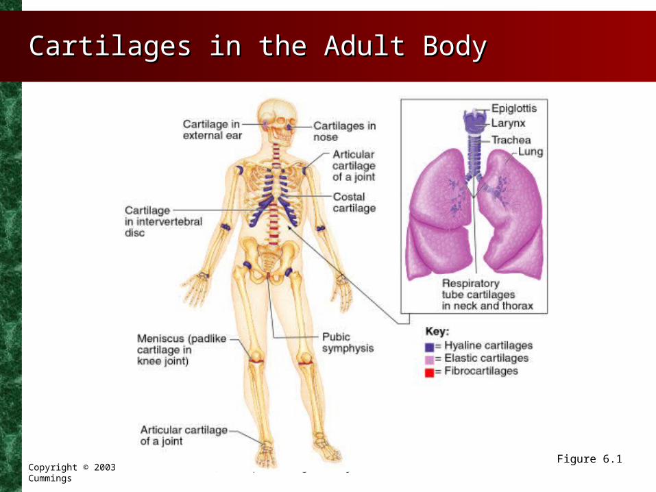
• Found throughout adult body
•Ear and nose
•Articular cartilages and costal cartilage
•Larynx and epiglottis
•Intervertebral discs and pubic symphysis
Copyright © 2003 Pearson Education, Inc. publishing as Benjamin Cummings
CartilageCartilage
• Is abundant in embryo
• Is surrounded by perichondrium
• Consists primarily of water
• Resilient tissue – springs back to original shape
Copyright © 2003 Pearson Education, Inc. publishing as Benjamin Cummings
Types of CartilageTypes of Cartilage
• Hyaline cartilage – (glass) – most abundant cartilage
• Provides support through flexibility
• Elastic cartilage – contains many elastic fibers
• Able to tolerate repeated bending
• Fibrocartilage – resists strong compression and strong tension
• An intermediate between hyaline and elastic cartilage
Copyright © 2003 Pearson Education, Inc. publishing as Benjamin Cummings
Cartilages in the Adult BodyCartilages in the Adult Body
Figure 6.1

Copyright © 2003 Pearson Education, Inc. publishing as Benjamin Cummings
Growth of CartilageGrowth of Cartilage
• Appositional growth
• Chondroblasts in surrounding perichondrium produce new cartilage
• Interstitial growth
• Chondrocytes within cartilage divide and secrete new matrix

Copyright © 2003 Pearson Education, Inc. publishing as Benjamin Cummings
BonesBones
• Organs that contain several types of tissues
• Dominated by bone connective tissue
• Contain nervous tissue and blood tissue
• Contain cartilage in articular cartilages
• Contain epithelial tissue lining blood vessels

Copyright © 2003 Pearson Education, Inc. publishing as Benjamin Cummings
Function of BonesFunction of Bones
• Support – provides hard framework
• Protection of underlying organs
• Movement – skeletal muscles use bones as levers
• Mineral storage – reservoir for important minerals
• Hemopoiesis- Blood-cell formation – bone contains red marrow

Copyright © 2003 Pearson Education, Inc. publishing as Benjamin Cummings
Classification of BonesClassification of Bones
• Long bones – longer than wide – a shaft plus ends
• Short bones – roughly cube-shaped
• Flat bones – thin and flattened, usually curved
• Irregular bones – various shapes, do not fit into other categories

Copyright © 2003 Pearson Education, Inc. publishing as Benjamin Cummings
Classification of BonesClassification of Bones
Figure 6.2

Copyright © 2003 Pearson Education, Inc. publishing as Benjamin Cummings
Gross Anatomy of BonesGross Anatomy of Bones
• Compact bone – dense outer layer of bone
• Spongy (cancellous) bone – internal network of bone

Copyright © 2003 Pearson Education, Inc. publishing as Benjamin Cummings
Structure of a Typical Long Bone F. Structure of a Typical Long Bone F. 5-15-1
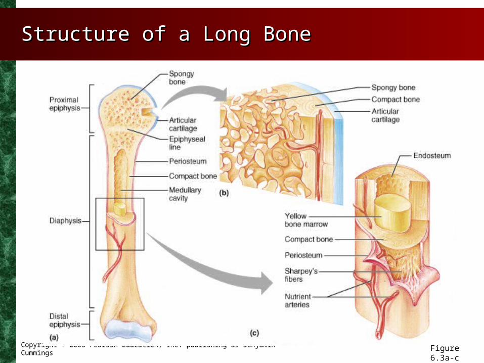
• Diaphysis – “shaft” of a bone
• Epiphysis – ends of a bone
• Blood vessels – well vascularized
• Medullary cavity – hollow cavity – filled with marrow
• Membranes – periosteum and endosteum
Copyright © 2003 Pearson Education, Inc. publishing as Benjamin Cummings
Structure of a Long BoneStructure of a Long Bone
Figure 6.3a-c

Copyright © 2003 Pearson Education, Inc. publishing as Benjamin Cummings
Structure of Short, Irregular, and Structure of Short, Irregular, and Flat BonesFlat Bones
Figure 6.4
Copyright © 2003 Pearson Education, Inc. publishing as Benjamin Cummings
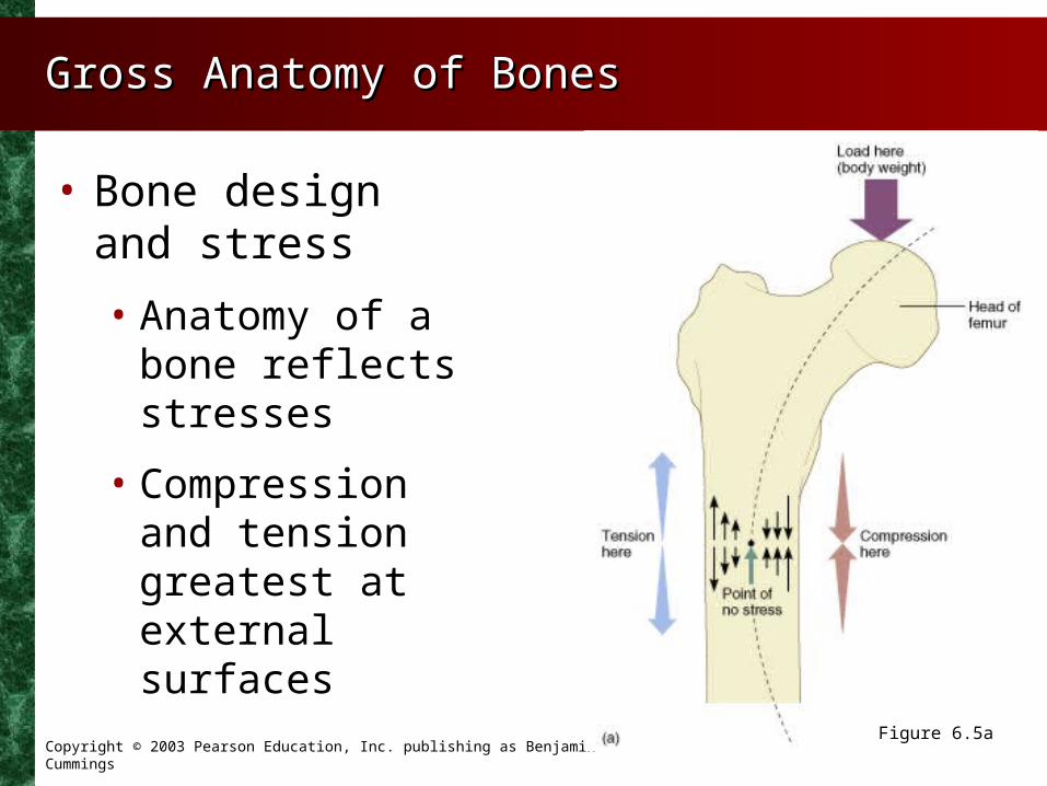
Gross Anatomy of BonesGross Anatomy of Bones
• Bone design and stress
• Anatomy of a bone reflects stresses
• Compression and tension greatest at external surfaces
Figure 6.5a

Copyright © 2003 Pearson Education, Inc. publishing as Benjamin Cummings
Microscopic Structure of Compact Microscopic Structure of Compact BonesBonesF. 5-2 & 5-3F. 5-2 & 5-3
Figure 6.6a, b