Convergent Rhythm Generation from Divergent Cellular Mechanisms
18
Systems/Circuits Convergent Rhythm Generation from Divergent Cellular Mechanisms Jason C. Rodriguez, Dawn M. Blitz, and Michael P. Nusbaum Department of Neuroscience, Perelman School of Medicine, University of Pennsylvania, Philadelphia, Pennsylvania 19104 Different modulatory inputs commonly elicit distinct rhythmic motor patterns from a central pattern generator (CPG), but they can instead elicit the same pattern. We are determining the rhythm-generating mechanisms in this latter situation, using the gastric mill (chewing) CPG in the crab (Cancer borealis) stomatogastric ganglion, where stimulating the projection neuron MCN1 (modulatory commissural neuron 1) or bath applying CabPK (C. borealis pyrokinin) peptide elicits the same gastric mill motor pattern, despite configuring different gastric mill circuits. In both cases, the core rhythm generator includes the same reciprocally inhibitory neurons LG (lateral gastric) and Int1 (interneuron 1), but the pyloric (food-filtering) circuit pacemaker neuron AB (anterior burster) is additionally necessary only for CabPK rhythm generation. MCN1 drives this rhythm generator by activating in the LG neuron the modulator-activated inward current (I MI ), which waxes and wanes periodically due to phasic feedback inhibition of MCN1 transmitter release. Each buildup of I MI enables the LG neuron to generate a self-terminating burst and thereby alternate with Int1 activity. Here we establish that CabPK drives gastric mill rhythm generation by activating in the LG neuron I MI plus a slowly activating transient, low-threshold inward current (I Trans-LTS ) that is voltage, time, and Ca 2 dependent. Unlike MCN1, CabPK maintains a steady I MI activation, causing a subthreshold depolarization in LG that facilitates a periodic postinhibitory rebound burst caused by the regular buildup and decay of the availability of I Trans-LTS . Thus, different modulatory inputs can use different rhythm-generating mechanisms to drive the same neuronal rhythm. Additionally, the same ionic current (I MI ) can play different roles under these different conditions, while different currents (I MI , I Trans-LTS ) can play the same role. Introduction Different modulatory inputs enable individual neuronal net- works to generate different output patterns by changing the in- trinsic and synaptic properties of network neurons (Dickinson, 2006; Briggman and Kristan, 2008; Doi and Ramirez, 2008; Raus- cent et al., 2009; Harris-Warrick, 2011; Marder, 2012). However, different modulatory inputs can also elicit the same activity pat- tern from that network (Saideman et al., 2007b). Determining how different modulatory pathways influence network activity is challenging, because these different pathways can converge onto the same direct inputs to a network (Di Prisco et al., 2000; Korn and Faber, 2005; Derjean et al., 2010; White and Nusbaum, 2011), comparably modulate the same network (Doi and Ramirez, 2010), distinctly alter multiple cellular and synaptic properties in the same circuit neurons (MacLean et al., 2003; Prinz et al., 2004a; Goaillard et al., 2009; Calabrese et al., 2011; Marder, 2012), and/or configure different circuits (Saideman et al., 2007b). The cellular mechanisms underlying the last of these processes are not determined in any system. We are determining the cellular mechanisms that enable two differently configured, network-driven central pattern generator (CPG) circuits to generate the same biphasic motor pattern, us- ing the isolated crab stomatogastric ganglion (STG; Marder and Bucher, 2007; Stein, 2009). These two gastric mill (chewing) cir- cuits are configured by the projection neuron MCN1 (modula- tory commissural neuron 1) and bath-applied CabPK (Cancer borealis pyrokinin) peptide (Saideman et al., 2007a,b). The core rhythm generator for both gastric mill circuits in- cludes the reciprocally inhibitory neurons LG (lateral gastric) and Int1 (interneuron 1). Rhythmic MCN1 transmitter release is also necessary for the MCN1– gastric mill rhythm, while the pyloric pacemaker neuron AB (anterior burster) is necessary for the CabPK rhythm. The cellular and synaptic mechanisms underly- ing MCN1-gastric mill rhythm generation are established (Cole- man et al., 1995; Bartos et al., 1999; DeLong et al., 2009a,b). A key MCN1 rhythm-generating mechanism is its activation of the modulator-activated, voltage-dependent inward current (I MI ) in the LG neuron, which waxes and wanes periodically due to rhyth- mic feedback inhibition of MCN1 transmitter release by LG. These events enable the LG neuron to periodically fire a self- terminating burst and alternate with Int1 activity. Here we identify two CabPK-activated currents in the LG neu- ron that are necessary and sufficient for gastric mill rhythm generation. These currents include I MI and a transient, low- threshold, slowly activating inward current (I Trans-LTS ). I Trans-LTS Received July 29, 2013; revised Sept. 17, 2013; accepted Oct. 7, 2013. Author contributions: J.C.R., D.M.B., and M.P.N. designed research; J.C.R. performed research; J.C.R. and M.P.N. analyzed data; J.C.R., D.M.B., and M.P.N. wrote the paper. This work was supported by National Institute of Neurological Disorders and Stroke Grant R37-NS 29436 (M.P.N.), National Science Foundation Grant IOS-1153417 (D.M.B.), and the Behavioral and Cognitive Neurosciences Training Grant T32-MH 17168. Correspondence should be addressed to Dr. Michael P. Nusbaum, Department of Neuroscience, 215 Stem- mler Hall, Perelman School of Medicine, University of Pennsylvania, Philadelphia, PA 19104-6074. E-mail: [email protected]. D.M. Blitz’s present address: Department of Biology, Miami University, 242 Pearson Hall, Oxford, OH 45056. DOI:10.1523/JNEUROSCI.3217-13.2013 Copyright © 2013 the authors 0270-6474/13/3318047-18$15.00/0 The Journal of Neuroscience, November 13, 2013 • 33(46):18047–18064 • 18047
Transcript of Convergent Rhythm Generation from Divergent Cellular Mechanisms
Systems/Circuits
Convergent Rhythm Generation from Divergent CellularMechanisms
Jason C. Rodriguez, Dawn M. Blitz, and Michael P. NusbaumDepartment of Neuroscience, Perelman School of Medicine, University of Pennsylvania, Philadelphia, Pennsylvania 19104
Different modulatory inputs commonly elicit distinct rhythmic motor patterns from a central pattern generator (CPG), but they caninstead elicit the same pattern. We are determining the rhythm-generating mechanisms in this latter situation, using the gastric mill(chewing) CPG in the crab (Cancer borealis) stomatogastric ganglion, where stimulating the projection neuron MCN1 (modulatorycommissural neuron 1) or bath applying CabPK (C. borealis pyrokinin) peptide elicits the same gastric mill motor pattern, despiteconfiguring different gastric mill circuits. In both cases, the core rhythm generator includes the same reciprocally inhibitory neurons LG(lateral gastric) and Int1 (interneuron 1), but the pyloric (food-filtering) circuit pacemaker neuron AB (anterior burster) is additionallynecessary only for CabPK rhythm generation. MCN1 drives this rhythm generator by activating in the LG neuron the modulator-activatedinward current (IMI ), which waxes and wanes periodically due to phasic feedback inhibition of MCN1 transmitter release. Each buildupof IMI enables the LG neuron to generate a self-terminating burst and thereby alternate with Int1 activity. Here we establish that CabPKdrives gastric mill rhythm generation by activating in the LG neuron IMI plus a slowly activating transient, low-threshold inward current(ITrans-LTS) that is voltage, time, and Ca 2� dependent. Unlike MCN1, CabPK maintains a steady IMI activation, causing a subthresholddepolarization in LG that facilitates a periodic postinhibitory rebound burst caused by the regular buildup and decay of the availability ofITrans-LTS. Thus, different modulatory inputs can use different rhythm-generating mechanisms to drive the same neuronal rhythm.Additionally, the same ionic current (IMI ) can play different roles under these different conditions, while different currents (IMI , ITrans-LTS) canplay the same role.
IntroductionDifferent modulatory inputs enable individual neuronal net-works to generate different output patterns by changing the in-trinsic and synaptic properties of network neurons (Dickinson,2006; Briggman and Kristan, 2008; Doi and Ramirez, 2008; Raus-cent et al., 2009; Harris-Warrick, 2011; Marder, 2012). However,different modulatory inputs can also elicit the same activity pat-tern from that network (Saideman et al., 2007b). Determininghow different modulatory pathways influence network activity ischallenging, because these different pathways can converge ontothe same direct inputs to a network (Di Prisco et al., 2000; Kornand Faber, 2005; Derjean et al., 2010; White and Nusbaum,2011), comparably modulate the same network (Doi andRamirez, 2010), distinctly alter multiple cellular and synapticproperties in the same circuit neurons (MacLean et al., 2003;Prinz et al., 2004a; Goaillard et al., 2009; Calabrese et al., 2011;Marder, 2012), and/or configure different circuits (Saideman et
al., 2007b). The cellular mechanisms underlying the last of theseprocesses are not determined in any system.
We are determining the cellular mechanisms that enable twodifferently configured, network-driven central pattern generator(CPG) circuits to generate the same biphasic motor pattern, us-ing the isolated crab stomatogastric ganglion (STG; Marder andBucher, 2007; Stein, 2009). These two gastric mill (chewing) cir-cuits are configured by the projection neuron MCN1 (modula-tory commissural neuron 1) and bath-applied CabPK (Cancerborealis pyrokinin) peptide (Saideman et al., 2007a,b).
The core rhythm generator for both gastric mill circuits in-cludes the reciprocally inhibitory neurons LG (lateral gastric) andInt1 (interneuron 1). Rhythmic MCN1 transmitter release is alsonecessary for the MCN1– gastric mill rhythm, while the pyloricpacemaker neuron AB (anterior burster) is necessary for theCabPK rhythm. The cellular and synaptic mechanisms underly-ing MCN1-gastric mill rhythm generation are established (Cole-man et al., 1995; Bartos et al., 1999; DeLong et al., 2009a,b). A keyMCN1 rhythm-generating mechanism is its activation of themodulator-activated, voltage-dependent inward current (IMI) inthe LG neuron, which waxes and wanes periodically due to rhyth-mic feedback inhibition of MCN1 transmitter release by LG.These events enable the LG neuron to periodically fire a self-terminating burst and alternate with Int1 activity.
Here we identify two CabPK-activated currents in the LG neu-ron that are necessary and sufficient for gastric mill rhythmgeneration. These currents include IMI and a transient, low-threshold, slowly activating inward current (ITrans-LTS). ITrans-LTS
Received July 29, 2013; revised Sept. 17, 2013; accepted Oct. 7, 2013.Author contributions: J.C.R., D.M.B., and M.P.N. designed research; J.C.R. performed research; J.C.R. and M.P.N.
analyzed data; J.C.R., D.M.B., and M.P.N. wrote the paper.This work was supported by National Institute of Neurological Disorders and Stroke Grant R37-NS 29436 (M.P.N.),
National Science Foundation Grant IOS-1153417 (D.M.B.), and the Behavioral and Cognitive Neurosciences TrainingGrant T32-MH 17168.
Correspondence should be addressed to Dr. Michael P. Nusbaum, Department of Neuroscience, 215 Stem-mler Hall, Perelman School of Medicine, University of Pennsylvania, Philadelphia, PA 19104-6074. E-mail:[email protected].
D.M. Blitz’s present address: Department of Biology, Miami University, 242 Pearson Hall, Oxford, OH 45056.DOI:10.1523/JNEUROSCI.3217-13.2013
Copyright © 2013 the authors 0270-6474/13/3318047-18$15.00/0
The Journal of Neuroscience, November 13, 2013 • 33(46):18047–18064 • 18047
exhibits voltage- and time-dependentproperties. CabPK-gastric mill rhythmgeneration results from IMI providing aconstant depolarizing drive that enablesperiodic postinhibitory rebound (PIR)bursting, triggered by ITrans-LTS. Therhythmic nature of the PIR burst genera-tion results from the time- and voltage-dependent properties of ITrans-LTS.Computational modeling and dynamic-clamp (DClamp) manipulations of thesetwo currents support their necessity andsufficiency for CabPK– gastric millrhythm generation, and reveal that the py-loric rhythm (AB)-timed influence on theLG neuron is necessary for triggeringeach PIR burst. Thus, distinct rhythm-generating mechanisms enable distinctcircuits to generate the same rhythmic ac-tivity. Additionally, the same ionic cur-rent (IMI) plays a different role underthese two conditions, whereas differentcurrents (IMI, ITrans-LTS) play a compara-ble role.
Materials and MethodsAnimals. Male Jonah crabs (C. borealis) werepurchased from commercial suppliers (FreshLobster, Marine Biological Laboratory) andmaintained in aerated, filtered artificial seawa-ter at 10 –12°C. Animals were cold anesthetizedby packing them in ice for at least 30 min beforedissection, after which the foregut was re-moved, in physiological saline at �4°C, andthe stomatogastric nervous system (STNS)isolated.
Solutions. C. borealis physiological salinecontained the following (in mM): 440 NaCl, 26MgCl2, 13 CaCl2, 11 KCl, 10 Trisma base, 5maleic acid, and 5 glucose, pH 7.4 –7.6. Allpreparations were superfused continuouslywith C. borealis saline (8 –12°C). CabPK-I orCabPK-II (Saideman et al., 2007a; Biotechnol-ogy Center, University of Wisconsin, Madison,WI) was diluted from a stock solution (10 �3
M)into physiological saline or voltage-clamp sa-line immediately before use. Bottles containingC. borealis saline and CabPK saline were con-nected to the same switching manifold for rapid solution changes. Ox-otremorine (OXO; 10 �5
M; Sigma), a muscarinic agonist, was applied inthe same manner.
For voltage-clamp experiments, tetrodotoxin (TTX; 10 �7M; Sigma),
picrotoxin (PTX; 10 �5M; Sigma), and tetraethylammonium chloride
(TEACl; 10 �2M; Sigma) were added to C. borealis saline (i.e., voltage-
clamp saline). These substances were used to suppress voltage-dependentNa � currents (TTX), glutamatergic inhibitory synaptic transmission(PTX), and a subset of K � currents (TEACl; Marder and Eisen, 1984;Golowasch and Marder, 1992). In some experiments, the microelectrodewas filled with a solution of CsCl (1 M; Sigma) and TEACl (1 M) toadditionally suppress a subset of K � currents. To test the sensitivity ofCabPK-influenced currents to extracellular Na �, in some experimentsNa � in the saline was replaced by NMDG � (n-methyl, D-glucamine;Fluka; Golowasch and Marder, 1992). The NMDG � was added to thesolution first and then neutralized with HCl before adding all other com-ponents. Additionally, in some experiments we used flufenamic acid(FFA; 10 �5
M; Sigma), an inhibitor of Ca 2�-activated, nonspecific cation
current (ICAN), dissolved in dimethylsulfoxide (DMSO) and addeddirectly to C. borealis saline. The final DMSO concentration neverexceeded 1%.
Electrophysiology. Electrophysiology experiments were performed us-ing standard techniques for this system (Beenhakker and Nusbaum,2004). In brief, the isolated STNS (Fig. 1A) was pinned into a siliconeelastomer (Sylgard 184, K.R. Anderson)-lined Petri dish. Extracellularnerve recordings were obtained using pairs of stainless steel wire elec-trodes (reference and recording) whose ends were pressed into theSylgard-coated dish. A differential AC amplifier (Model 1700, AM Sys-tems) amplified the voltage difference between the reference wire, in themain bath compartment, and the recording wire, isolated with a sectionof an individual nerve from the main bath compartment by petroleumjelly (Vaseline, Lab Safety Supply). This signal was then further amplifiedand filtered (Model 410 Amplifier; Brownlee Precision). For extracellularnerve stimulation, the pair of wires used to record nerve activity wasplaced into a stimulus isolation unit (Model SIU5; Astromed/Grass In-struments) connected to a stimulator (Model S88; Astro-Med/GrassInstruments).
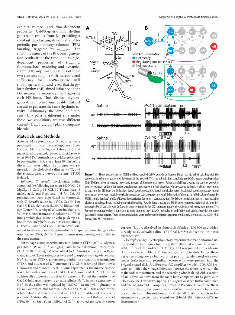
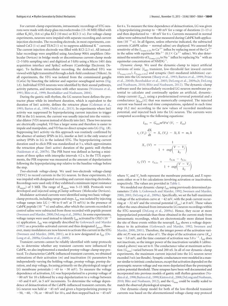
Figure 1. The projection neuron MCN1 and bath-applied CabPK peptide configure different gastric mill circuits but elicit thesame gastric mill motor pattern. A, Schematic of the isolated STNS, including its four ganglia [paired CoGs, oesophageal ganglion(OG), STG] plus their connecting nerves and a subset of the peripheral nerves. Paired parallel lines crossing the superior oesopha-geal nerves (sons) and inferior oesophageal nerves (ions) represent their bisection, which occurred at the start of each experimentto separate the STG from the CoGs. dgn, dorsal gastric nerve; dvn, dorsal ventricular nerve; lgn, lateral gastric nerve; lvn, lateralventricular nerve; mvn, medial ventricular nerve; stn, stomatogastric nerve. B, Schematic of the gastric mill circuit configured byMCN1 stimulation (top) and CabPK peptide superfusion (bottom). t bars, excitation; filled circles, inhibition; resistors, nonrectifyingelectrical coupling; diode, rectifying electrical coupling. Parallel lines crossing the MCN1 axon represent additional distance be-tween the MCN1 soma in each CoG and its axon terminals in the STG. Numbers in parentheses indicate the copy number per STNSfor each neuron type when it is present as more than one copy. C, MCN1 stimulation and CabPK bath application elicit the samegastric mill motor pattern. These two manipulations were performed in different preparations. From Saideman et al. (2007b). PRO,Protraction; RET, retraction.
18048 • J. Neurosci., November 13, 2013 • 33(46):18047–18064 Rodriguez et al. • Rhythm Generation by Distinct Mechanisms
For current-clamp experiments, intrasomatic recordings of STG neu-rons were made with sharp glass microelectrodes (15–30 M�) filled witheither K2SO4 (0.6 M) plus KCl (10 mM) or KCl (1 M). For voltage-clampexperiments, neurons were impaled with separate recording and currentinjection electrodes. The recording electrode, in most experiments, con-tained CsCl (1 M) and TEACl (1 M) to suppress additional K � currents.The current injection electrode was filled with KCl (2.5 M). All intracel-lular recordings were amplified using Axoclamp 900A amplifiers (Mo-lecular Devices) in bridge mode or discontinuous current-clamp mode(2–5 kHz sampling rate) and digitized at 5 kHz using a Micro 1401 dataacquisition interface and Spike2 software (Cambridge Electronic De-sign). To facilitate intracellular recording, the desheathed STG wasviewed with light transmitted through a dark-field condenser (Nikon). Inall experiments, the STG was isolated from the commissural ganglia(CoGs) by bisecting the inferior and superior oesophageal nerves (Fig.1A). Individual STNS neurons were identified by their axonal pathways,activity patterns, and interactions with other neurons (Weimann et al.,1991; Blitz et al., 1999; Beenhakker and Nusbaum, 2004).
During the gastric mill rhythm, the LG neuron burst defines the pro-tractor phase while its interburst duration, which is equivalent to theduration of Int1 activity, defines the retractor phase (Coleman et al.,1995; Bartos et al., 1999; Diehl et al., 2013). In experiments where Int1activity was suppressed by hyperpolarizing current injection to triggerPIR in the LG neuron, the current was usually injected into the ventric-ular dilator (VD) neuron instead of directly into Int1. These two neuronsare electrically coupled, VD has a larger soma and therefore is easier toimpale and manipulate, and VD has no direct synapse onto LG (Fig. 1B).Suppressing Int1 activity via this approach was routinely confirmed bythe absence of unitary IPSPs in LG, insofar as Int1 is the only source ofunitary IPSPs in LG in the isolated STG. The hyperpolarizing currentduration used to elicit PIR was standardized at 5 s, which approximatesthe retraction phase (Int1 active) duration of the gastric mill rhythm(Saideman et al., 2007b). The PIR burst was defined as having a mini-mum of three spikes with interspike intervals �2 s. In the TTX experi-ments, the PIR response was measured as the amount of depolarizationfollowing the hyperpolarizing step relative to the baseline voltage beforethe step.
Two-electrode voltage-clamp. We used two-electrode voltage-clamp(TEVC) to record currents in the LG neuron. In these experiments, LGwas impaled with designated recording and current-injecting electrodes.Recordings were used only if they exhibited a minimum input resistance(Rinput) of 5 M�. The range of Rinput was 5–15 M�. Protocols weredeveloped and injected using pClamp software (Molecular Devices).
Modulator-activated currents were identified using two basic voltage-clamp protocols, including ramps and steps. IMI was isolated by injectingvoltage ramps into LG (�90 to 0 mV at 75 mV/s) in the presence ofCabPK peptide (10 �6
M) and saline, after which the currents recorded incontrol saline were subtracted from those recorded with peptide present(Swensen and Marder, 2000; DeLong et al., 2009a). In some experiments,voltage ramps were used instead to identify IMI activated by OXO (10 �5
M) application. IMI was originally described by Golowasch and Marder(1992) as a proctolin-activated current and thus designated Iproct. How-ever, many modulators are now known to activate this current in the STG(Swensen and Marder, 2000, 2001), so it is now designated as IMI (De-Long et al., 2009a; Grashow et al., 2009).
Transient currents cannot be reliably identified with ramp protocolsso, to determine whether any transient currents were influenced byCabPK, we also implemented a standard prestep voltage-clamp step pro-tocol. To characterize CabPK-influenced transient currents, we obtainedestimations of their activation (m) and inactivation (h) parameters byindependently varying the holding voltage, prestep voltage, prestep du-ration, and step voltage, focusing primarily on the physiological range ofLG membrane potentials (�65 to �30 mV). To measure the voltagedependence of activation, LG was hyperpolarized to a prestep voltage of�80 mV for 10 s followed by a step depolarization to a voltage between�65 and 0 mV, in 5 mV increments. To determine the voltage depen-dence of deinactivation of the CabPK-influenced transient currents, theLG neuron was held at �45 mV and given a hyperpolarizing prestep to�50, �60, �70, or �80 mV for 10 s, and then stepped back to �45 mV
for 6 s. To measure the time dependence of deinactivation, LG was givena hyperpolarizing prestep to �80 mV for a range of durations (1–13 s)and then depolarized to �40 mV for 6 s. Currents measured in normalsaline were subtracted from those measured during CabPK bath applica-tion (10 �6
M). In all figures, unless otherwise indicated, the subtractedcurrents (CabPK saline � normal saline) are displayed. We assessed thesensitivity of the ITrans-LTS to Ca 2� influx by replacing most of the Ca 2�
in the saline with equimolar Mn 2� (0.1� Ca 2� saline). We also deter-mined the sensitivity of ITrans-LTS to Na � influx by replacing Na � with anequimolar concentration of NMDG �.
Dynamic clamp. We used the dynamic-clamp to inject artificialversions of ionic [IMI, transient, low-threshold fast inward current(ITrans-LTF), ITrans-LTS] and synaptic (Int1-mediated inhibition) cur-rents into the LG neuron (Sharp et al., 1993; Bartos et al., 1999; Prinzet al., 2004b; Beenhakker et al., 2005; DeLong et al., 2009a,b; DeLongand Nusbaum, 2010; Blitz and Nusbaum, 2012). The dynamic-clampsoftware used the intracellularly recorded LG neuron membrane po-tential to calculate and continually update an artificial, dynamic-clamp current (Idyn), using a predetermined reversal potential and aconductance [gdyn(t)] that was numerically computed. The injectedcurrent was based on real-time computations, updated in each timestep (0.2 ms) according to the new values of recorded membranepotential, and injected back into the LG neuron. The currents werecomputed according to the following equations:
Idyn � Gmaxmphqnr �V1 � Esyn�
tx �V2�dX
dt� X �V2� � X; X � m, h
X �V� �1
1 � exp�V � Vx
kX�
tX �V� � tX,Lo �tX,Hi � tX,Lo
1 � exp�V � Vx
kX�
where V1 and V2 both represent the membrane potential, and X repre-sents either m or h for calculations involving activation or inactivation,respectively. The values are provided in Table 1.
We modeled our dynamic-clamp IMI using previously determined pa-rameters (Table 1; Golowasch and Marder, 1992; Swensen and Marder,2000, 2001; DeLong et al., 2009a). Specifically, we set the half-maximumvoltage of the activation curve at �42 mV, with the peak current occur-ring at �32 mV and the reversal potential (Esyn) at 0 mV. These valuesreflect the ones obtained from intraneurite LG neuron recordings withinthe STG neuropil (DeLong et al., 2009a). Hence, they occur at morehyperpolarized potentials than those obtained in the current study fromintrasomatic recordings, which are electrotonically more distant fromthe site of these events within the neuropil. IMI shows a voltage depen-dence to its activation (Golowasch and Marder, 1992; Swensen andMarder, 2000, 2001). Therefore, the integer power of the activation vari-able m( P) was set to a value of 1. The slope of the activation curve (Km)was �5.0 mV, and the time constant of activation was 5.0 s �1. IMI doesnot inactivate, so the integer power of the inactivation variable h (abbre-viated q above) was set to 0. The conductance value at maximum activa-tion (Gmax) varied between 50 and 200 nS. In all of our dynamic-clampexperiments, the maximum current injected into the LG neuron neverexceeded 3 nA (see Results). Synaptic conductances were modeled in a man-ner similar to intrinsic conductances, except that activation depended on thepresynaptic neuron voltage and was more depolarized than the presynapticaction potential threshold. These synapses have been well documented andincorporated into previous models of gastric mill rhythm generation (Na-dim et al., 1998; Bartos et al., 1999; Kintos et al., 2008; DeLong and Nusbaum,2010; Blitz and Nusbaum, 2012), and the Gmax could be readily scaled tomatch the observed physiological synapses.
Our dynamic-clamp model for both of the low-threshold transientcurrents was based on the aforementioned voltage-clamp step protocol
Rodriguez et al. • Rhythm Generation by Distinct Mechanisms J. Neurosci., November 13, 2013 • 33(46):18047–18064 • 18049
experiments. The results from activation protocols were manually fit toHodgkin–Huxley equations using HHfit (version 3.2) software devel-oped by the Nadim laboratory [New Jersey Institute of Technology(NJIT) and Rutgers University, Newark, NJ; available at http://stg.rutgers.edu/software/]. Occasionally, the resting Vm and action potentialthreshold coordinately varied between preparations, possibly due to im-palement quality. Therefore, the dynamic-clamp parameters were linkedto the resting Vm. Table 1 contains a full parameter set for a neuronresting at �60 mV.
We used two versions of the dynamic-clamp on a personal computer(PC) running Windows XP/7 and a NI PCI-6070-E data acquisitionboard (National Instruments). The first version was developed in theNadim laboratory (NJIT and Rutgers University, Newark, NJ; available athttp://stg.rutgers.edu/software/). The second version was developed byE. Brady Trexler (Fishberg Department of Neuroscience, Mt. SinaiSchool of Medicine; freely available through Gotham Scientific:http://gothamsci.com/NetClamp/). Dynamic-clamp current injectionswere performed while recording in single-electrode, DCC mode (sam-pling rates, 2–5 kHz) or with separate voltage recording and current-injecting electrodes.
Data analysis. Data were collected onto a computer, with later play-back onto a chart recorder (Everest; Astro-Med). Acquisition onto thecomputer (sampling rate, 5 kHz) used the Spike2 data acquisition andanalysis system (Cambridge Electronic Design). Some analyses, includ-ing CabPK– gastric mill rhythm parameters, were conducted on the dig-itized data using a custom-written Spike2 program (The Crab Analyzer:freely available at http://www.neurobiologie.de/spike2).
Voltage-clamp data analysis was performed using PClamp (version 9;Molecular Devices), Spike2 (CED), and Igor Pro (Wavemetrics) soft-ware. For ramps, total neuron currents were determined by averaging 10ramps in each condition and subtracting the control from the experi-mental condition. For prestep protocols, the protocols were run once ineach condition and the control currents were subtracted before analysis.
For gastric mill rhythm analyses, unless otherwise stated, each data pointin a dataset was derived by determining the mean for the analyzed parameterfrom 10 consecutive gastric mill cycles. One gastric mill cycle was defined asextending from the onset of consecutive LG neuron action potential bursts(Beenhakker and Nusbaum, 2004; Wood et al., 2004). Thus, the gastric millcycle period was measured as the duration (in seconds) between the onset oftwo successive LG neuron bursts. The protractor phase was measured as theLG neuron burst duration, while the retractor phase was measured as the LGneuron interburst duration. The gastric mill rhythm-timed LG neuron burstduration was defined as the duration (in seconds) between the onset of thefirst and last action potential within an impulse burst, during which nointerspike interval was 1.5 s (approximately one pyloric cycle period dur-ing the CabPK–gastric mill rhythm and briefer than the duration of eachgastric mill phase; Saideman et al., 2007b). The intraburst firing rate of theLG neuron was defined as the number of action potentials � 1, divided bythe burst duration.
Data were plotted with Igor Pro (version 6.10A). Figures were pro-duced using CorelDraw (version 13.0 for Windows). Statistical analyseswere performed with Microsoft Excel and SigmaStat 3.0 (SPSS). Com-parisons were made to determine statistical significance using the pairedStudent’s t test or ANOVA with repeated-measures followed by the Stu-
dent–Newman–Keuls (SNK) post hoc test. In all experiments, the effect ofeach manipulation was reversible, and there was no significant differencebetween the premanipulation and postmanipulation groups. Data areexpressed as the mean � SE.
Gastric mill model. We constructed a computational model of theCabPK-gastric mill rhythm generator modified from an existingconductance-based model of the MCN1– gastric mill rhythm generator(Nadim et al., 1998; Beenhakker et al., 2005; DeLong et al., 2009a,b). Thepreviously published version modeled the LG, Int1, and MCN1 neuronsas having multiple compartments separated by an axial resistance, witheach compartment possessing intrinsic and/or synaptic conductances.The parameters of the CabPK– gastric mill rhythm generator model werebased on both previously published voltage-clamp analyses in STG neu-rons (including LG) and on the LG neuron voltage-clamp results ob-tained in this study (Golowasch and Marder, 1992; Swensen and Marder,2000, 2001; DeLong et al., 2009a). To mimic the effects of CabPK bathapplication to the biological system, we added IMI to the LG neurondendrite compartment as an intrinsic (nonsynaptically activated) cur-rent (Table 2). This approach was based on the fact that CabPK excitesLG by activating IMI (this study) and that CabPK was constantly presentduring its application. To more realistically mimic the biological system,in this version of the model we modified the CabPK-activated GMI in theLG neuron dendrite compartment to include a voltage dependence (Ta-ble 2). Based on data collected in this study, we also added a CabPK-activated ITrans-LTS to the LG neuron (Table 2). The time and voltagedependence of ITrans-LTS were empirically determined in the presentstudy with voltage-clamp analyses.
Simulations were performed on a PC with the freely available UbuntuLinux operating system (www.ubuntu.com). We used the Network sim-ulation software developed in the Nadim laboratory (http://stg.rutgers.edu/software/network.htm). This included using a fourth-order Runge–Kutta numerical integration method with time steps of 0.05 and 0.01 ms.Results were visualized by plotting outputted data points using the freelyavailable Gnuplot software package (www.gnuplot.info). In most figuresshowing the model output, we present conductance ( g ) instead of theassociated current ( I) to more clearly display the trajectory during thegastric mill retractor and protractor phases. The main difference betweeng and I is that the former lacks the fast transient changes that occur in thelatter during each LG neuron action potential (DeLong et al., 2009a). Inparticular, the relatively slow kinetics of the CabPK-activated conduc-tances make them insensitive to these fast transient changes in voltage.
The presentation of currents in the model and dynamic-clamp figuresrepresent different conventions. Specifically, the model output uses thestandard voltage-clamp convention, whereas the dynamic-clamp outputuses the standard current-clamp convention. For example, depolarizingcurrent has a downward trajectory in the model output figures but has anupward trajectory in the dynamic-clamp output figures.
ResultsIn the isolated crab STG, tonic MCN1 stimulation and bath-applied CabPK (�10�7
M) elicit comparable gastric mill motorpatterns, despite configuring different gastric mill circuits (Fig.1B; Saideman et al., 2007b). MCN1 does not contain CabPK, and
Table 1. Dynamic-clamp conductances
Neuron Conductances Parameters V1/2 (mV) k (mV) tlo (ms) thi (ms) Exp ( p, q, r) Erev Gmax
LG (biological) ITrans-LTS m �50 �3 500 500 1 10 0.6h �58 0.8 3500 1500 1
IMI m �50 �5 5 5 1 10 0.1Int1 (model) Nav m �42 �5 1.5 0.45 3 45 7.5
h �52 9.2 10 2.4 1KV m �26 �9 27 3 4 �70 30
h �16 1.5 20 200 2Leak �60 0.012
Int1 LG Synapse m �40 �1 200 200 1 �80 2LG Int1 Synapse m �30 �0.1 200 200 1 �80 15
V1/2 , half-activation voltage; Erev , reversal potential.
18050 • J. Neurosci., November 13, 2013 • 33(46):18047–18064 Rodriguez et al. • Rhythm Generation by Distinct Mechanisms
the CabPK– gastric mill rhythm can occur without MCN1 activ-ity. CabPK is present in two or three pairs of CoG projectionneurons that innervate the STG, although these neurons are notphysiologically identified (Saideman et al., 2007a). However,bath-applied peptide can mimic the actions resulting from itsneuronal release. For example, in the crab STG, bath applicationof the peptide proctolin (10�6
M) and direct stimulation of themodulatory proctolin neuron (MPN) elicit comparable re-sponses from the pyloric CPG, despite the fact that MPN containsa small-molecule cotransmitter (Nusbaum and Marder, 1989a,b;Blitz et al., 1999).
There are also at least several additional gastric mill motorpatterns in C. borealis, each distinct from the pattern elicited byMCN1 and CabPK, and driven by a different input pathway(Beenhakker and Nusbaum, 2004; Blitz et al., 2004; Christie et al.,2004; White and Nusbaum, 2011). These different rhythms allshare the same basic structure, which includes a biphasic motorpattern exhibiting rhythmic alternating bursting of protraction-
and retraction-related neurons across an overlapping range ofcycle periods (�5–20 s; Fig. 1C). They differ in the relative tim-ing, intensity, duration, and pattern of activity in the componentneurons. There are seven gastric mill motor neurons, includingfour protractor motor neurons and three retractor motor neu-rons, plus a single retraction-timed interneuron (Int1; Fig. 1B).As discussed below, the pyloric CPG pacemaker neuron AB alsoinfluences these gastric mill rhythms (Fig. 1B). Like the gastricmill rhythm, the pyloric (filtering of chewed food) rhythm isgenerated in the STG (Marder and Bucher, 2007).
A core component of the rhythm generator for the MCN1–and CabPK– gastric mill rhythms is the half-center formed by thereciprocally inhibitory protraction neuron LG and retractionneuron Int1 (Saideman et al., 2007b; Fig. 1B). The biphasicrhythm generated by these two neurons is then imposed on theother gastric mill neurons by synaptic actions from the rhythmgenerator plus the influences of MCN1 or CabPK. Under baselineconditions, the LG neuron is silent (Fig. 1C), and Int1 is sponta-
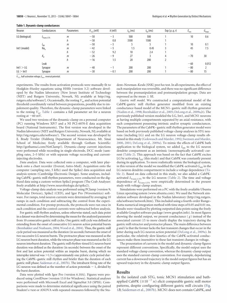
Figure 2. Bath-applied CabPK peptide elicits a sustained, subthreshold depolarization in the isolated LG neuron by activating the voltage-dependent IMI. A, With Int1 neuron activity suppressedby hyperpolarizing current injection, bath-applied CabPK elicited a sustained, subthreshold depolarization in the LG neuron. B, With Int1 neuron exhibiting its normal pyloric-timed burst pattern,thereby providing rhythmic inhibition to LG, bath-applied CabPK initially caused a gradual increase in the amplitude of the subthreshold, pyloric-timed oscillations in LG. Note that the oscillationpeaks became more depolarized, while the membrane potential of the trough was not changed. CabPK superfusion was begun immediately before the start of this trace. Subsequently, the gastricmill rhythm commenced. C, I–V plots of CabPK-influenced current in LG, obtained using TEVC and a voltage ramp protocol, during focal pressure application (5 psi, 1 s) of CabPK (10 �4
M) undercontrol conditions (black) and during CCAP (10 �6
M) bath application (red). Each curve represents the difference current (CabPK � control or CCAP condition) as indicated. Solid curves represent themean values for each condition; broken lines represent three individual experiments. D, Injection of artificial IMI ( gMI � 100 nS) into LG, via the dynamic-clamp, in a preparation where Int1 activitywas weak ( 5 Hz) caused a sustained depolarization comparable to that resulting from CabPK application.
Table 2. CabPK– gastric mill network model parameters
Neuron Conductances Parameters V1/2 (mV) k (mV) tlo (ms) thi (ms) Exp ( p, q, r) Erev Gmax
LG ITrans-LTS m �55 �3 500 500 1h �63 0.8 3500 1500 1
IMI m �60 �5 5 5 1Int1 Nav m �42 �5 1.5 0.45 3 45 7.5
h �52 9.2 10 2.4 1KV m �26 �9 27 3 4 �70 30
h �16 1.5 20 200 2Leak �60 0.012
Int1 LG Synapse m �40 �1 200 200 1 �80 2LG Int1 Synapse m �30 �0.1 200 200 1 �80 15
V1/2 , half-activation voltage; Erev , reversal potential.
Rodriguez et al. • Rhythm Generation by Distinct Mechanisms J. Neurosci., November 13, 2013 • 33(46):18047–18064 • 18051
neously active, exhibiting a pyloricrhythm-timed activity pattern due to in-hibitory input that it receives from the ABneuron (Fig. 1B; Bartos et al., 1999; Saide-man et al., 2007b). The pivotal event forenabling gastric mill rhythm generation isthe acquisition by the LG neuron of theability to fire rhythmic bursts.
The cellular and synaptic mechanismsunderlying MCN1 activation of the gas-tric mill rhythm generator are known(Coleman et al., 1995; Bartos et al., 1999;DeLong et al., 2009a). In brief, during theMCN1– gastric mill rhythm, there is arhythmic release of the MCN1 cotransmit-ters, which include the peptides proctolinand CabTRP Ia (C. borealis tachykinin-related peptide Ia) plus GABA (Blitz et al.,1999). MCN1 uses only CabTRP Ia to influ-ence LG (slow excitation) and only GABA toinfluence Int1 (fast excitation; Wood et al.,2000; Stein et al., 2007). MCN1 cotransmit-ter release is rhythmic, even when MCN1 istonically active, because its STG terminals(MCN1STG) receive ionotropic synaptic in-hibition from LG (Fig. 1B; Coleman andNusbaum, 1994). Thus, during retraction,continuous MCN1 release of CabTRP Iadrives a steady buildup of IMI in LG thateventually is sufficient to enable LG tofire an action potential burst (DeLong etal., 2009a). During protraction, whenMCN1STG cotransmitter release is inhibitedby LG, there is a steady decline in theamount of IMI in LG until it can no longersustain the LG neuron burst. This rhyth-mic activation of IMI in LG appears to besufficient to drive the gastric millrhythm across the physiological range ofMCN1 firing frequencies (DeLong et al.,2009a,b; DeLong and Nusbaum, 2010).MCN1-driven gastric mill rhythm gener-ation is also facilitated by the pyloricrhythm (cycle period, �1 s), becauseevery LG neuron burst initiates, after suf-ficient IMI has accrued, during a pyloric-timed depolarization (i.e., disinhibition)that results from AB neuron inhibition ofInt1 (Bartos et al., 1999; DeLong et al.,2009a). These disinhibitions reduce theMCN1– gastric mill cycle period by reduc-ing the amount of IMI-mediated depolarization needed to enableLG to fire a burst. However, this rhythm does persist, with alonger cycle period, when there is no pyloric rhythm (Bartos etal., 1999).
The cellular and synaptic mechanisms underlying the CabPK–gastric mill rhythm were not known, although it was determinedpreviously that activity in LG, Int1, and AB was necessary toenable this rhythm (Saideman et al., 2007b). Additionally, as dur-ing the MCN1– gastric mill rhythm, it appeared that directCabPK excitation of the LG neuron was a pivotal event forrhythm generation. Thus, we identified CabPK-influenced ioniccurrents in LG.
CabPK activates three voltage-dependent inward currents inthe LG neuronCabPK application (10�6
M) provides a depolarizing drive to LGfrom its resting potential (�57.5 � 1.5 mV; n � 8). For example,under the most reduced conditions, with LG isolated from syn-aptic input, CabPK consistently elicited in LG a steady 5–10 mVdepolarization. This depolarizing response occurred when LGwas isolated by either hyperpolarizing Int1 (depolarizing re-sponse, 8.5 � 1.1 mV; n � 8; Fig. 2A) or suppressing all glutama-tergic inhibitory input to LG by bath-applying PTX (10�5
M;depolarizing response, 10.7 � 0.54 mV; n � 11). The CabPK-mediated depolarization moved the LG neuron membrane
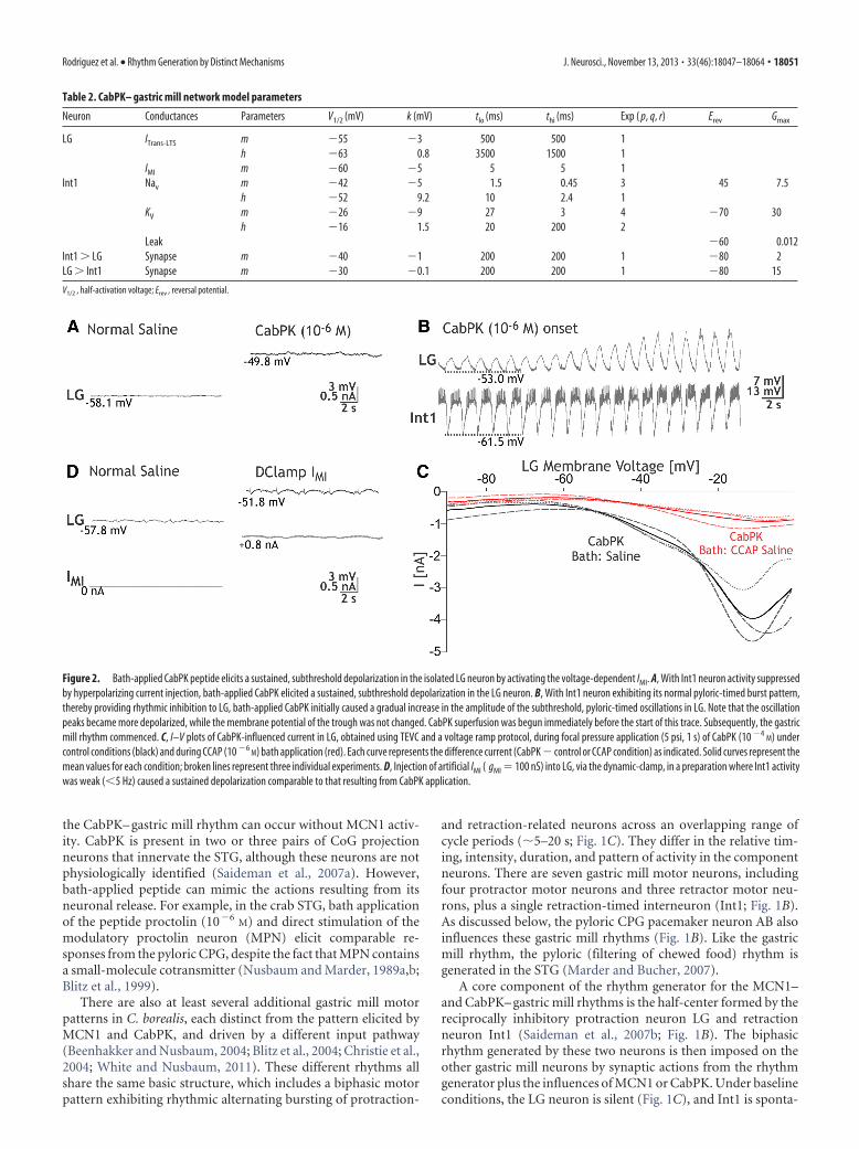
Figure 3. CabPK application influences both transient and sustained voltage-dependent inward currents in the LG neuron. A,Example raw current traces recorded during TEVC using a voltage step protocol (Vhold ��80 mV) to the indicated step potentials,during superfusion with voltage-clamp saline under control and CabPK conditions (see Materials and Methods). Note that, duringthe two more depolarized steps (�50 and �35 mV) in the presence of CabPK, there was a relatively large amplitude transientinward current, while during all three steps there was a reduction in the amplitude of the sustained outward current relative to thecontrol condition. B, Top, CabPK-influenced currents (CabPK condition � control condition) resulting from a voltage step protocolduring TEVC included a low threshold, fast transient current (ITrans-LTF), ITrans-LTS, and sustained current (IMI). ITrans-LTS was predom-inantly responsible for the transient current, while IMI was predominantly responsible for the reduced outward current in A.ITrans-LTF did not exhibit a distinct, separate peak in this experiment (Figs. 4A, 5A), but is evident as an initial steep inward slopebefore the shallower rising slope representing ITrans-LTS. Entire voltage step protocol (hold at �80 mV, 10 s; step to test voltage, 6 s)is shown. Bottom, Expansion of the current traces to highlight the events occurring during each voltage step. C, I–V plot of thecurrent (mean � SE) at the peak of ITrans-LTS for the step protocol used in experiments such as that in B (n � 9).
18052 • J. Neurosci., November 13, 2013 • 33(46):18047–18064 Rodriguez et al. • Rhythm Generation by Distinct Mechanisms
potential closer to its spike threshold, which was not changed byCabPK (saline, �42.4 � 1.3 mV; n � 9; CabPK, �44.5 � 1.4 mV;n � 9, p � 0.15). When Int1 was active, the LG neuron membranepotential exhibited subthreshold, pyloric-timed oscillations thatexhibited a more depolarized peak in the presence of CabPK(saline, �57.5 � 1.4 mV; n � 10; CabPK, �48.7 � 1.7 mV; n �10, p 0.01; Fig. 2B). These depolarized peaks remained sub-threshold before the onset of the gastric mill rhythm, as well asduring the ensuing gastric mill retraction phase. In contrast,CabPK did not alter the membrane potential at the trough ofthese LG neuron oscillations (saline, �59.8 � 1.8 mV; n � 10;CabPK, �59.1 � 1.7 mV; n � 10, p � 0.4; Fig. 2B; see below).Thus, the increased pyloric-timed oscillation amplitude was dueto a more depolarized peak.
Based on the assumption that the sustained depolarizing drivein the LG neuron during CabPK application resulted fromCabPK influence on a persistent current, we isolated CabPK-sensitive currents using a voltage ramp protocol (see Materialsand Methods) in TEVC. Difference currents between CabPK andcontrol solutions revealed a voltage-dependent, inward net cur-rent at potentials more hyperpolarized than 0 mV (Fig. 2C). Thisinward current exhibited a small, relatively constant amplitude atmembrane potentials more hyperpolarized than approximately�60 mV, whereas in the depolarizing direction from approxi-mately �60 mV, the I–V plot for this current displayed a trajec-tory reminiscent of voltage-dependent inward currents.Specifically, it displayed a steadily increasing inward current thatpeaked at �8.1 � 1.4 mV (peak amplitude: �4.4 � 0.6 nA, n �7), after which the amplitude steadily decreased (Fig. 2C). It wasnot possible to determine its reversal potential, likely due to aninability to completely clamp the residual, relatively large K�
currents at more depolarized potentials (DeLong et al., 2009a).This I–V relationship was comparable to that of the previouslyidentified IMI, which is activated by several different neuromodu-lators in crab STG neurons including the LG neuron (Golowaschand Marder, 1992; Swensen and Marder, 2000, 2001; DeLong etal., 2009a).
To further establish that the CabPK-activated, voltage-dependent inward current in the LG neuron that we identified involtage ramp protocols was IMI, we performed an occlusion ex-periment with a known IMI activator in the LG neuron, crusta-cean cardioactive peptide (CCAP; DeLong et al., 2009a). CCAPapplication occludes the ability of the MCN1 peptide CabTRP Iato activate IMI in LG (DeLong et al., 2009a). In these experiments,CabPK (10�4
M) was first pressure applied onto the desheathedSTG neuropil while recording LG in TEVC. CCAP (10�6
M) wasthen bath applied to activate IMI, during which time CabPK(10�4
M) was again puffed onto the STG neuropil. As shown inFigure 2C, the maximal CabPK-activated current amplitude wasdecreased substantially during CCAP bath application (CabPKpre-CCAP: �4.2 � 0.6 nA; CabPK during CCAP application:�0.9 � 0.1 nA; CabPK post-CCAP: �3.5 � 0.9; p � 0.01: CabPKpre-CCAP or CabPK post-CCAP vs CabPK during CCAP: p �0.33, CabPK pre-CCAP vs CabPK post-CCAP; one-wayrepeated-measures ANOVA with SNK post hoc test; n � 3, F(2,8)
� 16.8). This occlusion effect of CCAP thus supported the hy-pothesis that the aforementioned CabPK-activated inward cur-rent in the LG neuron was IMI. In contrast to the CabPKcondition, in normal saline IMI was either not expressed or waspresent at low levels, insofar as the LG neuron resting potential innormal saline was approximately �60 mV (see above), and evenmodest levels of modulator-activated IMI elicit a more depolar-
ized LG neuron membrane potential (Kirby and Nusbaum, 2007;DeLong et al., 2009a).
IMI is also sensitive to changes in extracellular Ca 2� (Golow-asch and Marder, 1992). Specifically, replacing most of the extra-cellular Ca 2� (0.1� normal Ca 2�) in the saline with additionalMg 2�, to maintain the total divalent cation concentration, lin-earizes the I–V curve for IMI at hyperpolarized potentials. Thislinearization in reduced Ca 2�/added Mg 2� saline also occurredfor the CabPK-sensitive current (current measured at �90 mV:CabPK saline, �0.21 � 0.3 nA; CabPK with reduced Ca 2� saline,�5.67 � 1.3 nA; n � 3, p 0.05), further supporting the hypoth-esis that CabPK activates IMI in LG neuron.
To test the hypothesis that IMI was responsible for the afore-mentioned, CabPK-mediated depolarization in LG, we used theDClamp to inject an artificial version of IMI into LG in normalsaline (Fig. 2D). Doing so using DClamp conductances compa-rable to those identified in voltage-clamp (50 –300 nS), while Int1was silent or only weakly active, consistently depolarized the LGneuron resting potential to the same extent as CabPK application(CabPK: 10.68 � 0.5 mV; DClamp IMI: 10.3 � 2 mV; n � 6, p �0.4; Fig. 2A,D).
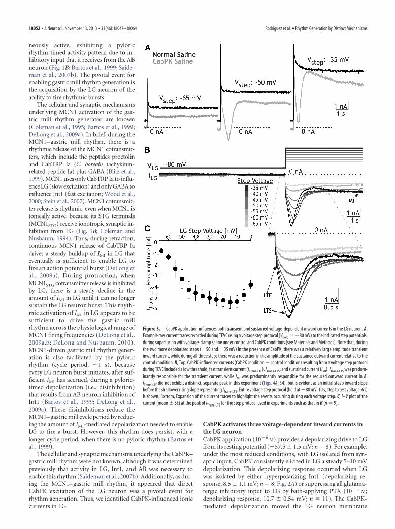
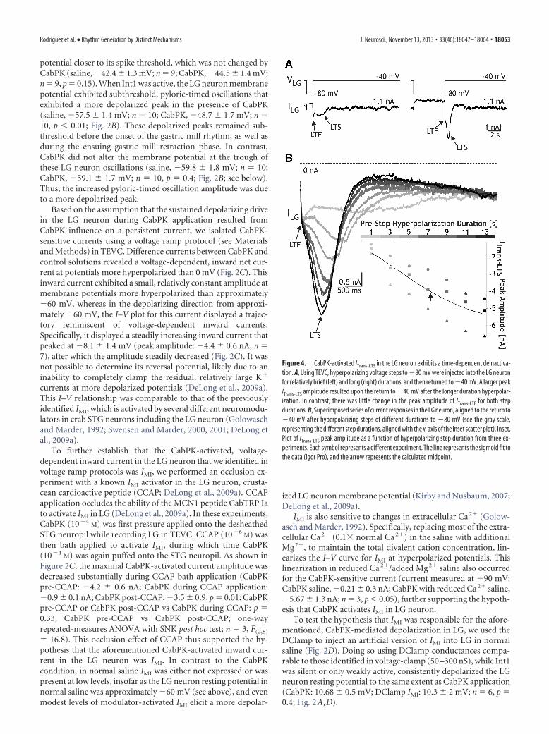
Figure 4. CabPK-activated ITrans-LTS in the LG neuron exhibits a time-dependent deinactiva-tion. A, Using TEVC, hyperpolarizing voltage steps to �80 mV were injected into the LG neuronfor relatively brief (left) and long (right) durations, and then returned to �40 mV. A larger peakITrans-LTS amplitude resulted upon the return to �40 mV after the longer duration hyperpolar-ization. In contrast, there was little change in the peak amplitude of ITrans-LTF for both stepdurations. B, Superimposed series of current responses in the LG neuron, aligned to the return to�40 mV after hyperpolarizing steps of different durations to �80 mV (see the gray scale,representing the different step durations, aligned with the x-axis of the inset scatter plot). Inset,Plot of ITrans-LTS peak amplitude as a function of hyperpolarizing step duration from three ex-periments. Each symbol represents a different experiment. The line represents the sigmoid fit tothe data (Igor Pro), and the arrow represents the calculated midpoint.
Rodriguez et al. • Rhythm Generation by Distinct Mechanisms J. Neurosci., November 13, 2013 • 33(46):18047–18064 • 18053
CabPK also activated other voltage-dependent inward currents in the LG neu-ron. These additional currents were notevident with our voltage ramp protocol,but they were present during a TEVC volt-age step protocol. Their absence duringour voltage ramp manipulations waslikely due to their time-dependent inacti-vation (see below). We identified theseother currents using a prestep hyperpolar-ization (�80 mV) whose duration wassimilar to the gastric mill retraction phase(see Materials and Methods). Using thisapproach, with relatively prolonged depo-larizing steps (6 s) comparable to the gas-tric mill protraction phase during whichLG is depolarized and spiking, we identi-fied three inward currents (Figs. 3, 4, 5).These currents included (1) ITrans-LTF, (2)ITrans-LTS, and (3) sustained inward cur-rent. The ITrans-LTF was not evident in theraw current recordings obtained duringCabPK superfusion (Fig. 3A), due to over-lap with the capacitative current, but wasreadily evident in the difference currenttraces (Figs. 3B, 4,5). In contrast, ITrans-LTS
was identifiable in both the raw CabPKand difference currents (Figs. 3, 4, 5), butwas not evidently expressed under controlconditions (Fig. 3A). Last, in the rawCabPK recordings the sustained inwardcurrent was evident as a smaller ampli-tude outward current relative to thecontrol recordings (Fig. 3A).
The CabPK-activated, sustained in-ward current was predominantly IMI.During the last 3 s of the voltage step, weconsistently observed a voltage-dependent,time-independent inward current, as antic-ipated from our voltage ramp experimentsthat identified CabPK activation of IMI (Fig.3B). There were three features of this sus-tained current in the voltage step protocols,however, that were distinct from IMI mea-sured from the voltage ramp protocol, asfollows: (1) the peak amplitude was smaller (step: �1.6 � 0.4 nA,n � 9; ramp: �4.6 � 0.4 nA; n � 9, p 0.01); (2) the currentexhibited less voltage dependence at depolarized potentials (data notshown); and (3) in some recordings, particularly with steps moredepolarized than �40 mV, a reduced inward current or small out-ward current was evident immediately following ITrans-LTS, relativeto the current amplitude at the end of the step (Figs. 3A,B, 4B, 5B).These features suggested that CabPK also activated a voltage- andtime-dependent outward current. We did not, however, further iso-late and characterize this additional component insofar as it did notappear to be necessary for the CabPK actions on gastric mill rhythmgeneration (see below).
The fast transient inward current exhibited a relatively rapidtime to peak (32.9 � 1.9 ms; n � 16) and small peak amplitude(�0.5 nA), which occurred at approximately �10 mV. It exhib-ited an apparent voltage threshold of approximately �45 mV(range, �50 to �30 mV; n � 16). Only an approximate peakcurrent amplitude is provided for ITrans-LTF because we could not
isolate this current from the other two CabPK-activated inwardcurrents, and these other currents appeared to contribute sub-stantially to the fast transient peak. In this context, it is notewor-thy that IMI reaches its peak current level relatively quickly inresponse to a depolarizing voltage step (Fig. 5A). We did notfurther characterize ITrans-LTF, insofar as it was not necessary forthe ability of CabPK to enable gastric mill rhythm generation (seebelow).
ITrans-LTS exhibited a longer time to peak than ITrans-LTF (timeto peak at �45 mV: 633 � 48 ms, n � 9, p 0.01) as well as alarger peak amplitude (�5.3 � 0.6 nA, measured at �15 � 3.1mV; n � 9; Fig. 3B,C). It exhibited a voltage threshold of approx-imately �55 mV (range, �60 to �50 mV; n � 9), and its ampli-tude increased with depolarization up to approximately �30 mV(n � 9; Fig. 3C). Note that this reported peak amplitude valueincludes the CabPK-activated IMI amplitude and, at the moredepolarized steps, likely also includes the aforementionedvoltage- and time-dependent outward current. The unusually
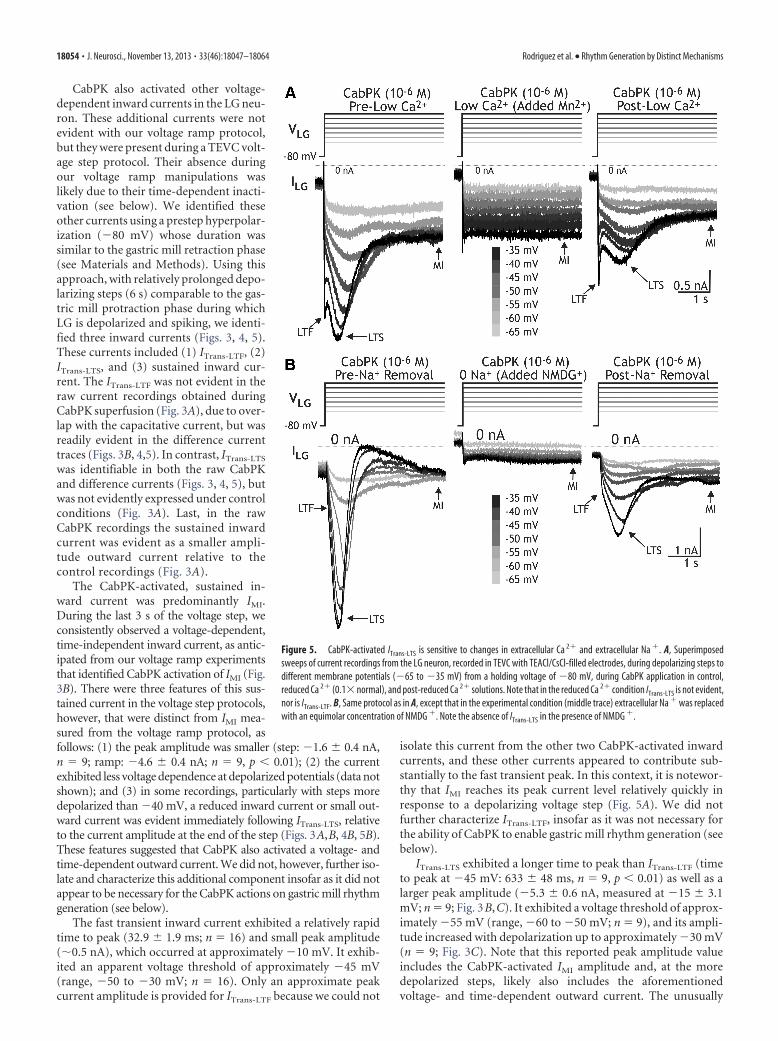
Figure 5. CabPK-activated ITrans-LTS is sensitive to changes in extracellular Ca 2� and extracellular Na �. A, Superimposedsweeps of current recordings from the LG neuron, recorded in TEVC with TEACl/CsCl-filled electrodes, during depolarizing steps todifferent membrane potentials (�65 to �35 mV) from a holding voltage of �80 mV, during CabPK application in control,reduced Ca 2� (0.1� normal), and post-reduced Ca 2� solutions. Note that in the reduced Ca 2� condition ITrans-LTS is not evident,nor is ITrans-LTF. B, Same protocol as in A, except that in the experimental condition (middle trace) extracellular Na � was replacedwith an equimolar concentration of NMDG �. Note the absence of ITrans-LTS in the presence of NMDG �.
18054 • J. Neurosci., November 13, 2013 • 33(46):18047–18064 Rodriguez et al. • Rhythm Generation by Distinct Mechanisms
shallow slope of the I–V curve between �40 and �10 mV likelyresults, at least partly, from the contribution of this outward cur-rent (Fig. 3C). The ITrans-LTS voltage threshold and time to peaksuggested that this current was likely to be activated during theCabPK– gastric mill rhythm, during which time the LG neuronmembrane potential exhibits rhythmic oscillations between ap-proximately �70 and �40 mV (Saideman et al., 2007b). Wecould not measure the full time course of the ITrans-LTS decay tothe baseline because it merged into the sustained inward current,which persisted for the remainder of each step (Figs. 3B, 4A).ITrans-LTS exhibited a reversal potential that was more depolarizedthan 0 mV (n � 16 each; Fig. 3C, ITrans-LTS), suggesting that it isprimarily carried by ions with positive equilibrium potentials(e.g., Ca 2� and/or Na�). Both of the CabPK-activated transientcurrents were clearly distinct from IMI, because IMI exhibits notime-dependent decrease in amplitude (Fig. 5; Golowasch andMarder, 1992).
ITrans-LTS not only exhibited the property of inactivation, but italso exhibited deinactivation. This deinactivation was sensitive tothe time and voltage range that occurs in the LG neuron duringthe CabPK– gastric mill rhythm. We therefore characterized thisproperty by varying parameters of the voltage step protocol involtage-clamp experiments. To determine the time dependenceof this property, we maintained LG at a holding potential of �40mV and systematically hyperpolarized it to �80 mV for a range ofdurations (1 to 13 s), stepping the voltage back to �40 mV aftereach hyperpolarization. We then measured the maximum ampli-tude of the slow transient inward current after the return to �40mV (Fig. 4). These data were then fit with a sigmoid curve (IgorPro), from which two parameters were identified, including themidpoint and slope. There was a relatively long time dependencefor ITrans-LTS deinactivation (midpoint, 7.4 � 0.4 s; slope, 2.4 �0.2 s�1; n � 3; Fig. 4B).
We determined the voltage dependence of the ITrans-LTS dein-activation by varying the prestep voltage across a range (�75 to�50 mV) of membrane potentials, while maintaining the prestepduration (8 s) and subsequent step potential (�45 mV; midpoint,�60.9 � 2 mV; slope, �5.3 � 0.5 mV�1, n � 3). This midpointvalue was well within the normal LG neuron membrane potentialrange (�55 to �70 mV) during the gastric mill retraction phase(Saideman et al., 2007b). Thus, based on the time and voltagedependence of this transient current in LG, and the LG neuronmembrane potential trajectories during the gastric mill rhythm(Saideman et al., 2007a,b; this study), it likely exhibits consider-able inactivation during the course of each gastric mill protrac-tion phase and deinactivation during the retractor phase. Thesecorrelations support the hypothesis that the CabPK-activatedslow transient inward current helps enable the LG neuron togenerate a periodic burst during a pyloric-timed membrane po-tential depolarization produced by the combination of IMI andthe periodic (AB-mediated) removal of synaptic inhibition fromInt1 (see below).
Both CabPK-activated transient inward currents were extra-cellular Ca 2� dependent. Replacing most of the extracellularCa 2� with Mn 2�, a Ca 2� channel antagonist (Turrigiano et al.,1996), consistently resulted in no measureable fast (n � 4) orslow transient inward currents (peak ITrans-LTS at �50 mV:CabPK alone, �1.6 � 0.1 nA; CabPK with 0.1 mM Ca 2�/10.9 mM
Mn 2�, 0 � 0 nA; CabPK post-reduced Ca 2�, �0.8 � 0.02 nA;n � 4; Fig. 5A). This manipulation had only a moderate effect onIMI (Fig. 5A), due to the divalent cation sensitivity of this currentbeing approximately equivalent for Ca 2� and Mn 2� (Golowaschand Marder, 1992). In addition, replacing extracellular Na� with
a nonpermeant ionic species (NMDG�) also consistently re-sulted in no measureable ITrans-LTS (maximum amplitude: con-trol, �3.8 � 0.9 nA; NMDG saline, 0 � 0 nA, n � 7; Fig. 5B). Incontrast, in all four of these NMDG� experiments where therewas a discernible ITrans-LTF peak in the control recordings, thispeak persisted in the NMDG� condition (data not shown). In thethree experiments where there was no distinguishable ITrans-LTF
peak in the control recording, it was not possible to determinewhether it was influenced by the NMDG� substitution (Fig. 5B).
In sum, these results suggested that ITrans-LTS is permeable toboth Na� and Ca 2�, is a Ca 2�-sensitive INa, or is an ICAN, al-though we also did not rule out the possibility that NMDG� actsinstead as an inhibitor of this current. ICAN was identified previ-ously in C. borealis, both in the stomatogastric nervous systemand cardiac ganglion, where it exhibited a reversal potential (Vrev)of approximately �30 mV and was insensitive to changes in ex-tracellular Na� but sensitive to caffeine application (10�2
M),which stimulates intracellular Ca 2� release, and the ICAN antag-onist FFA (10�5
M; Zhang et al., 1995; Kadiri et al., 2011; Ransdellet al., 2013). However, bath-applied FFA did not alter any of theCabPK-activated currents (n � 2). Insofar as ITrans-LTS exhibiteda Vrev 0 mV, sensitivity to extracellular Na� and insensitivity toFFA, it was not likely to be an ICAN.
CabPK-activated transient inward currents enablepostinhibitory rebound in the LG neuronTo determine how the CabPK-activated currents might contrib-ute to LG neuron burst generation during the CabPK– gastricmill rhythm, we examined intrinsic properties in LG. Specifically,we found that brief hyperpolarizing current injections into LGwere followed by passive responses during saline superfusion butelicited a PIR burst during CabPK superfusion. Under controlconditions (saline), we depolarized LG to a membrane potentialthat was comparable to its CabPK-mediated baseline potential(approximately �50 mV) using either direct depolarizing cur-rent injection or DClamp IMI injection. Despite this depolarizedbaseline potential, following a period of hyperpolarization therewas no evidence of PIR (n 10; Fig. 6). In contrast to its passiveresponse during saline superfusion, LG neuron hyperpolariza-tion from its CabPK-mediated depolarized resting potential wasconsistently followed by a PIR burst (Fig. 6). Specifically, during
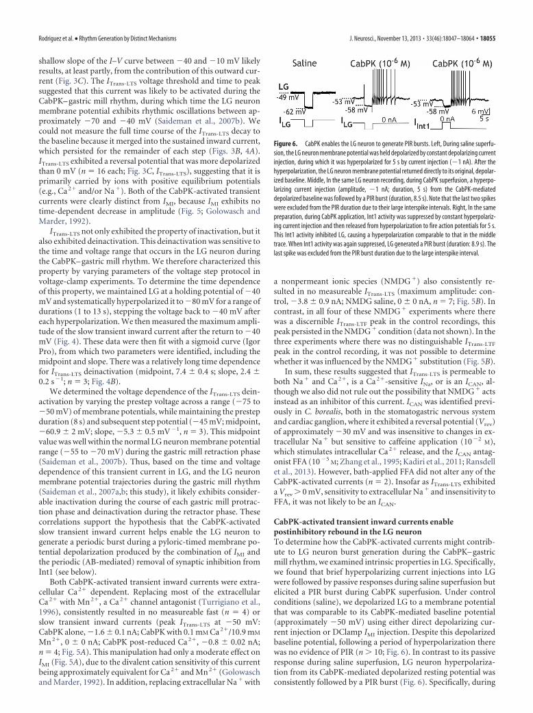
Figure 6. CabPK enables the LG neuron to generate PIR bursts. Left, During saline superfu-sion, the LG neuron membrane potential was held depolarized by constant depolarizing currentinjection, during which it was hyperpolarized for 5 s by current injection (�1 nA). After thehyperpolarization, the LG neuron membrane potential returned directly to its original, depolar-ized baseline. Middle, In the same LG neuron recording, during CabPK superfusion, a hyperpo-larizing current injection (amplitude, �1 nA; duration, 5 s) from the CabPK-mediateddepolarized baseline was followed by a PIR burst (duration, 8.5 s). Note that the last two spikeswere excluded from the PIR duration due to their large interspike intervals. Right, In the samepreparation, during CabPK application, Int1 activity was suppressed by constant hyperpolariz-ing current injection and then released from hyperpolarization to fire action potentials for 5 s.This Int1 activity inhibited LG, causing a hyperpolarization comparable to that in the middletrace. When Int1 activity was again suppressed, LG generated a PIR burst (duration: 8.9 s). Thelast spike was excluded from the PIR burst duration due to the large interspike interval.
Rodriguez et al. • Rhythm Generation by Distinct Mechanisms J. Neurosci., November 13, 2013 • 33(46):18047–18064 • 18055
CabPK superfusion, injecting a modesthyperpolarizing current (�1 nA) into LG,which caused a 5–10 mV hyperpolariza-tion, consistently elicited a PIR burstwhen the current injection was termi-nated (PIR burst: duration, 5.14 � 0.2 s;number of spikes: 8.22 � 0.2; n � 9). Weused hyperpolarizing durations (5 s) andamplitudes that were similar to those ex-perienced by LG during the gastric millretraction phase. The trough of the sub-threshold LG neuron oscillations duringretraction ranged from �55 to �65 mVacross experiments (Saideman et al.,2007a,b).
The LG neuron also readily exhibitedPIR bursts during CabPK application afteran episode of synaptic inhibition from Int1(n � 5; Fig. 6). To establish Int1 mediatedPIR in LG, Int1 activity was suppressed viahyperpolarizing current injection and peri-odically released from this hyperpolariza-tion to fire action potentials for 5 s,comparable to its active period during thegastric mill rhythm. The resulting inhibitionin LG caused it to hyperpolarize by 10.2 �1.5 mV (n � 5), comparable to its responseto Int1 during gastric mill retraction (Saide-man et al., 2007a,b). At the end of each in-hibitory episode, LG generated a PIR burstcomparable to those resulting from hyper-polarizing current injection (PIR burst du-ration, 5.3 � 1 s; number of spikes, 8.2 �0.6; n � 5; p 0.5 for both parameters).
PIR bursts are often driven at leastpartly by the hyperpolarization-activatedinward current (Ih; McCormick and Bal,1997; Sekirnjak and du Lac, 2002; Robin-son and Siegelbaum, 2003; Sangrey andJaeger, 2010; Engbers et al., 2011; Felixet al., 2011). However, Ih did not appearto contribute to the CabPK-enabled PIRbursts in LG insofar as neither hyperpo-larizing current injection nor synapticinhibition from Int1 revealed any evi-dence of a depolarizing sag potentialduring saline superfusion (n 10) orCabPK application (n � 18/20; Figs. 6,7). Similarly, there was no evidence inour voltage-clamp experiments for a sagcurrent (n 10). However, the CabPK-activated ITrans-LTS, with its voltage- andtime-dependent properties of inactiva-tion and deinactivation, was a candidatefor the ionic current underlying PIRburst generation.
We first assessed the contribution of this CabPK-activatedtransient inward current to PIR in LG by simplifying the prepa-ration with TTX (10�7
M) saline to silence all neurons. There wasno evidence for a PIR response after LG was hyperpolarized inTTX saline either from its resting potential (�59.1 � 1.1 mV; n �18) or from a depolarized membrane potential (�47.4 � 1 mV;n � 13; Fig. 7A). In contrast, during CabPK (10�6
M) application
under this condition, LG again exhibited a maintained depolar-ization (�48.4 � 1.2 mV, n � 18) from which it displayed PIR inresponse to hyperpolarizing pulses (�1 nA), albeit without asso-ciated action potentials (n � 18; Fig. 7A). Thus, this CabPK-mediated PIR did not require activation of TTX-sensitive INa.However, these PIR events were briefer than the PIR bursts thatoccurred during normal CabPK saline (2.0 � 0.1 s; n � 18; p
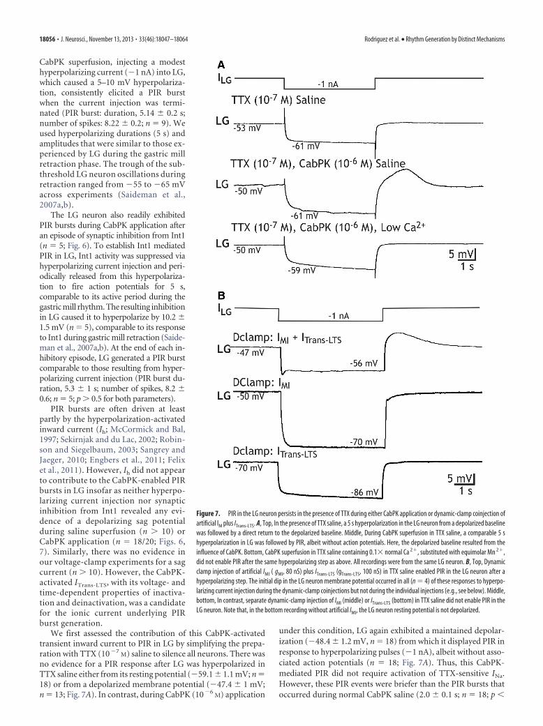
Figure 7. PIR in the LG neuron persists in the presence of TTX during either CabPK application or dynamic-clamp coinjection ofartificial IM plus ITrans-LTS. A, Top, In the presence of TTX saline, a 5 s hyperpolarization in the LG neuron from a depolarized baselinewas followed by a direct return to the depolarized baseline. Middle, During CabPK superfusion in TTX saline, a comparable 5 shyperpolarization in LG was followed by PIR, albeit without action potentials. Here, the depolarized baseline resulted from theinfluence of CabPK. Bottom, CabPK superfusion in TTX saline containing 0.1� normal Ca 2�, substituted with equimolar Mn 2�,did not enable PIR after the same hyperpolarizing step as above. All recordings were from the same LG neuron. B, Top, Dynamicclamp injection of artificial IMI ( gMI, 80 nS) plus ITrans-LTS (gTrans-LTS, 100 nS) in TTX saline enabled PIR in the LG neuron after ahyperpolarizing step. The initial dip in the LG neuron membrane potential occurred in all (n � 4) of these responses to hyperpo-larizing current injection during the dynamic-clamp coinjections but not during the individual injections (e.g., see below). Middle,bottom, In contrast, separate dynamic-clamp injection of IMI (middle) or ITrans-LTS (bottom) in TTX saline did not enable PIR in theLG neuron. Note that, in the bottom recording without artificial IMI, the LG neuron resting potential is not depolarized.
18056 • J. Neurosci., November 13, 2013 • 33(46):18047–18064 Rodriguez et al. • Rhythm Generation by Distinct Mechanisms
0.01), suggesting that a TTX-sensitive INa might prolong thisresponse.
We tested the role in PIR generation played by the IMI-associated depolarization in the LG neuron during CabPK appli-cation. To this end, during CabPK application in TTX saline, weinjected constant amplitude hyperpolarizing current to returnthe LG neuron membrane potential to its pre-CabPK resting po-tential. That is, we eliminated the effect of CabPK-activated IMI,which underlies the steady LG neuron depolarization (see above).From that resting potential, we again injected hyperpolarizingcurrent pulses as above. Doing so reduced the PIR amplitude to
�25% of the control amplitude (control:Vm � �51.1 � 2 mV; PIR amplitude,7.2 � 0.4 mV; hyperpolarized: Vm ��59.1 � 2 mV; PIR amplitude, 1.8 � 0.7mV; n � 7; p 0.01), supporting the hy-pothesis that the IMI-mediated depolar-ization in LG neuron strengthens PIRgeneration mediated by activation of thetransient inward current (see below).
We next assessed whether this CabPK-enabled PIR event in TTX saline wasCa 2�-sensitive by applying CabPK afterreplacing most of the Ca2� (0.1� normal)in the saline with an equimolar concentra-tion of Mn2�. Under these conditions, thePIR amplitude was reversibly reduced (pre-control, 7.5 � 0.7 mV; reduced Ca2�/addedMn2� saline, 0.4 � 0.1 mV; n � 6, p 0.01;Fig. 7A). These results suggested that theCabPK-elicited PIR was mediated by activa-tion of the CabPK-activated low-thresholdinward currents.
We tested the hypothesis that theCabPK-activated IMI plus ITrans-LTS wassufficient to enable PIR in LG. To this end,in TTX saline, we injected into LG adynamic-clamp version of these two cur-rents. This manipulation did indeed en-able LG to express PIR in response to thesame hyperpolarizing current injectionsused in the presence of CabPK (PIR am-plitude, 8.7 � 1.5 mV; PIR duration,4.5 � 0.03 s; n � 4; Fig. 7B). Performingthe same manipulation using only theDClamp version of IMI or ITrans-LTS didnot elicit PIR (n � 4; Fig. 7B). The absenceof PIR during the latter manipulation likelyresulted from the absence of a depolarizedLG neuron resting potential, which in turnlimited the activation of the transient in-ward current after the hyperpolarizing cur-rent injection. Thus, the ability of CabPK toenable the LG neuron to generate PIR burstsapparently results from its coactivation ofIMI, to depolarize LG, and ITrans-LTS, to pro-vide the drive for the PIR burst.
CabPK-activated inward currents in theLG neuron are necessary for gastric millrhythm generationWe tested the ability of the identifiedCabPK-activated inward currents in the
LG neuron to enable gastric mill rhythm generation in a compu-tational model, after which we tested the predictions of the modelin the biological system. We developed a computational model ofthe CabPK– gastric mill rhythm generator (LG, Int1, and AB neu-rons) in which LG contained CabPK-activated IMI plus ITrans-LTS
(see Materials and Methods; Table 2). This model was based onone of three previously published models (CabPK Mechanism 1)that were focused on distinct candidate mechanisms for CabPK-mediated gastric mill rhythm generation (Kintos et al., 2008). Asshown in Figure 8A, our model produced gastric mill rhythm-likealternating bursting in LG and Int1. The model rhythm exhibited
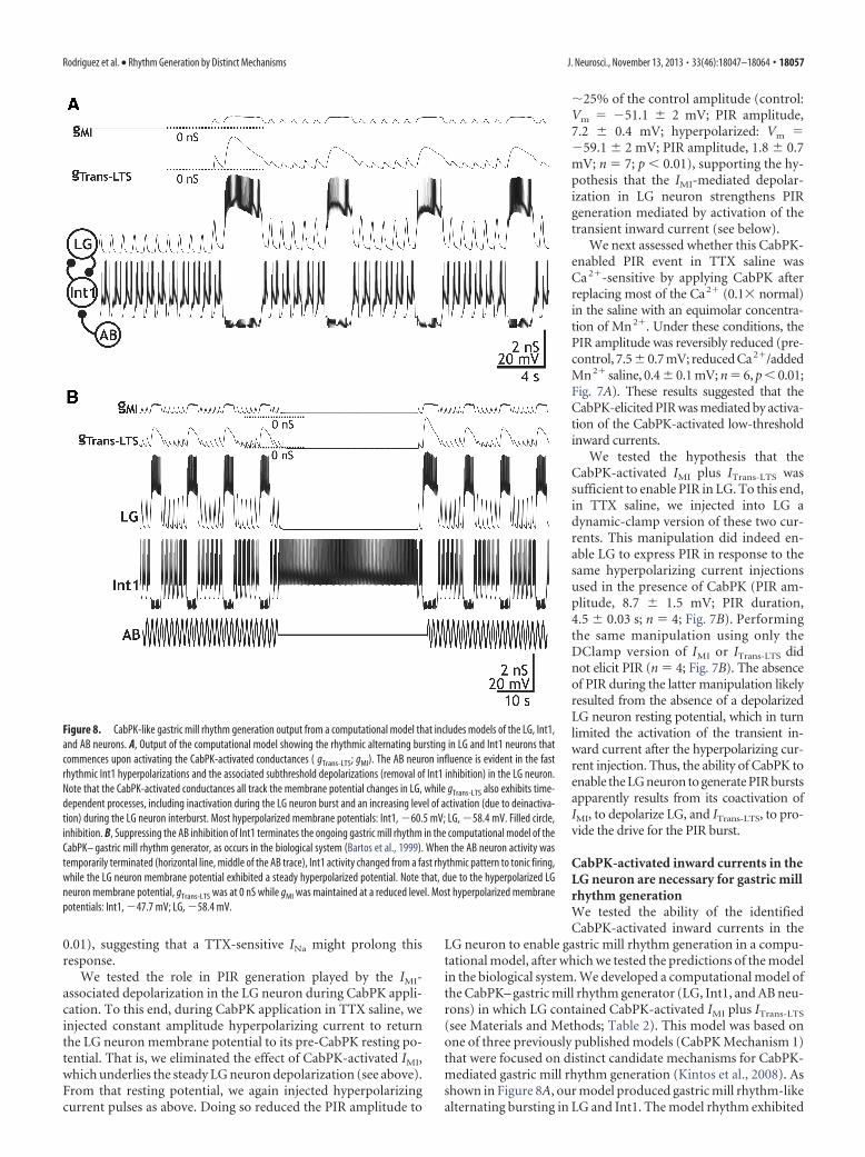
Figure 8. CabPK-like gastric mill rhythm generation output from a computational model that includes models of the LG, Int1,and AB neurons. A, Output of the computational model showing the rhythmic alternating bursting in LG and Int1 neurons thatcommences upon activating the CabPK-activated conductances ( gTrans-LTS; gMI). The AB neuron influence is evident in the fastrhythmic Int1 hyperpolarizations and the associated subthreshold depolarizations (removal of Int1 inhibition) in the LG neuron.Note that the CabPK-activated conductances all track the membrane potential changes in LG, while gTrans-LTS also exhibits time-dependent processes, including inactivation during the LG neuron burst and an increasing level of activation (due to deinactiva-tion) during the LG neuron interburst. Most hyperpolarized membrane potentials: Int1, �60.5 mV; LG, �58.4 mV. Filled circle,inhibition. B, Suppressing the AB inhibition of Int1 terminates the ongoing gastric mill rhythm in the computational model of theCabPK– gastric mill rhythm generator, as occurs in the biological system (Bartos et al., 1999). When the AB neuron activity wastemporarily terminated (horizontal line, middle of the AB trace), Int1 activity changed from a fast rhythmic pattern to tonic firing,while the LG neuron membrane potential exhibited a steady hyperpolarized potential. Note that, due to the hyperpolarized LGneuron membrane potential, gTrans-LTS was at 0 nS while gMI was maintained at a reduced level. Most hyperpolarized membranepotentials: Int1, �47.7 mV; LG, �58.4 mV.
Rodriguez et al. • Rhythm Generation by Distinct Mechanisms J. Neurosci., November 13, 2013 • 33(46):18047–18064 • 18057
a cycle period (10.8 � 2 � 10�4 s), LGneuron burst duration (2.75 � 1 � 10�4
s), and LG neuron interburst duration(8.09 � 1 � 10�4 s) similar to the biolog-ical CabPK– gastric mill rhythm (cycle pe-riod, 11.96 � 1.1 s; LG neuron burstduration, CabPK, 3.2 � 0.3 s; LG neuroninterburst duration, 8.8 � 1.1 s; n � 12).Additionally, the average protraction andretraction duty cycles (DCs; fraction ofthe cycle) were comparable (protractionDC: model, 0.25; biological, 0.27; retractionDC: model, 0.75; biological, 0.73). Notethat, during the model rhythm, gMI andgTrans-LTS followed the LG neuron voltagetrajectory (Fig. 8A), insofar as their activa-tion was voltage dependent. The deinactiva-tion (h) state of gTrans-LTS also tracked the LGneuron membrane potential, rising duringthe retractor phase when LG was rhythmi-cally hyperpolarized by Int1 inhibition.
The model CabPK–gastric mill rhythmwas also comparable to the biologicalrhythm in that it was suppressed by elimi-nating the AB inhibition of Int1 (Fig. 8B). Inthe biological system, this manipulation ter-minates the CabPK–gastric mill rhythm,but not the MCN1–gastric mill rhythm(Bartos et al., 1999; Saideman et al., 2007b).As discussed above, during the gastric millrhythm each fast rhythmic depolarization(disinhibition) in the LG neuron resultsfrom the fast rhythmic AB inhibition of Int1unmasking the depolarizing drive in LG dueto CabPK-activated IMI and ITrans-LTS.When the AB inhibition of Int1 is sup-pressed, Int1 fires tonically (Bartos et al.,1999). Selectively silencing AB in the modelCabPK–gastric mill rhythm-generating cir-cuit did cause Int1 to fire tonically and elim-inated the rhythmic disinhibitions in LGthat normally provide the trigger for eachLG burst (Fig. 8B). Silencing AB also resulted in the Int1 inhibitiondominating the LG neuron membrane potential, keeping LG toohyperpolarized to activate sufficient gMI and gTrans-LTS (Fig.8B).
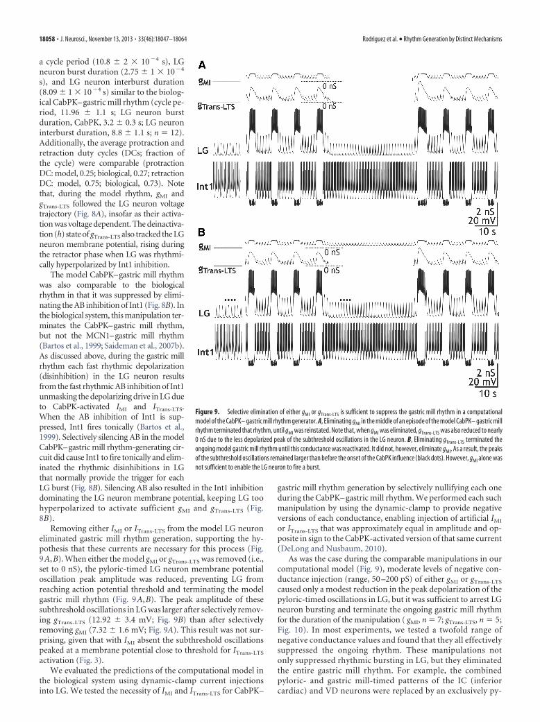
Removing either IMI or ITrans-LTS from the model LG neuroneliminated gastric mill rhythm generation, supporting the hy-pothesis that these currents are necessary for this process (Fig.9A,B). When either the model gMI or gTrans-LTS was removed (i.e.,set to 0 nS), the pyloric-timed LG neuron membrane potentialoscillation peak amplitude was reduced, preventing LG fromreaching action potential threshold and terminating the modelgastric mill rhythm (Fig. 9A,B). The peak amplitude of thesesubthreshold oscillations in LG was larger after selectively remov-ing gTrans-LTS (12.92 � 3.4 mV; Fig. 9B) than after selectivelyremoving gMI (7.32 � 1.6 mV; Fig. 9A). This result was not sur-prising, given that with IMI absent the subthreshold oscillationspeaked at a membrane potential close to threshold for ITrans-LTS
activation (Fig. 3).We evaluated the predictions of the computational model in
the biological system using dynamic-clamp current injectionsinto LG. We tested the necessity of IMI and ITrans-LTS for CabPK–
gastric mill rhythm generation by selectively nullifying each oneduring the CabPK– gastric mill rhythm. We performed each suchmanipulation by using the dynamic-clamp to provide negativeversions of each conductance, enabling injection of artificial IMI
or ITrans-LTS that was approximately equal in amplitude and op-posite in sign to the CabPK-activated version of that same current(DeLong and Nusbaum, 2010).
As was the case during the comparable manipulations in ourcomputational model (Fig. 9), moderate levels of negative con-ductance injection (range, 50 –200 pS) of either gMI or gTrans-LTS
caused only a modest reduction in the peak depolarization of thepyloric-timed oscillations in LG, but it was sufficient to arrest LGneuron bursting and terminate the ongoing gastric mill rhythmfor the duration of the manipulation ( gMI, n � 7; gTrans-LTS, n � 5;Fig. 10). In most experiments, we tested a twofold range ofnegative conductance values and found that they all effectivelysuppressed the ongoing rhythm. These manipulations notonly suppressed rhythmic bursting in LG, but they eliminatedthe entire gastric mill rhythm. For example, the combinedpyloric- and gastric mill-timed patterns of the IC (inferiorcardiac) and VD neurons were replaced by an exclusively py-
Figure 9. Selective elimination of either gMI or gTrans-LTS is sufficient to suppress the gastric mill rhythm in a computationalmodel of the CabPK– gastric mill rhythm generator. A, Eliminating gMI in the middle of an episode of the model CabPK– gastric millrhythm terminated that rhythm, until gMI was reinstated. Note that, when gMI was eliminated, gTrans-LTS was also reduced to nearly0 nS due to the less depolarized peak of the subthreshold oscillations in the LG neuron. B, Eliminating gTrans-LTS terminated theongoing model gastric mill rhythm until this conductance was reactivated. It did not, however, eliminate gMI. As a result, the peaksof the subthreshold oscillations remained larger than before the onset of the CabPK influence (black dots). However, gMI alone wasnot sufficient to enable the LG neuron to fire a burst.
18058 • J. Neurosci., November 13, 2013 • 33(46):18047–18064 Rodriguez et al. • Rhythm Generation by Distinct Mechanisms
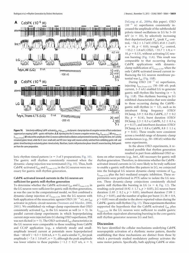
loric rhythm-timed pattern (n � 3 of 3 preparations; Fig. 10).The gastric mill rhythm consistently resumed when thedynamic-clamp injection was terminated (Fig. 10). Thus, bothCabPK-activated IMI and ITrans-LTS in the LG neuron were nec-essary for gastric mill rhythm generation.
CabPK-activated inward currents in the LG neuron aresufficient for gastric mill rhythm generationTo determine whether the CabPK-activated IMI and ITrans-LTS inthe LG neuron were sufficient for gastric mill rhythm generation,as was the case in the computational model, we first combined adynamic-clamp injection of ITrans-LTS into the LG neuron withbath application of the muscarinic agonist OXO (10�5
M), an IMI
activator in pyloric circuit neurons (Swensen and Marder, 2000,2001). We established via voltage-clamp experiments that OXOconsistently activated IMI in the LG neuron as well (n � 5). Inparallel current-clamp experiments in which hyperpolarizingcurrent steps were injected into LG during OXO superfusion, PIRwas not elicited (n � 5). The OXO-activated IMI characteristics inthe LG neuron were comparable to those resulting from CabPKand CCAP application (e.g., a relatively steady and small-amplitude inward current at potentials more hyperpolarizedthan �60 mV (�0.3 � 0.04 nA, n � 5), and a similar Vm for peakamplitude (�7.6 � 3.8 mV, n � 5), although the peak amplitudewas lower relative to these peptides (�1.2 � 0.27 nA, n � 5;
DeLong et al., 2009a; this paper). OXO(10�5
M) superfusion consistently in-creased the amplitude of the subthresholdpyloric-timed oscillations in LG by 5–10mV (n � 10), by selectively increasingtheir depolarized peak Vm (peak Vm: con-trol, �54.2 � 1.7 mV; OXO, 45.8 � 2 mV;n � 10, p 0.01; trough Vm: control,�55.2 � 1.8 mV; OXO, �55.7 � 1.9; n �10, p � 0.13), without activating LG neu-ron bursting (Fig. 11A). This result wascomparable to that occurring duringCabPK applications with dynamic-clamp nullification of ITrans-LTS, when theonly CabPK-activated inward current in-fluencing the LG neuron membrane po-tential was IMI (Fig. 10B).
During OXO (10�5M) superfusion,
injecting Idyn,Trans-LTS (50 –100 nS; peakcurrent, 1–3 nA) enabled LG to generategastric mill rhythm-like bursting (n � 5;Fig. 11B). This rhythmic bursting in LGexhibited characteristics that were similarto those occurring during the CabPK–gastric mill rhythm (n � 12), such as itsintraburst firing frequency (OXO/DClamp, 3.9 � 0.6 Hz; CabPK, 2.7 � 0.2Hz; p � 0.14), burst duration (OXO/DClamp, 2.3 � 0.3 s; CabPK, 3.2 � 0.3 s;p � 0.17), and interburst duration (OXO/DClamp, 6.4 � 0.8 s; CabPK, 8.8 � 1.1 s;p � 0.41). These results were consistentacross a twofold range of dynamic-clampconductances (e.g., 50 –100 nS) within thesame experiments.
In the above OXO experiments, it re-mained possible that rhythm generationresulted in part from additional OXO ac-
tions on other neurons (e.g., Int1, AB) necessary for gastric millrhythm generation. Therefore, to determine whether the CabPK-activated inward currents in LG were likely to be truly sufficientto enable a gastric mill rhythm-like pattern in LG, we coinjectedinto the biological LG neuron dynamic-clamp versions of IMI,ITrans-LTS plus the Int1-mediated synaptic inhibition. These ex-periments were performed in PTX saline to isolate the LG neu-ron. These dynamic-clamp coinjections consistently elicitedgastric mill rhythm-like bursting in LG (n � 4; Fig. 12). Theresulting cycle period (8.92 � 1.7 s, p 0.05), LG neuron burstduration (1.83 � 0.2 s, p 0.05), interburst duration (7.09 �0.8 s, p 0.05), and the number of spikes per burst (11.38 � 0.9,p 0.05) were all similar to the above-reported values during theCabPK– gastric mill rhythm (Fig. 13). These experiments thereforesupported the hypothesis that the CabPK-activated IMI plusITrans-LTS in the LG neuron were sufficient to enable gastricmill rhythm-equivalent alternating bursting in the core gastricmill rhythm generator neurons LG and Int1.
DiscussionWe have identified the cellular mechanisms underlying CabPKneuropeptide activation of a rhythmic motor pattern, therebyestablishing that these mechanisms are distinct from those bywhich a previously studied modulatory pathway activates thesame motor pattern. Specifically, bath applying CabPK or stim-
Figure 10. SelectivelynullifyingCabPK-activatedgMI orgTrans-LTS viadynamic-clampinjectionofanegativeversionofthatconductancesuppressed an ongoing CabPK– gastric mill rhythm. A, B, Injecting into the LG neuron a negative version of gMI [Idyn,MI (LG)] (A) or gTrans-LTS
[ITrans-LTS (LG)](B)reducedtheamplitudeoftheLGneuronsubthresholdoscillationsandpreventeditsbursting.Duringthistime,Int1activityremained pyloric timed, while the IC (mvn: small unit) and VD (mvn: large unit) neuron activity switched from exhibiting gastric mill- andpyloric-timed bursting to exclusively pyloric-timed activity. Black bars, Gastric mill protraction phase-timed IC neuron bursting. Both panelsare from the same preparation.
Rodriguez et al. • Rhythm Generation by Distinct Mechanisms J. Neurosci., November 13, 2013 • 33(46):18047–18064 • 18059
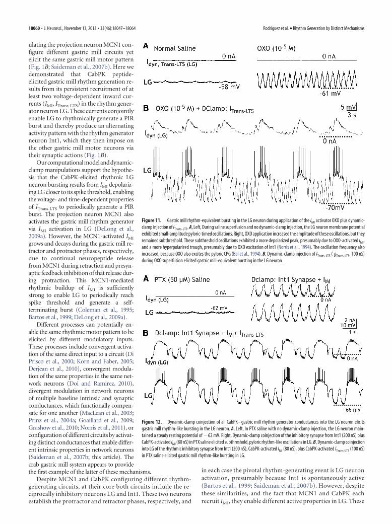
ulating the projection neuron MCN1 con-figure different gastric mill circuits yetelicit the same gastric mill motor pattern(Fig. 1B; Saideman et al., 2007b). Here wedemonstrated that CabPK peptide-elicited gastric mill rhythm generation re-sults from its persistent recruitment of atleast two voltage-dependent inward cur-rents (IMI, ITrans-LTS) in the rhythm gener-ator neuron LG. These currents conjointlyenable LG to rhythmically generate a PIRburst and thereby produce an alternatingactivity pattern with the rhythm generatorneuron Int1, which they then impose onthe other gastric mill motor neurons viatheir synaptic actions (Fig. 1B).
Ourcomputationalmodelanddynamic-clamp manipulations support the hypothe-sis that the CabPK-elicited rhythmic LGneuron bursting results from IMI depolariz-ing LG closer to its spike threshold, enablingthe voltage- and time-dependent propertiesof ITrans-LTS to periodically generate a PIRburst. The projection neuron MCN1 alsoactivates the gastric mill rhythm generatorvia IMI activation in LG (DeLong et al.,2009a). However, the MCN1-activated IMI
grows and decays during the gastric mill re-tractor and protractor phases, respectively,due to continual neuropeptide releasefrom MCN1 during retraction and presyn-aptic feedback inhibition of that release dur-ing protraction. This MCN1-mediatedrhythmic buildup of IMI is sufficientlystrong to enable LG to periodically reachspike threshold and generate a self-terminating burst (Coleman et al., 1995;Bartos et al., 1999; DeLong et al., 2009a).
Different processes can potentially en-able the same rhythmic motor pattern to beelicited by different modulatory inputs.These processes include convergent activa-tion of the same direct input to a circuit (DiPrisco et al., 2000; Korn and Faber, 2005;Derjean et al., 2010), convergent modula-tion of the same properties in the same net-work neurons (Doi and Ramirez, 2010),divergent modulation in network neuronsof multiple baseline intrinsic and synapticconductances, which functionally compen-sate for one another (MacLean et al., 2003;Prinz et al., 2004a; Goaillard et al., 2009;Grashow et al., 2010; Norris et al., 2011), orconfiguration of different circuits by activat-ing distinct conductances that enable differ-ent intrinsic properties in network neurons(Saideman et al., 2007b; this article). Thecrab gastric mill system appears to providethe first example of the latter of these mechanisms.
Despite MCN1 and CabPK configuring different rhythm-generating circuits, at their core both circuits include the re-ciprocally inhibitory neurons LG and Int1. These two neuronsestablish the protractor and retractor phases, respectively, and
in each case the pivotal rhythm-generating event is LG neuronactivation, presumably because Int1 is spontaneously active(Bartos et al., 1999; Saideman et al., 2007b). However, despitethese similarities, and the fact that MCN1 and CabPK eachrecruit IMI, they enable different active properties in LG. These
Figure 11. Gastric mill rhythm-equivalent bursting in the LG neuron during application of the IMI activator OXO plus dynamic-clamp injection of ITrans-LTS. A, Left, During saline superfusion and no dynamic-clamp injection, the LG neuron membrane potentialexhibited small-amplitude pyloric-timed oscillations. Right, OXO application increased the amplitude of these oscillations, but theyremained subthreshold. These subthreshold oscillations exhibited a more depolarized peak, presumably due to OXO-activated IMI,and a more hyperpolarized trough, presumably due to OXO excitation of Int1 (Norris et al., 1994). The oscillation frequency alsoincreased, because OXO also excites the pyloric CPG (Bal et al., 1994). B, Dynamic-clamp injection of ITrans-LTS ( gTrans-LTS, 100 nS)during OXO superfusion elicited gastric mill-equivalent bursting in the LG neuron.
Figure 12. Dynamic-clamp coinjection of all CabPK– gastric mill rhythm generator conductances into the LG neuron elicitsgastric mill rhythm-like bursting in the LG neuron. A, Left, In PTX saline with no dynamic-clamp injection, the LG neuron main-tained a steady resting potential of �62 mV. Right, Dynamic-clamp coinjection of the inhibitory synapse from Int1 (200 nS) plusCabPK-activated IMI (80 nS) in PTX saline elicited subthreshold, pyloric rhythm-like oscillations in LG. B, Dynamic-clamp coinjectioninto LG of the rhythmic inhibitory synapse from Int1 (200 nS), CabPK-activated IMI (80 nS), plus CabPK-activated ITrans-LTS (100 nS)in PTX saline elicited gastric mill rhythm-like bursting in LG.
18060 • J. Neurosci., November 13, 2013 • 33(46):18047–18064 Rodriguez et al. • Rhythm Generation by Distinct Mechanisms

different properties result from the distinct temporal dynam-ics of IMI activation by these two pathways, the likelihood thatMCN1 activates more IMI than CabPK (see below), and the factthat only CabPK appears to activate ITrans-LTS (Bartos et al., 1999;DeLong et al., 2009a; this article).
PIR bursts often involve the complementary influence ofmultiple voltage-dependent inward currents, as during CabPKmodulation of the LG neuron (McCormick and Bal, 1997;Sekirnjak and du Lac, 2002; Angstadt et al., 2005; Sangrey andJaeger, 2010; Wang et al., 2011; Engbers et al., 2011; Felix et al.,2011; Zheng and Raman, 2011). One inward current that oftencontributes to the initial PIR depolarization is Ih (McCormickand Bal, 1997; Sekirnjak and du Lac, 2002; Robinson and Sieg-elbaum, 2003; Pirtle et al., 2010; Sangrey and Jaeger, 2010;Engbers et al., 2011; Felix et al., 2011). The Ih-mediated re-bound depolarization facilitates activation of other voltage-dependent inward currents that elicit or strengthen the PIRburst. These latter currents often have time- and voltage-dependent properties similar to those of CabPK-activatedITrans-LTS. In many neurons, the additional PIR-generatingcurrent is some type of ICa (McCormick and Bal, 1997; Ang-stadt et al., 2005; Sangrey and Jaeger, 2010; Wang et al., 2011;Engbers et al., 2011; Felix et al., 2011). The role of Ih in PIRbursts is similar to that of CabPK-activated IMI in LG, whichdepolarizes the LG neuron after a period of inhibition andenables sufficient ITrans-LTS activation to generate the PIRburst. Also, similar to the CabPK comodulation of IMI andITrans-LTS, the complementary PIR-generating currents insome other systems can be comodulated (Harris-Warrick etal., 1995; Angstadt et al., 2005; Wang et al., 2011).
MCN1 and CabPK activation of the gastric mill rhythm gen-erator involves both divergent actions of the same ionic current(IMI) and a conserved function mediated by distinct ionic cur-rents (IMI, ITrans-LTS; Fig. 14). As discussed above, IMI is the burst-generating conductance during MCN1 rhythm generation,whereas during CabPK rhythm generation there is insufficientIMI to directly enable LG neuron bursting. In this latter condition,IMI instead facilitates burst generation by ITrans-LTS. The sharedrole of IMI and ITrans-LTS during MCN1 and CabPK rhythm gen-eration, respectively, results from each current exhibiting aphase-dependent growth and decay process that is pivotal to de-termining the LG neuron burst (protraction) and interburst (re-traction) durations (Fig. 14; Bartos et al., 1999; DeLong et al.,2009a; this article). The growth and decay process for IMI duringMCN1 stimulation results from its synaptic regulation, as dis-cussed above. In contrast, the availability of CabPK-activatedITrans-LTS grows and decays during gastric mill retraction and pro-traction, respectively, due to its voltage- and time-dependent
Figure 13. The range of the gastric mill rhythm-related parameters in the LG neuron arecomparable during the biological CabPK– gastric mill rhythm and when artificial versionsof synaptic inhibition plus the CabPK-activated conductances are coinjected into LG. Dis-tribution of mean values is shown for the LG neuron burst duration, interburst duration,and number of spikes per burst during CabPK– gastric mill rhythms (filled circles: n � 11)and dynamic-clamp-elicited gastric mill rhythm-like bursting in LG (n � 4). All error bars(SEM) are smaller than the associated circles. N.S., Not significantly different ( p 0.05).
Figure 14. MCN1 stimulation and CabPK superfusion activate different conductances toperform the same function, and activate the same conductance to perform different func-tions, during gastric mill rhythm generation. A, Rhythmic LG neuron burst generationresults from the rhythmic buildup and decay of MCN1-activated gMI (left) and CabPK-activated gTrans-LTS (right). During MCN1 stimulation, the buildup occurs during retractionand results from continual MCN1 activation of gMI, while the decay during protractionresults from LG neuron presynaptic inhibition of MCN1 transmitter release. During CabPKapplication, the buildup during retraction results from the accumulation of deinactivationin gTrans-LTS, while the decay during protraction results from the time-dependent inacti-vation of this conductance. B, The gMI in LG is the burst-generating conductance duringthe MCN1– gastric mill rhythm, whereas it provides a sustained but subthreshold depo-larizing drive that facilitates PIR burst generation by ITrans-LTS during the CabPK– gastricmill rhythm. Note that the different gMI trajectories during protraction result from gMI
being both voltage- and synaptic inhibition-dependent during the MCN1– gastric millrhythm, whereas during the CabPK– gastric mill rhythm it is only voltage-dependent. RET,Retraction; PRO, protraction.
Rodriguez et al. • Rhythm Generation by Distinct Mechanisms J. Neurosci., November 13, 2013 • 33(46):18047–18064 • 18061

properties. During retraction, when the LG neuron is hyperpo-larized by rhythmic synaptic inhibition from Int1, ITrans-LTS ex-hibits a buildup of deinactivation, which increases its availability,while during protraction it first enables and then limits the LGneuron burst duration due to its depolarization- and time-dependent inactivation.
Despite these differences in the rhythm-generating process,during both gastric mill rhythms the LG neuron burst initiatesduring a pyloric rhythm-timed depolarization that resultsfrom AB neuron inhibition of Int1 (Bartos et al., 1999; Saide-man et al., 2007b). However, these pyloric-timed depolariza-tions are necessary only for CabPK rhythm generation(Saideman et al., 2007b). This is because, when the pyloricrhythm is suppressed during MCN1 stimulation, the MCN1-activated IMI amplitude continues to grow during each pro-longed retraction phase until it eventually becomes largeenough to enable LG to escape from Int1 inhibition and gen-erate a burst (Bartos et al., 1999). In contrast, CabPK-activatedIMI alone is insufficient to enable the LG neuron to depolarizeand trigger ITrans-LTS activation in the presence of persistentInt1 inhibition.
The phase transitions occurring during rhythmic alternat-ing bursting by reciprocally inhibitory neurons commonlyresult from the inhibited neuron either escaping from con-tinuing inhibition (“escape” mode) or waiting until it is re-leased from that inhibition (“release” mode) (Skinner et al.,1994; Marder and Calabrese, 1996; McCormick and Bal,1997). Each mode can also mediate the same transition underdifferent conditions (Bartos et al., 1999; Sorensen et al., 2004;Kristan et al., 2005). During the MCN1– gastric mill and Cab-PK– gastric mill rhythms, the protraction-to-retraction tran-sition appears to occur via the release mechanism, as the LGneuron burst self-terminates and releases Int1 from inhibi-tion. In contrast, for reasons discussed above, during theretraction-to-protraction transition the CabPK rhythm onlyoperates via the release mode, whereas the MCN1 rhythm canoperate either in its normal release mode or, if the pyloricrhythm is very slow or suppressed, in the escape mode.
A neural network might have the ability to configure dis-tinct circuits generating the same activity pattern because itneeds to generate the same core behavior under different con-ditions. These differently configured circuits, however, mightbe differentially sensitive to particular inputs. This, for exam-ple, is evident for the pyloric pacemaker neuron AB influenceon these two gastric mill circuits (i.e., it is a necessary rhythm-generating component only for the CabPK– gastric mill cir-cuit). Such differential sensitivity likely also occurs in circuitsthat generate a consistent output pattern despite extensive,albeit compensatory, changes in the baseline intrinsic and syn-aptic conductances of circuit neurons (Prinz et al., 2004a;Grashow et al., 2009, 2010; Calabrese et al., 2011; Guttierez etal., 2013). Two interesting tests of this hypothesis for the gas-tric mill circuit will be the influence of the gastro-pyloric re-ceptor neuron, a muscle proprioceptor, and the peptidehormone CCAP on the CabPK-gastric mill rhythm. Both ofthese inputs regulate the MCN1– gastric mill rhythm via ac-tions that involve MCN1, which does not participate in theCabPK rhythm (Beenhakker et al., 2005; DeLong et al.,2009a,b).
This study highlights an alternative consequence to the wellestablished flexibility in neural circuit output imparted by
their neuromodulatory inputs. Here, neuromodulation con-figures distinct circuits/mechanisms that generate a conservedoutput rather than generating distinct outputs. As part of thisprocess, different modulatory pathways activate the sameionic current in the same neuron but use it in different ways,and they use different currents in the same neuron to performthe same function (Fig. 14). Whether the conserved outputpattern resulting from these distinct mechanisms belies latentdifferences in responsiveness to the same perturbation re-mains to be determined.
ReferencesAngstadt JD, Grassmann JL, Theriault KM, Levasseur SM (2005) Mech-
anisms of postinhibitory rebound and its modulation by serotonin inexcitatory swim motor neurons of the medicinal leech. J Comp PhysiolA Neuroethol Sens Neural Behav Physiol 191:715–732. CrossRefMedline
Bal T, Nagy F, Moulins M (1994) Muscarinic modulation of a pattern-generating network: control of neuronal properties. J Neurosci 14:3019 –3035. Medline
Bartos M, Manor Y, Nadim F, Marder E, Nusbaum MP (1999) Coordina-tion of fast and slow rhythmic neuronal circuits. J Neurosci 19:6650 –6660. Medline
Beenhakker MP, Nusbaum MP (2004) Mechanosensory activation of a mo-tor circuit by coactivation of two projection neurons. J Neurosci 24:6741–6750. CrossRef Medline
Beenhakker MP, DeLong ND, Saideman SR, Nadim F, Nusbaum MP (2005)Proprioceptor regulation of motor circuit activity by presynaptic inhibi-tion of a modulatory projection neuron. J Neurosci 25:8794 – 8806.CrossRef Medline
Blitz DM, Nusbaum MP (2012) Modulation of circuit feedback specifiesmotor circuit output. J Neurosci 32:9182–9193. CrossRef Medline
Blitz DM, Christie AE, Coleman MJ, Norris BJ, Marder E, Nusbaum MP(1999) Different proctolin neurons elicit distinct motor patternsfrom a multifunctional neuronal network. J Neurosci 19:5449 –5463.Medline
Blitz DM, Beenhakker MP, Nusbaum MP (2004) Different sensory systemsshare projection neurons but elicit distinct motor patterns. J Neurosci24:11381–11390. CrossRef Medline
Briggman KL, Kristan WB (2008) Multifunctional pattern-generating cir-cuits. Annu Rev Neurosci 31:271–294. CrossRef Medline
Calabrese RL, Norris BJ, Wenning A, Wright TM (2011) Coping with vari-ability in small neuronal networks. Integr Comp Biol 51:845– 855.CrossRef Medline
Christie AE, Stein W, Quinlan JE, Beenhakker MP, Marder E, Nusbaum MP(2004) Actions of a histaminergic/peptidergic projection neuron onrhythmic motor patterns in the stomatogastric nervous system of the crabCancer borealis. J Comp Neurol 469:153–169. CrossRef Medline
Coleman MJ, Nusbaum MP (1994) Functional consequences of compart-mentalization of synaptic input. J Neurosci 14:6544 – 6552. Medline
Coleman MJ, Meyrand P, Nusbaum MP (1995) Presynaptic inhibition me-diates a switch between two modes of synaptic transmission. Nature 378:502–505. CrossRef Medline
DeLong ND, Nusbaum MP (2010) Hormonal modulation of sensorimotorintegration. J Neurosci 30:2418 –2427. CrossRef Medline
DeLong ND, Kirby MS, Blitz DM, Nusbaum MP (2009a) Parallel regulationof a modulator-activated current via distinct dynamics underlies co-modulation of motor circuit output. J Neurosci 29:12355–12367.CrossRef Medline
DeLong ND, Beenhakker MP, Nusbaum MP (2009b) Presynaptic inhibi-tion selectively weakens peptidergic cotransmission in a small motor sys-tem. J Neurophysiol 102:3492–3504. CrossRef Medline
Derjean D, Moussaddy A, Atallah E, St-Pierre M, Auclair F, Chang S, Ren X,Zielinski B, Dubuc R (2010) A novel neural substrate for the transfor-mation of olfactory inputs into motor output. PLoS Biol 8:e1000567.CrossRef Medline
Dickinson PS (2006) Neuromodulation of central pattern generators in in-vertebrates and vertebrates. Curr Opin Neurobiol 16:604 – 614. CrossRefMedline
18062 • J. Neurosci., November 13, 2013 • 33(46):18047–18064 Rodriguez et al. • Rhythm Generation by Distinct Mechanisms
Diehl F, White RS, Stein W, Nusbaum MP (2013) Motor circuit-specificburst patterns drive different muscle and movement patterns. J Neurosci33:12013–12029. CrossRef Medline
Di Prisco GV, Pearlstein E, Le Ray D, Robitaille R, Dubuc R (2000) A cellularmechanism for the transformation of a sensory input into a motor com-mand. J Neurosci 20:8169 – 8176. Medline
Doi A, Ramirez JM (2008) Neuromodulation and the orchestration of therespiratory rhythm. Respir Physiol Neurobiol 164:96 –104. CrossRefMedline
Doi A, Ramirez JM (2010) State-dependent interactions between excitatoryneuromodulators in the neuronal control of breathing. J Neurosci 30:8251– 8262. CrossRef Medline
Engbers JD, Anderson D, Tadayonnejad R, Mehaffey WH, Molineux ML,Turner RW (2011) Distinct roles for I(T) and I(H) in controlling thefrequency and timing of rebound spike responses. J Physiol 589:5391–5413. CrossRef Medline
Felix RA 2nd, Fridberger A, Leijon S, Berrebi AS, Magnusson AK (2011)Sound rhythms are encoded by postinhibitory rebound spiking in thesuperior paraolivary nucleus. J Neurosci 31:12566 –12578. CrossRefMedline
Goaillard JM, Taylor AL, Schulz DJ, Marder E (2009) Functional conse-quences of animal-to-animal variation in circuit parameters. Nat Neuro-sci 12:1424 –1430. CrossRef Medline
Golowasch J, Marder E (1992) Proctolin activates an inward current whosevoltage dependence is modified by extracellular Ca 2�. J Neurosci 12:810 –817. Medline
Grashow R, Brookings T, Marder E (2009) Reliable neuromodulation fromcircuits with variable underlying structure. Proc Natl Acad Sci U S A 106:11742–11746. CrossRef Medline
Grashow R, Brookings T, Marder E (2010) Compensation for variable in-trinsic neuronal excitability by circuit-synaptic interactions. J Neurosci30:9145–9156. CrossRef Medline
Guttierez GJ, O’Leary T, Marder E (2013) Multiple mechanisms switch anelectrically coupled, synaptically inhibited neuron between competingrhythmic oscillators. Neuron 77:845– 858. CrossRef Medline
Harris-Warrick RM (2011) Neuromodulation and flexibility in central pat-tern generator networks. Curr Opin Neurobiol 21:685– 692. CrossRefMedline
Harris-Warrick RM, Coniglio LM, Levini RM, Gueron S, Guckenheimer J(1995) Dopamine modulation of two subthreshold currents producesphase shifts in activity of an identified motoneuron. J Neurophysiol 74:1404 –1420. Medline
Kadiri LR, Kwan AC, Webb WW, Harris-Warrick RM (2011) Dopamine-induced oscillations of the pyloric pacemaker neuron rely on release ofcalcium from intracellular stores. J Neurophysiol 106:1288 –1298.CrossRef Medline
Kintos N, Nusbaum MP, Nadim F (2008) A modeling comparison of pro-jection neuron- and neuromodulator-elicited oscillations in a central pat-tern generating network. J Comput Neurosci 24:374 –397. CrossRefMedline
Kirby MS, Nusbaum MP (2007) Peptide hormone modulation of a neuro-nally modulated motor circuit. J Neurophysiol 98:3206 –3220. CrossRefMedline
Korn H, Faber DS (2005) The Mauthner cell half a century later: a neu-robiological model for decision-making? Neuron 47:13–28. CrossRefMedline
Kristan WB Jr, Calabrese RL, Friesen WO (2005) Neuronal control of leechbehavior. Prog Neurobiol 76:279 –327. CrossRef Medline
MacLean JN, Zhang Y, Johnson BR, Harris-Warrick RM (2003) Activity-independent homeostasis in rhythmically active neurons. Neuron 37:109 –120. CrossRef Medline
Marder E (2012) Neuromodulation of neuronal circuits: back to the future.Neuron 76:1–11. CrossRef Medline
Marder E, Bucher D (2007) Understanding circuit dynamics using the sto-matogastric nervous system of lobsters and crabs. Annu Rev Physiol 69:291–316. CrossRef Medline
Marder E, Calabrese RL (1996) Principles of rhythmic motor pattern gen-eration. Physiol Rev 76:687–717. Medline
Marder E, Eisen JS (1984) Transmitter identification of pyloric neurons:electrically coupled neurons use different transmitters. J Neurophysiol51:1345–1361. Medline
McCormick DA, Bal T (1997) Sleep and arousal: thalamocortical mecha-nisms. Annu Rev Neurosci 20:185–215. CrossRef Medline
Nadim F, Manor Y, Nusbaum MP, Marder E (1998) Frequency regulationof a slow rhythm by a fast periodic input. J Neurosci 18:5053–5067.Medline
Norris BJ, Coleman MJ, Nusbaum MP (1994) Recruitment of a projectionneuron determines gastric mill motor pattern selection in the stomato-gastric nervous system of the crab, Cancer borealis. J Neurophysiol 72:1451–1463. Medline
Norris BJ, Wenning A, Wright TM, Calabrese RL (2011) Constancy andvariability in the output of a central pattern generator. J Neurosci 31:4663– 4674. CrossRef Medline
Nusbaum MP, Marder E (1989a) A modulatory proctolin-containing neu-ron (MPN). I. Identification and characterization. J Neurosci 9:1591–1599. Medline
Nusbaum MP, Marder E (1989b) A modulatory proctolin-containing neu-ron (MPN). II. State-dependent modulation of rhythmic motor activity.J Neurosci 9:1600 –1607. Medline
Pirtle TJ, Willingham K, Satterlie RA (2010) A hyperpolarization-activatedinward current alters swim frequency of the pteropod mollusk Clionelimacina. Comp Biochem Physiol A Mol Integr Physiol 157:319 –327.CrossRef Medline
Prinz AA, Bucher D, Marder E (2004a) Similar network activity fromdisparate circuit parameters. Nat Neurosci 7:1345–1352. CrossRefMedline
Prinz AA, Abbott LF, Marder E (2004b) The dynamic clamp comes of age.Trends Neurosci 27:218 –224. CrossRef Medline
Ransdell JL, Temporal S, West NL, Leyrer ML, Schulz DJ (2013) Character-ization of inward currents and channels underlying burst activity in mo-toneurons of crab cardiac ganglion. J Neurophysiol 110:42–54. CrossRefMedline
Rauscent A, Einum J, Le Ray D, Simmers J, Combes D (2009) Opposingaminergic modulation of distinct spinal locomotor circuits and theirfunctional coupling during amphibian metamorphosis. J Neurosci 29:1163–1174. CrossRef Medline
Robinson RB, Siegelbaum SA (2003) Hyperpolarization-activated cation cur-rents: from molecules to physiological function. Annu Rev Physiol 65:453– 480. CrossRef Medline
Saideman SR, Ma M, Kutz-Naber KK, Cook A, Torfs P, Schoofs L, Li L,Nusbaum MP (2007a) Modulation of rhythmic motor activity by pyro-kinin peptides. J Neurophysiol 97:579 –595. CrossRef Medline
Saideman SR, Blitz DM, Nusbaum MP (2007b) Convergent motor pat-terns from divergent circuits. J Neurosci 27:6664 – 6674. CrossRefMedline
Sangrey T, Jaeger D (2010) Analysis of distinct short and prolonged compo-nents in rebound spiking of deep cerebellar nucleus neurons. Eur J Neu-rosci 32:1646 –1657. CrossRef Medline
Sekirnjak C, du Lac S (2002) Intrinsic firing dynamics of vestibular nucleusneurons. J Neurosci 22:2083–2095. Medline
Sharp AA, O’Neil MB, Abbott LF, Marder E (1993) The dynamic clamp:artificial conductances in biological neurons. Trends Neurosci 16:389 –394. CrossRef Medline
Skinner FK, Kopell N, Marder E (1994) Mechanisms for oscillation andfrequency control in reciprocally inhibitory model neural networks.J Comput Neurosci 1:69 – 87. CrossRef Medline
Sorensen M, DeWeerth S, Cymbalyuk G, Calabrese RL (2004) Using a hy-brid neural system to reveal regulation of neuronal network activity by anintrinsic current. J Neurosci 24:5427–5438. CrossRef Medlineb
Stein W (2009) Modulation of stomatogastric rhythms. J Comp Physiol A Neu-roethol Sens Neural Behav Physiol 195:989–1009. CrossRef Medline
Stein W, DeLong ND, Wood DE, Nusbaum MP (2007) Divergent co-transmitter actions underlie motor pattern activation by a modulatoryprojection neuron. Eur J Neurosci 26:1148 –1165. CrossRef Medline
Swensen AM, Marder E (2000) Multiple peptides converge to activate thesame voltage-dependent current in a central pattern-generating circuit.J Neurosci 20:6752– 6759. Medline
Swensen AM, Marder E (2001) Modulators with convergent cellular actionselicit distinct circuit outputs. J Neurosci 21:4050 – 4058. Medline
Turrigiano GG, Marder E, Abbott LF (1996) Cellular short-term memoryfrom a slow potassium conductance. J Neurophysiol 75:963–966.Medline
Rodriguez et al. • Rhythm Generation by Distinct Mechanisms J. Neurosci., November 13, 2013 • 33(46):18047–18064 • 18063

Wang D, Grillner S, Wallen P (2011) 5-HT and dopamine modulatesCaV1.3 calcium channels involved in postinhibitory rebound in the spinalnetwork for locomotion in lamprey. J Neurophysiol 105:1212–1224.CrossRef Medline
Weimann JM, Meyrand P, Marder E (1991) Neurons that form multiplepattern generators: identification and multiple activity patterns of gastric/pyloric neurons in the crab stomatogastric system. J Neurophysiol 65:111–122. Medline
White RS, Nusbaum MP (2011) The same core rhythm generator underliesdifferent rhythmic motor patterns. J Neurosci 31:11484 –11494. CrossRefMedline
Wood DE, Stein W, Nusbaum MP (2000) Projection neurons with shared
cotransmitters elicit different motor patterns from the same neural cir-cuit. J Neurosci 20:8943– 8953. Medline
Wood DE, Manor Y, Nadim F, Nusbaum MP (2004) Intercircuit control viarhythmic regulation of projection neuron activity. J Neurosci 24:7455–7463. CrossRef Medline
Zhang B, Wootton JF, Harris-Warrick RM (1995) Calcium-dependent plateaupotentials in a crab stomatogastric ganglion motor neuron. II. Calcium-activated slow inward current. J Neurophysiol 74:1938–1946. Medline
Zheng N, Raman IM (2011) Prolonged postinhibitory rebound firing in thecerebellar nuclei mediated by group I metabotropic glutamate receptorpotentiation of L-type calcium currents. J Neurosci 31:10283–10292.CrossRef Medline
18064 • J. Neurosci., November 13, 2013 • 33(46):18047–18064 Rodriguez et al. • Rhythm Generation by Distinct Mechanisms