Connective Tissue Found everywhere in the body Includes the most abundant and widely distributed...
30
Connective Tissue • Found everywhere in the body • Includes the most abundant and widely distributed tissues • Functions – Binds body tissues together – Supports the body – Provides protection
-
Upload
mariah-west -
Category
Documents
-
view
214 -
download
0
Transcript of Connective Tissue Found everywhere in the body Includes the most abundant and widely distributed...

Connective Tissue
• Found everywhere in the body
• Includes the most abundant and widely distributed tissues
• Functions– Binds body tissues together– Supports the body– Provides protection

Connective Tissue Characteristics
• Variations in blood supply– Some tissue types are well vascularized– Some have poor blood supply or are
avascular
• Extracellular matrix– Non-living material that surrounds living cells

Extracellular Matrix
• Two main elements1. Ground substance – mostly water along with
adhesion proteins and polysaccharide molecules
2. Fibers
• Produced by the cells called fibroblasts• Three types
– Collagen fibers– Elastic fibers– Reticular fibers

Connective Tissue Types
1. Areolar Tissue– Most widely distributed connective tissue– Soft, pliable. A type of “Loose Connective
Tissue”– Contains all fiber types
– Binds skin to underlying organs– Fills spaces between muscles– Common connective tissue beneath epithelium
Figure 3.19e


Connective Tissue Types
2. Adipose tissue (fat)– Matrix is an areolar tissue in which fat
globules predominate– Many cells contain large lipid deposits
(Adipocytes)– Functions
• Insulates the body• Protects some organs (kidneys)• Serves as a site of energy storage


Connective Tissue Types
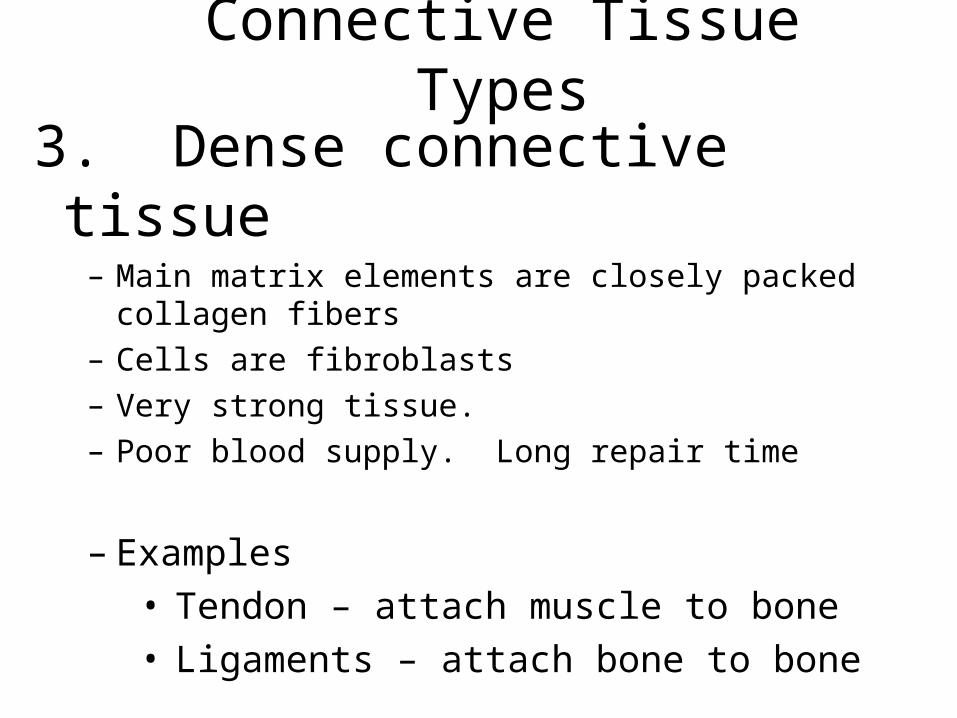
3. Dense connective tissue– Main matrix elements are closely packed collagen fibers– Cells are fibroblasts– Very strong tissue. – Poor blood supply. Long repair time
– Examples• Tendon – attach muscle to bone• Ligaments – attach bone to bone


Connective Tissue Types
4. Cartilage. A rigid connective tissue composed of cells called Chondrocytes surrounded by a rubbery matrix.
3 types, each with a different matrix
Figure 3.19b

Hyaline Cartilage
Most common cartilageComposed of: Abundant collagen fibers in a rubbery matrix
Locations:Entire fetal skeleton is hyaline cartilageEnds of bonesSoft part of nose
Elastic cartilage– Provides elasticity. More Elastin fibers– Example: supports the external ear

Fibrocartilage– Highly compressible, composed of tightly
packed collagen fibers
– Example: forms cushion-like discs between adjacent vertebrae
Figure 3.19c


Connective Tissue Types
5. Bone (osseous tissue)– Composed of:
• Bone cells called Osteocytes, in lacunae (cavities)
• Hard matrix of calcium phosphate cementing large numbers of collagen fibers together
– Used to protect and support the body– Site of blood cell formation– Store Calcium– Highly organized, excellent blood supply,
very active tissue, heals rapidly.


Connective Tissue Types
6. Blood– Blood cells surrounded by fluid matrix– Fibers are visible during clotting– Functions as the transport vehicle for
materials


Muscle Tissue
• Composed of elongated cells
• Have the ability to contract or shorten and produce movement
• Three types– Skeletal muscle– Cardiac muscle– Smooth muscle

Muscle Tissue Types
1. Skeletal muscle
– Can be controlled voluntarily– Attached to bones– Cells are long and threadlike (called fibers)– Cells are striated– Cells have more than one nucleus
(multinucleated)
Figure 3.20a


Muscle Tissue Types
2. Smooth muscle– Involuntary muscle (cannot be
consciously controlled)– Surrounds hollow organs (stomach,
intestines, blood vessels)– Attached to other smooth muscle cells– No visible striations– One nucleus per cell


Muscle Tissue Types
3. Cardiac muscle
– Found only in the heart– Function is to pump blood (involuntary)– Cells attached to other cardiac muscle
cells at intercalated disks– Cells are striated– One nucleus per cell– Does not need nervous stimulation to
contract.


Nervous Tissue
• Neurons and nerve support cells (neuroglia)
• Function is to send impulses to other areas of the body
– Irritability– Conductivity
Figure 3.21