Congenital cerebral cavernous malformations in an infant ......Cavernous malformations were...
3
Advances in Pediatric Research Quon et al. 2014 | 1:8 1 Congenital cerebral cavernous malformations in an infant: A case report Jennifer L. Quon 1 , Ryan A. Grant 1 , Andrea G. Asnes 2 , Michael L. DiLuna 1* 1 Department of Neurosurgery, Section of Pediatric Neurosurgery, Yale University School of Medicine, New Haven, CT, USA 2 Department of Pediatrics, Yale University School of Medicine, New Haven, CT, USA Abstract Abusive head trauma is a leading cause of infant death, but other causes of intracranial hemorrhage must be systematically excluded. Here we present a case of multiple hemorrhagic cavernous malformations that was initially thought to be indicative of abusive head trauma. Using a clinical decision-making framework, we provide insight into child abuse diagnostics. Citation: Quon JL, Grant RA, Asnes AG, DiLuna ML (2014) Congenital cerebral cavernous malformations in an infant: A case report. Adv Pediatr Res 1:8. doi:10.12715/apr.2014.1.8 Received: October 29, 2014; Accepted: December 22, 2014; Published: December 31, 2014 Copyright: © 2014 Quon et al. This is an open access article distributed under the terms of the Creative Commons Attribution License, which permits unrestricted use, distribution, and reproduction in any medium, provided the original work is properly cited. Competing interests: The authors have declared that no competing interests exist. * Email: [email protected] Introduction Abusive head trauma (AHT) in infants is a leading cause of infant death [1]. Unfortunately, physicians often underreport AHT due to uncertainty about the diagnosis, as well as hesitation to engage in legal proceedings [2]. Furthermore, physician bias towards certain races, socioeconomic groups, or family structures can impact reporting, regardless of the actual incidence of abuse amongst these groups [3]. Findings from the history, physical exam, or radiography that should prompt a consideration of AHT include an inadequate or inconsistent history in the setting of significant trauma, retinal hemorrhages, subdural hemorrhage, skull fractures, and skeletal fractures of varying ages [4-7]. While intracranial hemorrhage and altered mental status should raise suspicion for abuse, other causes must be thoroughly explored and excluded. We present a case of multiple hemorrhagic cavernous malformations that initially prompted concern for abusive head trauma. Case report A 7-month-old African-American male, otherwise healthy, presented with vomiting and somnolence. He was at his grandparents’ home prior to presentation and was later found to be lethargic. On presentation, he was bradycardic, moderately hypertensive, unresponsive, had minimally responsive but equally round pupils of 4 mm, a bulging tense anterior fontanelle, symmetric withdrawal of all extremities to noxious stimuli, increased tone, and diffuse hyperreflexia. There was no superficial evidence of trauma. A non-contrast head computed tomography (CT) scan demonstrated a large, 2-cm left occipital hemorrhage, as well as additional hemorrhagic contusions in the left parietal and frontal lobes, and basal ganglia (Fig. 1). His coagulation factors and platelet count were normal. A right frontal ventriulostomy was placed with an opening intracranial pressure (ICP) of 16 mm Hg, with improvement in the neurologic examination. A CT angiogram of the head did not reveal an underlying vascular etiology. The infant’s social history was significant for 19 year-old, unmarried parents. He
Transcript of Congenital cerebral cavernous malformations in an infant ......Cavernous malformations were...
Advances in Pediatric Research Quon et al. 2014 | 1:8 1
Congenital cerebral cavernous malformations in an infant: A case report Jennifer L. Quon 1, Ryan A. Grant 1, Andrea G. Asnes 2, Michael L. DiLuna 1*
1 Department of Neurosurgery, Section of Pediatric Neurosurgery, Yale University School of Medicine, New Haven, CT, USA 2 Department of Pediatrics, Yale University School of Medicine, New Haven, CT, USA
Abstract Abusive head trauma is a leading cause of infant death, but other causes of intracranial hemorrhage must be systematically excluded. Here we present a case of multiple hemorrhagic cavernous malformations that was initially thought to be indicative of abusive head trauma. Using a clinical decision-making framework, we provide insight into child abuse diagnostics. Citation: Quon JL, Grant RA, Asnes AG, DiLuna ML (2014) Congenital cerebral cavernous malformations in an infant: A case report. Adv Pediatr Res 1:8. doi:10.12715/apr.2014.1.8
Received: October 29, 2014; Accepted: December 22, 2014; Published: December 31, 2014
Copyright: © 2014 Quon et al. This is an open access article distributed under the terms of the Creative Commons Attribution License, which permits unrestricted use, distribution, and reproduction in any medium, provided the original work is properly cited.
Competing interests: The authors have declared that no competing interests exist. * Email: [email protected]
Introduction Abusive head trauma (AHT) in infants is a leading cause of infant death [1]. Unfortunately, physicians often underreport AHT due to uncertainty about the diagnosis, as well as hesitation to engage in legal proceedings [2]. Furthermore, physician bias towards certain races, socioeconomic groups, or family structures can impact reporting, regardless of the actual incidence of abuse amongst these groups [3].
Findings from the history, physical exam, or radiography that should prompt a consideration of AHT include an inadequate or inconsistent history in the setting of significant trauma, retinal hemorrhages, subdural hemorrhage, skull fractures, and skeletal fractures of varying ages [4-7].
While intracranial hemorrhage and altered mental status should raise suspicion for abuse, other causes must be thoroughly explored and excluded. We present a case of multiple hemorrhagic cavernous malformations that initially prompted concern for abusive head trauma.
Case report A 7-month-old African-American male, otherwise healthy, presented with vomiting and somnolence. He was at his grandparents’ home prior to presentation and was later found to be lethargic. On presentation, he was bradycardic, moderately hypertensive, unresponsive, had minimally responsive but equally round pupils of 4 mm, a bulging tense anterior fontanelle, symmetric withdrawal of all extremities to noxious stimuli, increased tone, and diffuse hyperreflexia. There was no superficial evidence of trauma. A non-contrast head computed tomography (CT) scan demonstrated a large, 2-cm left occipital hemorrhage, as well as additional hemorrhagic contusions in the left parietal and frontal lobes, and basal ganglia (Fig. 1). His coagulation factors and platelet count were normal. A right frontal ventriulostomy was placed with an opening intracranial pressure (ICP) of 16 mm Hg, with improvement in the neurologic examination. A CT angiogram of the head did not reveal an underlying vascular etiology. The infant’s social history was significant for 19 year-old, unmarried parents. He
Advances in Pediatric Research Quon et al. 2014 | 1:8 2
lived with his mother and her family, but frequently visited his father and paternal grandparents.
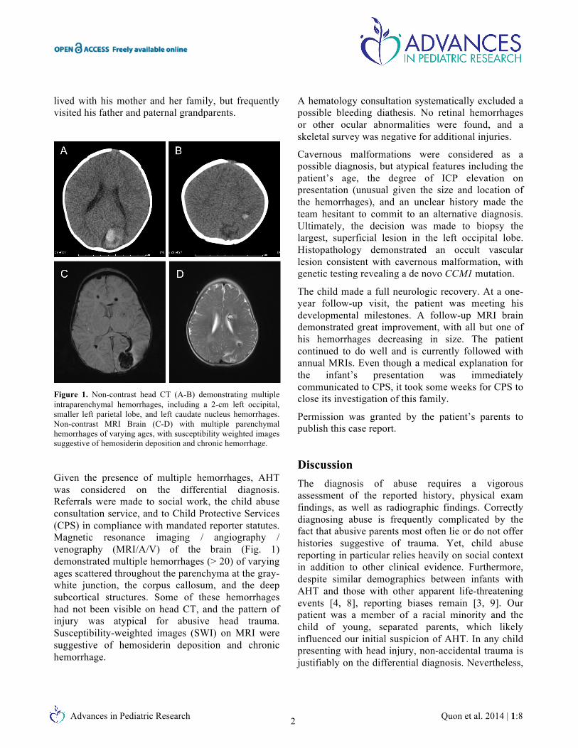
Figure 1. Non-contrast head CT (A-B) demonstrating multiple intraparenchymal hemorrhages, including a 2-cm left occipital, smaller left parietal lobe, and left caudate nucleus hemorrhages. Non-contrast MRI Brain (C-D) with multiple parenchymal hemorrhages of varying ages, with susceptibility weighted images suggestive of hemosiderin deposition and chronic hemorrhage.
Given the presence of multiple hemorrhages, AHT was considered on the differential diagnosis. Referrals were made to social work, the child abuse consultation service, and to Child Protective Services (CPS) in compliance with mandated reporter statutes. Magnetic resonance imaging / angiography / venography (MRI/A/V) of the brain (Fig. 1) demonstrated multiple hemorrhages (> 20) of varying ages scattered throughout the parenchyma at the gray-white junction, the corpus callosum, and the deep subcortical structures. Some of these hemorrhages had not been visible on head CT, and the pattern of injury was atypical for abusive head trauma. Susceptibility-weighted images (SWI) on MRI were suggestive of hemosiderin deposition and chronic hemorrhage.
A hematology consultation systematically excluded a possible bleeding diathesis. No retinal hemorrhages or other ocular abnormalities were found, and a skeletal survey was negative for additional injuries.
Cavernous malformations were considered as a possible diagnosis, but atypical features including the patient’s age, the degree of ICP elevation on presentation (unusual given the size and location of the hemorrhages), and an unclear history made the team hesitant to commit to an alternative diagnosis. Ultimately, the decision was made to biopsy the largest, superficial lesion in the left occipital lobe. Histopathology demonstrated an occult vascular lesion consistent with cavernous malformation, with genetic testing revealing a de novo CCM1 mutation.
The child made a full neurologic recovery. At a one-year follow-up visit, the patient was meeting his developmental milestones. A follow-up MRI brain demonstrated great improvement, with all but one of his hemorrhages decreasing in size. The patient continued to do well and is currently followed with annual MRIs. Even though a medical explanation for the infant’s presentation was immediately communicated to CPS, it took some weeks for CPS to close its investigation of this family.
Permission was granted by the patient’s parents to publish this case report.
Discussion The diagnosis of abuse requires a vigorous assessment of the reported history, physical exam findings, as well as radiographic findings. Correctly diagnosing abuse is frequently complicated by the fact that abusive parents most often lie or do not offer histories suggestive of trauma. Yet, child abuse reporting in particular relies heavily on social context in addition to other clinical evidence. Furthermore, despite similar demographics between infants with AHT and those with other apparent life-threatening events [4, 8], reporting biases remain [3, 9]. Our patient was a member of a racial minority and the child of young, separated parents, which likely influenced our initial suspicion of AHT. In any child presenting with head injury, non-accidental trauma is justifiably on the differential diagnosis. Nevertheless,

Advances in Pediatric Research Quon et al. 2014 | 1:8 3
the label of suspected child abuse remained in spite of objective evidence that suggested otherwise.
Why was the wrong diagnosis initially made? Cognitive psychology describes two processing pathways [10]: intuitive and analytic processing. Intuitive processing relies on subconscious patterns derived from past experiences [10, 11]. While this type of processing allows clinicians to more automatically recognize characteristic disease patterns, this method of thinking is also prone to cognitive errors [10]. Intuiting a diagnosis while evaluating clinical evidence can lead to “premature closure,” or accepting a diagnosis before it has been fully proven [12]. In contrast, analytic processing is more methodical, though necessarily requires more effort [10]. In this type of analysis, hypotheses are deliberated based on competing evidence [11]. Analytic failures occur less often than intuitive ones as the latter are more prone to bias [10, 13].
Recognizing the implicit biases in child abuse reporting can help systematize our diagnostic thinking. Our misjudgment came when we continued to suspect child abuse despite absent subdural and retinal hemorrhages and an unconvincing MRI. Intentionally employing analytical over intuitive thinking means relying on objective clinical evidence to distinguish abusive from other types of trauma. In this case, bias likely interfered with correct assessment of the objective data.
Child abuse medicine is both unique and revealing regarding a classic analysis of clinical decision-making. Social context is important and relevant, but contextual data must be separated from more objective clinical data in the diagnostic process [14]. A premature designation of abuse may interfere with the effective interpretation of objective evidence [14]. Our case demonstrates the need for “cognitive de-biasing” and the careful application of evidenced-based medicine. We propose that a rigorous commitment to the analytic mode of thinking is crucial in a field that necessarily engages one’s intuition that something is amiss.
References
1. Gerber P, Coffman K. Nonaccidental head trauma in infants. Childs Nerv Syst. 2007;23:499-507.
2. Chadwick DL, Castillo EM, Kuelbs C, Cox SA, Lindsay SP. Missed and missing cases of abusive injuries: the magnitude and the measurement of the problem. Child Abuse Negla. 2010;34:943-50.
3. Jenny C, Hymel LCKP, Ritzen A, Reinert SE, Hay TC. Analysis of missed cases of abusive head trauma. JAMA. 1999;282:621-9.
4. Guenther E, Powers A, Srivastava R, Bonkowsky JL. Abusive head trauma in children presenting with an apparent life-threatening event. J Pediatr. 2010;157:821-5.
5. Piteau SJ, Ward MG, Barrowman NJ, Plint AC. Clinical and radiographic characteristics associated with abusive and nonabusive head trauma: a systematic review. Pediatrics. 2012;130:315-23.
6. Levin AV. Retinal hemorrhage in abusive head trauma. Pediatrics. 2010;126:961-70.
7. Hymel KP, Makoroff KL, Laskey AL, Conaway MR, Blackman JA. Mechanisms, clinical presentations, injuries, and outcomes from inflicted versus noninflicted head trauma during infancy: results of a prospective, multicentered, comparative study. Pediatrics. 2007;119:922-9.
8. Rubin DM, Christian CW, Bilaniuk LT, Zazyczny KA, Durbin DR. Occult head injury in high-risk abused children. Pediatrics. 2003;111:1382-6.
9. Lane WG, Rubin DM, Monteith R, Christian CW. Racial differences in the evaluation of pediatric fractures for physical abuse. JAMA. 2002;288:1603-9.
10. Croskerry P. From mindless to mindful practice - cognitive bias and clinical decision making. New Eng J Med. 2013;368:2445-8.
11. Thammasitboon S, Cutrer WB. Diagnostic decision-making and strategies to improve diagnosis. Curr Probl Pediatr Adolesc Health Care. 2013;43:232-41.
12. Nendaz M, Perrier A. Diagnostic errors and flaws in clinical reasoning: mechanisms and prevention in practice. Swiss Med Wkly. 2012;142:w13706.
13. Croskerry P. The importance of cognitive errors in diagnosis and strategies to minimize them. Acad Med. 2003;78:775-80.
14. LeBlanc V, Brooks L, Norman G. Believing is seeing: The influence of a diagnostic hypothesis on the interpretation of clinical features. Acad Med. 2002;77:S67-9.