cerebral cavernous malformations - SAIVMs
12
Clinical course of untreated cerebral cavernous malformations Version 1.2 1 Statistical Analysis Plan Statistical Analysis Plan Clinical course of untreated cerebral cavernous malformations: an individual patient data meta-analysis Background The disease A cerebral cavernous malformation (CCM) is a small round cluster of thin-walled, dilated blood vessels, packed together with no intervening brain tissue. As a result of their angio- architecture, CCM are prone to bleed. Although the quantity of blood leaking out tends to be small because the blood flow is very slow, even a small intracranial haemorrhage (ICH) can result in a clinically significant neurological deficit, especially when the CCM is located in the brainstem or another eloquent area. CCM detection has increased alongside greater use of brain magnetic resonance imaging (MRI). 1, 2 In particular, this can lead to many asymptomatic CCM being detected since the prevalence of CCMs is 0.16-0.39% of the population. 3, 4 The magnitude and predictors of the risk of ICH in the clinical course of untreated CCM are important to patients and clinicians because estimates of prognosis inform decisions about whether to treat CCM. CCMs constitute a challenging condition to study. The diagnosis may be subject to detection bias, since it relies on brain imaging of the right type being performed at the right time, and clinicians tend to investigate the cause of ICH in young, normotensive adults, whereas ICH in older, hypertensive adults is less likely to be further investigated. If patients with CCM who are treated conservatively have minor symptoms they may be less likely to report them (if they know they will not be treated, and their clinicians in turn may be less likely to arrange radiographic investigation) or more likely to report them (if they are anxious for the treatment decision to be changed). Therefore, because some new focal neurological deficits (FND) may have been undetected ICHs, reporting standards recommend combining proven ICH and FND into a composite outcome. 5, 6 The risk of intracranial haemorrhage from CCM Several studies have described the risk of ICH in the clinical course of untreated CCM. 6-9 However, the main limitations of these studies have been small sample size and short follow-up. Most of these studies were retrospective hospital-based series, without clearly- defined diagnostic criteria or outcome events, 10 and some restrict inclusion to selected participants according to the anatomical location of the CCM or whether its cause is genetic/sporadic. Comparison of the risks of ICH between these studies is problematic because different statistical methods have been used to calculate risk. In many papers, authors have calculated the risk of ICH assuming that the CCM is congenital, whereas it is now accepted that lesions do occur de novo during lifetime. 11-13 In some studies, the risk per lesion has been estimated rather than that per patient. In other studies, the total number of ICHs in
Transcript of cerebral cavernous malformations - SAIVMs
Clinical course of untreated cerebral cavernous malformations Version 1.2
1 Statistical Analysis Plan
Statistical Analysis Plan Clinical course of untreated cerebral cavernous malformations: an individual patient data meta-analysis Background
The disease
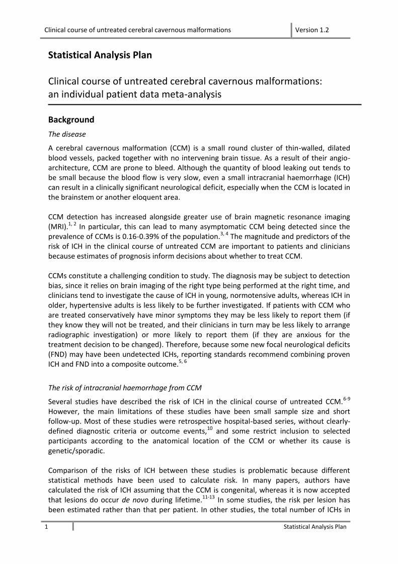
A cerebral cavernous malformation (CCM) is a small round cluster of thin-walled, dilated blood vessels, packed together with no intervening brain tissue. As a result of their angio-architecture, CCM are prone to bleed. Although the quantity of blood leaking out tends to be small because the blood flow is very slow, even a small intracranial haemorrhage (ICH) can result in a clinically significant neurological deficit, especially when the CCM is located in the brainstem or another eloquent area. CCM detection has increased alongside greater use of brain magnetic resonance imaging (MRI).1, 2 In particular, this can lead to many asymptomatic CCM being detected since the prevalence of CCMs is 0.16-0.39% of the population.3, 4 The magnitude and predictors of the risk of ICH in the clinical course of untreated CCM are important to patients and clinicians because estimates of prognosis inform decisions about whether to treat CCM. CCMs constitute a challenging condition to study. The diagnosis may be subject to detection bias, since it relies on brain imaging of the right type being performed at the right time, and clinicians tend to investigate the cause of ICH in young, normotensive adults, whereas ICH in older, hypertensive adults is less likely to be further investigated. If patients with CCM who are treated conservatively have minor symptoms they may be less likely to report them (if they know they will not be treated, and their clinicians in turn may be less likely to arrange radiographic investigation) or more likely to report them (if they are anxious for the treatment decision to be changed). Therefore, because some new focal neurological deficits (FND) may have been undetected ICHs, reporting standards recommend combining proven ICH and FND into a composite outcome.5, 6
The risk of intracranial haemorrhage from CCM
Several studies have described the risk of ICH in the clinical course of untreated CCM.6-9 However, the main limitations of these studies have been small sample size and short follow-up. Most of these studies were retrospective hospital-based series, without clearly-defined diagnostic criteria or outcome events,10 and some restrict inclusion to selected participants according to the anatomical location of the CCM or whether its cause is genetic/sporadic. Comparison of the risks of ICH between these studies is problematic because different statistical methods have been used to calculate risk. In many papers, authors have calculated the risk of ICH assuming that the CCM is congenital, whereas it is now accepted that lesions do occur de novo during lifetime.11-13 In some studies, the risk per lesion has been estimated rather than that per patient. In other studies, the total number of ICHs in
Clinical course of untreated cerebral cavernous malformations Version 1.2
2 Statistical Analysis Plan
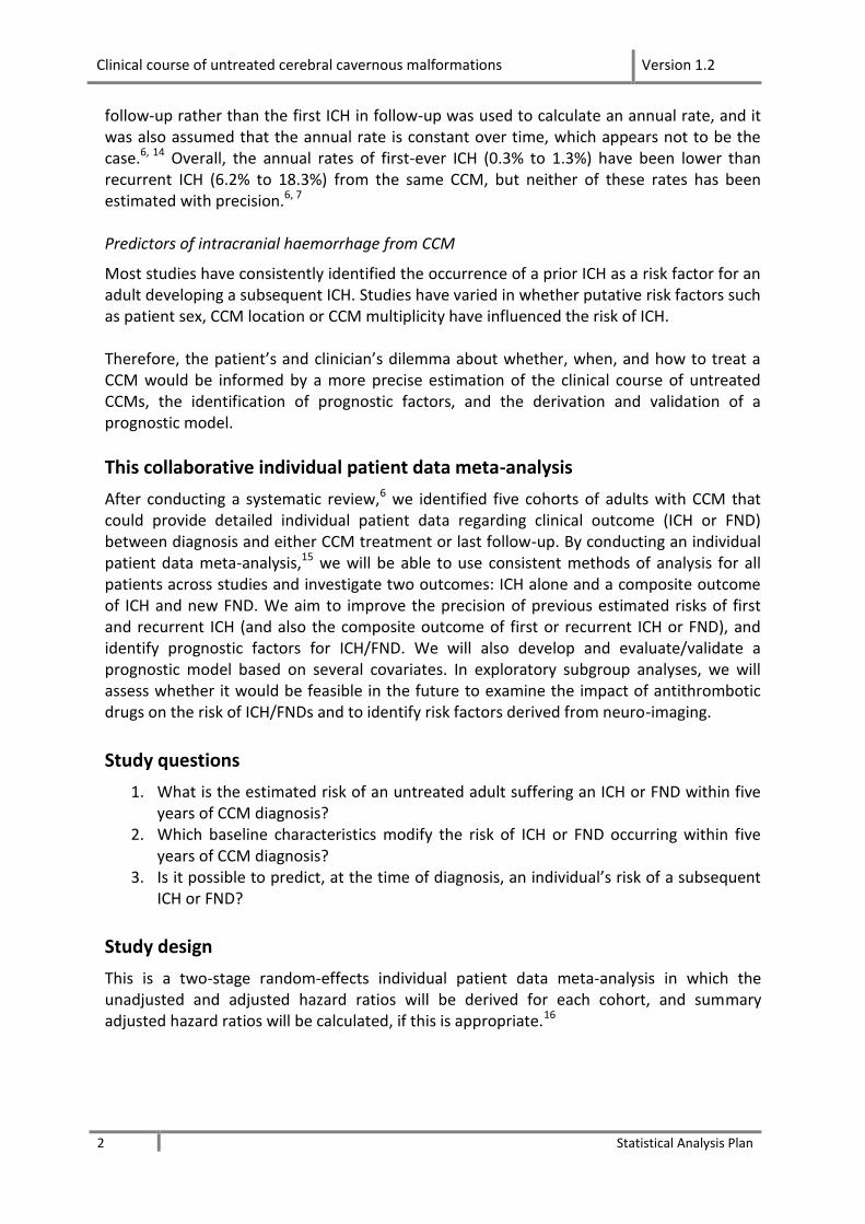
follow-up rather than the first ICH in follow-up was used to calculate an annual rate, and it was also assumed that the annual rate is constant over time, which appears not to be the case.6, 14 Overall, the annual rates of first-ever ICH (0.3% to 1.3%) have been lower than recurrent ICH (6.2% to 18.3%) from the same CCM, but neither of these rates has been estimated with precision.6, 7 Predictors of intracranial haemorrhage from CCM
Most studies have consistently identified the occurrence of a prior ICH as a risk factor for an adult developing a subsequent ICH. Studies have varied in whether putative risk factors such as patient sex, CCM location or CCM multiplicity have influenced the risk of ICH. Therefore, the patient’s and clinician’s dilemma about whether, when, and how to treat a CCM would be informed by a more precise estimation of the clinical course of untreated CCMs, the identification of prognostic factors, and the derivation and validation of a prognostic model.
This collaborative individual patient data meta-analysis
After conducting a systematic review,6 we identified five cohorts of adults with CCM that could provide detailed individual patient data regarding clinical outcome (ICH or FND) between diagnosis and either CCM treatment or last follow-up. By conducting an individual patient data meta-analysis,15 we will be able to use consistent methods of analysis for all patients across studies and investigate two outcomes: ICH alone and a composite outcome of ICH and new FND. We aim to improve the precision of previous estimated risks of first and recurrent ICH (and also the composite outcome of first or recurrent ICH or FND), and identify prognostic factors for ICH/FND. We will also develop and evaluate/validate a prognostic model based on several covariates. In exploratory subgroup analyses, we will assess whether it would be feasible in the future to examine the impact of antithrombotic drugs on the risk of ICH/FNDs and to identify risk factors derived from neuro-imaging.
Study questions
1. What is the estimated risk of an untreated adult suffering an ICH or FND within five years of CCM diagnosis?
2. Which baseline characteristics modify the risk of ICH or FND occurring within five years of CCM diagnosis?
3. Is it possible to predict, at the time of diagnosis, an individual’s risk of a subsequent ICH or FND?
Study design
This is a two-stage random-effects individual patient data meta-analysis in which the unadjusted and adjusted hazard ratios will be derived for each cohort, and summary adjusted hazard ratios will be calculated, if this is appropriate.16
Clinical course of untreated cerebral cavernous malformations Version 1.2
3 Statistical Analysis Plan
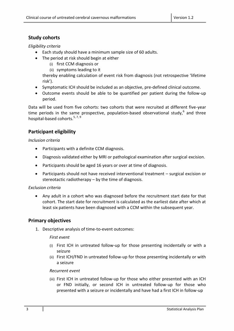
Study cohorts
Eligibility criteria
Each study should have a minimum sample size of 60 adults.
The period at risk should begin at either (i) first CCM diagnosis or (ii) symptoms leading to it
thereby enabling calculation of event risk from diagnosis (not retrospective ‘lifetime risk’).
Symptomatic ICH should be included as an objective, pre-defined clinical outcome.
Outcome events should be able to be quantified per patient during the follow-up period.
Data will be used from five cohorts: two cohorts that were recruited at different five-year time periods in the same prospective, population-based observational study,6 and three hospital-based cohorts.5, 7, 9
Participant eligibility
Inclusion criteria
Participants with a definite CCM diagnosis.
Diagnosis validated either by MRI or pathological examination after surgical excision.
Participants should be aged 16 years or over at time of diagnosis.
Participants should not have received interventional treatment – surgical excision or stereotactic radiotherapy – by the time of diagnosis.
Exclusion criteria
Any adult in a cohort who was diagnosed before the recruitment start date for that cohort. The start date for recruitment is calculated as the earliest date after which at least six patients have been diagnosed with a CCM within the subsequent year.
Primary objectives
1. Descriptive analysis of time-to-event outcomes:
First event
(i) First ICH in untreated follow-up for those presenting incidentally or with a seizure
(ii) First ICH/FND in untreated follow-up for those presenting incidentally or with a seizure
Recurrent event
(iii) First ICH in untreated follow-up for those who either presented with an ICH or FND initially, or second ICH in untreated follow-up for those who presented with a seizure or incidentally and have had a first ICH in follow-up
Clinical course of untreated cerebral cavernous malformations Version 1.2
4 Statistical Analysis Plan
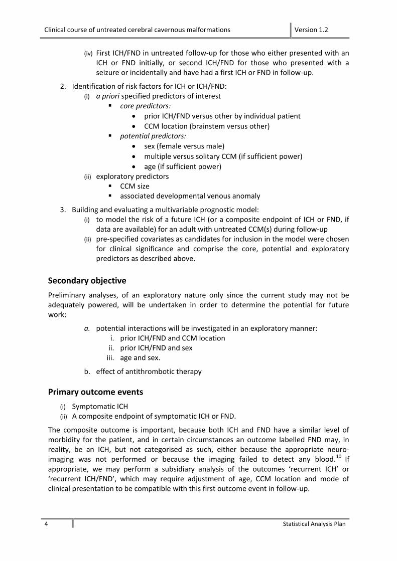
(iv) First ICH/FND in untreated follow-up for those who either presented with an ICH or FND initially, or second ICH/FND for those who presented with a seizure or incidentally and have had a first ICH or FND in follow-up.
2. Identification of risk factors for ICH or ICH/FND: (i) a priori specified predictors of interest
core predictors:
prior ICH/FND versus other by individual patient
CCM location (brainstem versus other) potential predictors:
sex (female versus male)
multiple versus solitary CCM (if sufficient power)
age (if sufficient power) (ii) exploratory predictors
CCM size associated developmental venous anomaly
3. Building and evaluating a multivariable prognostic model: (i) to model the risk of a future ICH (or a composite endpoint of ICH or FND, if
data are available) for an adult with untreated CCM(s) during follow-up (ii) pre-specified covariates as candidates for inclusion in the model were chosen
for clinical significance and comprise the core, potential and exploratory predictors as described above.
Secondary objective
Preliminary analyses, of an exploratory nature only since the current study may not be adequately powered, will be undertaken in order to determine the potential for future work:
a. potential interactions will be investigated in an exploratory manner: i. prior ICH/FND and CCM location
ii. prior ICH/FND and sex iii. age and sex.
b. effect of antithrombotic therapy
Primary outcome events
(i) Symptomatic ICH (ii) A composite endpoint of symptomatic ICH or FND.
The composite outcome is important, because both ICH and FND have a similar level of morbidity for the patient, and in certain circumstances an outcome labelled FND may, in reality, be an ICH, but not categorised as such, either because the appropriate neuro-imaging was not performed or because the imaging failed to detect any blood.10
If appropriate, we may perform a subsidiary analysis of the outcomes ‘recurrent ICH’ or ‘recurrent ICH/FND’, which may require adjustment of age, CCM location and mode of clinical presentation to be compatible with this first outcome event in follow-up.
Clinical course of untreated cerebral cavernous malformations Version 1.2
5 Statistical Analysis Plan
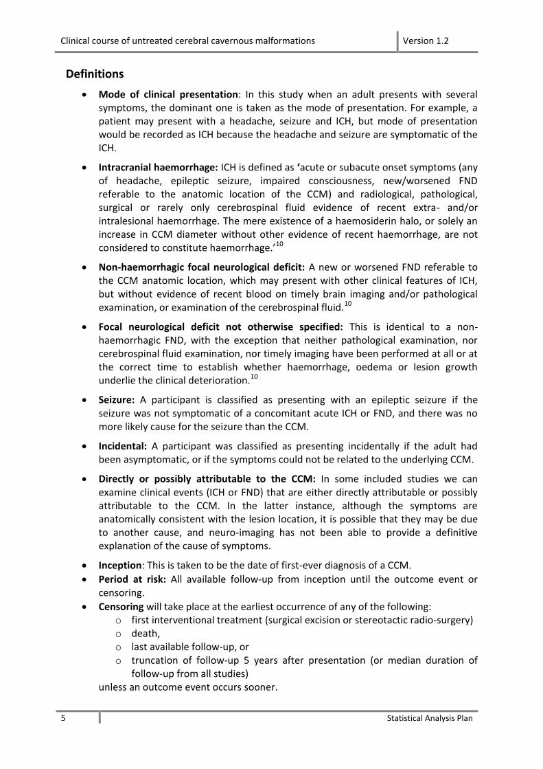
Definitions
Mode of clinical presentation: In this study when an adult presents with several symptoms, the dominant one is taken as the mode of presentation. For example, a patient may present with a headache, seizure and ICH, but mode of presentation would be recorded as ICH because the headache and seizure are symptomatic of the ICH.
Intracranial haemorrhage: ICH is defined as ‘acute or subacute onset symptoms (any of headache, epileptic seizure, impaired consciousness, new/worsened FND referable to the anatomic location of the CCM) and radiological, pathological, surgical or rarely only cerebrospinal fluid evidence of recent extra- and/or intralesional haemorrhage. The mere existence of a haemosiderin halo, or solely an increase in CCM diameter without other evidence of recent haemorrhage, are not considered to constitute haemorrhage.’10
Non-haemorrhagic focal neurological deficit: A new or worsened FND referable to the CCM anatomic location, which may present with other clinical features of ICH, but without evidence of recent blood on timely brain imaging and/or pathological examination, or examination of the cerebrospinal fluid.10
Focal neurological deficit not otherwise specified: This is identical to a non-haemorrhagic FND, with the exception that neither pathological examination, nor cerebrospinal fluid examination, nor timely imaging have been performed at all or at the correct time to establish whether haemorrhage, oedema or lesion growth underlie the clinical deterioration.10
Seizure: A participant is classified as presenting with an epileptic seizure if the seizure was not symptomatic of a concomitant acute ICH or FND, and there was no more likely cause for the seizure than the CCM.
Incidental: A participant was classified as presenting incidentally if the adult had been asymptomatic, or if the symptoms could not be related to the underlying CCM.
Directly or possibly attributable to the CCM: In some included studies we can examine clinical events (ICH or FND) that are either directly attributable or possibly attributable to the CCM. In the latter instance, although the symptoms are anatomically consistent with the lesion location, it is possible that they may be due to another cause, and neuro-imaging has not been able to provide a definitive explanation of the cause of symptoms.
Inception: This is taken to be the date of first-ever diagnosis of a CCM.
Period at risk: All available follow-up from inception until the outcome event or censoring.
Censoring will take place at the earliest occurrence of any of the following: o first interventional treatment (surgical excision or stereotactic radio-surgery) o death, o last available follow-up, or o truncation of follow-up 5 years after presentation (or median duration of
follow-up from all studies) unless an outcome event occurs sooner.

Clinical course of untreated cerebral cavernous malformations Version 1.2
6 Statistical Analysis Plan
Handling missing data
During the process of cleaning each dataset, problems of missing data will be addressed by contacting the clinician responsible for providing the data and where possible will be resolved. In cases where this is not possible, missing values will be imputed, if appropriate, using a multiple imputation technique.
Completeness of follow-up
We will use one method of reporting completeness of follow-up,17 which calculates the total actual follow-up accrued as a percentage of the total follow-up that was potentially available, before death or the end of the five-year period for the analyses.
Heterogeneity
Heterogeneity is an important consideration, both from the clinical and statistical perspectives, and in this study there are three potential sources of heterogeneity. (i) Study design
An example of heterogeneity of study design is that patients in a population-based study may be more likely to present incidentally than patients in a hospital-based study; to account for this source of heterogeneity, the total dataset will be stratified by study design – i.e. population-based cohort vs hospital-based cohort. In addition, length of follow-up is an important consideration as outcome events may require several years of follow-up. (ii) Baseline characteristics
Of particular concern are patient age, sex, mode of clinical presentation, CCM location and multiplicity, as these may influence outcome: for example, patients in a tertiary referral neurosurgery unit may be more likely to harbour brainstem lesions, and patients in an institute specialising in genetics may be more likely to have the familial form of the disease, with multiple lesions, than patients in general hospitals or the community. (iii) Hazard ratios
The final type of heterogeneity is that of hazard ratios (see section ‘Identifying and quantifying heterogeneity in hazard ratios’ below); this might make pooling (and the derivation of an overall prognostic model) questionable.18, 19
Baseline characteristics
A table of baseline characteristics, stratified by mode of initial clinical presentation, will be presented for each cohort, together with a similar table for the combined cohorts.
Age at start of follow-up (either CCM diagnosis or other inception point)1
Sex
1 These variables may vary, according to whether analyses are time to first/second event (for adults who
present with a seizure or incidentally, and then have an outcome event during follow-up)
Clinical course of untreated cerebral cavernous malformations Version 1.2
7 Statistical Analysis Plan
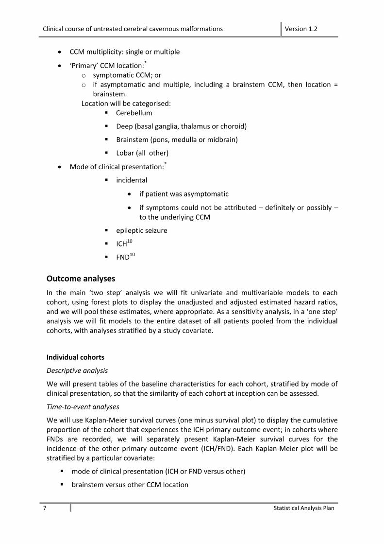
CCM multiplicity: single or multiple
‘Primary’ CCM location:* o symptomatic CCM; or o if asymptomatic and multiple, including a brainstem CCM, then location =
brainstem. Location will be categorised:
Cerebellum
Deep (basal ganglia, thalamus or choroid)
Brainstem (pons, medulla or midbrain)
Lobar (all other)
Mode of clinical presentation:*
incidental
if patient was asymptomatic
if symptoms could not be attributed – definitely or possibly – to the underlying CCM
epileptic seizure
ICH10
FND10
Outcome analyses
In the main ‘two step’ analysis we will fit univariate and multivariable models to each cohort, using forest plots to display the unadjusted and adjusted estimated hazard ratios, and we will pool these estimates, where appropriate. As a sensitivity analysis, in a ‘one step’ analysis we will fit models to the entire dataset of all patients pooled from the individual cohorts, with analyses stratified by a study covariate.
Individual cohorts
Descriptive analysis
We will present tables of the baseline characteristics for each cohort, stratified by mode of clinical presentation, so that the similarity of each cohort at inception can be assessed.
Time-to-event analyses
We will use Kaplan-Meier survival curves (one minus survival plot) to display the cumulative proportion of the cohort that experiences the ICH primary outcome event; in cohorts where FNDs are recorded, we will separately present Kaplan-Meier survival curves for the incidence of the other primary outcome event (ICH/FND). Each Kaplan-Meier plot will be stratified by a particular covariate:
mode of clinical presentation (ICH or FND versus other)
brainstem versus other CCM location
Clinical course of untreated cerebral cavernous malformations Version 1.2
8 Statistical Analysis Plan
sex (female versus male)
multiple versus solitary CCM.
We will use the log-rank test to compare the survival plots for each of these stratifications. The length of follow-up period displayed graphically will be up to five years, but it will be determined by the numbers at risk at specific time points. Univariate analyses
In both the univariate and multivariable analyses of time to one or other primary outcome, we will use the Cox proportional-hazards regression model, provided that the proportional-hazards assumption is fulfilled. The following covariates will be entered in univariate analyses to determine the unadjusted hazard ratio (and 95% confidence interval) for each putative predictor.
Putative risk factors
Covariates of a priori interest
o Core covariates
Mode of clinical presentation - for survival analyses, mode of clinical presentation will be dichotomised:
ICH or FND at presentation (x = 1) vs incidental or seizure at presentation (x = 0)
Location - for survival analyses location will be dichotomised: brainstem location (x = 1) vs other location (x = 0)
o Other covariates of a priori interest
Age at start of follow-up (as a continuous variable) CCM multiplicity: multiple (x = 1) vs single (x = 0) Sex: female (x = 1) vs male (x = 0)
Other potential covariates, depending on whether they have been recorded
CCM size antithrombotic therapy: never (x = 0) vs ever (x =1)
Design covariate – for multivariable analyses only
cohort
If antithrombotic therapy is included as a potential covariate, then it may need to be modelled as a time-varying covariate.
Multivariable analyses
The hazard ratios and 95% confidence intervals will be examined for each covariate in the univariate analyses. In the multivariable analysis, since there is inconsistency about the role of sex as a putative risk factor for ICH or FND, we will force all the covariates of a priori interest apart from sex into the model, regardless of their statistical significance in the

Clinical course of untreated cerebral cavernous malformations Version 1.2
9 Statistical Analysis Plan
univariate analyses, because they are of clinical significance; then, adjusting for these four covariates, sex will be included in the model, to ascertain its impact on the outcome.
The following interactions will also be explored in terms of their potential for future research:
prior ICH/FND and CCM location
prior ICH/FND and sex
age and sex
Sensitivity analysis
The data from the five cohorts will be pooled and the descriptive analysis, time-to-event analyses, univariate and multivariable analyses described above will be repeated on the entire dataset, stratified by the covariate ‘study’.
Forest plots
Estimates of the unadjusted and adjusted hazard ratios for the five a priori risk factors for i. ICH
ii. ICH or FND will be presented in forest plots, together with the 95% confidence interval for each covariate. The hazard ratios will be adjusted for factors imbalanced at baseline and/or known or suspected to influence CCM prognosis. If appropriate, we will pool all studies and calculate unadjusted and adjusted pooled estimates of the hazard ratios.
Identifying and quantifying heterogeneity in hazard ratios
To identify any heterogeneity between the cohorts in the hazard ratios, we will
compute Q – the weighted sum of squares of deviation of each effect size from the mean, on a standardized scale;
estimate τ2 – variance of the true effect sizes (estimated by T2);
compute I2 statistic – proportion of observed dispersion that is real, rather than spurious (a measure of inconsistency across the findings of the studies which reflects the extent of overlap of confidence intervals).
Prognostic model
If heterogeneity between the cohorts is sufficiently modest to enable a prognostic model to be built, patient data in all cohorts will be pooled to form a single dataset. Assuming the proportional-hazards assumption is not violated, the Cox proportional-hazards regression model will be used to model the risk of a future ICH (and FND, if the data are available) for an adult with CCM during untreated follow-up.20, 21 Pre-specified covariates in the model will be selected for their clinical significance and will be chosen in order from the five covariates of a priori interest listed above, namely, prior ICH/FND, brainstem location, age (as a continuous variable), lesion multiplicity, and sex. Other potential covariates include lesion size, and whether the adult has received antithrombotic therapy. If heterogeneity between the cohorts is substantial, but the proportional-hazards assumption is not violated, then the
Clinical course of untreated cerebral cavernous malformations Version 1.2
10 Statistical Analysis Plan
Cox proportional-hazards regression will be stratified by a ‘study’ covariate, with covariates of a priori interest included in the model.22-24 If appropriate, we will round the regression coefficients, estimate the baseline event risk, and then develop a prognostic index by calculating a prognostic score for each adult in the dataset. A frequency distribution of the individual scores will be examined, and two cut-off points at arithmetically convenient points (e.g. 25% with worst prognosis, and 25% with best prognosis) will be used to separate the dataset into high-, medium- and low-risk categories.21, 25 A Kaplan-Meier plot, stratified by these three risk categories, will then be calculated to illustrate the degree of discrimination achieved by the index. If the proportional-hazards assumption is seriously violated, then a multivariable logistic regression model will be used, with occurrence of ICH (or FND, if data are available) within the first five years of follow-up as the dependent variable.
Model validation
Internal validation
It will probably not be possible to validate this model by splitting our current dataset in half, as we will have insufficient outcome events in each portion. However, if it is not possible to use one or more cohorts in developing the model, then these could possibly be used to validate it.
If the model is built using data from all the cohorts, then the internal–external cross-evaluation technique will be used to validate the model internally and adjust the model for overfitting using a shrinkage factor.26, 27 Using this method, the model will be fitted on four of the five cohorts and validated on the fifth, for five occurrences (so that each model is omitted from the validation process in turn).27, 28 We will examine the predictive performance of the model by assessing its calibration and discrimination. Calibration compares the level of agreement between, for example, the model estimation of the probability of an adult experiencing an ICH within five years and the observed frequency of ICH.29 Discrimination describes how well the model splits into those who have the outcome of interest and those who do not. Harrell’s concordance index (c-index) will be used to quantify the discriminative ability of the model and calibration will be assessed using plots and the modified Hosmer and Lemeshow test for survival analysis.30
Exploratory analyses
We will explore the potential for future research, depending on the data provided by the different studies.
Whether an interaction exists between each of the following pairs of covariates: o prior ICH / FND and CCM location o prior ICH /FND and sex o age and sex
Effect of antithrombotic therapy on adults with untreated CCM.
Clinical course of untreated cerebral cavernous malformations Version 1.2
11 Statistical Analysis Plan
References
1. Brown RD, Wiebers DO, Torner JC, Ofallon WM. Frequency of intracranial hemorrhage as a presenting symptom and subtype analysis: A population-based study of intracranial vascular malformations in Olmsted County, Minnesota. Journal of Neurosurgery. 1996; 85(1): 29-32.
2. Al-Shahi R, Bhattacharya JJ, Currie DG, Papanastassiou V, Ritchie V, Roberts RC, et al. Prospective, Population-Based Detection of Intracranial Vascular Malformations in Adults: The Scottish Intracranial Vascular Malformation Study (SIVMS). Stroke. 2003; 34(5): 1163-9.
3. Morris Z, Whiteley WN, Longstreth WT, Jr, Weber F, Lee Y-C, Tsushima Y, et al. Incidental Findings on Brain Magnetic Resonance Imaging: systematic review and meta-analysis. British Medical Journal. 2009; 339: b3016.
4. Del Curling O, Jr, Kelly DL, Elster AD, Craven TE. An Analysis of the Natural History of Cavernous Angiomas. Journal of Neurosurgery 1991; 75: 702-8.
5. Porter PJ, Willinsky RA, Harper W, Wallace MC. Cerebral cavernous malformations: natural history and prognosis after clinical deterioration with or without hemorrhage. Journal of Neurosurgery. 1997; 87(2): 190-7.
6. Al-Shahi Salman R, Hall JM, Horne MA, Moultrie F, Josephson CB, Bhattacharya JJ, et al. Untreated clinical course of cerebral cavernous malformations: a prospective, population-based cohort study. Lancet Neurology. 2012.
7. Flemming KD, Link MJ, Christianson TJH, Brown RD, Jr. Prospective hemorrhage risk of intracerebral cavernous malformations. Neurology. 2012; 78: 632-6.
8. Flemming KD, Link MJ, Christianson TJH, Brown RD. Use of antithrombotic agents in patients with intracerebral cavernous malformations. Journal of Neurosurgery 2013; 118(1): 43-6.
9. Schneble H-M, Soumare A, Herve D, Bresson D, Guichard J-P, Riant F, et al. Antithrombotic therapy and bleeding risk in a prospective cohort study of patients with cerebral cavernous malformations. Stroke. 2012; 43: 3196-9.
10. Al-Shahi Salman R, Berg MJ, Morrison L, Awad IA, on behalf of the Angioma Alliance Scientific Advisory B. Hemorrhage from Cavernous Malformations of the Brain: Definition and Reporting Standards. Stroke. 2008; 39(12): 3222-30.
11. Zabramski JM, Wascher TM, Spetzler RF, Johnson B, Golfinos J, Drayer BP, et al. The natural history of familial cavernous malformations: results of an ongoing study. Journal of Neurosurgery. 1994; 80(3): 422-32.
12. Kattapong VJ, Hart BL, Davis LE. Familial cerebral cavernous angiomas: clinical and radiologic studies. Neurology. 1995; 45: 492-7.
13. Detwiler PW, Porter RW, Zabramski JM. De novo formation of a central nervous system cavernous malformation: implications for predicting risk of hemorrhage. Journal of Neurosurgery. 1997; 87: 629-32.
14. Barker II FG, Amin-Hanjani S, Butler WE, Lyons S, R.G. O, Chapman PH, et al. Temporal clustering of hemorrhages from untreated cavernous malformations of the central nervous system. Neurosurgery. 2001; 49: 15-25.
15. Riley RD, Lambert PC, Abo-Zaid G. Meta-analysis of individual participant data: rationale, conduct, and reporting. BMJ. 2010; 340: c221.
16. Riley RD, Higgins JPT, Deeks JJ. Interpretation of random effects meta-analyses. BMJ. 2011; 342: 964-7.
17. Clark TG, Altman DG, De Stavola BL. Quantification of the completeness of follow-up. Lancet. 2002; 359: 1309-10.
18. Borenstein M, Hedges L, JHiggins J, Rothstein H. Introduction to Meta-Analysis. Chichester, UK: John Wiley; 2009.
19. Ioannidis J, Patsopoulos N, Rothstein H. Reasons or excuses for avoiding meta-analysis in forest plots. BMJ. 2008; 336: 1413-5.
Clinical course of untreated cerebral cavernous malformations Version 1.2
12 Statistical Analysis Plan
20. Harrell FE, Lee KL, Mark DB. Multivariable prognostic models: Issues in developing models, evaluating assumptions and adequacy, and measuring and reducing errors. Statistics in Medicine. 1996; 15(4): 361-87.
21. Machin D, Cheung YB, Parmar MKB. Survival analysis: a practical approach. 2nd ed. Chichester, UK: John Wiley; 2006.
22. Hosmer DWJ, Lemeshow S, May S. Applied Survival Analysis:. 2nd ed. Hoboken, NJ: John Wiley; 2008.
23. Steyerberg EW. Clinical Prediction Models. New York: Springer; 2009 24. Fibrinogen, Studies, Collaboration. Measures to assess the prognostic ability of the stratified Cox
proportional hazards model. Statistics in Medicine. 2009; 28(3): 389-411. 25. Leonard RCF, Hayward RL, Prescott RJ, Wang J-X. Identification of discrete prognostic groups in
low-grade Hodgkin's lymphoma. Annals of Oncology 1991; 2: 655-62. 26. Debray TPA, Moons KGM, Ahmed I, Koffijberg H, Riley RD. Framework for developing,
implementing, and evaluating clinical prediction models in an individual participant data meta-analysis. Statistics in Medicine. 2012; 32 (18): 3158-80.
27. Royston P, Parmar MKB, Sylvester R. Construction and validation of a prognostic model across several studies, with an application in superficial bladder cancer. Statistics in Medicine. 2004; 23: 907-26.
28. May M, Royston P, Egger M, Justice AC, Sterne JAC. Development and validation of a prognostic model for survival time data: application to prognosis of HIV positive patients treated with antiretroviral therapy. Statistics in Medicine. 2004; 23: 2375-98.
29. Moons KGM, Kengne AP, Woodward M, Royston P, Vergouwe Y, Altman DG, et al. Risk prediction models: I. Development, internal validation, and assessing the incremental value of a new (bio)marker. Heart. 2012; 98(9): 683-90.
30. May S, Hosmer DWJ. Simplified method of calculating an overall goodness-of-fit test for the Cox proportional hazards model. Lifetime Data Analysis. 1998; 4: 109-20.