ENDOVASCULAR TREATMENT OF CEREBRAL … · 3150 endovascular treatment of cerebral arteriovenous...
8
3150 ENDOVASCULAR TREATMENT OF CEREBRAL ARTERIOVENOUS MALFORMATIONS AND DURAL ARTERIOVENOUS MALFORMATIONS TRATAMIENTO ENDOVASCULAR DE LAS MALFORMACIONES ARTERIOVENOSAS CEREBRALES Y DE LAS MALFORMACIONES ARTERIOVENOSAS DURALES Juan Carlos Puentes 1 Franco Ruales 2 Héctor Restrepo 3 SUMMARY Objective: To report our experience with the endovascular management of cerebral arteriovenous malformations and dural arteriovenous fistulas. Methods: Fifty patients (66% males and 34% females) with cerebral arteriovenous malformations and dural arteriovenous fistulas were treated in our department between September 2007 and April of 2010 using endovascular therapy. A total of 84 endovascular procedures were performed. N-butyl cyanoacrylate Histoacryl ® was the embolic material used in 76% of the cases; Onyx ® alone was used in 20%, Onyx ® and coils combined in 3.6%. Results: The most common symptoms were headache, epileptic seizures and intracranial hemorrhage. Thirty-two percent of the patients were cured using embolization as the only therapeutic technique, meaning that a higher number of patients were cured in just one session. Thirty-eight percent of all patients underwent a surgical resection or radiosurgical treatment after nodal size reduction through endovascular treatment. The overall reported complications were 5.9%. Conclusion: Cerebral arteriovenous malformations and dural arteriovenous fistulas are complex lesions with a potential risk for intracranial hemorrhages. Endovascular therapy is safe and effective, and together with surgery and radiosurgery, is an essential component of the multimodal approach to this problem. RESUMEN Objetivo: Reportar una experiencia en el manejo endovascular de las malformaciones arteriovenosas cerebrales y las fistulas arteriovenosas durales. Método: Se recolectó una muestra por conveniencia desde septiembre del 2007 hasta abril del 2010. Fueron tratados 50 pacientes, 34 con diagnóstico de malformación arteriovenosa cerebral y 16 con diagnóstico de fístula arteriovenosa dural, mediante terapia endovascular. De ellos el 66% corresponde a hombres, y el 34% a mujeres. Se realizaron en total 84 sesiones de embolización según una técnica endovascular para cada material de embolización. Se utilizó N-butil cianoacrilato, Histoacryl ® en el 76% de los procedimientos; Onyx ® en el 20%, y una combinación de estos con poroespirales en el 3,6% de los procedimientos. Resultados: Los síntomas más frecuentes de manifestación fueron cefalea, convulsión y hemorragia intracerebral. En el 32% de los casos se alcanzó la curación con el manejo endovascular como técnica única, logrando mayor número de curaciones con una sola sesión de embolización. El 38% de los pacientes fueron enviados a radioterapia o a manejo quirúrgico complementario luego de disminuir el tamaño de la lesión KEY WORDS (MeSH) Intracranial arteriovenous malformations Arteriovenous fistula Endovascular procedures PALABRAS CLAVE (DeCS) Malformaciones arteriove- nosas cerebrales Fístula arteriovenosa Procedimientos endovascu- lares 1 Endovascular neurosurgeon, Department of Radiology and Diagnostic Imaging, and Neurosurgery Department, Fundación Cardioinfantil- Cardiology Institute, and San Ignacio’s Hospital, Bogotá, Colombia 2 Radiology and Diagnostic Imaging Resident, Fundación Cardioinfantil-Cardiology Institute, and Colegio Mayor Nuestra Señora del Rosario, Bogotá, Colombia. 3 Epidemiologist physician, Research Department, Fundación Cardioinfantil- Cardiology Institute, Bogotá, Colombia.
Transcript of ENDOVASCULAR TREATMENT OF CEREBRAL … · 3150 endovascular treatment of cerebral arteriovenous...
3150
ENDOVASCULAR TREATMENT OF CEREBRAL ARTERIOVENOUS MALFORMATIONS AND DURAL ARTERIOVENOUS MALFORMATIONSTRATAMIENTO ENDOVASCULAR DE LAS MALFORMACIONES ARTERIOVENOSAS CEREBRALES y DE LAS MALFORMACIONES ARTERIOVENOSAS DURALES
Juan Carlos Puentes1
Franco Ruales2 Héctor Restrepo3
Summary Objective: To report our experience with the endovascular management of cerebral
arteriovenous malformations and dural arteriovenous fistulas. Methods: Fifty patients (66% males and 34% females) with cerebral arteriovenous malformations and dural arteriovenous fistulas were treated in our department between September 2007 and April of 2010 using endovascular therapy. A total of 84 endovascular procedures were performed. N-butyl cyanoacrylate Histoacryl® was the embolic material used in 76% of the cases; Onyx® alone was used in 20%, Onyx® and coils combined in 3.6%. Results: The most common symptoms were headache, epileptic seizures and intracranial hemorrhage. Thirty-two percent of the patients were cured using embolization as the only therapeutic technique, meaning that a higher number of patients were cured in just one session. Thirty-eight percent of all patients underwent a surgical resection or radiosurgical treatment after nodal size reduction through endovascular treatment. The overall reported complications were 5.9%. Conclusion: Cerebral arteriovenous malformations and dural arteriovenous fistulas are complex lesions with a potential risk for intracranial hemorrhages. Endovascular therapy is safe and effective, and together with surgery and radiosurgery, is an essential component of the multimodal approach to this problem.
reSumenObjetivo: Reportar una experiencia en el manejo endovascular de las malformaciones
arteriovenosas cerebrales y las fistulas arteriovenosas durales. Método: Se recolectó una muestra por conveniencia desde septiembre del 2007 hasta abril del 2010. Fueron tratados 50 pacientes, 34 con diagnóstico de malformación arteriovenosa cerebral y 16 con diagnóstico de fístula arteriovenosa dural, mediante terapia endovascular. De ellos el 66% corresponde a hombres, y el 34% a mujeres. Se realizaron en total 84 sesiones de embolización según una técnica endovascular para cada material de embolización. Se utilizó N-butil cianoacrilato, Histoacryl® en el 76% de los procedimientos; Onyx® en el 20%, y una combinación de estos con poroespirales en el 3,6% de los procedimientos. Resultados: Los síntomas más frecuentes de manifestación fueron cefalea, convulsión y hemorragia intracerebral. En el 32% de los casos se alcanzó la curación con el manejo endovascular como técnica única, logrando mayor número de curaciones con una sola sesión de embolización. El 38% de los pacientes fueron enviados a radioterapia o a manejo quirúrgico complementario luego de disminuir el tamaño de la lesión
Key words (meSH) Intracranial arteriovenous
malformationsArteriovenous fistulaEndovascular procedures
Palabras clave (DeCS)Malformaciones arteriove-
nosas cerebralesFístula arteriovenosa Procedimientos endovascu-
lares
1Endovascular neurosurgeon, Department of Radiology
and Diagnostic Imaging, and Neurosurgery Department,
Fundación Cardioinfantil-Cardiology Institute, and
San Ignacio’s Hospital, Bogotá, Colombia
2Radiology and Diagnostic Imaging Resident, Fundación
Cardioinfantil-Cardiology Institute, and Colegio Mayor Nuestra Señora del Rosario,
Bogotá, Colombia. 3Epidemiologist physician,
Research Department, Fundación Cardioinfantil-
Cardiology Institute, Bogotá, Colombia.

3151Rev Colomb Radiol. 2011; 22(2): 3150-7
original articles
por medio endovascular. Las complicaciones alcanzaron el 5,9%. Conclusiones: Las malformaciones arteriovenosas cerebrales y fístulas arteriovenosas durales son lesiones complejas que conllevan un riesgo potencial de sangrado para los pacientes con sus consecuencias. El tratamiento endovascular es seguro y efectivo, y se constituye en parte fundamental dentro del esquema de manejo multidisciplinario con la radiocirugía y cirugía.
IntroductionThe most frequent vascular malformations of the brain, and the most
significant because of the concomitant risk of bleeding, are cerebral arte-riovenous malformations and dural arteriovenous fistulas. The former are lesions in which a nest of vessels creates pathological communications between arteries and veins, without an intervening capillary bed. They are probably the result of a congenital abnormality of blood vessels emerging in fetal life; however, they may be associated with hereditary syndromes – including Rendu-Osler-Weber disease (hereditary hemo-rrhagic telangiectasia) and Wyburn-Mason syndrome.
Their incidence and prevalence are unknown, although there are data derived from autopsy series or limited population studies in diffe-rent published series, with figures ranging between 0.8 and 2.1 cases for every 100,000 inhabitants. Several classifications have been deve-loped, but the most widely used is the Spetzler-Martin classification that defines the risk associated with the surgical treatment and provides uniformity to the different reports. It classifies arteriovenous malfor-mations on the basis of their size, location and venous drainage (1-3).
When arteriovenous malformations are symptomatic, they gives rise to different clinical manifestations such as headache, seizures or neurological decline secondary to cerebral hemorrhage, and they are more frequent among young adults between the third and fourth decades of life. Over 50% of cerebral arteriovenous malformations manifest with intracranial bleeding, although intraparenchymal he-morrhage is the most common. They can also produce subaracnoid or intraventricular bleeding. Available studies on the natural history of arteriovenous malformations suggest that the annual risk of bleeding is 2-3%, while the risk of rebleeding during the first year after the first bleeding episode ranges between 6% and 18% (4-8).
On the other hand, dural arteriovenous fistulas are direct connec-tions between arteries and veins (dural sinuses or cortical veins) locali-zed inside dural layers, without an intervening capillary network. They account for 10-15% of all intracranial vascular malformations and they may show up at any place of dural coverage of the brain, although the most frequent sites are the cavernous sinus and the transverse venous sinus. Patients may either remain asymptomatic or experience various mild- to-fatal symptoms, including bleeding (9-11).
Treatment of cerebral arteriovenous malformations and dural arteriovenous fistulas includes endovascular management, stereo-tactic radiosurgery, surgery, or conservative treatment. The role of endovascular management may be defined in accordance with speci-fic situations: curative therapy with embolization only; preoperative management using embolization before complete surgical excision, or before radiosurgery, and palliative embolization designed to diminish symptoms associated with the arteriovenous shunting or to eliminate elements that increase the risk of bleeding within the malformation complex (12-15).
Presented here is a descriptive case series study using a convenience sample including a total of 50 patients treated between September 2007 and April 2010. Upon admission, these patients had an angiographic diagnosis of cerebral arteriovenous malformation or dural arteriovenous
fistula, and they underwent curative endovascular management, or en-dovascular management before radiosurgery or surgery.
materials and methodsThis descriptive case series study with a convenience sample inclu-
ded 50 patients treated between September 2007 and April 2010. Upon admission, they had an angiographic diagnosis of cerebral arteriovenous malformation or dural arteriovenous fistula, and later they were taken to endovascular management. The endovascular treatment was performed by the same surgeon (JCP) in all cases. General anesthesia was given in all cases where Onyx® was used as embolization material, and in pediatric cases.
A guide wire was inserted through a femoral access for microcathe-terization of arterial feeders using flow microcatheters placed inside the vascular nest (Magic, Balt Montmorency; Sonic, Bal Montmorency; Marathon, ev3Irvine, CA). Two different embolization agents were used – Histocryl® (B Braun, Melsungen GA) and Onyx® (ev3 Inc., Irvine, CA), in some cases with platinum coils each, in accordance with protocols established for each embolization material (16-19).
A form with boxes for each of the study variables was used in the Access 2007® software during recruitment. Later, with the help of a data-base transformer, the Access database was migrated to the SPSS statistical package, which was used from then on for all the statistical analyses.
The database was initially filtered in order to find and correct extreme out-of-range data points, classified as normal in accordance with the type of variables. The statistical analysis was performed after this initial purge. The analysis consisted of a description of the study population based on descriptive measurements, and the corresponding analysis was performed in relation to the variable types.
Central trend measurements were taken as averages, means and medians with their corresponding confidence intervals, percentages and absolute and relative frequencies in accordance with the variables. Later, using contingency tables, dichotomy-type variables were compared in order to determine the degree of correlation with the variables defined as outcomes.
resultsThe case series study of endovascular treatment in cerebral ar-
teriovenous malformations and dural fistulas analyzed 50 patients, 34 of whom presented arteriovenous malformations and 16 dural arteriovenous fistulas. Cases were categorized according to gender and it was found that 66% were males and 34% females. In terms of age, 26% of patients were above 50, and in terms of symptoms at entry, the main symptoms were headache (32%), seizures (30%) and intracranial bleeding (30%) (Table 1). They were also classified according to the site of the lesion, and it was found that 26% of the lesions were localized in the frontal region, followed by the temporal region (20%). Symptoms were compared in relation to the location of the lesion, and it was determined that patients with frontal lesions had seizures (Figure 1).

Endovascular Treatment of Cerebral Arteriovenous Malformations and Dural Arteriovenous Malformations. Puentes J., Ruales F., Restrepo H.3152
Figure 1. Relationship between symptoms on entry and location of the lesions, in patients with cerebral arteriovenous malformations and dural arteriovenous fistulas.
Table 1. SymptomsValid Frequency Percentage Valid
percentageCumulative percentage
Seizure 15 30.0 30.0 30.0
Bleeding 15 30.0 30.0 60.0
Headache 16 32.0 32.0 92.0
Tinnitus 2 4.0 4.0 96.0
Vertigo 1 2.0 2.0 98.0
Proptosis 1 2.0 2.0 100.0
Total 50 100.0 100.0
Table 2. Cure per session contingency
SessionsCure
Totalyes no Lost
0Count 1 0 3 4
% of total 2.0 0.0 6.0 8.0
ICount 10 3 9 22
% of total 20.0 6.0 18.0 44.0
IICount 2 7 3 12
% of total 4.0 14.0 6.0 24.0
IIICount 3 7 0 10
% of total 6.0 14.0 0.0 20.0
IVCount 0 2 0 2
% of total 0.0 4.0 0.0 4.0
TotalCount 16 19 15 50
% of total 32.0 38.0 30.0 100.0 Overall, there were 84 embolization sessions. Forty-four per cent
of patients received only one session, 24% received two, and 20% of cases received three. Only 4% of the study population went through four sessions. Results show an overall cure rate with embolization of 32%; of this, 43.7% was in dural arteriovenous fistulas and 26.5% in arteriovenous malformations
By gender, 66% of the embolizations were performed in males and 34% in females. A statistically significant relationship (p < 0.05) was found when analyzing the percentage of cure of the various lesions previously classified according to their size and complexity in relation to the number of sessions. A higher cure rate was achieved with a single embolization session, where a 20% cure rate was attained with a single session in smaller lesions, compared to a 6% cure rate when up to three sessions were required for more complex lesions (Table 2).
When discriminating the percentage of cure in relation to the site of the lesion, it was determined that occipital lesions were cured in 57% of cases. However, for lesions in the frontal location, the cure rate was 37%, due in part to the complexity of frontal lesions (p > 0.05). Thirty-eight per cent of patients were referred for multidisciplinary management with radiosurgery or surgery.
Of the total population, ten patients are still under treatment, wai-ting for a new embolization session. Five patients dropped out from treatment and were lost to follow-up. In terms of the percentage of cure in relation to gender, 26% of males were cured compared with 6% of women, although this was not statistically significant (p > 0.05).
Microcatheterization failed in four patients because of highly tortuous arterial feeders that prevented adequate microcatheter placement. In terms of the material used for the 84 sessions, Histoacryl® (n-butyl-2cyanoa-crylate) was used in 76% of cases (Figures 2 and 3); Onyx® was used in 20% of cases (Figures 4 and 5); and coils were used in 3.6% of cases.
The Spetzler-Martin classification of arteriovenous malformations was used (1). Of the 34 patients with arteriovenous malformations, 2.9% were classified as grade I, 29.4% as grade II, 47.1% as grade III, and 20.6% as grade IV. No grade V arteriovenous malformations were found in this population. In terms of gender, arteriovenous malforma-tions grades II, III and IV were more frequent among men, and grade I malformations were found more frequently in females (p > 0.05).
A statistically significant relationship was found when the severity of the arteriovenous malformations was assessed using the Spetzler-Martin classification in accordance with the location of the lesions (p <0.05). Thus, grade II malformations were found to be more frequent in the temporal region, while grade III and IV lesions were found to be more frequent in the frontal region; however, the sample size does not permit final conclusions (Table 3).
The symptom at entry was assessed in relation to the Spetzler-Martin classification and it was shown that the greater the degree of arteriovenous malformation, the greater the severity of the symptoms (Figure 6). Likewise, when the percentage of cure was assessed in rela-tion to the degree of chronicity in our population, it was found that the greater the severity according to the Spetzler-Martin classification, the lower the percentage of cure using only endovascular management. This is due to the complexity of major lesions that require multidisciplinary assessment and management (Table 4).
On the other hand, dural arteriovenous fistulas were classified ac-cording to Cognard (Table 5). Of 16 patients treated, 50% were grade I, 12.5% grade II, 6.3% grade III, 25% grade IV, and 6.3% grade V (Table 6). The percentage of cure was analyzed in relation to the severity of the lesions and it was found that 5 out of 8 patients with grade I fistulas and 2 out of 4 patients with grade IV fistulas were cured with the endovas-cular treatment (Table 7). When the degree of severity of the fistulas was analyzed in relation to the site, it was found that the majority of grade I fistulas were localized in the tentorium and the sigmoid sinuses, while the largest number of grade IV fistulas were localized in the temporal region.
3153Rev Colomb Radiol. 2011; 22(2): 3150-7
original articles
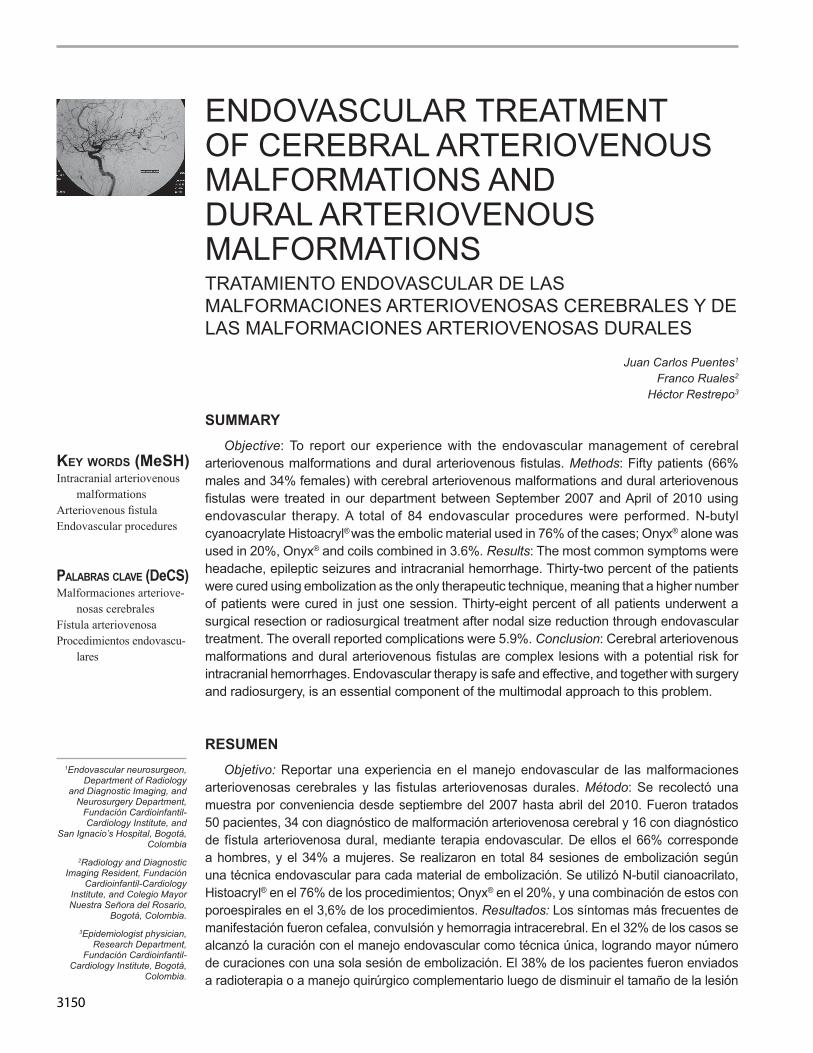
Figure 2. Nineteen year-old patient with an epileptic manifestation and a history of failed surgical removal of the arteriovenous malformation. (a) Lateral view. (b) Microcatheter placed in the malformation nest. (c) Embolization with cyanoacrylate through the pedicle in the pericallous artery. (d) Lesion cured after one embolization session.
ba
dc
Table 4. Cure contingency as per the Spetzler-Martin classification
CureClasificación Spetzler-Martin
TotalI II III IV
yesCount 1 5 3 0 9
% of total 2.9 14.7 8.8 0.0 26.5
NoCount 0 2 10 5 17
% of total 0.0 5.9 29.4 14.7 50.0
LostCount 0 3 3 2 8
% of total 0.0 8.8 8.8 5.9 23.5
TotalCount 1 10 16 7 34
% of total 2.9 29.4 47.1 20.6 100.0
Table 5. Arteriovenous fistulas according to Cognard’s classification
Type Venous drainageI Antegrade flow towards the dural sinus
II
Drainage towards the dural sinus with reflux towards:IIa – the sinus onlyIIb – the cortical veins onlyIIa+IIb – the sinus and the cortical veins
III Direct drainage to the cortical veins with no ectasia
IV Drainage to cortical veins with venous ectasia*
V Drainage to perimedullary leptomeningeal veins
*Caliber of 5 mm or greater than 3 times their normal diameter
Tabla 3. Contingencia
Category and Location
Spetzler-Martin classification Total
I II III IV
Frontal
Count 0 3 6 3 12
% of total
0.0 8.8 17.6 8.8 35.3
Temporal
Count 0 4 2 1 7
% of total
0.0 11.8 5.9 2.9 20.6
Parietal
Count 0 1 2 3 6
% of total
0.0 2.9 5.9 8.8 17.6
Cerebellar
Count 0 1 3 0 4
% of total
0.0 2.9 8.8 0.0 11.8
Occipital
Count 1 1 0 0 2
% of total
2.9 2.9 0.0 0.0 5.9
Thalamic
Count 0 0 3 0 3
% of total
0.0 0.0 8.8 0.0 8.8
Total
Count 1 10 16 7 34
% of total
2.9 29.4 47.1 20.6 100.0
Endovascular Treatment of Cerebral Arteriovenous Malformations and Dural Arteriovenous Malformations. Puentes J., Ruales F., Restrepo H.3154
ba
dc
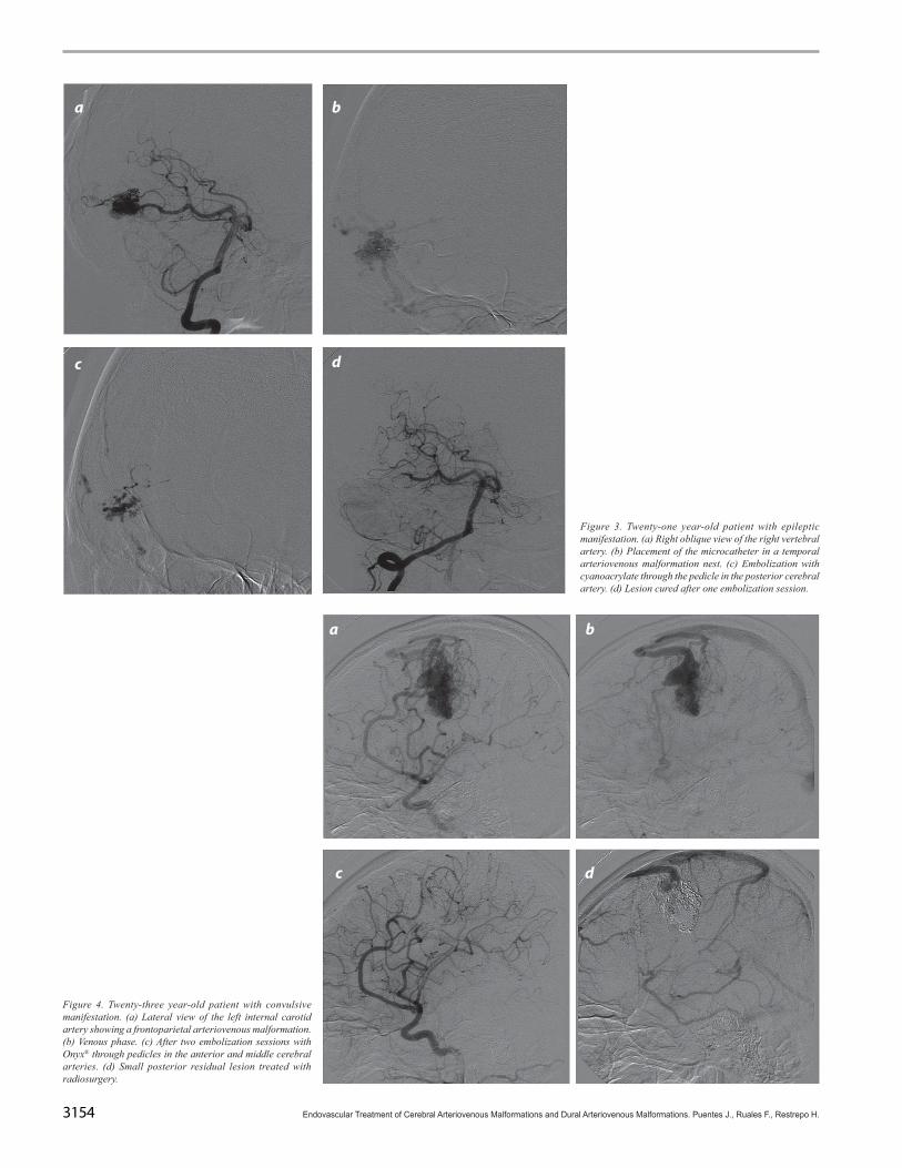
Figure 4. Twenty-three year-old patient with convulsive manifestation. (a) Lateral view of the left internal carotid artery showing a frontoparietal arteriovenous malformation. (b) Venous phase. (c) After two embolization sessions with Onyx® through pedicles in the anterior and middle cerebral arteries. (d) Small posterior residual lesion treated with radiosurgery.
ba
dc
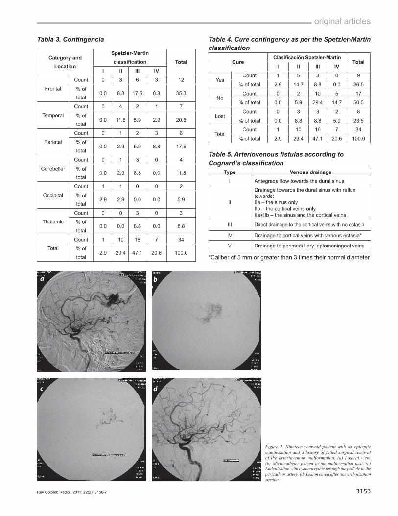
Figure 3. Twenty-one year-old patient with epileptic manifestation. (a) Right oblique view of the right vertebral artery. (b) Placement of the microcatheter in a temporal arteriovenous malformation nest. (c) Embolization with cyanoacrylate through the pedicle in the posterior cerebral artery. (d) Lesion cured after one embolization session.

3155Rev Colomb Radiol. 2011; 22(2): 3150-7
original articles
Figure 5. Forty-three year-old patient with manifestations of headache and tinnitus. (a) Lateral view of the left external carotid artery showing a grade I dural arteriovenous fistula. (c) After two embolization sessions with Onyx® through pedicles in the occipital and left middle meningeal artery. (d) Exclusion of the dural arteriovenous fistula resulting in symptom resolution.
Table 6. Grades of arteriovenous fistulas
Frecuency Percentage Valid Percentage
Cumulative Percentage
Valid
I 8 16.0 50.0 50.0
II 2 4.0 12.5 62.5
III 1 2.0 6.3 68.8
IV 4 8.0 25.0 93.8
V 1 2.0 6.3 100.0
Total 16 32.0 100.0
Lost System 34 68.0
Total 50 100.0
Tabla 7. Contingencia curación por fístula
CureFistula
TotalI II III IV V
yesCount 5 0 0 2 0 7
% of total 31.3 0.0 0.0 12.5 0.0 43.8
NoCount 1 0 0 1 0 2
% of total 6.3 0.0 0.0 6.3 0.0 12.5
LostCount 2 2 1 1 1 7
% of total 12.5 12.5 6.3 6.3 6.3 43.8
TotalCount 8 2 1 4 1 16
% of total 50.0 12.5 6.3 25.0 6.3 100.0
Figure 6. Correlation between the degree of severity according to the Spetzler-Martin classifcation, and the symptom at entry.
ba
dc
Endovascular Treatment of Cerebral Arteriovenous Malformations and Dural Arteriovenous Malformations. Puentes J., Ruales F., Restrepo H.3156
There were complications during the embolization procedure in five cases (10%) as part of the 84 sessions performed (5.9%). Two patients had thromboembolic complications not directly associated with the embolization material; they were managed using intra-arterial thrombolysis resulting in complete recanalization in one case and partial recanalization in the other, which left a an infarction sequela in Broca’s area. There was intracerebral bleeding in one patient two hours after embolization of the dural arteriovenous fistula, requiring surgical management, and there were 2 cases of transient cerebellar edema with complete symptom resolution afterwards. In the six-month follow-up period after the complication, two of the patients had a score of 2 on Rankin’s Modified Scale. Complications were more frequent among males (8%) as compared to females (2%), and there were no deaths.
DiscussionCerebral arteriovenous malformations and dural arteriovenous fistu-
las require complex treatment. A multi-disciplinary team consisting of a neurosurgeon, a neurointerventionist and and radiotherapist is required in order to assess and manage patients (20,21). All treated cases were discussed in a decision-making meeting with the participation of the three specialties. In cases of cerebral arteriovenous malformations, Spetzler-Martin malformations grades I and II with deep localization and arterial feeder vessels, and malformations grades III and IV, were indications for endovascular treatment after supplementary manage-ment with radiosurgery or surgery.
The protocol used for the treatment of cerebral arteriovenous malformations includes endovascular management as primary therapy, provided the anatomy of the lesion allows the selective placement of the microcatheter for an adequate injection of the embolization material. The treatment was performed in several sessions depending on the bulk of the lesion, taking care of not exceeding 30% of the embolized volume in each session. Two different embolization materials were used primarily - Histoacryl and Onyx® - the former for most posterior fossa arteriovenous malformations in lesions with distal tortuous arte-rial feeders, and in malformations with a high fistulous component. In determining the concentration of the material, the anatomy of the nest, the morphology of the pedicle, the shunt velocity and the characteristics of the drainage veins were all taken into consideration.
The three primary forms of clinical manifestations in cerebral arteriovenous malformations correlate with those described in pre-vious studies, but there was no clinical preponderance of intracerebral bleeding as the initial manifestation. Frontal lobe lesions tended to be associated with epilepsy as a manifestation. In our series, the cure rate with embolization only for cerebral arteriovenous malformations was 26.5%. This percentage is consistent with reports for embolization with Histoacryl, ranging between 9% and 40%, and with Onyx®, ranging between 18% and 49% (22,23).
A logical finding was a higher cure rate in small arteriovenous malformations with a single arterial pedicle with only one embolization session. However, there were residual lesions left behind after endo-vascular surgery that required stereotactic surgery as a complementary treatment in 32% of patients. Follow-up angiography is pending because it must be performed 24 months after treatment according to the protocol. With this, the percentage of cure is expected to increase in patients treated under the multidisciplinary approach. Twenty per cent of patients are still in the endovascular treatment protocol and are
waiting for a new embolization session, while 10% of patients dropped out from the treatment plan of their own accord or were not authorized by their insurance to continue with the treatment in our hospital.
There is a controversy at present regarding the indication for initia-ting treatment in cases of cerebral arteriovenous malformation found incidentally in asymptomatic patients. There is concern on whether starting treatment might modify the natural history and increase the risk of bleeding. The ARUBA is an ongoing randomized multicentric study designed to answer this question. It compares two groups of patients, one receiving the best available treatment between radiosurgery, em-bolization or surgery, and the second undergoing observation (24). All of the patients in our group had some form of clinical manifestation.
The reported rate of complications in endovascular treatment of cerebral arteriovenous malformations ranges between 3% and 25%. Reported mortality rates associated with embolization are 2% or less, according to published studies. In our series, there were complications in five patients (10% of the population), three of which where directly associated with the material and the embolization technique used (25).
All cases of dural arteriovenous malformations were indications for endovascular treatment. The embolization material was selected depending on the possibility of a supraselective placement of the microcatheter, where the goal was to obliterate the fistula site and the start of the drainage vein. The embolization material used in the majority of cases was Onyx®. In our series, the rate of cure achieved with embolization only was 43.7% in dural fistulas. There were two complications in our group with embolization of dural arteriovenous malformations: one case of spontaneous intracranial bleeding in the immediate postoperative period, related to a deleterious effect on one drainage vein, requiring an urgent craniotomy that left a neurological sequela in the form of a visual field defect; in the second case there was a transient cerebellar syndrome with no sequela from cerebellar edema, accounting for a morbidity rate of 9%.
ConclusionsCerebral arteriovenous malformations and dural fistulas are com-
plex lesions that entail a potential risk of bleeding, with all its conse-quences for the patient. The endovascular treatment with embolization is a fundamental component of the multidisciplinary management, together with radiosurgery and surgery. In our setting, we may use endovascular treatment safely and effectively, as long as it is done in a context where the treatment is used rationally when indicated and where practitioners are familiar with the strengths of the multidisci-plinary approach.
references1. Spetzler RF, Martin NA. A proposed grading system for arteriovenous malforma-
tions. J Neurosurg. 1986;65:476-83.2. Cognard C, Gobin YP, Pierot L, et al. Cerebral dural arteriovenous fistulas: clinical
and angiographic correlation with a revised classification of venous drainage. Ra-diology. 1995;194:671-80.
3. Forsting M. Intracranial vascular malformations and aneurysms from diagnostic work-up to endovascular therapy. Heidelberg: Springer-Verlag; 2004.
4. Strozyk D, Noqueira RG, Lavine SD. Endovascular treatment of intracranial arte-riovenous malformation. Neurosurg Clin N Am. 2009;20:399-418.
5. Neumaier-Probst E. Dural arteriovenous fistulas. Klin Neuroradiol. 2009;19:91-100. Epub 2009 May 15.
6. Mohr JP, Moskowitz A, Ascheim D, et al. A Randomized Multicenter Clinical Trial of Unruptured Brain AVMs (ARUBA). Clinical Protocol. New York: National Insti-tute of Neurological Disorders and Stroke National Institutes of Health; 2008.
3157Rev Colomb Radiol. 2011; 22(2): 3150-7
original articles
7. Pierot L, Januel AC, Herbreteau D, et al. Endovascular treatment of brain arterio-venous malformations using onyx: results of a prospective, multicenter study. J Neuroradiol. 2009;36:147-52. Epub 2009 Feb 14.
8. Cognard C, Januel AC, Silva NA Jr, et al. Endovascular treatment of intracranial dural arteriovenous fistulas with cortical venous drainage: new management using Onyx. AJNR Am J Neuroradiol. 2008;29:235-41. Epub 2007 Nov 7.
9. Mounayer C, Hammami N, Piotin M, et al. Nidal embolization of brain arte-riovenous malformations using Onyx in 94 patients. AJNR Am J Neuroradiol. 2007;28:518-23.
10. Linfante I, Wakhloo AK. Brain aneurysms and arteriovenous malformations: advancements and emerging treatments in endovascular embolization. Stroke. 2007;38:1411-7. Epub 2007 Feb 22.
11. Weber W, Kis B, Siekmann R, et al. Endovascular treatment of intracranial arte-riovenous malformations with onyx: technical aspects. AJNR Am J Neuroradiol. 2007;28:371-7.
12. Hartmann A, Mast H, Choi JH, et al. Treatment of arteriovenous malformations of the brain. Curr Neurol Neurosci Rep. 2007;7:28-34.
13. Richling B, Killer M, Al-Schameri AR, et al. Therapy of brain arteriovenous mal-formations: multimodality treatment from a balanced standpoint. Neurosurgery. 2006;59(5 Suppl 3):S148-57; discussion S3-13.
14. Gailloud P. Endovascular treatment of cerebral arteriovenous malformations. Tech Vasc Interv Radiol. 2005;8:118-28.
15. Hussain MS, Qureshi Al, Kirmani JF, et al. Update on endovascular treatment of cerebrovascular diseases. J Endovasc Ther. 2004;11 Suppl 2:II32-42.
16. Cockroft KM, Hwang SK, Rosenwasser RH. Endovascular treatment of cerebral arteriovenous malformations: indications, techniques, outcome, and complications. Neurosurg Clin N Am. 2005;16:367-80.
17. Smith JL, Garg B. Treatment of arteriovenous malformations of the brain. Curr Neurol Neurosci Rep. 2002;2:44-9.
18. Martin NA, Khanna R, Doberstein C, et al. Therapeutic embolization of arteriove-nous malformations: the case for and against. Clin Neurosurg. 2000;46:295-318.
19. Richling B, Killer M. Endovascular management of patients with cerebral arterio-venous malformations. Neurosurg Clin N Am. 2000;11:123-45.
20. De Oliveira E, Tedeschi H, Raso J. Comprehensive management of arteriovenous malformations. Neurol Res. 1998;20:673-83.
21. Geibprasert S, Ponqpech S, Jiarakongmun P, et al. Radiologic assessment of brain arteriovenous malformations: what clinicians need to know. Radiographics. 2010;30:483-501.
22. Kiyosue H, Hori Y, Okahara M, et al. Treatment of intracranial dural arteriovenous fistulas: current strategies based on location and hemodynamics, and alternative techniques of transcatheter embolization. Radiographics. 2004;24:1637-53.
23. Ogilvy CS, Stieg P, Awad I, et al. Recommendations for the management of intra-cranial arteriovenous malformations: a statement for healthcare professionals from a special writing group of the Stroke Council, American Stroke Association. Stroke. 2001;32;1458-71.
24. Fiorella D, Albuquerque FC, Woo HH, et al. The role of neuroendovascular therapy for the treatment of brain arteriovenous malformations. Neurosurgery. 2006;59:S163-77.
25. Picard L, Bracard S, Anxionnat A, et al. Brain AVM embolization. Retrospec-tive study concerning 728 patients followed between 1984 and 2004. Interv Neuroradiol.2005;11(S1)45-50.
Correspondence
Franco Ruales Departamento de Radiología e Imágenes Diagnósticas Fundación Cardio Infantil-Instituto de Cardiología Calle 163 A No. 13B-60 Bogotá, Colombia [email protected] [email protected]
Received for evaluation: September 6, 2010Accepted for publication: May 5, 2011