congenital atresia of the esophagus: with tracheo-esophageal fistula
8
CONGENITAL ATRESIA OF THE ESOPHAGUS: WITH TRACHEO-ESOPHAGEAL FISTULA ROLLIN A. DANIEL, JR., M.D. NASHVILLE, TENN. FROM THE DEPARTMENT OF SURGERY, VANDERBILT UNIVERSITY MEDICAL SCHOOL, NASHVILLE, TENN. DURING THE LAST THREE YEARS seven infants with congenital atresia of the esophagus were admitted to the Vanderbilt University Hospital. A tracheo-esophageal fistula, Vogt's type 3B1 was present in each case. All of these patients were operated upon. The operative procedures have varied, l)ut in all cases the fistula has been exposed at operation. An end-to-end aniastomosis of the segments was attempted in three cases and it was possible to bring the ends of the segments together in two of them. One patient survived and is living. This child is six months old. A summary of the cases is presented in Table I, and the one case ill which a successful anastomosis was effected is reported in detail. Case Report.-R. L. C., male, born November 28, I943, in the Vanderbilt Unii- versity Hospital, at term. He was the first child of a healthy and normal mother, age 33. At birth the infant weighed 6 lbs., 7.75 ounces. He breathed spontaneously, and physical examination revealed no abnormalities. Thirty-six hours after birth it was noted that the child had an unusually large amount of mucous in his mouth and throat, and 56 hours after birth the following note was made by the Pediatric intern: "Baby having a large amount of mucous and is vomiting practically all fluids offered. Believe this baby should be investigated for possibility of tracheo-esophageal fistula." The infant was given 50 cc. of blood plasma and 50 cc. of IO per cent glucose intra- venously and 200 CC. of normal saline solution subcutaneously, and a catheter was passed into the pharynx. The tube could be inserted for a distance of only six inc,es, where it met an obstruction. The infant was then taken to the Roentgenologic Department, where a swallow of radiopaque oil revealed the esophagus to end in a blind pouch about two inches below its origin. The presence of large amounts of gas in the gastro-intestinal tract confirmed the diagnosis of congenital atresia of the esophagus, with tracheo-esophageal fistula. The patient was transferred to the Surgical Service. At this time his weight was 5 lbs. 12.5 ounces. The breath sounds were rough throughout both lung fields but there was no evidence of consolidation. The patient showed no evidence of de- hydration. He was given 25 cc. of blood plasma and 25 cc. of IO per cent glucose solution intravenously, and IOO cc. of normal saline subcutaneously, and taken to the operating room 84 hours after birth. Operation.-The skin of the posterior neck and back was prepared with soap, sterile water and alcohol. The skin and subcutaneous tissues about the line of incision were infiltrated with 0.5 per cent procaine solution. No other anesthesia was used throughout the procedure. An incision about six centimeters long was made about one centimeter to the left of the spinus processes, extending between the spine and the border of the scapula. The 3rd, 4th, and sth ribs were divided and segments of the ribs about one centimeter in length were excised. The ribs were retracted forward. The intercostal muscles were divided and the parietal pleura was carefully stripped from the surface of the trans- verse processes and the spine. This dissection was carried down until the mediastinum The author wishes to express his appreciation to Dr. Mary Clark of the Department of Pediatrics, for her untiring efforts in the postoperative care of this patient. 764
Transcript of congenital atresia of the esophagus: with tracheo-esophageal fistula

CONGENITAL ATRESIA OF THE ESOPHAGUS:WITH TRACHEO-ESOPHAGEAL FISTULA
ROLLIN A. DANIEL, JR., M.D.NASHVILLE, TENN.
FROM THE DEPARTMENT OF SURGERY, VANDERBILT UNIVERSITY MEDICAL SCHOOL, NASHVILLE, TENN.
DURING THE LAST THREE YEARS seven infants with congenital atresiaof the esophagus were admitted to the Vanderbilt University Hospital. Atracheo-esophageal fistula, Vogt's type 3B1 was present in each case. Allof these patients were operated upon. The operative procedures have varied,l)ut in all cases the fistula has been exposed at operation. An end-to-endaniastomosis of the segments was attempted in three cases and it was possibleto bring the ends of the segments together in two of them. One patientsurvived and is living. This child is six months old.
A summary of the cases is presented in Table I, and the one case illwhich a successful anastomosis was effected is reported in detail.
Case Report.-R. L. C., male, born November 28, I943, in the Vanderbilt Unii-versity Hospital, at term. He was the first child of a healthy and normal mother,age 33. At birth the infant weighed 6 lbs., 7.75 ounces. He breathed spontaneously,and physical examination revealed no abnormalities. Thirty-six hours after birthit was noted that the child had an unusually large amount of mucous in his mouth andthroat, and 56 hours after birth the following note was made by the Pediatric intern:"Baby having a large amount of mucous and is vomiting practically all fluids offered.Believe this baby should be investigated for possibility of tracheo-esophageal fistula."The infant was given 50 cc. of blood plasma and 50 cc. of IO per cent glucose intra-venously and 200 CC. of normal saline solution subcutaneously, and a catheter waspassed into the pharynx. The tube could be inserted for a distance of only sixinc,es, where it met an obstruction. The infant was then taken to the RoentgenologicDepartment, where a swallow of radiopaque oil revealed the esophagus to end in ablind pouch about two inches below its origin. The presence of large amounts ofgas in the gastro-intestinal tract confirmed the diagnosis of congenital atresia ofthe esophagus, with tracheo-esophageal fistula.
The patient was transferred to the Surgical Service. At this time his weightwas 5 lbs. 12.5 ounces. The breath sounds were rough throughout both lung fieldsbut there was no evidence of consolidation. The patient showed no evidence of de-hydration.
He was given 25 cc. of blood plasma and 25 cc. of IO per cent glucose solutionintravenously, and IOO cc. of normal saline subcutaneously, and taken to the operatingroom 84 hours after birth.
Operation.-The skin of the posterior neck and back was prepared with soap, sterilewater and alcohol. The skin and subcutaneous tissues about the line of incision wereinfiltrated with 0.5 per cent procaine solution. No other anesthesia was used throughoutthe procedure.
An incision about six centimeters long was made about one centimeter to the leftof the spinus processes, extending between the spine and the border of the scapula. The3rd, 4th, and sth ribs were divided and segments of the ribs about one centimeter inlength were excised. The ribs were retracted forward. The intercostal muscles weredivided and the parietal pleura was carefully stripped from the surface of the trans-
verse processes and the spine. This dissection was carried down until the mediastinum
The author wishes to express his appreciation to Dr. Mary Clark of the Departmentof Pediatrics, for her untiring efforts in the postoperative care of this patient.
764

ATRESIA OF ESOPHAGUS
.0 .
co '40.
.o S.
o34 0
o
4)@._o
> do
cd ci
I, .0>
0 -.
gC 3
4Cj
_ .-
0d .X
o 0 0..0 bo'
0 0
go> >
4)J0
04)
o M
con
,U4)0.0
00 40 4
0 O
.X .E.n _
0
<0
.4co'@
5bE .5 -65E4C )>
o'4 0 04 24)
t.~- .)44 4)
4J 0 2.0 -
Co V > v 0 0~~~~~~~~ ..0. w
'0
0Cd~~~~~~~~
> 0 .CCd cdC,>S
4J 410~~~44W 4J ~ ~ ~~~)
4)0 '4~ 0
0)
C.4 d) 0,0 a
0 0 0 ; 4j~~~~~ .
o
Cd> 0
0 b
a 0
'4 0 d
'~0
4) 0.
'4 4)
- 0.
0U
O'4 '4Z
O2o
cd Cd 0
2
04C-
0 0
cd4)
,0 0.
w
.'00
gL M :o M
'0...@~~~.-
*
,,g
-
to 00
0 40
765
Volume 120Number 5
0
3 4).
.-'
)4 :
> g
0 .n0. ze
E E)....oe *,
C
r. *-: 0
_o
0 _
.0
4) o
04) 4)X~ E.0 0
o
'4@ 4)
o. .? o
0
00
.0
VL)
co
00
CUU)
4.)
.44CC
0Go0
4)
-
tiid
0.1
'4
4)co
0
02
C0e
. -
0
4)
A
00
st
.0
0
>
be
0
-'[4 .2
o
0. ~o
4
cd E Cs
4)00
> r. 0
F~=*_ d X
oe0.
(14
olIs0

ROLLIN A. DANIEL, JR. Annals of SurgeryNovember. 1944
was reached. Three upper intercostal arteries were divided between clamps and wereligated with fine silk. In this way the arch and upper portion of the descending aortacould be mobiliod and retracted forward. After a rather lengthy search, the upperblind pouch of the esophagus was located and its lower portion was freed. A tractionsuture of silk wgs placed through the lower end of this pouch. The trachea was thenexposed. The right and left main stem bronchi were identified and the lower segmentof the esophagus was found attached to the carina (Fig. i). it was treed and aligature of fine )ilk was placed around the lower end of the esophagus close to the carina.A. No. io F. catheter was placed through the infant's nose into the upper segment ofthe esophagus. A very smalf opening was then made through the lower end of the
FIri. I.-sketch showing the deformity observed in Case 6. (Posteriorview)
blind pouch of the esophagus.. The catheter was passed through this opening intothe wound. The exposed portion of the lower segment of the esophagus was ofextremely small caliber and had a very thin wall. With the scissors a small nick wasmade in the side of the wall of the lower segment about five millimeters below the silkligature (Fig. g). The end of the catheter was then threaded through this openinginto the lumen of the esophagus and passed downward for a distance of about sixinches. Another ligature of fine silk was then placed around the esophagus and wastied tightly over the lower segment of the esophagus and the enclosed rubber catheterabout three millimeters below the opening through which the catheter was placed (Fig. 3).The lower segment of the esophagus. was then divided completely between the twoligatures. Slight upward traction was placed on the catheter. In this way the lower
766

Volume 120Number 5 ATRESIA OF ESOPHAGUS
segment could be pulled upward a little way. Interrupted fine silk sutures were placedin the wall of the upper segment and through the wall of the lower segment belowthe ligature so that when these sutures were tied the lower segment was inbricatedinto the upper segment (Fig. 4). Interrupted fine silk sutures were placed in this manneraround the entire circumference of the esophagus. Toward the end of this procedure asmall hole was inadvertently made in the parietal pleura, so that a pneumothoraxwas created.
NAMRS \\N",
FIG. 2.-The upper intercostal arteries have been divided between ligatures and the arch of theaorta displaced. A traction suture fixes the end of the upper pouch, into which has been passed a smallcatheter. The lower segment has been ligated just below the fistula and an opening has been madethrough the side of the wall of the lower segment.
FIG. 3.-The catheter has been passed through a small opening at the lower end of the upper pouchand through the opening in the wall of the lower segment, downward into the stomach. The lowersegment has not yet been completely divided.
A small rubber tissue wick was placed into the wound and its end was left aboutone centimeter lateral to the esophagus. The surface of the wound was dusted with twograms of sulfanilamide powder. The soft tissues of the wound were brought togetherand sutured in layers with interrupted fine silk sutures. Traction on the catheterwas maintained while closure of the wound was effected. A Logan's bow was appliedto the child's face and the catheter was strapped to the bow with adhesive in sucha way as to maintain the traction which had been applied to the catheter.
767

ROLLIN A. DANIEL, JR. Annals of SurgeryNovember, 1944
The child was given oxygen throughout the procedure. There were short periodsof cyanosis but at the end of the procedure the child's general appearance was fairly good.
Postoperative Course.-Immediately upon return to the ward, the patient was placedin a warm incubator crib in the Trendelenburg position. Oxygen was given by meansof a mask placed near the face. Twenty cubic centimeters of blood plasma and 20 CC.of IO per cent glucose solution were administered intravenously, and 6o cc. of normalsaline were given subcutaneously. Fluids were administered in this manner every fourhours during the next two days.
'~~~~~~'FIG. 4.-The long segment has been completely divided
and is being imbricated into the upper segment. Consider-able traction has been placed on the catheter to pull thelower segment upward slightly.
Forty-eight hours after the operation it was noted that there was no tension onthe catheter, and that it could be moved up and down through the esophagus.
Feedings of IO per cent glucose every two hours were begun, through the catheter,48 hours after operation. Two days later he was begun on I5 cc. of skimmed milkformula every two hours.
Occasional episodes of cyanosis followed the accumulation of mucous in themouth and throat. Scattered moist rales were heard over both lung fields, and aroentgenogram of the chest, made seven days after operation, showed some scattered,hazy areas of decreased penetration throughout both lung bases.
On the seventh postoperative day the esophageal catheter became obstructed, anddespite repeated attempts it could not be reopened. It was thought to be inadvisableto remove the catheter because of the belief that a stricture at the site of anastomosiswas almost certain to occur. Gastrostomy was performed and small feedings of formulaand IO per cent glucose solution were begun 24 hours after operation. Feedings were
768

Volume 120 ATRESIA OF ESOPHAGUSNumber 5
gradually inicreased in amount until the normal calculated quantity of formula wasgiven, in I2 daily feedings, ten days following gastrostomy.
During this period of time the esophageal catheter was left in place. Two weeksafter gastrostomy was performed an attempt was made to deliver the end of thecatheter through the gastrostomy wound. The tube could not be found, evell bydirect vision through a small esophagascope inserted through the gastrostomy wound.Repeated attempts were made to find the catheter during the succeeding ten days, whenthis was accomplished by means of a simple hooked probe. A braided silk string wastied to the end of the catheter, the catheter removed through the nose and the string leftthrough the esophagus.
Dilatations were beguni with a No. 12 F. retrograde bougie the following day, fiveweeks after the original operation. Five days later a No. i6 F. bougie was passed withlittle resistance. The child then suddenly developed a severe diarrhea. Innoculationof the corneae of a rabbit revealed a virus, recently described by Buddingh and Dodd2as the cause of severe stomatitis and diarrhea in infants. Cultures from the mouth of theintern caring for the patient revealed the presence of the virus and indicated the sourceof the infection.
Dilatations were not carried out then for ten days. During this time the patient'sdiarrhea had become very severe. In spite of the frequent administration of blood plasmathe patient became edematous. When regular dilatations were begun again, a No. I4 F.bougie was passed with some difficulty.
After two weeks the diarrhea subsided completely. Since then the child hasgained weight progressively. Dilatations have been continued once or twice weekly; andat the present time the patient weighs i8 pounds. He appears to be developing normallyand is taking all of his feedings by mouth. A No. 30 F. bougie is being passed throughthe esophagus at intervals of one week. Roentgenologic examination reveals a narrow-ing at the site of the anastomosis, but there is no delay of thin barium feedings at thesite of the stricture.
DISCUSSION OF CASES.-In attemptiing to evaluate the cases which havebeen operated upon, several factors are apparent. In the first case observed,a tight-fitting mask and positive pressure anesthesia were employed. Duringthe course of the operative procedure the stomach became greatly distendedwith gas. It should be realized that this is bound to occur, since the stomachcommunicates with the trachea. The same thing occurred at the time of thesecond operation upon Case 4. In this case the esophagus had been ligatedwith umbilical tape at the esophageal hiatus of the diaphragm through anabdominal incision. Twenty-five days afterward the infant coughed up asmall amount of the feeding which had been given through the gastrostomytube. At operation, and later at autopsy, it was proven that the esophagushad recanalized. The tape ligature was found lying within the lumen of thecardiac end of the stomach at autopsy.
Thus, a transpleural incision is not desirable if one expects to resort tothe use of the tightly-fitting mask and positive pressure anesthesia. Further-more, simple ligation of the lower segment of the esophagus, particularly atits lower end, is likely to be followed by recanalization of the esophagus twoor three weeks later.
A posterior retropleural incision was employed in five of these cases. Infour of these the incision was made on the right side, the azygos vein being
769

ROLLIN A. DANIEL, JR. Annals ofSurver,
doubly ligated and divided. We feel that this is preferable to an incisionon the left side, where the arch of the aorta lies in the way. The one suc-cessful operation reported here was accomplished through an incision madeon the left side, however.
The position of the fistula between the lower segment and the trachea isvariable. In two of the cases reported here the fistula was directly at thecarina. In the remaining cases the fistula was in the posterior wall of thetrachea, somewhat above the bifurcation. The length of the upper segmentvaries also. In Case 7, the upper segment was unusually short, and tensionon the suture line was extreme. Exteriorization of the upper segment andgastrostomy should have been resorted to in this case when it became apparentthat the anastomosis could not be satisfactorily accomplished. The distancebetween the two segments in Case 6 was all of two centimeters. The uppersegment was fairly long and although the fistula was at the carina and theupper end of the lower segment was extremely small, it was possible to "tele-scope" it into the upper segment because of the mobility of the upper segmentand the slight upward displacement of the lower segment which was effectedby means of traction on the catheter.
In Case 5 the operative procedure was too great. Cervical esophagostomyand gastrostomy should have been deferred and performed at another time.
COMMENT: There have been approximately 400 cases of this type of con-genital abnormality reported. The condition is undoubtedly more commonthan this number would seem to indicate.
There have been two methods of approach to the surgical treatment ofthis condition. The first aims to interrupt the continuity of the lower segmentof the esophagus and to create an esophagostomy by bringing the upper segmentto the skin of the neck. The construction of a subcutaneous epithelial-linedtube to communicate between the upper segment of the esophagus and thestomach is undertaken later. Such a plan obviously requires several opera-tions. The second plan is directed toward the establishment of continuityof the esophagus within the thorax. The latter plan, accomplished in asingle operation, is the ideal one.
Leven,3 in I94I, reported the first successful operation for congenitalesophageal atresia, with tracheo-esophageal fistula. This child was operatedupon in November, I939, and is still alive. A gastrostomy was first performed,followed at successive operations by extrapleural ligation of the fistula andcervical esophagostomy. An external esophagus has since been constructed.Since that time several additional successful procedures of this type havebeen reported (Ladd, Humphreys, Haight).
In June, 1943, Haight and Towsley4 reported the first successful operationfor the establishment of the continuity of the esophagus. Since then, sevenadditional successful operations have been reported, five by Haight,5 one byHumphreys,6 and one by Ladd.7
It is our belief that the fistula should be exposed at operation in all casesand that it should be closed and the lqwer segment of the esophagus divided
770
Volume 120Number 5 ATRESIA OF ESOPHAGUS
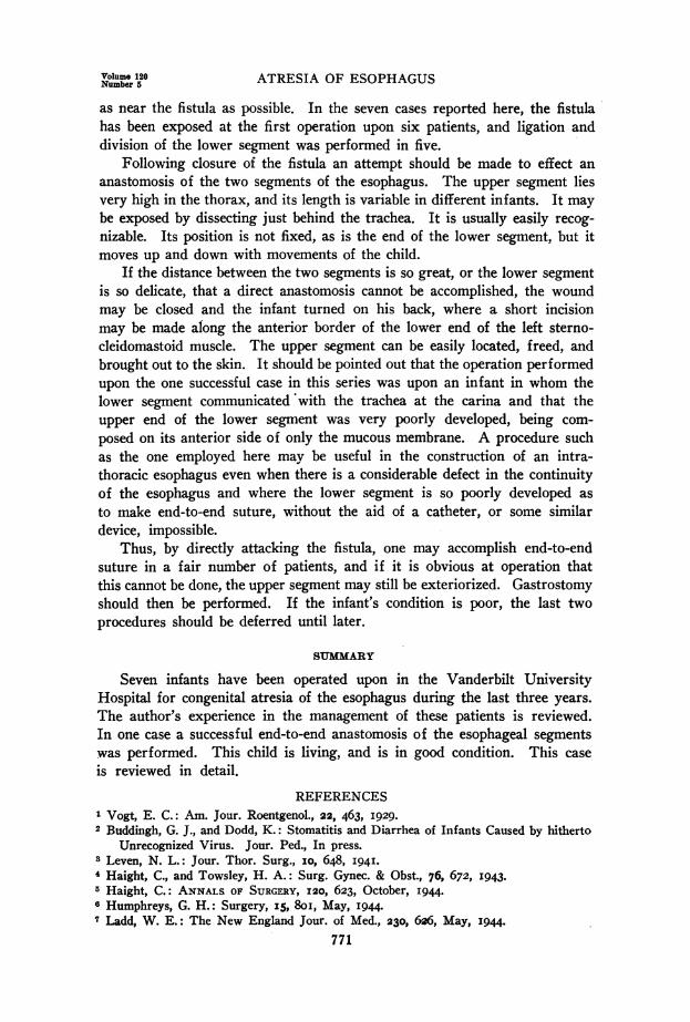
as near the fistula as possible. In the seven cases reported here, the fistulahas been exposed at the first operation upon six patients, and ligation anddivision of the lower segment was performed in five.
Following closure of the fistula an attempt should be made to effect ananastomosis of the two segments of the esophagus. The upper segment liesvery high in the thorax, and its length is variable in different infants. It maybe exposed by dissecting just behind the trachea. It is usually easily recog-nizable. Its position is not fixed, as is the end of the lower segment, but itmoves up and down with movements of the child.
If the distance between the two segments is so great, or the lower segmentis so delicate, that a direct anastomosis cannot be accomplished, the woundmay be closed and the infant turned on his back, where a short incisionmay be made along the anterior border of the lower end of the left sterno-cleidomastoid muscle. The upper segment can be easily located, freed, andbrought out to the skin. It should be pointed out that the operation performedupon the one successful case in this series was upon an infant in whom thelower segment communicated with the trachea at the carina and that theupper end of the lower segment was very poorly developed, being com-posed on its anterior side of only the mucous membrane. A procedure suchas the one employed here may be useful in the construction of an intra-thoracic esophagus even when there is a considerable defect in the continuityof the esophagus and where the lower segment is so poorly developed asto make end-to-end suture, without the aid of a catheter, or some similardevice, impossible.
Thus, by directly attacking the fistula, one may accomplish end-to-endsuture in a fair number of patients, and if it is obvious at operation thatthis cannot be done, the upper segment may still be exteriorized. Gastrostomyshould then be performed. If the infant's condition is poor, the last twoprocedures should be deferred until later.
SUMMARY
Seven infants have been operated upon in the Vanderbilt UniversityHospital for congenital atresia of the esophagus during the last three years.The author's experience in the management of these patients is reviewed.In one case a successful end-to-end anastomosis of the esophageal segmentswas performed. This child is living, and is in good condition. This caseis reviewed in detail.
REFERENCESI Vogt, E. C.: Am. Jour. Roentgenol., 22, 463, I929.2 Buddingh, G. J., and Dodd, K.: Stomatitis and Diarrhea of Infants Caused by hitherto
Unrecognized Virus. Jour. Ped., In press.s Leven, N. L.: Jour. Thor. Surg., IO, 648, I94I.4 Haight, C., and Towsley, H. A.: Surg. Gynec. & Obst., 76, 672, I943.5 Haight, C.: ANNALS. OF SURGERY, I20, 623, October, I944.Humphreys, G. H.: Surgery, I5, 8oi, May, 1944.
7 Ladd, W. E.: The New England Jour. of Med., 230, 6X6, May, 1944.771