Confocal(Microscopy(( &((...
59
Confocal Microscopy & Superresolu3on Colin Sheppard Nano7Physics Department Italian Ins3tute of Technology (IIT) Genoa, Italy [email protected]
Transcript of Confocal(Microscopy(( &((...
Confocal(Microscopy((&((
Superresolu3on(Colin(Sheppard(
Nano7Physics(Department(Italian(Ins3tute(of(Technology((IIT)(
Genoa,(Italy([email protected](

Imaging(using(a(detector(array(Can generate an image with a lens and a detector array
detector array
wide-field detection

Another(way(of(genera3ng(an(image:(using(a(scanning(system(
• Detector does not image, only collects light. • Magnification of image is ratio of size of image to amplitude of scan. • Independent of probe diameter.
single element detector

Imaging(with(a(focused(probe(

Equivalence(of(scanning(and(conven3onal(microscopes(
• Based on Principle of Reciprocity!• Holds even with loss or multiple scattering! (but not inelastic scattering)!• First shown for electron microscopes!
Pogany & Turner, Acta Cryst. A24 103 (1968)!Cowley, App. Phys. Lett. 15 58 (1969)!Zeitler & Thomson, Optik 31 258 (1970)!Welford, J. Microscopy 96 105 (1972)!Barnett, Optik 38 585 (1973)!Engel, Optik 41 117 (1974)!Kermisch, J. Opt. Soc. Am. 67 1357 (1977)!Sheppard, Optik 78, 39-43 (1986); J. Opt. Soc. Am. A 3, 755-756 (1986)!

Scanning(vs.(conven3onal(microscope(
Scanning Equivalent
Conventional
Confocal
Conventional with image scanning
Scanning
or CCD detector

Confocal(imaging:(schema3c(diagram(

Hamilton(DK,(Wilson(T,(Sheppard(CJR((
Experimental((observa3ons(of(the(depth7discrimina3on(proper3es(of(scanning((
microscopes(
Opt.%Le(s.6,(6257626((1981)(
Op3cal(sec3oning(

Confocal(microscopy(•(Advantages(( ( (Op3cal(sec3oning(
– 3D(imaging(
– Surface(profiling"
( ( (Reduced(sca]ered(light(– Imaging(through(sca]ering(media,(e.g.(3ssue(
( ( (Improved(resolu3on(
•(Reflec3on(( ( ( (–(Industrial(applica3ons,(surface(profiling(( ( ( (–(Sca]ering(media,(3ssue(
•(Fluorescence((–(Autofluorescence(or(labelled((–(Fixed(or(living(

Autofocus(and(surface(profile(

Autofocus(and(surface(profile(
Isometric view

Coherent(Imaging(

Confocal(Imaging((not(fluorescence)(
I (xd , yd ) = h1(x, y)t(x − xs , y − ys )h2 (xd − x, yd − y)dxdy∫∫2
xd, yd!
after sample xs, ys are scan coordinates
• Pinhole: xd, yd = 0:! I = h1(x, y)h2 (−x,−y)( )⊗ t(x, y)2
• h2 even:!
• Coherent microscope, with heff = h1h2
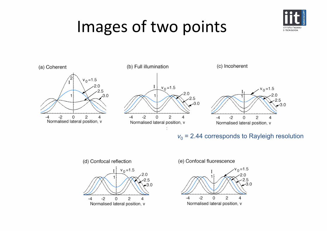
Images(of(two(points(
v0 = 2.44 corresponds to Rayleigh resolution

Marvin(Minsky(1957(

Goldman,(1940(
Spaltlampenphotographie und –photometrie, Ophthalmologica 98, 257-270 (1940).
lens
cornea
slit
film
Slit-scanning confocal with angular gating object

Z(Koana(1942(

Petrán(1968(˘
Egger & Petrán, Science 157, 306 (1967) ˘
Many parallel confocal microscopes
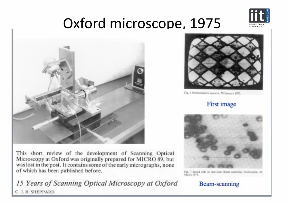
Oxford(microscope,(1975(
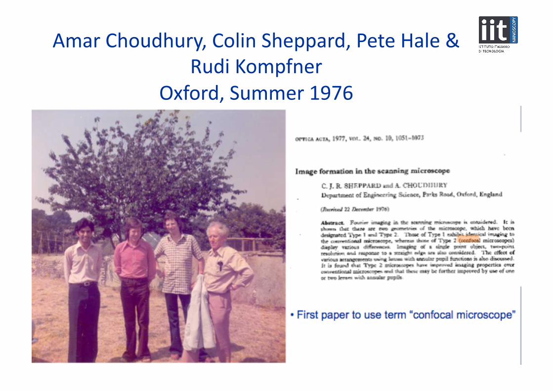
Amar(Choudhury,(Colin(Sheppard,(Pete(Hale(&(Rudi(Kompfner(
Oxford,(Summer(1976(
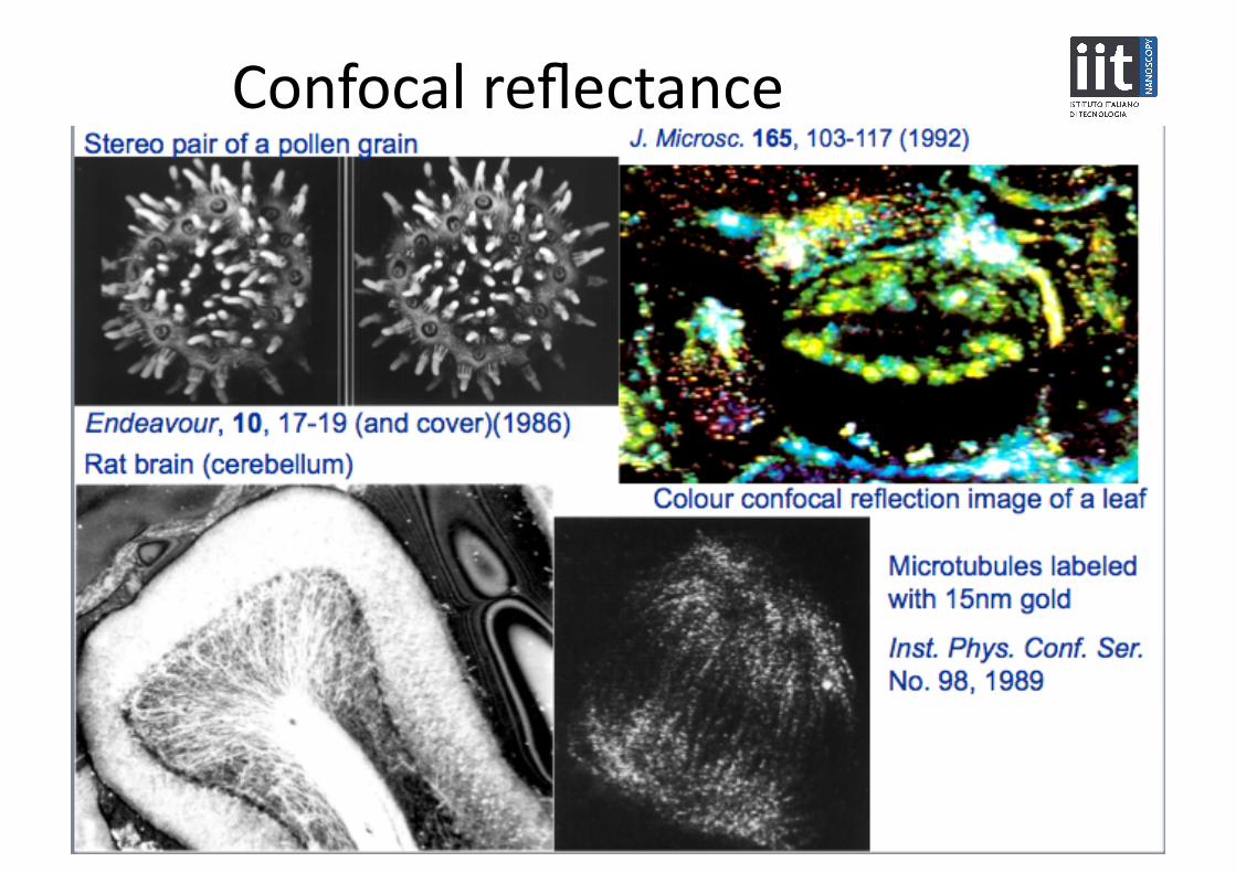
Confocal(reflectance(

Cox(IJ,(Sheppard(CJR((1983)(Digital(image(processing(of(confocal(images,(Image%&%Vision%Compu5ng(1,(52756((1983)(
conventional! confocal!
confocal!autofocus!
surface!profile!
Confocal(microscope(with(computer(

Commercializa3on(of(confocal(microscope(

Confocal(imaging(through(sca]ering(medium(((confocal(ga3ng)(
M Gu, T Tannous, CJR Sheppard

Limita3ons(of(confocal(microscopy(• (Speed(
– Illuminate(only(one(spot(at(a(3me(– In(fluorescence,(speed(limited(by(satura3on(of(fluorophore(– Solu3on:(illuminate(by(more(than(one(spot(
• Spinning(disk(• Line(illumina3on(• Structured(illumina3on((fringe(projec3on)(
• (Size(– Endoscopic(microscopy(
• Cost(• (Resolu3on(
– 4Pi(microscopy(– STED(– Localiza3on(microscopy((PALM/STORM)(– Structured(illumina3on/Image(scanning(microscopy(
• (Penetra3on(– Coherence(ga3ng(– Two/three(photon(– Focal(modula3on(microscopy((FMM)(

37D(imaging(methods(
• Confocal • Digital deconvolution • Coherence probe/ optical coherence tomography (OCT) • Multiphoton microscopy:
2-photon fluorescence, SHG • Structured illumination

Lukosz,(1963((
W Lukosz, M Marchand Optica Acta 10, 241-255 (1963)
Structured illumination (or fringe projection)
Optical reconstruction using a second grating

Op3cal(sec3oning(in(line(illumina3on(or(aperture(
array(microscopes(
• Confocal, decays as 1/z2
• Line illumination, decays as 1/z • Aperture array, tends to a constant (cross-talk)

Strength(of(background(
slope – 2
slope – 3 slope – 5/2
1 d
width of divider
Using D-shaped pupils for illumination and detection, sectioning is improved

Two7photon(microscopy(
• Signal(propor3onal(to(square(of(illumina3on(intensity(– Op3cal(sec3oning(with(no(pinhole(– Signal(increased(using(pulsed(laser(

Mul3photon(microscopy(

SHG(image((in(blue)(
(of(collagen(in(mouse(dermis(
Cox G, Xu P, Sheppard CJR, Ramshaw J (2003)
Characterization of the Second Harmonic Signal from Collagen, Proc. SPIE 4963, 32-40

Harmonic(microscopy(of(my(arm(

OTF(for(confocal(fluorescence(Cut-off doubled but response is very weak
Even weaker (or negative) for finite-sized pinhole
Suggests possibility to use pupil filters to increase magnitude of OTF!

Superresolu3on(• Classical theory
Transfer function is band-limited
• Toraldo di Francia (1952): Resolution is not a fundamental limit
• Methods of Lukosz, Lohmann (~1960) Capacity for information transfer is invariant, not bandwidth Increase bandwidth using different polarizations, wavelengths etc.
• Cox and Sheppard (1985) Information capacity, but include noise (Shannon)
�
C = 1+ Bx Lx( )∏ 1+ By Ly( ) 1+ Bz Lz( ) 1+ Bt Lt( ) log2(1+ SNR)
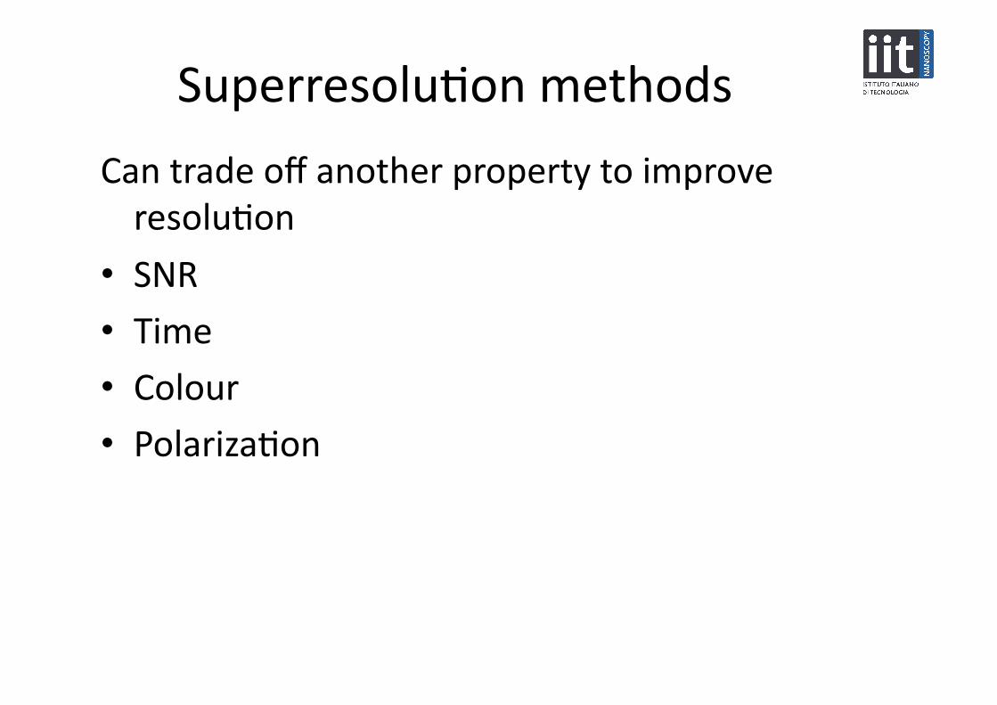
Superresolu3on(methods(
Can(trade(off(another(property(to(improve(resolu3on(
• SNR(• Time(
• Colour(• Polariza3on(

Dis3nguish(between(different(classes(of(‘superresolu3on’(• Class(3:(Improve(spa3al(frequency(response,(but(cut7off(unchanged(
– 27point(resolu3on(improved(– Some3mes(called(ultra7resolu3on,(or(hyper7resolu3on(
• image(filtering(• simple(digital(deconvolu3on((Wiener(filtering,(nearest(neighbour)(
• superresolving(filters((masks),(superoscilla3ons(
• Class(2:(Cut7off(increased,(but(the(effec3ve(NA(is(s3ll(<(n%• polariza3on,(etc.(• synthe3c(aperture(
• Class(1b:(Cut7off(increased,(and(the(effec3ve(NA(>(n%• structured(illumina3on(• confocal(• source/detector(arrays((ISM)(• solid(immersion(lens((SIL)(• nonlinear(imaging(
• Class(1a:(Cut7off(increased,(and(the(effec3ve(NA(is(unlimited(• STED(• saturated(SIM(• localiza3on(microscopy((PALM/STORM)((• near7field(microscope((SNOM,(photon(tunneling(microscope)(• deconvolu3on(with(constraints(

Comparison(of(different(imaging(methods(
1999 OTF PSF

Comparison(of(4Pi(and(I5M(
Hell

3D(Spa3al(Frequency(cut7offs(
Abbe (incoherent)
Coherent
Confocal fluorescence or Structured illumination
Maximum 4/λ (4n/λ in medium, e.g 6/λ )
Maximum possible with propagating waves, sphere radius 4n/λ
no missing cone

Focal(modula3on(microscopy(
Reference signal
Image signal • Detect beat frequency • Only get a signal from the focal region, where the 2 beams cross
f1
f2

Chondrocytes(from(chicken(car3lage(

Image(of(a(point(object(
The intensity image of a point object with a point detector, representing the intensity point spread function IPSF.
(a) confocal (b) D-shaped
(c) FMM

Integrated(intensity((background)(
The variations of the integrated intensity of FMM, compared with confocal microscope with circular apertures and with D-shaped apertures, for a point detector.
Decays as 1/z3

Source/Detector(arrays(• (Tandem(scanning,(Petrán((1968)(
• (Singular(value(decomposi3on((Bertero(&(Pike,(1982)(
• (‘Type(3’:(Maximum(signal(in(detector(plane((Reinholz,(1987)(
• "Pixel"reassignment"(Sheppard,"1988)(• (Subtrac3ve(imaging((Cogswell(&(Sheppard(1990,(and(others)("
• (Source/detector(arrays((Benedep(1996)(
• (Programmable(array(microscope((PAM)((Hanley,(1998)(
• (Structured(illumina3on((
( ((Lukosz,(1963;(Gustafsson,(2000)(

Offset(pinhole(
• Point spread function gets narrower • Intensity decreases • But increased side lobes • And effective psf shifts sideways
PSF:

Gives(the(image(of(a(shired(object(point(

Offset(pinhole(&(reassignment(
offset pinhole after reassignment
• Integrate without reassignment: same as conventional • Integrate with reassignment (to centre of illumination and detection):
PSF sharpened and signal improved
conventional given by envelope

Pixel(reassignment(
Optical transfer function
function of 2xs
product of rescaled OTFs (not convolution of OTFs as for confocal)

Image(scanning(microscopy(

Integra3on(over(finite(region(
4(1−16 / 3π 2 ) = 1.84
Maximum of point spread function for large vdmax is
(4 elements gives ~1.4)
• Maximum is >1 • Super-concentration • Beats classical limit of étendue
(0.72 AU)
(1 AU)
Ipeak = 1 for conventional
peak intensity goes above 1!
(magic number)
(1st zero of Airy disc)
Resolution and signal strength improve as vdmax increases
half-width = 1 for conventional 0.72 0.65

Unnormalized(OTF(

Zeiss(Airyscan(

Nonlinear(imaging(• (Incoherent(versus(coherent(imaging((square(law)(
• (Saturable(absorber(sharpens(point(spread(func3on((((Choudhury,(1977)(
• (Nonlinearity(of(detec3on(in(op3cal(storage((Braat,(1980s)(• (Nonlinear(effects(in(lithography(• (Mul3photon(imaging((SHG,(27photon(fluorescence)(
• (S3mulated(emission(deple3on(microscopy((STED)((Hell)(
• (Satura3on(in(structured(illumina3on((Heinzmann,(Gustafsson)(
• PALM,(STORM(
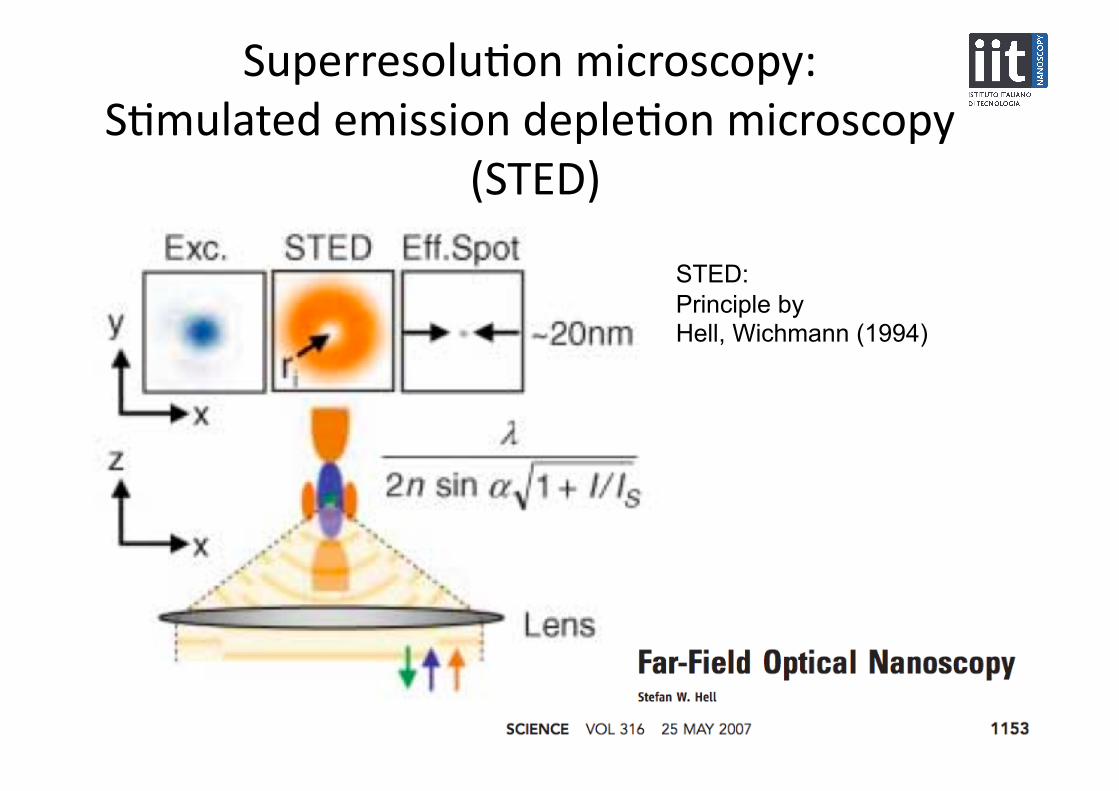
Superresolu3on(microscopy:((S3mulated(emission(deple3on(microscopy(
(STED)(
STED: Principle by Hell, Wichmann (1994)

Nonlinear(structured(illumina3on(microscopy(
• Heintzmann,(Jovin(&(Cremer((2002)(
• Gustafsson((2005)(
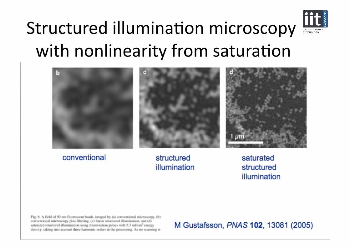
Structured(illumina3on(microscopy((with(nonlinearity(from(satura3on(

Localiza3on(microscopy(• Lidke(et%al.%Opt%Exp.%13,(7052((2005)(Blinking(of(quantum(dots(
• Betzig(et%al.%Science%313,(1642((2006)(PALM(
• Hess(et%al.%Biophys.%J.%91,(4258((2006)(FPALM(
• Rust(et%al.%Nature%Methods%3,(793((2006)(STORM(

Conclusions(• (Dis3nguish(between(true(superresolu3on(and(others(• (Some(methods(can(give(improvement(in(resolu3on(with(an(
unchanged(spa3al(frequency(cut7off(• Pupil(filters(
• (Some(methods(can(increase(spa3al(frequency(cut7off(by(factor(of(two(
• Confocal(• Structured(illumina3on(
• Source/detector(arrays(• (Some(methods(can(give(further(increased(bandwidth(
• Nonlinear(or(switching(• Near(field(