COMPLEXATION OF NOVEL THIOMERS AND INSULIN TO …
37
COMPLEXATION OF NOVEL THIOMERS AND INSULIN TO PROTECT AGAINST IN VITRO ENZYMATIC DEGRADATION – TOWARDS ORAL INSULIN DELIVERY C.O. Ibie, R.M. Knott, C.J. Thompson * School of Pharmacy and Life Sciences, Robert Gordon University, Garthdee Road, Aberdeen, AB10 7GJ, UK *[email protected]; +441224262561 ABSTRACT A significant barrier to oral insulin delivery is its enzymatic degradation in the gut. Nano-sized polymer-insulin polyelectrolyte complexes (PECS) have been developed to protect insulin against enzymatic degradation. Poly(allylamine) (Paa) was trimethylated to yield QPaa. Thiolation of Paa and QPaa was achieved by attaching either N-acetylcysteine (NAC), or thiobutylamidine (TBA) ligands (Paa-NAC/QPaa-NAC and Paa-TBA/QPaa-TBA thiomers). PEC formulations were prepared in Tris buffer (pH 7.4) at various polymer:insulin mass ratios (0.2:1-2:1). PECS were characterised by %transmittance of light and photon correlation spectroscopy. Insulin complexation efficiency and enzyme-protective effect of these complexes was determined by HPLC. Complexation with insulin was found to be optimal at mass ratios of 0.4-1:1 for all
Transcript of COMPLEXATION OF NOVEL THIOMERS AND INSULIN TO …

COMPLEXATION OF NOVEL THIOMERS AND INSULIN TO PROTECT AGAINST IN
VITRO ENZYMATIC DEGRADATION – TOWARDS ORAL INSULIN DELIVERY
C.O. Ibie, R.M. Knott, C.J. Thompson*
School of Pharmacy and Life Sciences,
Robert Gordon University,
Garthdee Road,
Aberdeen,
AB10 7GJ, UK
*[email protected]; +441224262561
ABSTRACT
A significant barrier to oral insulin delivery is its enzymatic degradation in the gut.
Nano-sized polymer-insulin polyelectrolyte complexes (PECS) have been developed to
protect insulin against enzymatic degradation. Poly(allylamine) (Paa) was
trimethylated to yield QPaa. Thiolation of Paa and QPaa was achieved by attaching
either N-acetylcysteine (NAC), or thiobutylamidine (TBA) ligands (Paa-NAC/QPaa-NAC
and Paa-TBA/QPaa-TBA thiomers). PEC formulations were prepared in Tris buffer (pH
7.4) at various polymer:insulin mass ratios (0.2:1-2:1). PECS were characterised by
%transmittance of light and photon correlation spectroscopy. Insulin complexation
efficiency and enzyme-protective effect of these complexes was determined by HPLC.
Complexation with insulin was found to be optimal at mass ratios of 0.4-1:1 for all
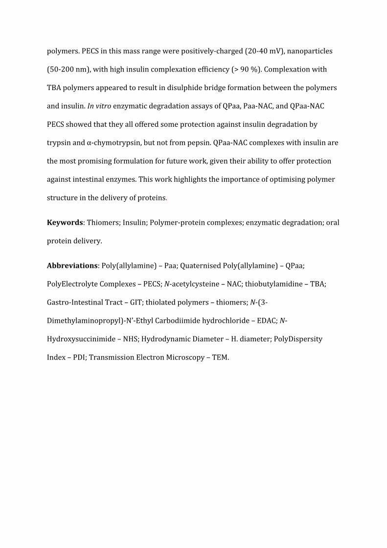
polymers. PECS in this mass range were positively-charged (20-40 mV), nanoparticles
(50-200 nm), with high insulin complexation efficiency (> 90 %). Complexation with
TBA polymers appeared to result in disulphide bridge formation between the polymers
and insulin. In vitro enzymatic degradation assays of QPaa, Paa-NAC, and QPaa-NAC
PECS showed that they all offered some protection against insulin degradation by
trypsin and α-chymotrypsin, but not from pepsin. QPaa-NAC complexes with insulin are
the most promising formulation for future work, given their ability to offer protection
against intestinal enzymes. This work highlights the importance of optimising polymer
structure in the delivery of proteins.
Keywords: Thiomers; Insulin; Polymer-protein complexes; enzymatic degradation; oral
protein delivery.
Abbreviations: Poly(allylamine) – Paa; Quaternised Poly(allylamine) – QPaa;
PolyElectrolyte Complexes – PECS; N-acetylcysteine – NAC; thiobutylamidine – TBA;
Gastro-Intestinal Tract – GIT; thiolated polymers – thiomers; N-(3-
Dimethylaminopropyl)-N’-Ethyl Carbodiimide hydrochloride – EDAC; N-
Hydroxysuccinimide – NHS; Hydrodynamic Diameter – H. diameter; PolyDispersity
Index – PDI; Transmission Electron Microscopy – TEM.
INTRODUCTION
Oral delivery of insulin used for the management of Type 1 Diabetes is one of the major
long term goals of diabetes research. The limited treatment options for maintenance of
blood glucose levels (parenteral insulin), predisposes those with diabetes to
physiological stress and pain due to multiple daily injections.1 Also, parenteral
administration of insulin results in abnormal systemic distribution of insulin which has
been associated with the occurrence of peripheral hyperinsulinaemia and insulin
resistance causing hypoglycaemia, weight gain, neuropathy, retinopathy,
atherosclerosis and hypertension.2 The need to eliminate these drawbacks as well as
offer individuals affected by diabetes a better quality of life has prompted researchers
to explore other routes for the delivery of insulin.
The oral route offers an excellent alternative being the easiest and most convenient
route of drug administration and also demanding less time and effort from medical
personnel and carers.3 Also, on oral administration exogenous insulin distribution
mimics the natural physiological fate of insulin in the body thereby closely replicating
the direct delivery of endogenous insulin to the liver, where its effects of suppressing or
facilitating hepatic glucose production are fundamental in sustaining glucose
homeostasis.4 However, the bioavailability of orally administered insulin is markedly
low (estimated to be less than 1 %) due to proteolytic digestion in the gut and limited
absorption of insulin from the gut lumen due to its high hydrophilicity and low log P
value.5 Retaining the integrity and bioactivity of insulin during formulation and
manufacturing procedures presents an additional difficulty in achieving oral
administration. Disturbances in the primary, secondary or tertiary structure of insulin

(due to processing conditions) can lead to its deactivation and/or denaturation
resulting in loss of pharmacological activity.6
Current research in this area has been focused on the development of novel oral
formulations of insulin that can overcome these barriers and maximise the
bioavailability of oral insulin. Some approaches involve co-administration with known
absorption enhancers like fatty acids and protease inhibitors, while others involve the
use of smart polymers or carrier systems that can protect insulin from proteolytic
digestion while also mediating its absorption across the gastro-intestinal tract (GIT)
epithelium.7-9
Recent advances in the design of functional polymeric drug delivery systems have led to
the utilisation of polyelectrolyte complexes (PECS) for protein delivery. PECS are
formed spontaneously by electrostatic interaction between oppositely charged
polyelectrolytes (i.e. a polymer and protein) in aqueous solution.10,11 Indeed, PECS
containing insulin and other proteins have already been the subject of research by a
number of groups.11-16 These complexes are typically positively charged nanoparticles,
with hydrodynamic sizes between 100-400 nm in aqueous media. Polyelectrolyte
complexation has the advantage of being a more benign process than other methods of
manufacturing nanoparticles, which usually require the use of aggressive processing
conditions and organic solvents. Formulation of proteins as PECS can also impart
improved characteristics, i.e. enhanced absorption and reduced degradation, via
modification to the polymeric carrier, rather than to the protein itself; thereby,
preserving the original structure of the protein.15 Hence, rational optimisation of
polymer structure to provide a robust network to facilitate optimum complexation,
enzymatic protection and GI absorption of proteins is required.

Polyallylamine (Paa) was previously modified by both hydrophobic (palmitoyl, cetyl
and cholesteryl ligands) and hydrophilic (quaternary ammonium salt) moieties for use
in oral insulin delivery by our group.17 Quaternised Paa (QPaa) complexation with
insulin resulting in spherical nano-sized PECS which exhibited good insulin loading
efficiency, protected insulin from tryptic degradation and promoted uptake of insulin by
Caco-2 cells.16,17 These modified Paa polymers were, however, limited in their
protective effect against α-chymotrypsin and pepsin-mediated degradation of insulin.17
Subsequent work has, therefore, been based on developing a new set of polymers which
overcome this limitation: thiolated version of Paa (thiomers).16,18
Thiomers have also been found to be able to maximise the amount of oral insulin
available for absorption by chelating the metal ions of endogenous proteases thereby
curtailing their activity.12,19-21 Thiolated polymers have also been observed to be strong
mucoadhesives as they contain reactive thiol groups capable of forming disulphide
bonds with cysteine-rich domains of mucus glycoproteins.14,22 Mucoadhesive dosage
forms also promote oral absorption by adhering tightly to the intestinal mucosa,
prolonging the residence time of the active molecule at its absorption site and creating a
local concentration gradient that maximises uptake of the drug through the intestinal
mucosa.19
The aim of this study was to develop and evaluate a set of thiolated Paa polymers which
were used to form PECS with insulin. This work reports on evaluation of the impact of
Paa quaternisation and thiolation on insulin complexation efficiency, particle size and
morphology, enzyme-protective capacity and mucoadhesive characteristics of the
resultant polymer-insulin PEC. For the first time, we have demonstrated the significance

of the overall polymer structure and the type of thiolation of Paa plays an important
role in protecting insulin from enzymatic degradation.
MATERIALS AND METHODS
Materials
Poly(allylamine hydrochloride) (average Mw = 15 kDa) was converted to its free base,
as previously described.13 Tris(hydroxymethyl)aminomethane (Tris base) (≥ 99%),
iodomethane, N-(3-Dimethylaminopropyl)-N’-ethyl carbodiimide hydrochloride
(EDAC), N-hydroxysuccinimide (NHS), N-acetylcysteine (NAC), 2-iminothiolane
hydrochloride, sodium borohydride, insulin (27 units per mg/Umg-1, from bovine
pancreas), tris(hydroxymethyl)aminomethane (Tris base) (≥ 99%), pepsin (3640 Umg-1,
from porcine gastric mucosa), α-chymotrypsin (TLCK treated, Type VII from bovine
pancreas, 40 Umg-1), trypsin (TPCK treated, from bovine pancreas, 11,004 Umg-1) and
porcine gastric mucin (crude type II) were all purchased from Sigma-Aldrich UK.
Mercodia Bovine insulin ELISA kit and sample buffer were obtained from Diagenics Ltd.
UK.
Trifluoroacetic acid (TFA) and Acetonitrile (HPLC grade) were purchased from Fischer
Scientific, UK. Other chemicals used were of analytical grade.
Polymer synthesis
The method used for the quaternisation and purification of Paa (QPaa) was adapted
from the previous work of Thompson et al. (n=3).13

The methods used for the thiolation of Paa/QPaa were adapted from that used by other
groups14,19, but have also been reported and described in detail in a previous
publication by our group.18 Briefly, thiolation of Paa/QPaa using N-acetylcysteine (n=3)
was carried out by dissolving NAC (250 mg, 1.53 mmol) in 100 ml deionised water
containing EDAC/NHS (250 mmol) at pH 4-5 and stirring for 1 hr under nitrogen before
adding the Paa (250 mg) and leaving the reaction to proceed for 5 hrs at room
temperature. These polymers are designated as Paa-NAC and QPaa-NAC. Thiolation
with 2-iminothiolane hydrochloride (n=3) involved reacting the polymer (500 mg) with
2-iminothiolane hydrochloride (400 mg) in 50 ml deionised water and the reaction
mixture left stirring under nitrogen for 14 hrs in the dark. These polymers are
designated as Paa-TBA and QPaa-TBA.
All thiolated polymers were subsequently recovered by dialysis against dilute (0.1 M)
hydrochloric acid at 4°C and subsequently freeze-dried.
The degree of polymer quaternisation was obtained by elemental analysis, while the
level of polymer thiolation was determined by iodometric titration of free thiol groups
(estimation of total amount of thiol groups added onto the polymer backbone was done
after disulphide bond reduction with sodium borohydride) as previously reported.18
Preparation of polymer:insulin complexes
Formulations of polymer:insulin complexes were prepared in glass vials at room
temperature by mixing equal volumes (2 mL) of polymer (polymers used include Paa,
Paa-TBA, Paa-NAC, QPaa, QPaa-TBA and QPaa-NAC) with insulin, both previously
dissolved in pH 7.4 Tris buffer, as per previous work.23 For optimal formulations of
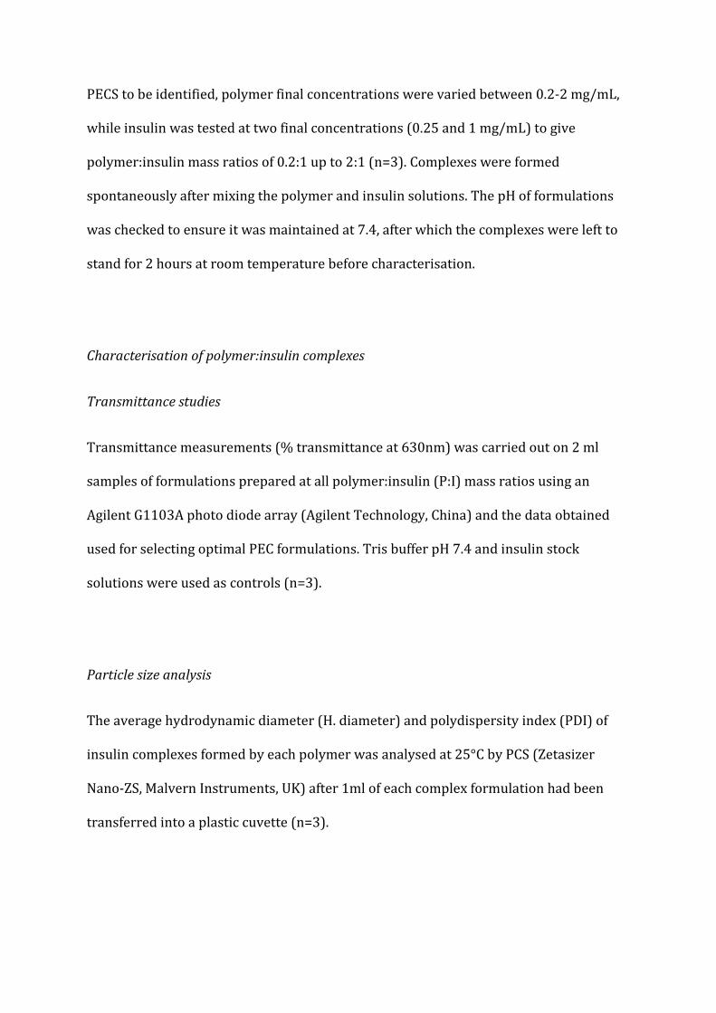
PECS to be identified, polymer final concentrations were varied between 0.2-2 mg/mL,
while insulin was tested at two final concentrations (0.25 and 1 mg/mL) to give
polymer:insulin mass ratios of 0.2:1 up to 2:1 (n=3). Complexes were formed
spontaneously after mixing the polymer and insulin solutions. The pH of formulations
was checked to ensure it was maintained at 7.4, after which the complexes were left to
stand for 2 hours at room temperature before characterisation.
Characterisation of polymer:insulin complexes
Transmittance studies
Transmittance measurements (% transmittance at 630nm) was carried out on 2 ml
samples of formulations prepared at all polymer:insulin (P:I) mass ratios using an
Agilent G1103A photo diode array (Agilent Technology, China) and the data obtained
used for selecting optimal PEC formulations. Tris buffer pH 7.4 and insulin stock
solutions were used as controls (n=3).
Particle size analysis
The average hydrodynamic diameter (H. diameter) and polydispersity index (PDI) of
insulin complexes formed by each polymer was analysed at 25°C by PCS (Zetasizer
Nano-ZS, Malvern Instruments, UK) after 1ml of each complex formulation had been
transferred into a plastic cuvette (n=3).
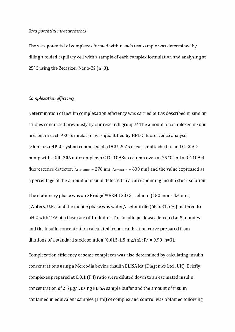
Zeta potential measurements
The zeta potential of complexes formed within each test sample was determined by
filling a folded capillary cell with a sample of each complex formulation and analysing at
25°C using the Zetasizer Nano-ZS (n=3).
Complexation efficiency
Determination of insulin complexation efficiency was carried out as described in similar
studies conducted previously by our research group.23 The amount of complexed insulin
present in each PEC formulation was quantified by HPLC-fluorescence analysis
(Shimadzu HPLC system composed of a DGU-20As degasser attached to an LC-20AD
pump with a SIL-20A autosampler, a CTO-10ASvp column oven at 25 ºC and a RF-10Axl
fluorescence detector: λexcitation = 276 nm; λemission = 600 nm) and the value expressed as
a percentage of the amount of insulin detected in a corresponding insulin stock solution.
The stationery phase was an XBridgeTm BEH 130 C18 column (150 mm x 4.6 mm)
(Waters, U.K.) and the mobile phase was water/acetonitrile (68.5:31.5 %) buffered to
pH 2 with TFA at a flow rate of 1 mlmin-1. The insulin peak was detected at 5 minutes
and the insulin concentration calculated from a calibration curve prepared from
dilutions of a standard stock solution (0.015-1.5 mg/mL; R2 = 0.99; n=3).
Complexation efficiency of some complexes was also determined by calculating insulin
concentrations using a Mercodia bovine insulin ELISA kit (Diagenics Ltd., UK). Briefly,
complexes prepared at 0.8:1 (P:I) ratio were diluted down to an estimated insulin
concentration of 2.5 µg/L using ELISA sample buffer and the amount of insulin
contained in equivalent samples (1 ml) of complex and control was obtained following

the method outlined by the manufacturer (Diagenics Ltd. UK). The insulin content of
both control and complex samples (n=3) was determined using the values obtained
from a calibration plot (n=3; R2 = 0.98) prepared from insulin standards provided in the
kit (0.05-3 µg/L).
Transmission Electron Microscopy (TEM)
The morphology of polymer, insulin complexes was visualised using a LEO 912 energy
filtering transmission electron microscope at 100/120kV. Formvar/Carbon-coated 200
mesh copper grids were glow discharged and complex solutions dried down to a thin
layer onto a hydrophilic support film. Aqueous methylamine vanadate (1 %) (Nanovan;
Nanoprobes, Stony Brook, NY, USA) was applied and the set-up air dried before imaging
.23
In vitro enzymatic degradation studies
Trypsin (6.4 mg/mL, 2.7 x 10⁻⁴ M), test complex suspensions and insulin-alone control
(0.25 mg/mL) (each 4.5 ml) solutions were prepared separately in pH 8 Tris buffer and
incubated in a water bath at 37°C for 2 hours. Trypsin (0.05 ml) was then added into
each complex solution (n=3). Aliquots of the mixture (0.2 ml) were withdrawn every 30
minutes and mixed with ice cold TFA solution (0.015 ml, 0.1 % v/v) to stop enzymatic
activity. The experiment was conducted for 4 hours at 37°C with samples being
analysed by HPLC, as described earlier. The same process was repeated with α-
chymotrypsin (5 mg/mL, 2.0 x 10⁻⁴ M) in pH 8 Tris buffer (n=3).

Peptic degradation studies was carried out by dissolving pepsin (0.1 mg/mL, 2.8 x10⁻⁶
M) in 0.01 M HCl after which complex, insulin-alone control (0.25 mg/mL) and enzyme
solutions were then buffered to pH 2 with a drop of 5 M HCl and incubated in a water
bath at 37°C for 2 hrs (n=3). Pepsin (0.016 ml) was added into the complex solutions
and samples (0.15 ml) were drawn from each mixture every 30 minutes and put into ice
cold Tris base (0.15 ml; 0.1 M) to halt enzyme activity.
Results obtained for PECS in all enzymatic degradation studies were expressed as % of
non-degraded insulin present in each sample over time, in order to compare with the
insulin-alone controls as per a previous study.17
In vitro evaluation of mucoadhesive capacity of complexes
Assessment of the mucoadhesive capacity of each polymer, insulin PEC formulation was
determined by measurement of the amount of mucin adsorbed by the complexes using
an established mucin adsorption assay method.18 Experiments were carried out by
mixing 1 ml of mucin solution (Tris buffer pH 7.4; 1 mgml-1) with a 0.25 ml sample of
each PEC formulation. Controls were prepared by mixing 0.25 ml of Tris buffer pH 7.4
with 1 ml (1 mgml-1) mucin solution. An additional control sample containing mucin
and 0.25 ml of insulin in Tris buffer pH 7.4 (0.25 mgml-1) was also prepared. These
mixtures were subsequently incubated at 37°C on a shaking water bath for 5 hrs.
All control and test samples were subsequently transferred into separate Eppendorf
tubes and centrifuged at 10,000 rpm for 30 minutes, and the concentration of mucin in
each supernatant measured by UV spectrometry at 251 nm. Percentage (%) of total
mucin adsorbed to complexes was obtained as shown below18:
Percentage (%) of total mucin adsorbed to each sample of polymer was calculated as
shown below:
% mucin adsorption (Mad) = [Mo – Ms] / Mo × 100
Where,
MO = concentration of free mucin in control supernatant
MS = concentration of free mucin in the sample supernatant

RESULTS AND DISCUSSION
Polymer synthesis
Quaternisation of Paa was confirmed by 1H-NMR and average degree of Paa
quaternisation determined by elemental analysis and found to be 72 ± 2 mol%, as per
previous publications.13,16,18 Subsequent thiolation of both Paa and QPaa yielded two
types of thiolated derivatives-the thiobutylamidine (TBA) conjugates (Paa-TBA and
QPaa-TBA) and N-acetylcysteine (NAC) conjugates (Paa-NAC and QPaa-NAC). Based on
results of iodometric titration the thiol content of each thiomer was obtained and is
displayed in table 1. Further characterisation of these polymers including IR analysis,
thermal analysis, mucoadhesive characteristics and biocompatibility have been
described in these publications.16,18
Characterisation of polymer:insulin complexes
Transmittance studies
For each polymer, turbidimetric analysis was used alongside particle size data to select
optimal polymer:insulin mass ratios and insulin stock concentration levels for the
formulation of stable PECS. Greater clarity (higher % transmittance) and lack of
precipitation was deemed to be indicative stable complexation.17,23 The optimisation
process was done for all polymers tested, but only the data for the parent polymers, Paa
and QPaa is shown as this is representative of their thiolated derivatives.
Results of the formulation optimisation process carried out at various P:I mixing ratios
showed that at low P:I mass ratios (specifically 0.2:1 at either insulin stock

concentration) PEC formulations had low transmittance values (Figure 1) and were
observed to be turbid suggesting the attractive forces between polymer and insulin
were weak at this P:I mixing ratio. After 4 hrs, these formulations were observed to be
unstable separating into a bottom layer composed of flocculated precipitates and a clear
supernatant layer. This was likely due to the excess of insulin in solution causing a
marked charge neutralisation of the polymer on complexation which in turn resulted in
these complexes precipitating.
Increasing the P:I mass ratio to 0.4:1 or 0.8:1 stabilised the formulations facilitating the
formation of clear PECS containing soluble, non-aggregated complexes which displayed
higher transmittance values and showed no visible signs of precipitation.
However, at higher P:I mass ratios of 2:1, PEC formulations were observed to be
translucent displaying a corresponding decrease in transmittance as can be observed in
Figure 1. This suggest that at the higher P:I mass ratios (2:1) where there is an excess of
the polymer, compression of the electrical double layer around suspended particles may
reduce the magnitude of the repulsive barrier between dispersed particles promoting
PEC aggregation and causing a corresponding decrease in transmittance values.
Raising the concentration level of the insulin stock solution used from 0.5 to 2 mg/mL
did not noticeably change the dynamics of the complexation process across the different
P:I mass ratios combinations used. The stability profile of complexes followed a similar
trend with that obtained at 0.5 mg/mL: PECS at the upper and lower margins of the
mass ratio combinations (0.2:1 and 2:1) having relatively lower transmittance values
than other formulations and PECS prepared at 0.2:1 containing visible aggregates. More
importantly, each formulation prepared using the higher insulin stock concentration (2
mg/mL) was relatively more turbid than the corresponding formulation prepared using

the lower insulin stock (0.5 mg/mL) resulting in a relative decrease in transmittance
values with increase in insulin stock concentration (Figure 1). Hence, although stable
formulations (0.4-1:1) prepared using this higher insulin stock concentration (2
mg/mL) were free of aggregation/precipitation the PEC suspension was translucent.
This translucent appearance of complexes could be a direct effect of the higher
concentration of complexes in the suspension and/or as a result of intercomplex
associations as interparticulate distances fall allowing attractive forces to predominate
promoting PEC aggregation.
These results indicate that in Tris buffer pH 7.4, P:I mass ratios between 0.4-1:1 were
optimal formulations for the preparation of polymer, insulin PECS. This is in agreement
with the results of similar work which reports the formation of unstable aggregates at
low P:I mass ratios presumed to be as a result of excess of the interacting protein.24
Particle size analysis
Results showed that mixing each polymer solution with insulin (in Tris buffer pH 7.4)
resulted in the spontaneous formation of nanoparticles (Figure 2). PEC formulations
prepared at low P:I ratio of 0.2:1, which were observed to be turbid and precipitated
after 4 hrs, displayed large particle sizes and high PDI values at 2 hrs.
The formation of small, compact nano-sized complexes of relatively narrow size
distribution at P:I mass ratios of 0.4:1 shows that interactive forces were sufficiently
strengthened by an increase in polymer concentration facilitating optimal P:I
complexation. Particle size data also showed that complexes were relatively larger at
2:1 P:I ratio, confirming that the fall in transmittance observed at this mass ratio could
be related to increase in PEC sizes as suspected. The excess concentrations of either
interacting polyelectrolyte results in increase in PEC sizes which may be attributed to
the screening effect of excess charged polyelectrolyte on the repulsive charges exhibited
by dispersed particles compressing the double layer and reducing the charge barrier to
the point at which aggregation sets in.25 This could also be a direct result of an increase
in particle count as polyelectrolyte concentration increases resulting in a reduction in
interparticulate distances (since suspension volume was kept constant) which causes
attractive forces to predominate leading to adjacent complexes associating to form
larger aggregates.
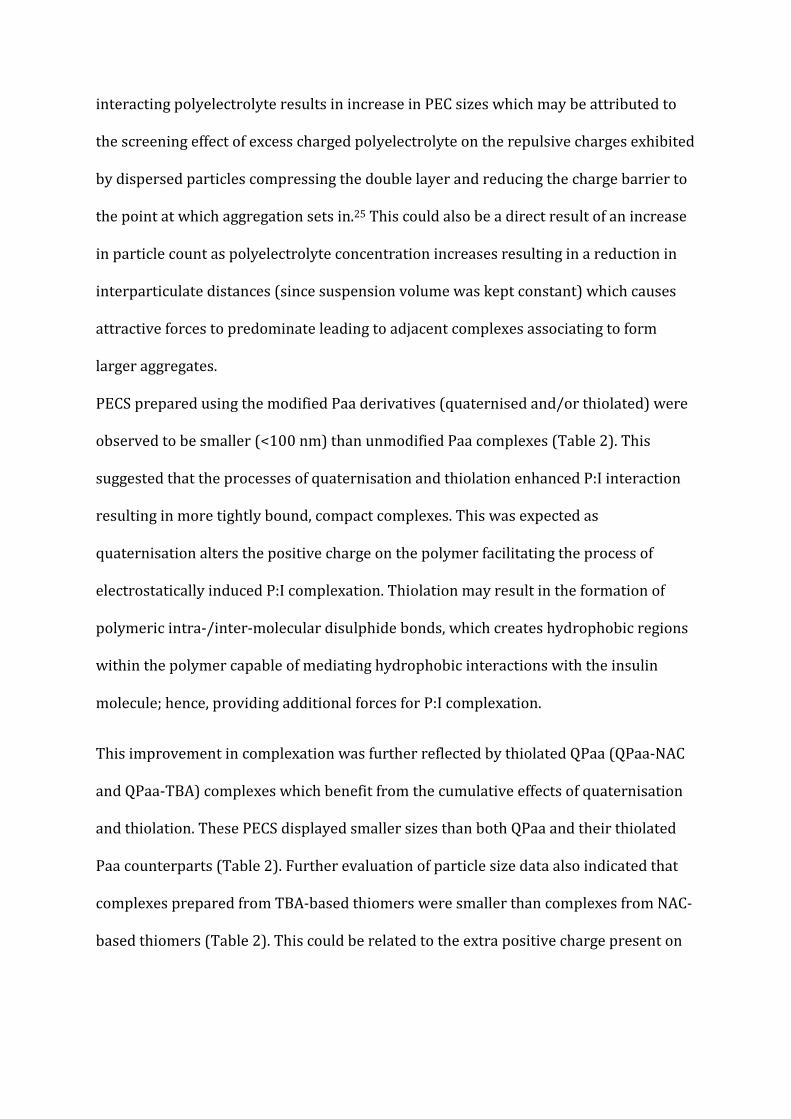
PECS prepared using the modified Paa derivatives (quaternised and/or thiolated) were
observed to be smaller (<100 nm) than unmodified Paa complexes (Table 2). This
suggested that the processes of quaternisation and thiolation enhanced P:I interaction
resulting in more tightly bound, compact complexes. This was expected as
quaternisation alters the positive charge on the polymer facilitating the process of
electrostatically induced P:I complexation. Thiolation may result in the formation of
polymeric intra-/inter-molecular disulphide bonds, which creates hydrophobic regions
within the polymer capable of mediating hydrophobic interactions with the insulin
molecule; hence, providing additional forces for P:I complexation.
This improvement in complexation was further reflected by thiolated QPaa (QPaa-NAC
and QPaa-TBA) complexes which benefit from the cumulative effects of quaternisation
and thiolation. These PECS displayed smaller sizes than both QPaa and their thiolated
Paa counterparts (Table 2). Further evaluation of particle size data also indicated that
complexes prepared from TBA-based thiomers were smaller than complexes from NAC-
based thiomers (Table 2). This could be related to the extra positive charge present on

the amidine bond of TBA-conjugates providing a site for additional electrostatic
interaction with insulin and hence condensing PECS even more.
The size of nanocomplexes for oral delivery is very important in optimising their
intestinal uptake as smaller-sized particles (< 300 nm) have been found to be favoured
in the processes like transcellular uptake by Peyer’s patches and paracellular uptake
through tight junctional spaces.26,27 Particle size has also been found to affect the
process of nanoparticle clearance by macrophages; as complexes with smaller sizes
(<150 nm) have been shown to exhibit a higher level of exocytosis.28
Based on results of the formulation optimisation process in 3.2.1 and 3.2.2., PECS
prepared at 0.8:1 P:I mass ratio, using the 0.5mg/mL insulin stock solution, were
identified as optimal formulation formulations for further testing. This was based on
their relatively small sizes and PDI values, as well as their relatively high
%transmittance. These values were, also, shown to be relatively stable over 3 days
incubation at room temperature (data not shown).
Zeta potential
Complexation of the polymers with insulin resulted in the production of positively
charged PECS at optimal P:I mass ratios (Table 2). The zeta potential of polymer, insulin
complexes was observed to range between approximately 28-38 mV. The positive
surface charge on complexes is beneficial in facilitating processes like particle
uptake/transport and mucoadhesion through electrostatic interaction with tight
junction proteins and the intestinal mucosa. Surface charge of nanoparticles also affects
their biodistribution.28 Nanoparticles with a slight negative charge have been shown to
accumulate better in tumour sites. While an elevation of positive surface charge was
found to be associated with an increased affinity for the negatively charged cell
membrane enhancing cellular uptake of cationic nanoparticles.28 The differences in the
zeta potential of separate polymer and insulin solutions from that of their complexes
indicate the role of electrostatic forces in the formation of these polymer-insulin
complexes (Table 2).18 NAC conjugates displayed lowest zeta potential values, Paa and
QPaa showed intermediate zeta potential values, while TBA conjugates had the highest
magnitude of surface charge. This may be due to the fact that there was a reduction in
surface charge of NAC-based thiomers due to substitution with the neutral amide
bond.18 However, complexes from TBA-based thiomers exhibited the highest zeta
potential probably due to the extra-cationic charge conferred by the permanently
protonated amidine bond.
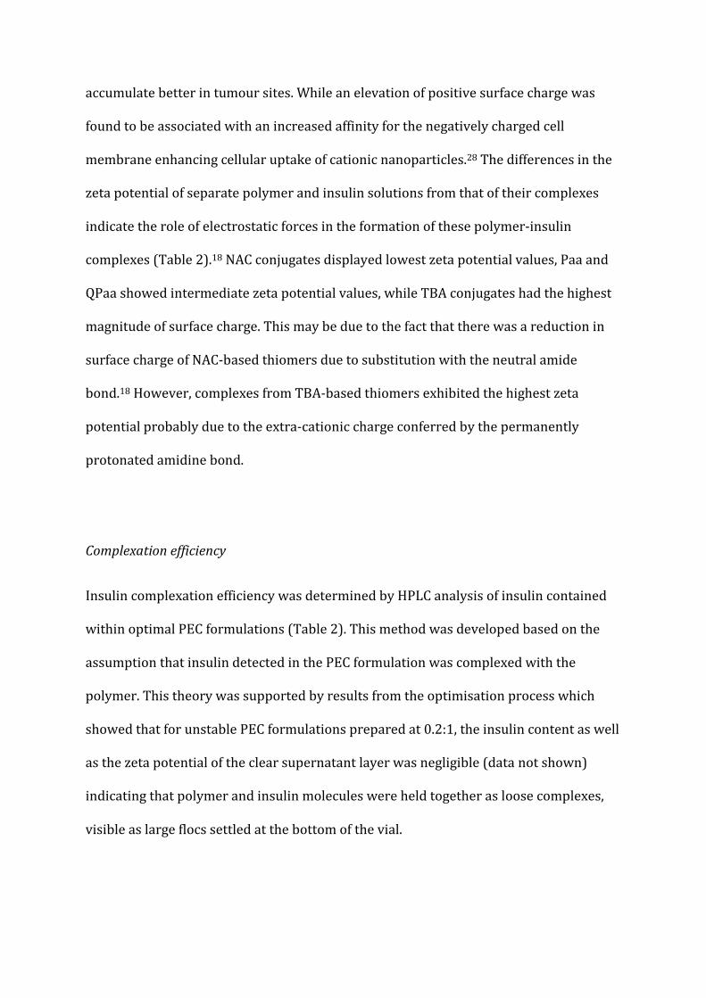
Complexation efficiency
Insulin complexation efficiency was determined by HPLC analysis of insulin contained
within optimal PEC formulations (Table 2). This method was developed based on the
assumption that insulin detected in the PEC formulation was complexed with the
polymer. This theory was supported by results from the optimisation process which
showed that for unstable PEC formulations prepared at 0.2:1, the insulin content as well
as the zeta potential of the clear supernatant layer was negligible (data not shown)
indicating that polymer and insulin molecules were held together as loose complexes,
visible as large flocs settled at the bottom of the vial.

Comparing HPLC analysis of insulin contained in all PEC formulations prepared at 0.8:1
P:I mass ratio using the 0.5mg/mL insulin stock and different Paa/QPaa thiolated
derivatives, insulin complexation efficiency was above 90% for all formulations as
shown in Table 2. However, for Paa-TBA/QPaa-TBA, insulin complexes, insulin
complexation efficiency results were obtained by an alternate ELISA method. This was
because the insulin peak detected by HPLC was absent in these PEC formulations
(Figure S1) leading to the assumption that the structure of insulin was altered on
complexation with the polymer thereby affecting the normal interaction of the protein
with the stationary phase of the column. The insulin content of these complexes was
observed to gradually decline from 100% after the complexes were made (t=0 hrs) to
approximately 10% after 2 hrs, proving that as insulin complexes with the polymer its
ability to be detected by HPLC is altered resulting in a fall in insulin concentration
recorded with time.
However, it was observed that acidification of Paa/QPaa-TBA insulin complexes using 2
M HCl resulted in a distinct insulin peak similar to that obtained on acidification of a
similar insulin control solution (Figure S2). This implies that the insulin complexed with
TBA-based thiomers could be recovered by acidification, hence insulin complexation
efficiency of these complexes was obtained by analysing this acidified peak and
comparing the results of the insulin complex with that of the acidified insulin control
(Figure S2). The fact that the effect of TBA conjugates on insulin may be reversed by
acidification suggests that this alteration in insulin characteristics may be caused by
covalent (thiol-disulphide) bonding between the thiol groups on the polymer and
insulin at pH 7.4, reversed by protonation of reactive thiolate ions on acidification.19 The
increased reactivity of the thiol groups on TBA conjugates compared to NAC conjugates

may be due to the cationic substructure of the amidine thiol-bearing moiety enhancing
the affinity of the polymer thiols for the cysteine groups on insulin. This sort of P:I
interaction could have deleterious effects on the conformation of the insulin chains and
may affect the ability of the protein to interact with its receptor.20 Techniques like
circular dichroism could be used in the future in investigating the nature of
conformational change in insulin complexed with TBA-conjugates.24
Confirmation of this reduction of insulin content of Paa-TBA insulin complexes was
carried out using an insulin ELISA kit. Results confirmed that after 2 hours, only 11.8 ±
4.2% of insulin was available as opposed to 97.8 ± 0.2% of insulin present in the control.
TEM
The morphology of different PECS prepared at 0.8:1 P:I mass ratio using the 0.5 mg/mL
insulin stock was elucidated using TEM (Figure 3). The TEM micrographs show that
quaternised polymers (QPaa, QPaa-NAC and QPaa-TBA) appeared to be capable of
forming nanoparticles having a single layer of polymer chains as their outer corona
(3B), while non-quaternised thiomers (Paa-NAC/Paa-TBA) appeared to form nano-
vesicles with a distinctive bilayered outer corona (3A). These nano-vesicle bilayers
appear to show darkened regions which suggests that these areas may have resisted
staining indicating they are likely more hydrophobic than other regions of the
complex.13
The formation of bilayer vesicles has been observed to be directly related to the
hydrophobic content of the polymer, with nano-vesicular self-assembly being initiated
by an attempt to minimise the high energy interaction between hydrophobic groups of

the polymer and the aqueous disperse phase while also maximising interfacial area by
sustaining low level interactions between hydrophilic groups and the disperse
phase.29,30 Water-soluble oppositely charged polyelectrolytes have also been found to
promote vesicle formation in non-vesicle forming water-soluble amphiphiles. This
phenomenon arises due to partial charge neutralization by the adsorption of one
polyelectrolyte onto the bilayer of the polymer of opposite charge, influencing the
balance of opposing forces of electrostatic repulsion and hydrophobic interaction in
favour of vesicle formation.31 Considering that modification of Paa by thiolation imparts
some level of hydrophobicity to the resultant constructs may suggest that interaction
with anionic insulin molecules can promote the formation of the vesicles observed in
the TEM micrographs through the aforementioned mechanism. TEM results further
emphasize the role of hydrophobic interactions arising from the intramolecular
disulphide bonds formed during the thiolation of the polymers in complexation with
insulin.
In vitro enzymatic degradation studies
The difficulties in analysing insulin concentration of TBA-conjugates, meant that
enzymatic degradation studies were only carried out on NAC-conjugate complexes. The
ability of QPaa, Paa-NAC and QPaa-NAC complexes (optimal formulations at 0.8:1, P:I,
mass ratio-0.5mg/mL insulin stock) to protect complexed insulin from degradation by
the serine proteases trypsin, α-chymotrypsin and pepsin was assessed. The results were
compared to that obtained for a similar control solution of free insulin and are
illustrated in Figure 4.

Results of exposure of polymer, insulin complexes to tryptic degradation showed that >
90% of insulin contained within the thiolated complexes was non-degraded after 4 hrs;
in comparison to less than 70% for the insulin control. The fact that 88% of undegraded
insulin was present in QPaa complexes after 4 hrs and 96% insulin available in a similar
formulation of QPaa-NAC after exposure to trypsin for the same time interval indicated
that thiolation further enhanced enzymatic protection. Paa-NAC and QPaa-NAC
complexes exhibited only slight variations in protection of insulin from the effects of
trypsin.
Thiomer, insulin complexes showed a different insulin degradation profile on exposure
to α-chymotrypsin. Thiolated QPaa (QPaa-NAC) insulin complexes showed increased
resistance to degradation by α-chymotrypsin than either QPaa or Paa-NAC complexes.
The amount of undegraded insulin available in QPaa-NAC complexes after 4 hrs was
observed to be 10% more than QPaa complexes, 15% more than Paa-NAC complexes,
and 30% more than was present in the insulin control. This may show a synergistic
effect was obtained from thiolation and quaternisation of the parent polymer, Paa, in
terms of protection of insulin from degradation by α-chymotrypsin. Bonds prone to
tryptic cleavage in insulin include the carboxyl terminus of B29-Lys and B22-Arg (in the
B-chain), these carboxyl groups are deprotonated and hence negatively charged at pH
7.4. It may, therefore, be that the electrostatic binding of positively charged polymer
molecules at this negative terminal shields susceptible bonds at this site from tryptic
attack.32 This sort of enzymatic protection/shielding of insulin by polymer-insulin
interactions have previously been reported by researchers working with modified
chitosan and Paa.33,34 Hence, this protective effect would be limited to sites on insulin
which are capable of coulombic interactions with compatible polymer chains. However,

two bonds prone to cleavage by α-chymotrypsin are enclosed within the hydrophobic
core of the insulin molecule31 and complexation with a less hydrophobic polymer like
QPaa may not allow for interaction of these regions with the polymer chains due to
differences in polarity. Hence, these areas may not be protected from the attacking
protease.
Conversely, polymer thiolation creates hydrophobic regions within the thiomer as seen
with the TEM photos displayed in Figure 3 due to intra-/intermolecular disulphide bond
formation. Hence, the additional protection of complexed insulin from proteolytic attack
by α-chymotrypsin after complexation with NAC-conjugates may be because these
thiomers are capable of protecting the bonds located within the hydrophobic core of
insulin. (Hydrophobic interactions between the thiomer and insulin occurring at this
region.) The ability of hydrophobic intractions to offer improved protection of insulin
associated within a PEC delivery system to degradation by serine proteases has been
previously reported.17,33
Consequently, QPaa-NAC which may incorporate charge-mediated and hydrophobic
interactions with insulin on complexation yields a cumulative protective effect being
able to shield more susceptible sites on the insulin molecule from proteolytic
degradation by α-chymotrypsin than either QPaa or Paa-NAC. This result is in
agreement with previous publications which showed that the ability of PECS formulated
from Paa-based polymers to protect complexed insulin from the effects of α-
chymotrypsin was enhanced when Paa was functionalised with both hydrophobic and
quaternary groups.17
No insulin PEC formulation protected insulin from degradation by pepsin, as insulin
present in complexes was observed to be completely degraded after 30 minutes of

exposure to pepsin. This is probably as a result of the numerous sites on the insulin
molecule susceptible to degradation by pepsin. Examples of sites susceptible to peptic
degradation include sites before leucine, phenylalanine, tyrosine, and tryptophan
(except if preceded by proline).35 Rapid peptic degradation of complexed insulin may
also be accelerated by acidification of the complexes: simulation of normal physiological
pH conditions for peptic activity was required, thereby making insulin positively
charged (below its pI of 5.5) and destabilizing electrostatic polymer-insulin interaction.
Amphiphillic Paa-insulin nanocomplexes which benefited from both hydrophobic and
electrostatic interactions were observed to show some protective effect from peptic
degradation.17 However, exposure of oral insulin formulations to the effect of pepsin can
be prevented by the use of enteric formulations which target the release of insulin to the
small intestine and distal parts of the GIT, hence curtailing insulin proteolysis by
enzymes operating within the gastric region of the gut.24
In vitro evaluation of mucoadhesive capacity of complexes
Assessment of mucin adsorption properties of polymer, insulin complexes prepared in
Tris buffer pH 7.4 at 0.8:1 P: I mass ratio using the 0.5 mgml-1 insulin stock solution
(Figure 5).
The mucin adsorption profile of insulin PECS showed that modified Paa-based
complexes showing better mucoadhesive properties than complexes prepared from the
unmodified Paa backbone. Thiolated Paa (Paa-NAC and Paa-TBA) PECS displayed the
greatest mucoadhesive properties, while thiolation of QPaa did not offer any substantial
improvement in the mucoadhesive profile of the quaternised backbone or its

complexes. High levels of quaternisation have been associated with decreased
mucoadhesivity due to reduced chain flexibility, interpenetration and steric hindrance
while lower levels of quaternisation reduce interactions with mucus, also causing
corresponding decreases in mucoadhesion.36,37 Optimisation of the level of
quaternisation has been found to be vital in facilitating paracellular transport and
mucoadhesion using quaternised chitosan.24,38 In summary, all complexes exhibited
good mucoadhesive properties with modified polymers performing better than the
unmodified Paa backbone.
CONCLUSION
The data collected from the above processes support the spontaneous formation of
spherical, nano-sized PECS with good insulin complexation efficiency on mixing optimal
mass ratios of Paa/QPaa and their thiolated derivatives with insulin in Tris buffer
solutions. Optimisation of the complexation process varying polyelectrolyte
stoichiometry showed that complexes prepared at 0.4-1:1 P:I mass ratio were
consistently stable for all polymers, with 0.8:1 showing the best overall stability profile.
Electrostatic–induced polymer, insulin complexation carried out using Paa-based
thiomers was found to result in the formation of nano-sized, positively-charged
complexes which were either in the form of nanoparticles or bilayered nanovessicles.
Complexation efficiency data obtained showed that interaction of TBA-based thiomers
with insulin in Tris buffer pH 7.4 may have altered the conformation of insulin limiting
its detection by HPLC. The complexation efficiency of TBA PECS was also confirmed
using an insulin ELISA assay. Assessment of the ability of QPaa, QPaa-NAC and Paa-NAC
PECS to shield complexed insulin from proteolytic degradation by trypsin, α-
chymotrypsin and pepsin showed that all formulations are effective against tryptic

degradation with about 30% more undegraded insulin being recovered from complexes
than an equivalent sample of free insulin. QPaa-NAC complexes were observed to offer
the best protection of complexed insulin from the effects of α-chymotrypsin containing
approximately 30% more undegraded insulin than the insulin control solution after 4
hours. PECS were, however, unable to protect insulin from the effects of pepsin. Overall,
this work will assist other groups in the rational design of polymers for insulin delivery.
Future work will involve further optimisation of the QPaa-NAC polymer, by increasing
the degree of thiol substitution to increase protection against intestinal enzymes, and
enteric coating of formulations.
ACKNOWLEDGEMENTS
The authors acknowledge the Institute for Health and Wellbeing Research, Robert
Gordon University and the Scottish Overseas Research Scholarship Awards Scheme for
providing the funding for this work.

REFERENCES
1. Carino, G.P. and Mathiowitz, E. (1999) Oral insulin delivery. Advanced Drug
Delivery Reviews, 35, 249-257.
2. Ehud Arbit, M.D and Kidron, M. (2009) Oral insulin: The rationale for this approach
and current developments. Journal of Diabetes Science and Technology, 3(3), 562-
567.
3. Narayani, R. and Panduruanga Rao, K. (1995) Hypoglycemic effect of gelatin
microspheres with entrapped insulin and protease inhibitor in normal and diabetic
rats. Drug Delivery, 2, 29-38.
4. Satake, S., Moore, M.C., Igawa, K., Converse, M., Farmer, B., Neal D.W. and
Cherrington A.D. (2002) Direct and indirect effects of insulin on glucose uptake and
storage by the liver. Diabetes, 51(6), 1663-1671.
5. Pauletti, G., Gangwar, S., Knipp, G.T., Nerurkar, M.M., Okuma, F.W., Tamura, K.,
Siahaan, T.J. and Borchardt, R.T. (1996) Structural requirements for intestinal
absorption of peptide drugs. Journal of Controlled Release, 41, 3-17.
6. Fix, A.J. (1996) Oral controlled release technology for peptides: status and future
prospects. Pharmaceutical Research, 13(12), 1760-1764.
7. Narayani, R. (2001) Oral delivery of insulin-making needles needless. Trends in
Biomaterials and Artificial Organs, 15 (1), 12-16.
8. Fasano, A. and Uzzau, S. (1997) Modulation of intestinal tight junctions by zona
occludens toxin permits enteral absorption of insulin and other macromolecules in
an animal model. Journal of Clinical Investigations, 99, 1158-1164.
9. Eaimtrakarn, S., Ramaprasad, Y.V., Konishi T., Yoshikawa Y., Shibata N., Takada K.
and Ohno, T. (2002) Absorption-enhancing effect of labrasol on the intestinal
absorption of insulin in rats. Journal of Drug Targeting, 10(3), 255-260.
10. Kubisa, P. (2004) Terminology of polymers containing ionizable or ionic groups and
of polymers containing ions. IUPAC recommendations (draft 23, December, 2004).
11. Mao, S., Germershaus, O., Fischer, D., Linn, T., Schnepf, R. and Kissel, T. (2005)
Uptake and transport of PEG-graft-trimethyl-chitosan copolymer-insulin
nanocomplexes by epithelial cells. Pharmaceutical Research, 22, 2058-2068.
12. Sadeghi, A.M.M., Dorkoosh, F.A., Avadi, M.R., Weinhold, M., Bayat, A., Delie, F., Gurny,
R., Larijani, B., Rafiee-Tehrani, M. and Junginger, H.E. (2008) Permeation enhancer
effect of chitosan and chitosan derivatives: comparison of formulations as soluble
polymers and nanoparticulate systems on insulin absorption in Caco-2 cells.
European Journal of Pharmaceutics and Biopharmaceutics, 70, 270-278.
13. Thompson, C.J., Ding, C., Qu, X., Yang, Z., Uchegbu, I.F., Tetley, L. and Cheng, W.P.
(2008) The effect of polymer architecture on the nano self-assemblies based on
novel comb-shaped amphiphillic poly(allylamine). Colloid and Polymer Science,
286, 1511-1526.
14. Yin, L., Ding, J., He, C., Cui, L., Tang, C. and Yin, C. (2009) Drug permeability and
mucoadhesion properties of thiolated trimethyl chitosan nanoparticles in oral
insulin delivery. Biomaterials, 30, 5691-5700.
15. Ibie, C. and Thompson, C.J. (2011) The oral delivery of proteins using interpolymer
polyelectrolyte complexes. Therapeutic Delivery, 2(12), 1611-1631.
16. Ibie, C. Knott R.M. and Thompson C.J. (2015a) In vitro evaluation of the effect of
polymer structure on uptake of novel polymer-insulin polyelectrolyte complexes by
human epithelial cells. International Journal of Pharmaceutics 479(1), 103-117.

17. Thompson, C.J., Tetley, L. and Cheng, W.P. (2010) The influence of polymer
architecture on the protective effect of novel comb-shaped amphiphillic
poly(allylamine) against in vitro enzymatic degradation of insulin - Towards oral
insulin delivery. International Journal of Pharmaceutics, 260(2), 229-237.
18. Ibie, C. Knott R.M. and Thompson C.J. (2015b) Synthesis, characterisation and in-
vitro evaluation of novel thiolated derivatives of Polyallylamine and Quaternised
Polyallylamine. Colloid and Polymer Science, 293(4), 1435-1536.
19. Bernkop-Schnurch, A., Krauland, A.H., Leitner, K. and Palmberger, T. (2004)
Thiomers: potential excipients for non-invasive peptide delivery systems. European
Journal of Pharmaceutics and Biopharmaceutics, 58, 253-263.
20. Hamman, J.H., Enslin, G.M.and Kotze, A.F. (2005) Oral delivery of peptide drugs-
barriers and developments. Biodrugs, 19(3), 165-177.
21. Sajeesh, S., Bouchemal, K., Sharma, C. P. and Vauthier, C. (2010) Thiol-functionalized
polymethacrylic acid based hydrogel microparticles for oral drug delivery.
European Journal of Pharmaceutics and Biopharmaceutics, 74, 209-218.
22. Bernkop-Schnurch, A., Hornof, M. and Zoidl, T. (2003) Thiolated polymers-thiomers:
synthesis and in vitro evaluation of chitosan-2-iminothiolane conjugates.
International Journal of Pharmaceutics, 260(2), 229-237.
23. Thompson, C.J., Tetley, L., Uchegbu, I.F. and Cheng, W.P. (2009) The complexation
between novel comb shaped amphiphillic polyallyamine and insulin – Towards oral
insulin delivery. International Journal of Pharmaceutics, 376, 216-227.
24. Simon, M., Wittmar, M., Bakowsky, U. and Kissel, T. (2004) Self-assembling
nanocomplexes from insulin and water-soluble branched polyesters, poly[(vinyl-3-
(diethylamino)-propylcarbamate-co-(vinylacetate)-co-(vinylalcohol)-graft-poly(L-

lactic acid): A novel carrier for transmucosal delivery of peptides. Bioconjugate
Chemistry, 15, 841-849.
25. Dautzenberg, H. (1997) Polyelectrolyte complex formation in highly aggregating
systems. 1. Effect of salt: polyelectrolyte complex formation in the presence of NaCl.
Macromolcules, 30, 7810-7815.
26. Brayden, D.J., Jepson, M.A. and Baird A.W. (2005) Keynote review: Intestinal Peyer’s
patch M cells and oral vaccine targeting. Drug Discovery Today, 10, 1145-1157.
27. Shakweh, M., Besnard, M., Nicolas, V. and Fattal, E. (2005) Poly(lactide-co-glycolide)
particles of different physicochemical properties and their uptake by Peyer’s
patches in mice. European Journal of Pharmaceutical Biopharmaceutics, 61, 1-13.
28. He, C, Hu, Y., Yin, L., Tang, C. and Yin, C. (2010) Effects of particle size and surface
charge on cellular uptake and biodistribution of polymeric nanoparticles.
Biomaterials, 31, 3657-3666.
29. Wang, W., Tetley, L. and Uchegbu, I.F. (2001) The level of hydrophobic substitution
and the molecular weight of amphiphillic poly-L-lysine-based polymers strongly
affects their assembly into polymeric bilayer vessicles. Journal of Colloid Interfacial
Science, 237, 200-207.
30. Uchegbu, F., Anderson, S. and Brownlie, A. (2006) Polymeric vessicles. In I. F.
Uchegbu and A.G. Schatzlein [Ed] Polymers in drug delivery. Taylor and Francis
group, Boca Raton, 131-153.
31. Kotz, J., Tiersch, B. and Bogen, I. (2000) Polyelectrolyte-induced vesicle formation in
lamellar liquid-crystalline model systems. Colloid Polymer Science, 278, 164-168.
32. Zhang, L., Jang, H., Zhu, W., Lin, W., Song, L., Wu. Q. and Ren. Y. (2008) Improving the
stability of insulin in solutions containing intestinal proteases in vitro. International
Journal of Molecular Sciences, 9, 2376-2387.

33. Jintapattanakit, A., Junyaprasert, V.B., Mao, S., Sitterberg, J., Bakowsky, U. and Kissel,
T. (2007) Peroral delivery of insulin using chitosan derivatives; a comparative study
of polyelectrolyte nanocomplexes and nanoparticles. International Journal of
Pharmaceutics, 342, 240-249.
34. Lin, A., Liu, Y., Huang, Y., Sun, J., Wu, Z., Zhang, X. and Ping, Q. (2008) Glycyrrhizin
surface-modified chitosan nanoparticles for hepatocyte-targeted delivery.
International Journal of Pharmaceutics, 359(1–2), 247–53.
35. Schilling, R.J. and Mitra, A.K. (1991) Degradation of insulin by trypsin and alpha-
chymotrypsin. Pharmaceutical Research, 8(6), 721-727.
36. Jonker, C., Hamman, J.H. and Kotze, A.F. (2002) Intestinal paracellular permeation
enhancement with quaternized chitosan: in situ and in vitro evaluation.
International Journal of Pharmaceutics, 238, 205-213.
37. Hamman, J.H., Schultz, C.M. and Kotze, A.F. (2003) N-trimethyl chitosan chloride:
optimum degree of quaternization for drug absorption enhancement across
epithelial cells. Drug Development and Industrial Pharmacy.
38. Thanou, M., Verhoef, J.C. and Junginger, H.E. (2001) Oral drug absorption
enhancement by chitosan and its derivatives. Advanced Drug Delivery Reviews, 52,
117-126.

Table 1: Total thiol content, free thiol and disulphide bond content of thiomers
(indicated values are mean ± S.D.) (n = 3). (as in Ibie et al., 2015b)
Polymer Free SH content
(µmol/g)
S-S bond content
(µmol/g)
Total thiol Substitution
(µmol/g)
Paa-NAC 60 ± 1.2 280 340 ± 4.1
QPaa-NAC 60 ± 4.3 220 280 ± 3.3
Paa-TBA 490 ± 18 590 1080 ± 28
QPaa-TBA 440 ± 21 560 1000 ± 31
Table 2: Comparative analysis of PEC properties showing polymer IC50 (mg/mL) values
and transmittance, %T (%), particle size (nm), PDI, zeta potential (mV) and
complexation efficiency (%) of different polymer, insulin PEC formulations prepared at
0.8:1 P: I mass ratio with the 0.5mg/mL insulin stock (Mean ± S.D.; n=3).
Polymer %T
(%)
Particle
Size
(nm)
PDI Zeta
potential
(mV)
Complexation
efficiency
(%)
Paa 93 ± 2 104.0 ± 4 0.45 ± 0.03 31.2 ± 2 84 ± 4
QPaa 94 ± 3 75.0 ± 9 0.24 ± 0.05 30.4 ± 1 94 ± 5
Paa-NAC 95 ± 2 74.8 ± 3 0.28 ± 0.03 29.2 ± 3 99 ± 9
QPaa-NAC 98 ± 2 71.1 ± 9 0.27 ± 0.01 28.7 ± 3 92 ± 12
Paa-TBA 98 ± 3 64.6 ± 9 0.43 ± 0.03 35.1 ± 6 95 ± 5
QPaa-TBA 99 ± 1 54.0 ± 7 0.26 ± 0.04 37.0 ± 2 98 ± 10

Figure 1: Transmittance (%) of Paa and QPaa insulin complexes prepared at varied P:I
mass ratios with 0.5 or 2 mg/mL insulin stock concentrations (mean ± S.D.; n=3).
0102030405060708090
100
0.2:1 0.4:1 0.8:1 1.0:1 2.0:1
% T
rans
mit
tanc
e
P : I mass ratioQPaa + 0.5mg/mL insulin Paa + 0.5mg/mL insulinPaa + 2 mg/mL insulin QPaa + 2mg/mL insulin

0.1
0.2
0.3
0.4
0.5
0.6
0
20
40
60
80
100
120
140
160
0.4:1 0.8:1 1.0:1 2.0:1
PDI
Part
icle
siz
e/nm
Polymer:insulin mass ratio
Paa + 0.5mg/ml insulin Size Paa + 2mg/ml insulin SizePaa + 0.5mg/ml insulin PDI Paa + 2mg/ml insulin PDI
0.1
0.2
0.3
0.4
0.5
0.6
0
20
40
60
80
100
120
140
160
0.4:1 0.8:1 1.0:1 2.0:1
PDI
Part
icle
siz
e/nm
Polymer:insulin mass ratioQPaa + 0.5mg/ml insulin Size QPaa + 2mg/ml insulin SizeQPaa + 0.5mg/ml insulin PDI QPaa + 2mg/ml insulin PDI
A
B

Figure 2: Particle size analysis of polymer, insulin complexes showing hydrodynamic
diameter and PDI of A)Paa, insulin and B) QPaa, insulin PECS prepared at varied P:I
mass ratios using 0.5 and 2 mg/mL insulin stock (n=3; mean ± S.D.).
Figure 3: TEM micrographs of polymer, insulin complexes A) nanovesicular Paa-NAC
complex with a bilayered outer corona B) QPaa-NAC nanoparticles with a single layer
outer corona.
60
65
70
75
80
85
90
95
100
0 30 60 90 120 150 180 210 240
Und
egra
ded
insu
lin/%
Time/minutesInsulin 0.5mg/ml QPaa Paa-NAC QPaa-NAC
A
Bilayered outer corona
Single layered corona B
A
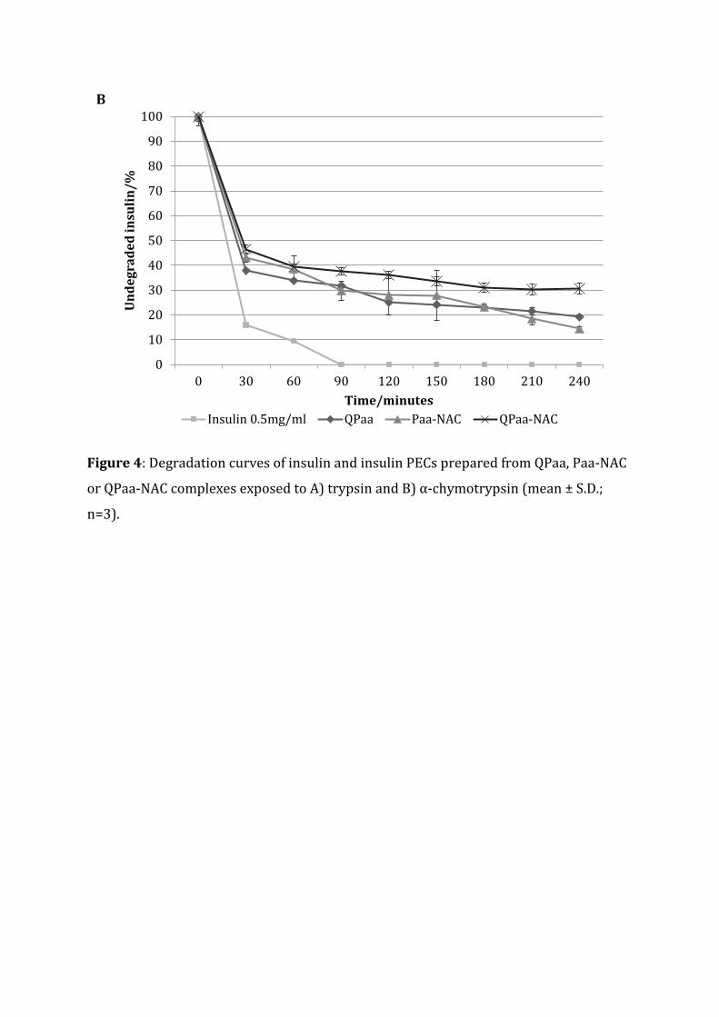
Figure 4: Degradation curves of insulin and insulin PECs prepared from QPaa, Paa-NAC
or QPaa-NAC complexes exposed to A) trypsin and B) α-chymotrypsin (mean ± S.D.;
n=3).
0
10
20
30
40
50
60
70
80
90
100
0 30 60 90 120 150 180 210 240
Und
egra
ded
insu
lin/%
Time/minutesInsulin 0.5mg/ml QPaa Paa-NAC QPaa-NAC
B

Figure 5: Mucin adsorption profile of polymer, insulin complexes (mean ± S.D.; n=3).
0
20
40
60
80
100M
ucin
ads
orbe
d (%
)
Polymers
Paa
Paa-NAC
Paa-TBA
QPaa
QPaa-NAC
QPaa-TBA