INCLUSION COMPLEXATION OF GEFITINIB WITH …
70
INCLUSION COMPLEXATION OF GEFITINIB WITH CYCLODEXTRINS Except where reference is made to the work of others, the work described in this thesis is my own or was done in collaboration with my advisory committee. This thesis does not include proprietary or classified information. _______________________________________________ Yann-Huei Phillip Lee Certificate of Approval: _____________________________ ____________________________ Daniel L. Parsons Jayachandra B. Ramapuram, Chair Professor Assistant Professor Pharmacal Sciences Pharmacal Sciences _____________________________ ____________________________ William R. Ravis George T. Flowers Professor Interim Dean Pharmacal Sciences Graduate School
Transcript of INCLUSION COMPLEXATION OF GEFITINIB WITH …

INCLUSION COMPLEXATION OF GEFITINIB WITH CYCLODEXTRINS
Except where reference is made to the work of others, the work described in this thesis is
my own or was done in collaboration with my advisory committee. This thesis does not
include proprietary or classified information.
_______________________________________________
Yann-Huei Phillip Lee
Certificate of Approval:
_____________________________ ____________________________
Daniel L. Parsons Jayachandra B. Ramapuram, Chair
Professor Assistant Professor
Pharmacal Sciences Pharmacal Sciences
_____________________________ ____________________________
William R. Ravis George T. Flowers
Professor Interim Dean
Pharmacal Sciences Graduate School

INCLUSION COMPLEXATION OF GEFITINIB WITH CYCLODEXTRINS
Yann-Huei Phillip Lee
A Thesis
Submitted to
the Graduate Faculty of
Auburn University
in Partial Fulfillment of the
Requirements of the
Degree of
Master of Science
Auburn, Alabama
August 9, 2008

iii
INCLUSION COMPLEXATION OF GEFITINIB WITH CYCLODEXTRINS
Yann-Huei Phillip Lee
Permission is granted to Auburn University to make copies of this thesis at its discretion,
upon request of individuals or institutions and at their expense. The author reserves all
publication rights.
______________________________
Signature of Author
______________________________
Date of Graduation

iv
THESIS ABSTRACT
INCLUSION COMPLEXATION OF GEFITINIB WITH CYCLODEXTRINS
Yann-Huei Phillip Lee
Master of Science, August 9, 2008
(B.S., Auburn University, 2004)
70 Typed Pages
Directed by Jayachandra Babu Ramapuram
Gefitinib, a very slightly soluble new drug, is used in the treatment of locally
advanced or metastatic non-small cell lung cancer (NSCLC) in patients who have
previously received chemotherapy. This study examined the complexation of gefitinib
with three selected cyclodextrins, β-cyclodextrin (βCD), hydroxypropyl-β-cyclodextrin
(HPβCD), and randomly methylated β-cyclodextrin (RMβCD), with the objective of
improving the solubility and dissolution of the drug.
Phase solubility studies were performed with the different cyclodextrins to
characterize the complexation in the liquid state. The complexation in the solid state was
characterized by differential scanning calorimetry and x-ray diffraction analyses. The
dissolution studies were performed using USP dissolution testing equipment and the
dissolution samples were analyzed by an UV spectrophotometric assay. The solubility of
gefitinib was significantly improved by all cyclodextrins (CDs) in the study. The

v
association rate constants (Ks) calculated from the phase solubility diagrams indicate that
gefitinib can form a stable inclusion complex with all three CDs. The freeze-dried
formulations showed substantial increases in the dissolution of gefitinib with all three
CDs compared to gefitinib alone, while the kneaded and physical mixtures showed no
improvement in the dissolution. Furthermore, addition of the hydrophilic polymers
polyvinyl pyrrolidone (PVP) and hydroxypropyl methyl cellulose (HPMC) markedly
enhanced the dissolution of gefitinib from CD complexes. The gefitinib-HPβCD (1:1)
complex yielded 50% dissolution in 1 hr whereas PVP or HPMC in association with the
complex increased the dissolution up to 95% within 1hr.
In conclusion, gefitinib can form a stable inclusion complex with all three
cyclodextrins as demonstrated by the liquid and solid state complexation studies.
HPβCD showed the greatest improvement in the dissolution of gefitinib followed by
RMβCD and βCD.

vi
ACKNOWLEDGEMENTS
I’d like to thank my advisor, Dr. Ramapuram for his guidance and support, the
professors in the School of Pharmacy for their input, my fellow graduate students for
their help, and my Mother, Father, and Brother for their love.

vii
Style manual: The AAPS Journal
Computer software: Microsoft Word 2000
viii
TABLE OF CONTENTS
SECTION
LIST OF FIGURES..…………………………………………………………..…
LIST OF TABLES………………………………………………………...............
INTRODUCTION……..………………………………………………………….
LITERATURE REVIEW…………………………………………………………
MATERIALS AND METHODS…………………………………………………
RESULTS AND DISCUSSION…………………………………………………
CONCLUSIONS………………………………………………………………….
REFERENCES….………………………………………………………………...
PAGE
ix
xi
1
7
30
35
52
54

ix
LIST OF FIGURES
1-1: The molecular structure of gefitinib…………………………………………
1-2: The cyclodextrin complexation equilibrium in water………………………...
1-3: The three naturally occurring cyclodextrins………………………………….
4-1: The standard curve for gefitinib absorbance…………………………………
4-2: Phase solubility diagram of gefitinib with βCD at room temperature……….
4-3: Phase solubility diagram of gefitinib with HPβCD at room temperature……
4-4: Phase solubility diagram of gefitinib with RMβCD at room temperature……
4-5: X-ray diffraction pattern of gefitinib, βCD, their physical mixture (1:1),
kneaded mixture (1:1), and inclusion complex (freeze dried, 1:1)……………
4-6: X-ray diffraction analysis of gefitinib, HPβCD, their physical mixture (1:1),
kneaded mixture (1:1), and inclusion complex (freeze dried, 1:1)……………
4-7: X-ray diffraction analysis of gefitinib, RMβCD, their physical mixture (1:1),
kneaded mixture (1:1), and inclusion complex (freeze dried, 1:1)……………
4-8: Differential scanning calorimetry analysis of gefitinib, HPβCD, their
kneaded mixture (1:1), and inclusion complex (freeze dried, 1:1)……………
PAGE
1
3
3
35
37
37
38
41
42
43
44
x
LIST OF FIGURES, CONT.
4-9: Differential scanning calorimetry analysis of gefitinib, RMβCD, their
kneaded mixture (1:1), and inclusion complex (freeze dried, 1:1)……………
4-10: Dissolution of gefitinib-βCD formulations…………………………………
4-11: Dissolution of gefitinib-HPβCD formulations……………………………...
4-12: Dissolution of gefitinib-RMβCD formulations……………………………..
4-13: Effect of PVP on the dissolution of gefitinib-HPβCD complex………........
4-14: Effect of HPMC on the dissolution of gefitinib-HPβCD complex………….
PAGE
46
48
49
49
50
50
xi
LIST OF TABLES
3-1: Various CD formulation compositions in the dissolution studies of gefitinib.
4-1: Gefitinib solubility curves with their calculated equations…………………
4-2: Average percent dissolved for various gefitinib-cyclodextrin systems……
PAGE
33
38
51

1
INTRODUCTION
Gefitinib is an anilinoquinazoline with the chemical name 4-Quinazolinamine, N-(3-
chloro-4- fluorophenyl) -7-methoxy-6- [3-4-morpholin) propoxy] and the following
structural formula:
Figure 1-1: The molecular structure of gefitinib.
It has the molecular formula C22H24ClFN4O3, a relative molecular mass of 446.9 and
is a brown-colored powder. Gefitinib is a free base with has pKa’s of 5.4 and 7.2 and
therefore ionizes progressively in solution as the pH falls. Gefitinib is soluble in aqueous
solvents at pH 1, but is practically insoluble above pH 7 and the solubility dropping
sharply between pH 4 and pH 6. In non-aqueous solvents, gefitinib is freely soluble in
glacial acetic acid and dimethylsulphoxide, soluble in pyridine, and sparingly soluble in
tetrahydrofuran, and slightly soluble in methanol, ethanol (99.5%), ethyl acetate, propan-
2-ol, and acetonitrile.

2
Gefitinib is a new drug used in the treatment of locally advanced non-small cell lung
cancer. There is no absolute data on the bioavailability of gefitinib in humans; while in
animal studies, a bioavailability of about 50% has been reported. Gefitinib is 90%
protein bound in vitro to serum albumin and α-1-acid glycoprotein, and has a mean
steady state volume of distribution of 1400 liters. Gefitinib is metabolized in the kidneys,
but renal elimination accounts for only 4% of the administered dose. The predominant
mode of excretion is via feces (approximately 86%). The elimination half-life ranges
from 6 to 49 hours, with no clear evidence of dose-dependency, and values being similar
after single doses or repeat administration. The bioavailability can be very well increased
by improving the solubility and dissolution of the drug. If the drug absorption is limited
by its solubility and dissolution, various approaches can be followed to improve these.
Some examples are: 1) solvent dispersion on an inert carrier, 2) micronization of drug
particles, 3) nanoparticle formation, 4) hot-melt extrusion, and 5) cyclodextrin
complexation. This research proposes to study the effect of cyclodextrin complexations
on gefitinib solubility and dissolution.
Cyclodextrins (CDs) are crystalline, cyclic oligosaccharides with a bucket-like
structure having a hydrophobic internal cavity and a hydrophilic exterior cavity. The
interior of the toroid is hydrophobic as a result of the electron rich environment provided
in large part by the glycosidic oxygen atoms. This structure allows the formation of
inclusion complexes in which lipophilic compounds are noncovalently bound within the
cavity. It is the interplay of atomic (Van der Waals), thermodynamic (hydrogen
bonding), and solvent (hydrophobic) forces that accounts for the stable complexes that
may be formed with chemical substances while in the apolar environment of the

3
cyclodextrin cavity. The complex exists in an equilibrium dependent upon the
concentrations of the cyclodextrin, the guest chemical and water.
Figure 1-2: The cyclodextrin complexation equilibrium in water.
Cyclodextrins have been employed in the pharmaceutical industry to increase the
aqueous solubility and stability of drugs and have been used in both parenteral and oral
drug delivery systems. There are three types of naturally occurring cyclodextrins—alpha,
beta, and gamma, seen in Figure 1-3.
Figure 1-3: The three naturally occurring cyclodextrins.
4
β-cyclodextrin (βCD) in particular is useful for complexation of average size
molecules, such as most drugs. The promising advantages of β-cyclodextrin as a drug
carrier are limited by its low aqueous solubility (1.8 g/100 ml). Therefore chemical
modification of βCD is used to produce products of very high water solubility (>50 g/100
ml) and minimal toxicity. Furthermore, the inclusion ability of βCD is amply magnified
due to chemical modification and presently about 50 cyclodextrin derivatives are
commercially available.
Since the usefulness of cyclodextrins vary according to molecular weight, cavity size,
intrinsic solubility in water and other solvents, safety, and cost, the study must match the
intended use of the drug with the appropriate cyclodextrin or cyclodextrin derivative.
Many different chemicals may be introduced into the cyclodextrin molecule by reaction
with the hydroxyl groups lining the upper and lower ridges of the toroid, for example,
methyl, hydroxypropyl, carboxymethyl, and acetyl. The randomness of position and type
of substitution causes the resultant modified cyclodextrins to be amorphous, which
contributes to its greatly enhanced aqueous solubility compared to the crystalline natural
form. One such modification, hydroxypropyl-β-cyclodextrin (HPβCD), has the best
balance of enhanced aqueous solubility, and the range of drugs which can form stable
complexes. It is very soluble in water, greater than 500 mg/ml at room temperature
compared to 18 mg/ml for βCD. Hydroxypropyl-β-cyclodextrin is also moderately
soluble in methanol and ethanol, which allows for greater drug incorporation into
powdered formulations using co-solvent techniques.
Inclusion complexes are entities comprising two or more molecules, where the
cyclodextrins are usually the host molecules, and include a guest molecule held only by

5
physical forces without covalent bonding. Cyclodextrins are capable of forming
complexes with compounds with a size compatible with the cyclodextrin’s inner cavity.
Geometric factors, rather than chemical, are key to determining which guest molecules
can be complexed. The complexed molecules are oriented inside the host to achieve the
maximum contact between the hydrophobic parts of the guest molecule to the cavity.
The driving force of complexation includes various effects, one of which is the
substitution of the energetically polar and apolar interaction between the included water
and the cyclodextrin cavity, and between water and the guest molecules, by the more
favorable interaction between the guest and the cavity. Another is the strain release on
complexation, and also the Van der Waals and hydrogen bonding interactions.
Stability and solubility of the complexes are entirely independent properties. It is not
the most insoluble inclusion complex which is the most stable one in solution. However,
a direct correlation exists between the complex stability and the enhancement of the guest
molecule’s poor solubility. With increasing temperatures, the complexes disassociate,
with association constant values to decrease rapidly.
We believe that gefitinib can form stable inclusion complexes with cyclodextrins to
improve the solubility and dissolution for this drug for enhanced absorption and oral
bioavailability.
The objectives of the present study are:
1) To characterize the inclusion complexation of gefitinib in the liquid state: Phase
solubility studies of gefitinib with three different cyclodextrins (β-cyclodextrin,
6
hydroxypropyl-β-cyclodextrin, and randomly methylated-β-cyclodextrin) were
conducted and stability rate constants of the complexes were calculated.
2) To characterize the inclusion complexation of gefitinib in the solid state: The
inclusion complexes of gefitinib with various cyclodextrins were prepared by
freeze drying and the formation of complexes in the solid state was confirmed by
differential scanning calorimetry (DSC) and x-ray diffraction analysis.
3) To study the dissolution improvement of gefitinib by complexation with
cyclodextrins: The dissolution studies were conducted according to USP
guidelines.
7
LITERATURE REVIEW
Gefitinib
Gefitinib is a tyrosine kinase inhibitor used for the treatment of non-small cell lung
cancer (NSCLC). It is taken in 250 mg oral doses once daily, higher doses showing no
improvement but increased toxicity. It has been approved for continued monotherapy in
patients with locally advanced/metastatic NSCLC who have failed both platinum and
docetaxel-based chemotherapies, or in patients who are or have benefited from gefitinib.
It has a molecular mass of 446.9 and is a brown-colored powder. Gefitinib has pKa’s of
5.4 and 7.2 and therefore ionizes progressively in solution as the pH falls. Gefitinib is
sparingly soluble at pH 1, but is practically insoluble above pH 7 and the solubility drops
sharply between pH 4 and pH 6. In non-aqueous solvents, gefitinib is freely soluble in
glacial acetic acid and dimethylsulphoxide, soluble in pyridine, and sparingly soluble in
tetrahydrofuran, and slightly soluble in methanol, ethanol (99.5%), ethyl acetate, propan-
2-ol, and acetonitrile.1
There is no absolute data on the bioavailability of gefitinib in humans; while in
animal studies, a bioavailability of about 50% has been reported. Gefitinib is 90%
protein bound in vitro to serum albumin and α-1-acid glycoprotein, and has a mean
steady state volume of distribution of 1400 liters. Gefitinib is metabolized in the kidneys,
but renal elimination accounts for only 4% of the administered dose. The predominant
mode of excretion is via feces (approximately 86%). The elimination half-life ranges
8
from 6 to 49 hours, with no clear evidence of dose-dependency and values being similar
after single doses or repeat administration.1 Furthermore, upon oral administration,
gefitinib induces gastro-intestinal adverse effects such as nausea, vomiting and diarrhea,
which are dose dependent. Clearly, there is a need to improve the dissolution of this drug
in order to improve the bioavailability.
Cyclodextrins
Cyclodextrins have well-known effects on drug solubility and dissolution,
bioavailability, safety, and stability. There are various factors influencing inclusion
complex formation and an understanding of these factors is necessary for proper handling
of these versatile materials. Some important considerations in selecting CDs in drug
formulation are their commercial availability, regulatory status, and patent status. CDs,
are expected to solve many problems associated with the delivery of different novel drugs
through different delivery routes.2
Cyclodextrins were called cellulosine when first described in 1891. Soon after,
Schardinger identified the three naturally occurring cyclodextrins: alpha, beta, and
gamma. These new compounds were referred to as Schardinger sugars. For 25 years
between 1911 and 1935, Pringsheim in Germany was the leading researcher in this area,
demonstrating that these sugars formed stable aqueous complexes with many other
chemicals. By the mid 1970's, each of the natural cyclodextrins had been structurally and
chemically characterized and many more complexes had been studied. The natural
cyclodextrins are produced from starch by the action of cyclodextrin glycosyltransferase,
an enzyme produced by several organisms, Bacillus macerans being the earliest source.3

9
Structurally, cyclodextrins consist of 6, 7, or 8 (α, β, and γ respectively) D-
glucopyranosyl units connected by alpha-(1,4) glycosidic linkages. The most stable three
dimensional molecular configuration for these non-reducing cyclic oligosaccharides takes
the form of a toroid with the larger and smaller opening of the toroid presenting
secondary and primary hydroxyl groups, respectively, to the solvent environment. The
interior of the toroid is hydrophobic as a result of the electron rich environment provided
in large part by the glycosidic oxygen atoms. It is the interplay of atomic (Van der
Waals), thermodynamic (hydrogen bonding), and solvent (hydrophobic) forces that
accounts for the stable complexes that may be formed with chemical substances while in
the apolar environment of the cyclodextrin cavity. The complex exists in an equilibrium
dependent upon the concentrations of the cyclodextrin, the guest chemical and water.
The rate at which the associated complex is formed is determined in large part by the
accessibility of the guest molecule to the cyclodextrin cavity and the magnitude of the net
thermodynamic driving force.3
Cyclodextrins are able to form water-soluble inclusion complexes with many
lipophilic water-insoluble drugs. In aqueous solutions drug molecules located in the
central cavity are in a dynamic equilibrium with free drug molecules. Furthermore,
lipophilic molecules in the aqueous complexation media will compete with each other for
a space in the cavity. Due to their size and hydrophilicity only insignificant amounts of
cyclodextrins and drug-cyclodextrin complexes are able to penetrate into lipophilic
biological barriers, such as intact skin.4

10
Cyclodextrin Toxicity
Natural cyclodextrins are practically not absorbed under normal physiological
conditions, and the metabolites produced are not unusual, and they are therefore
considered safe for oral use. In the U.S. since 1997, β-cyclodextrins have been
considered GRAS for certain food uses. However, β-cyclodextrin is not safe for
parenteral use, since it forms insoluble complexes in the kidneys, causing
nephrotoxicity.5
Chemically modified cyclodextrins were introduced partially in order to improve the
parenteral safety profile of β-cyclodextrin. Randomly methylated β-cyclodextrin has
been shown to have very good complexing ability, in addition to being very water
soluble. However, its lipophilicity can create irritation and hemolysis. Hydroxypropyl β-
cyclodextrin has been shown to be safe at very high oral doses, and observed to have no
irreversible adverse effects in i.v. doses up to 400 mg/Kg.5
Solubility and Dissolution Improvement
A study compared the physicochemical characteristics of the solid complexes
prepared by traditional methods (kneading, freeze-drying and spray-drying) and using a
supercritical fluid technology to enhance the solubility of Naproxen, a poorly soluble
anti-inflammatory drug, with β-cyclodextrin. In physical mixtures and kneaded systems,
the drug’s endothermic properties are present, suggesting incomplete encapsulation,
while freeze dried and spray dried systems show complexation.6
The effect of β-cyclodextrin on the aqueous solubility and dissolution rate of
celecoxib was investigated. Celecoxib, a specific inhibitor of cycloxygenase-2 (COX-2),

11
is a poorly water-soluble nonsteroidal anti-inflammatory drug with relatively low
bioavailability. The possibility of molecular arrangement of inclusion complexes of
celecoxib and β-cyclodextrin were studied using molecular modeling and structural
designing. A phase-solubility profile indicated that the solubility of celecoxib was
significantly increased in the presence of β-cyclodextrin. Solid complexes were prepared
by freeze drying, evaporation, and kneading methods and characterized using differential
scanning calorimetry, powder x-ray diffractometry, and scanning electron microscopy.
Freeze-dried complexes showed a higher dissolution rate than the other complexes.7
Solid dispersions of Celecoxib have also been prepared with hydroxypropyl-β-
cyclodextrin by various methods such as physical mixture, cogrinding, kneading, and
coevaporation. The dispersions were characterized by differential scanning calorimetry,
X-ray diffraction patterns, infrared spectroscopy, and nuclear magnetic resonance studies.
The DSC thermograms of the dispersions indicated the potential of a heat-induced
interaction between Celecoxib and cyclodextrin that could influence in vitro drug
dissolution. The dispersions exhibited faster rates of dissolution compared to that of
Celecoxib alone.8
The method of co-grinding with CDs was applied to a poorly water soluble drug,
ONO-8713 (solubility; 0.92 l g/ml in H2O at 25°C), as a method to prepare nanoparticles.
ONO-8713 was co-ground with various CDs in a vibration mill. When ONO-8713 was
co-ground with βCD, 85% of the drug was recovered as nanoparticles with a mean
particle size of 120 nm. Nanoparticle yield achieved 90% when hydroxypropyl-β-
cyclodextrin was used as a co-grinding additive. It was found that the amount of drug
nanoparticles depended on the characteristics of the CDs. This phenomenon was

12
probably due to the difference in the cavity size of CDs along with the variation of
substitution groups that affected the interaction between CDs and drug, and the affinity
between CD and drug. Zeta potential analysis suggested that the CD would form a layer
covering on the particle surface and alter the charge of the particles, improving the
stability and total yield of the nanoparticle.9
The inclusion behavior of sulfobutyl ether-7 derivative of β-cyclodextrin (SBE7βCD)
in solution and solid state was compared with that of natural β-cyclodextrin toward a
poorly water-soluble anti-inflammatory agent, rofecoxib (ROFX). A phase solubility
method was used to evaluate the stoichiometries and stability constants of ROFXβCD
and ROFX-SBE7βCD complexes. Solubility enhancement was much greater for the
rofecoxib-SBE7βCDcomplex compared to drug-βCD complex. The stability constant
obtained for the SBE7βCD inclusion complex of rofecoxib was higher. Finally,
dissolution profiles obtained suggest that SBE-βCD is more effective than β-cyclodextrin
in improving the pharmaceutical properties of rofecoxib.10
In another study, the influence of natural β-cyclodextrin and its hydrophilic
derivatives (HPβCD and SBE7βCD) on the in vitro dissolution rate, in vivo absorption,
and oral bioavailability of a poorly water soluble anti-inflammatory agent, valdecoxib
(VALD) was studied. Equimolar drug-cyclodextrin solid complexes were prepared by
kneading and coevaporation methods and characterized by infrared spectroscopy,
differential scanning calorimetry, and X-ray diffraction. In the liquid state, the
cyclodextrin complexes were studied using phase solubility analysis. Drug solubility and
dissolution rate in distilled water were notably improved by employing the CDs. It was

13
found that the cyclodextrin complexes of drug showed significant improvement in anti-
inflammatory activity.11
Parent β-cyclodextrin and 2-hydroxypropyl-β-cyclodextrin can form 1:1 solid
complexes with an orally active angiotensin-converting enzyme inhibitor, captopril,
while hydrophobic perbutanoyl-β-cyclodextrin (TBβCD) forms a solid dispersion or solid
solution with the drug. The binary system of captopril-HPβCD or captopril-TBβCD and
the ternary system of captopril-TBβCD-HPβCD in different molar ratios were prepared
by the kneading method, and the release behavior of the drug was investigated. The
release rate of captopril from the binary HPβCD system was rather fast, whereas that
from the binary TBβCD system was comparatively slower, the retarding effect being
dependent on the amounts of TBβCD. It was difficult to prolong plasma levels of
captopril by administering orally either the binary HPβCD or TBβCD system in dogs.
On the other hand, the ternary captopril-TBβCD-HPβCD system gave a plasma profile
comparable to that of a commercially available sustained release preparation.12
Complexes between members of a homologous series of alkylparabens and β-
cyclodextrin have been prepared by both kneading and co-precipitation methods and their
behavior studied by DSC, thermogravimetric, infrared and powder X-ray diffraction
techniques. Results showed that complexation did occur by both the kneading and co-
precipitation methods. DSC and IR techniques confirmed these results and
thermogravimetric indicated the presence and number of water molecules in each
complex.13
Salbutamol laurate is a novel salt form of the well-known bronchodilator, salbutamol
(albuterol). Its polymorphism and inclusion in 2-hydroxypropyl-β-cyclodextrin were
14
investigated by thermogravimetry, differential scanning calorimetry, infrared and powder
X-ray diffraction techniques. Two polymorphic forms of the salt were identified.
Conditions for inclusion complex formation between the salt and (2-hydroxypropyl)-β-
cyclodextrin, namely prolonged co-grinding and kneading, were established by a
combination of the above methods.14
Chemical analysis, DSC and solubility determinations have been applied to the study
of solid inclusion complexes of a pesticide, 2, 4-dichlorofenoxyacetic acid as guest, and
β-cyclodextrin as host, in order to produce experimental evidence of the inclusion process
and of the stoichiometry of the inclusion compound. Three processing methods were
studied and compared: physical (mechanical) mixing; kneading; and spray-drying. The
phase-solubility diagram of complex formation in solution has been also established. The
stoichiometric ratio of the complexes was found to be 1:1 by solubility and DSC, being
confirmed by chemical analysis. Spray-drying was found to be most suitable method for
preparing the complexes.15
A study aimed at developing a tablet formulation was based on an effective
flurbiprofen-cyclodextrin system, able to allow a rapid and complete dissolution of this
practically insoluble drug. Three different cyclodextrins were evaluated: the parent β-
cyclodextrin, and two amorphous, highly soluble β-cyclodextrin derivatives, i.e., methyl-
β-cyclodextrin and hydroxyethyl-β-cyclodextrin. Equimolar drug-cyclodextrin binary
systems prepared according to five different techniques (physical mixing, kneading,
sealed-heating, coevaporation, and colyophilization) were characterized by differential
scanning calorimetry, x-ray powder diffractometry, infrared, and optical microscopy.
The drug solubility improvement obtained by the different binary systems varied from a
15
minimum of 2.5 times up to a maximum of 120 times, depending on both the
cyclodextrin type and the system preparation method. All formulations containing drug-
cyclodextrin systems gave a higher drug dissolved amount than the corresponding ones
with drug alone. It can be reasonably expected that the obtained drug dissolution rate
improvement will result in an increase in its bioavailability, with the possibility of
reducing drug dosage and side effects.16
Microencapsulation of lemon oil was undertaken by kneading with α-cyclodextrin, at
an α-cyclodextrin to lemon oil ratio of 88:12 (w/w). The resulting paste samples of the
complex were vacuum- or spray-dried. Ten selected lemon oil flavor volatiles in the
complex were analyzed periodically after kneading. The results indicated that the levels
of these volatiles were not significantly different irrespective of mixing time or type of
drying (vacuum- or spray-drying) used. An optimum mixing time was found to be 15
min, at which time the maximum encapsulation of lemon oil was obtained in the complex
powder.17
For another study, the possibility of improving dehydroepiandrosterone (DHEA)
solubility and bioavailability by high-energy cogrinding with α-cyclodextrin (αCD) in the
presence or absence of different auxiliary substances was investigated. In all cases,
ternary products exhibited higher drug solubilizing properties than the binary DHEA-α-
cyclodextrin coground system. Glycine was the most effective component. The best
combinations were characterized by differential scanning calorimetry and X-ray powder
diffractometry and evaluated for dissolution rate. The presence of glycine favored
destruction of DHEA crystalline structure during cogrinding, as evidenced by the strong
reduction in both time and vibration frequency of milling necessary to obtain total drug

16
amorphization. Both ternary products showed better dissolution properties than the drug
alone, affording, respectively, a 40 and 60% increase of dissolution efficiency.18
The aim of another study was to enhance the low solubility of eflucimibe, a new
chemical entity to treat dyslipidemiae, by complexation with γ-cyclodextrin. The
complex was prepared using the kneading method. The interaction evolution was studied
during process by comparison of the semi-solid and physico-chemical states of the
product. The evolution of the semi-solid state was followed by torque measurement
while the evolution of physico-chemical state was studied by differential scanning
calorimetry, infrared spectroscopy and by determination of the drug solubilization profile.
The interaction, which occurs during the process, is characterized by a modification of
the product consistency and by a disappearance of the drug endothermic peaks, a
disappearance of a drug spectral band and an improvement in the drug solubilization
profile. Indeed, after complete interaction, the drug quantity solubilized in specific
conditions increased about 44-fold compared to those of untreated drug. Moreover, the
comparison of the physico-chemical and semi-solid states during the kneading process
clearly shows that when the interaction takes place, a solidification of the paste occurs.
The results suggested that the formation of new solid phase allows an enhancement of the
solubility of eflucimibe.19
The purpose of another study was to investigate the effect of moisture condition
during the cogrinding process on fine drug particle formation. Cogrinding of CDs and
pranlukast (PRK) hemihydrate was performed in various moisture conditions and the
formation of PRK submicron particles was investigated. The moisture content in the
cogrinding process significantly affected the fine particle formation. More than 90% of
17
PRK loaded transformed to submicron particles when coground with αCD, βCD or γCD
containing the specific amount of water for each CD system. Fine particle formation of
PRK was considered as a particular phenomenon to cyclodextrins, since the submicron
particles could not be formed when d-mannitol, lactose or microcrystalline cellulose
(MCC) was used as a cogrinding additive. Moreover, the appearance and disappearance
of fine particle formation was found to be reversible depending on the existence of water
during the grinding process.20
Glimepiride is one of the third generation sulfonylureas used for treatment of type 2
diabetes. Poor aqueous solubility and slow dissolution rate of the drug lead to
irreproducible clinical response or therapeutic failure in some cases due to subtherapeutic
plasma drug levels. Consequently, the rationale of the study was to improve the
biological performance of this drug through enhancing its solubility and dissolution rate.
Inclusion complexes of glimepiride in βCD, HPβCD and SBEβCD, with or without water
soluble polymers were prepared by the kneading method. Binary systems were
characterized by thermogravimetric analysis, IR spectroscopy and X-ray diffractometry.
Phase solubility diagrams revealed an increase in solubility of the drug upon cyclodextrin
addition. All the ternary systems containing βCD or HPβCD showed higher dissolution
efficiency compared to the corresponding binary systems. In conclusion, the association
of water soluble polymers with glimepiride-CD systems leads to great enhancement in
dissolution rate, increased duration of action and improvement of therapeutic efficacy of
the drug.21
Inclusion complexes of clofibrate with β-cyclodextrin were prepared by
coprecipitation, kneading and sealed heating methods and characterized by UV
18
spectrophotometry, differential scanning calorimetry, X-ray diffractometry and infrared
spectroscopy. All results were in keeping with the formation of a 1:1 inclusion complex.
Dissolution studies showed that clofibrate entrapped in inclusion complexes dissolved
much faster than uncomplexed liquid clofibrate. The complex prepared by the sealed
heating method showed the greatest improvement in dissolution rate. These results show
that a more easily manipulated, solid form of clofibrate may be obtained through
formation of its inclusion complex with βCD, and that complex formation simultaneously
enhances the solubility and dissolution rate of this drug.22
Inclusion complexes of tolbutamide with β-cyclodextrin and hydroxypropyl-β-
cyclodextrin were prepared using different methods: kneading, coprecipitation and
freeze-drying. Inclusion complexation in aqueous solution and in solid phase was studied
by the solubility method, X-ray diffractometry, thermal analysis and Raman
spectroscopy. The solubility of tolbutamide increased as a function of cyclodextrin
concentration, showing BS and AL type diagrams for β-cyclodextrin and hydroxypropyl-
β-cyclodextrin, respectively. The dissolution rate of tolbutamide from the inclusion
complexes was much more rapid than tolbutamide alone.23
The methods of obtaining and physicochemical properties of inclusion complexes of
amlodipine (AM) and felodipine (FL) with methyl-β-cyclodextrin (MβCD) clathrates
were studied. Solid complexes were obtained by two methods: kneading and
lyophilization with the drug and MβCD at the molar ratio of 1:1. The identity of the
obtained clathrates was confirmed by IR, 13C-NMR spectra and DSC measurements.
The process of AM and FL complexation with MβCD involve the aromatic ring, the
carbonyl groups in the ester bonds and the carbon atoms of the ring linked via the ester

19
bonds. One of the aims of complexation was to improve the drug solubility, so the
dissolution rate of the obtained clathrates was tested. As a result of the inclusion
complexes formation obtained both by kneading and lyophilization, the solubility of AM
increased 3 times. The inclusion complex formation with FL and MβCD brought the
most dramatic increase in FL solubility, which increased 16 times.24
The effects of hydroxypropyl-β-cyclodextrin and methyl-β-cyclodextrin on the
solubility of ketoconazole were examined. Products were prepared by physical mixing,
kneading and spray-drying in four molecular ratios. Kneaded products in a ratio of drug:
cyclodextrin (1:2) and spray-dried products showed the highest dissolution rate. Phase
solubility diagrams of ketoconazole with these cyclodextrins at 25 °C in water and
simulated intestinal medium were constructed. A solubility diagram of the AL type was
obtained with hydroxypropyl-β-cyclodextrin, and of the AP type with methyl-β-
cyclodextrin. The complexes were characterized by thermal methods. Multicomponent
systems were prepared with tartaric acid. The effects of water-soluble polymers, e.g.,
polyvinyl pyrrolidone, on the aqueous solubility of ketoconazole were investigated. The
particle size of ketoconazole (70 μm) is reduced to 12 μm by the preparation of spray-
dried products. As the solubility in water increased, the partition coefficient, surface
tension and wetting angle values decreased.25
The possible competitive displacement of a drug from its cyclodextrin-based
inclusion complex by a third substance was investigated by studying the dissolution
behavior of tolbutamide-β-cyclodextrin inclusion complex in demineralized water and in
aqueous solution of different surfactants. Physical mixtures and kneaded systems were
prepared in 1:1 and 1:2 drug-β-cyclodextrin mol/mol ratios and they were characterized

20
by hot-stage microscopy, differential scanning calorimetry, and X-ray powder
diffractometry. The release behavior of tolbutamide from its inclusion complex was
studied by dissolution of the binary systems in water and in aqueous solutions of three
surfactants: polysorbate 20, poloxyl 23-lauryl ether, and sodium lauryl sulfate. When
demineralized water was used as the dissolution medium, the fastest dissolution of
tolbutamide was obtained from the 1:2 kneaded system followed by the 1:1 kneaded
system. The presence of poloxyl 23-lauryl ether and sodium lauryl sulfate in the media
caused a decrement in the rate and extent of dissolution of the drug from both kneaded
systems in comparison with that obtained from the same systems in water. However, the
release of tolbutamide from the kneaded systems remains unaffected when polysorbate
20 was present in the dissolution medium. Results of this study suggest that the
simultaneous presence of β-cyclodextrin and surfactants of proper molecular structure in
a pharmaceutical formulation can yield an unexpected dissolution of the drug.26
Griseofulvin-cyclodextrin interactions were investigated in an aqueous environment
and in solid state. Two cyclodextrin derivatives (α-cyclodextrin and 2-hydroxypropyl α-
cyclodextrin) were used to prepare different physical mixtures and kneaded systems, and
the drug/cyclodextrin ratios were 1:1 and 1:2 mol/mol. Scanning electron microscopy
(SEM), hot-stage microscopy (HSM), differential scanning calorimetry (DSC),
thermogravimetry (TG), and X-ray powder diffractometry were employed to characterize
pure substances and their kneaded counterparts and all of the binary systems. The
solubility of griseofulvin was increased in accord with the quantity of cyclodextrin added.
HSM examination revealed that 2-hydroxypropyl-α-cyclodextrin was dissolved in the
droplets of melted griseofulvin, but did not show any interactions between melted

21
griseofulvin and α-cyclodextrin particles. The presence of the griseofulvin endothermic
peak in the DSC curves suggested the absence of any griseofulvin/cyclodextrin inclusion
compound in the solid state. In TG, data of weight loss owing to the dehydration of
cyclodextrins was similar for both kneaded systems and physical mixtures. X-ray
diffraction patterns exhibited the amorphous nature of 2-hydroxypropyl-α-cyclodextrin
and the crystalline nature of griseofulvin and binary systems. Griseofulvin dissolution
profiles from all binary systems showed an improvement in drug dissolution, which
indicates that a drug/cyclodextrin inclusion compound was formed in the aqueous
dissolution medium.27
Equimolar combinations of econazole, a very poorly water soluble antifungal agent,
with β-cyclodextrin and statistically substituted methyl-β-cyclodextrin were investigated
for both solid state characterization (differential scanning calorimetry, hot-stage
microscopy, infrared spectroscopy, scanning electron microscopy) and dissolution
properties (dispersed amount method). The influence of the preparation method (physical
mixing, ball-milling, kneading, and sealed-heating) on the physicochemical properties of
the products was evaluated. Kneading and sealed heating techniques led to amorphous
products in the case of systems with methyl-β-cyclodextrin, whereas crystalline drug was
still clearly detectable in all products with β-cyclodextrin. Independently of the
preparation technique, all combinations with methyl-β-cyclodextrin yielded better
performance than the corresponding ones with β-cyclodextrin. However, the influence of
the preparation method was clearly more marked for products with methyl-β-
cyclodextrin. In fact, the sealed-heated with the β-derivative showed an increase of drug
dissolution efficiency of 130% with respect to the corresponding physical mixture, in

22
comparison to the 70% increase obtained from that with β-cyclodextrin. Moreover,
whereas the difference in dissolution efficiency values between coground products was
only about 8% in favor of the β-derivative, it reached 80 and 90% between sealed-heated
and kneaded products, respectively.28
Binary systems of ketoprofen with native crystalline β-cyclodextrin and amorphous
statistically substituted methyl-β-cyclodextrin were investigated for both solid phase
characterization (differential scanning calorimetry, powder X-ray diffraction, infrared
spectroscopy, scanning electron microscopy) and dissolution properties (dispersed
amount and rotating disc methods). Grinding, kneading, sealed-heating and
colyophilization of equimolar combinations of ketoprofen with methyl-β-cyclodextrin, as
well as colyophilization of analogous combinations with β-cyclodextrin, led to
amorphous products. Crystalline drug, instead, was still clearly detectable in coground,
kneaded and sealed-heated products with β-cyclodextrin. Both the preparation method,
and even more the nature of the carrier, played an important role in the performance of
the system. Colyophilized and sealed-heated products showed the best dissolution
properties. However, independently of the preparation technique, all combinations with
methyl-β-cyclodextrin yield better performances than the corresponding ones with the β-
cyclodextrin. Moreover, the intrinsic dissolution rate of ketoprofen from a simple
physical mixture with the β-cyclodextrin derivative was five-fold higher than that from
the best product with the parent β-cyclodextrin.29
The complexing, solubilizing and amorphizing abilities toward ibuproxam (a poorly
water-soluble anti-inflammatory agent) of some randomly substituted amorphous β-
cyclodextrin derivatives were investigated and compared with those of the parent β-
23
cyclodextrin. Equimolar drug-cyclodextrin solid systems were prepared by blending,
cogrinding, coevaporation, and colyophilization. All the derivatives showed greater
solubilizing efficacies toward ibuproxam than the parent one, due to their higher water
solubility. Colyophilized products were in all cases the most effective, followed by
coground and coevaporated systems, whose dissolution efficiencies were over four times
higher than the corresponding physical mixtures and about 15 times higher than the pure
drug.30
The effect of ternary complexation of naproxen, a poorly water soluble anti-
inflammatory drug, with hydroxypropyl-β-cyclodextrin and the basic amino acid L-
arginine on drug dissolution properties has been investigated. Equimolar binary (drug-
cyclodextrin or drug-arginine) and ternary (drug-cyclodextrin-arginine) systems were
prepared by blending, cogrinding, coevaporation, and characterized by differential
scanning calorimetry, thermogravimetric analysis, FT-IR spectroscopy, X-ray
diffractometry. The dissolution behavior of naproxen from the different products was
evaluated by means of a continuous flow through method. The results of solid state
studies indicated the presence of strong interactions between the components in ternary
coevaporated and coground systems, which were both of totally amorphous nature. In
contrast, the presence of either free drug or free arginine was detected when the third
component (cyclodextrin or amino acid) was physically mixed, respectively, to the drug-
arginine binary system (as physical mixture, coevaporate, or co-ground product) or to the
drug-cyclodextrin binary system (as physical mixture, coevaporate, or coground product).
All ternary combinations were significantly more effective than the corresponding binary
drug-cyclodextrin and drug-arginine systems in improving the naproxen dissolution rate.
24
The best performance in this respect was given by the ternary coevaporate, with about a
15 times increase in terms of both drug relative dissolution rate and dissolution
efficiency. The synergistic effect of the simultaneous use of arginine and cyclodextrin on
the dissolution rate of naproxen was attributed to the combined effects of inclusion in
cyclodextrin and salt formation, as well as to a specific role played by arginine in this
interaction.31
The inclusion complex between sulfaproxyline (SP) and βCD was prepared by the
freeze-drying and the kneading method. Complex formation was confirmed in the solid
state by X-ray diffractometry and by infrared spectroscopy. The interaction between SP
and β-cyclodextrin in solution was studied by the solubility method and 13C-NMR
spectroscopy. Phase solubility studies in water revealed an AN type diagram.
Complexation was found to improve the dissolution rate of SP.32
The interactions of nabumetone (NAB) with α-cyclodextrin and γ-cyclodextrin were
studied in aqueous solution by means of phase-solubility analysis. Solid dispersions of
NAB with αCD, βCD, MβCD, and HPβCD were prepared by coevaporation and
kneading and also by coprecipitation in the case of γCD. X-ray diffractometry, thermal
analysis and infrared spectroscopy (FTIR) were used to study the possible complexation
of the drug with the different cyclodextrins. Solid dispersions of NAB with γCD showed
a remarkable improvement in the dissolution rate of NAB.33
The application of molecular modeling to the inclusion complexation of natural and
modified CDs with carboxylic acid derivatives as guest molecules was examined.
Information was available on the thermal behavior in the solid-state of benzoic acid (BA),
salicylic acid (SA), and various substituted aminosalicylic acids (3-aminosalicylic acid

25
(3-ASA), 4-aminosalicylic acid (4-ASA), and 5-aminosalicylic acid (5-ASA), as well as
on the thermal behavior of 1:1 molar ratio physical and kneaded mixtures of these acids
with each of three different cyclodextrins, βCD, HPβCD, and γCD. The thermal behavior
of the binary mixtures was modeled using stepwise multiple regression (SMR). Two
models for the prediction of the percentage mass loss and enthalpy of dehydration of the
physical mixtures were established with correlation coefficients of 0.79 and 0.92,
respectively. Decreased correlation in the thermal behavior of kneaded mixtures
indicated significant interaction and possible formation of inclusion complexes.34
An inclusion complex between warfarin and β-cyclodextrin was obtained to improve
the in vitro bioavailability of the drug in acidic media. Inclusion complexation in
solution was studied by the phase solubility technique. The apparent stability constant
was influenced by the pH of the medium ranging from 633.26 M-1
(at pH 1.2, where the
drug was in the unionized form) to 99.81M-1
(at pH 7.4, where the drug was in the
ionized form). Phase solubility study showed an AL-type diagram indicating the
formation of an inclusion complex in 1:1 molar ratio. Solid binary mixtures of the drug
with β-cyclodextrin were prepared by several methods (physical mixing, kneading, co-
evaporation, freeze-drying). Physicochemical characterizations were performed using
differential scanning calorimetry, powder X-ray diffractometry and dissolution studies.
Preparation method influenced the physicochemical properties of the binary mixtures.
An inclusion complex was obtained by freeze-drying, with a high solubility and drug
dissolution rate. The physical stability of the complex was also studied. After one year
storage in a glass container at room temperature no significant changes were detected in
the diffractogram, thermogram and dissolution profile of the freeze-dried product.35

26
The solubility of miconazole in water increased in the presence of CDs. The apparent
K values calculated from the phase solubility diagrams of βCD, hydroxypropyl-α-
cyclodextrin, hydroxyethyl-α-cyclodextrin, HPβCD, and αCD were 695 ± 39.6, 363 ±
34.1, 333 ± 18.5, 312 ± 31.0, 305 ± 27.6, and 293 ± 17.6 M-1, respectively. Solid 1:1
molar complexes were prepared by freeze-drying and kneading and characterized by UV
spectroscopy, differential scanning calorimetry, and electron microscopy. The
dissolution rate increased 28-255 fold and the solubility 9-55 fold. Oral bioavailability in
rats increased 2.3 fold by complexation with hydroxypropyl-α-cyclodextrin. Human
cadaver skin retained 2.6 fold more drug from the miconazole-cyclodextrrin complex and
hairless mice skin retained 8.4 fold more drug from the HPαCD complex than from
miconazole solution alone in 24 h.34
Solid complexes between gliclazide and βCD were prepared by kneading,
coprecipitation, neutralization, co-grinding and spray-drying. Characterization of
gliclazide-cyclodextrin inclusion complexes was performed using X-ray diffractometry
and cross polarizing/magic angle spinning ~3C-nuclear magnetic resonance
spectroscopy. These techniques clearly demonstrated the existence of solid-state
inclusion compound formation. The complexes obtained by neutralization and spray-
drying showed enhanced dissolution rates of gliclazide.37
Natural crystalline (α-, β-, γ-) and amorphous derivative (hydroxypropyl-β- and
methyl-β-) cyclodextrins were selected as potential carriers for obtaining, through a co-
grinding technique, a stable activated amorphous form of glyburide with improved
dissolution properties. Differential scanning calorimetry was used to investigate solid-
state modifications of the drug induced by cogrinding with the selected carriers in a high
27
energy vibrational micro-mill. X-ray powder diffraction and FTIR spectroscopy were
employed as additional techniques to support DSC data. Equimolar drug: cyclodextrin
physical mixtures were co-ground for different times (up to 60 min) at constant vibration
frequency (24 Hz). A progressive drug amorphization with increasing grinding time was
observed in all binary systems, but, interestingly, different degrees of sensitivity to the
mechanical-chemical activation were evident. In fact, blends with natural cyclodextrins,
despite the initial higher crystallinity than those with the amorphous derivatives, required
the same or shorter cogrinding times (60 min) to achieve complete drug amorphization.
Stability studies indicated no appreciable drug recrystallization in co-ground products
after 4 months storage in sealed containers at 25°C or 1 month at 25°C and 75% RH. No
stability differences were detected between products with natural or derivative
cyclodextrins. The results accounted for the suitability of cyclodextrin co-grinding
technique to obtain and stabilize glyburide in the activated amorphous form.38
An optimized kneading method for the preparation of lycopene-cyclodextrin binary
systems was developed leading to solubilization of lycopene in water and 5% (w/v)
dextrose solution. Storage stability characteristics of the binary systems were studied in
the lyophilized products. At 4°C, storage stability of lycopene-cyclodextrin binary
systems in water or 5% (w/v) aqueous dextrose solutions was limited Addition of the
antioxidant sodium metabisulfite increased the stability of lycopene-HPβCD binary
system in water at -20°C, the lyophilized lycopene-cyclodextrin binary systems were
stable for at least 2 weeks.39
The possibility of obtaining inclusion complexes between omeprazole (OME) and
γCD by kneading, spray-drying, coprecipitation, and freeze-drying was evaluated. All
28
methods led to the isolation of a true inclusion compound, as evidenced by DSC,
infrared, and X-ray diffractometry on powder (PXRD). Moreover, PXRD and scanning
electron microscopy (SEM) afforded data concerning crystallinity and surface
characteristics of the solid phases obtained. In all cases, a significant increase in the
release rate with respect to the drug alone was found, and it was attributed to the
formation of an inclusion compound. Among the solid phases obtained, the
coprecipitated product presented the highest dissolution rate.40
The inclusion complexes of β-cyclodextrin-isoproturon were prepared using
techniques such as kneading, coevaporation, and co-precipitation. These complexes were
characterized by UV, FTIR, and NMR spectroscopy. To investigate the stoichiometry of
the complexes, the phase solubility method was adopted. Most of the complexes showed
an increase in the dissolution rate of the herbicide. The best results were obtained when
inclusion complexes were prepared by the co-precipitation and co-evaporation methods.41
The complexation of gliclazide (GL) with a partially methylated β-cyclodextrin was
studied. Phase-solubility and 1H NMR spectroscopy were employed to investigate the
complexation behavior in solution and to demonstrate the complexation in liquid medium
with the participation of both azabicyclooctyl and tolyl moieties of GL in the inclusion
process. Solid systems prepared by kneading, co-grinding and spray drying were
checked using DSC and HSM to assess the formation of the inclusion compound.
Evidence of complexation was found for the co-ground and spray-dried systems.42
γ-Cyclodextrin and dimethyl-β-cyclodextrin were used as solubilizing agents for the
very poorly water-soluble drug clotrimazole, an imidazole derivative antifungal agent.
Solid products were prepared by physical mixing, kneading, precipitation and spray-

29
drying in 1:1 and 1:2 drug:cyclodextrin molar ratios. Drug interactions were studied by
thermoanalytical methods such as DSC, X-ray diffractometry, and FTIR. The results
demonstrated the formation of inclusion complexes in some products.43
A physical mixture and a kneaded product containing sulfadimidine and β-
cyclodextrin were prepared. The morphologies of these products and of the
sulfadimidine bulk substance were studied by scanning electron microscopy. The
thermoanalytical behavior of the samples was studied. The existence of an inclusion
complex in the products could not be proved. However, an increase in dissolution rate
was observed. The reason in the case of the physical is the regular distribution of the
active agent crystals and the β-cyclodextrin crystals, and in the case of the kneaded
product in the formation of new recrystallized particles.44

30
MATERIALS AND METHODS
Materials
Gefitinib was from Sequoia Research Products Ltd, United Kingdom. The glacial
acetic acid came from Fisher Scientific, PA. The β-cyclodextrin was from ISP
Technologies, Inc., NJ, while the hydroxypropyl-β-cyclodextrin and the randomly
methylated-β cyclodextrin were from Cyclodextrin Technologies Development, Inc., FL.
Gefitinib Standard Curve
A standard curve was prepared to calculate the concentrations of gefitinib in the
solubility as well as the dissolution study samples. Gefitinib is freely soluble in glacial
acetic acid, so a series of samples with known concentrations were made. First, a 5
mg/mL stock solution of gefitinib in glacial acetic acid was prepared by adding 50 mg of
gefitinib to 10 mL of acetic acid. This stock solution was diluted into a series of 10 mL
solutions in increasing concentration, at 2, 5, 10, 25, 40, 50, 60, 75, 90, and 100 μg/mL.
Samples from each solution were UV spectrophotometrically analyzed (DU460;
Beckman, CA) at 340 nm. A plot was then made showing concentration of gefitinib vs.
UV absorbance.
31
The Effect of Cyclodextrins on Gefitinib’s Solubility: Phase Solubility Studies
Phase solubility studies were conducted according to the method of Higuchi and
Connors.45
Solubility studies were performed by preparing samples of 10 mL distilled
water with gefitinib and cyclodextrin. Approximately 5 mg of gefitinib were added to
each 10 ml of distilled water. Increasing amounts of each cyclodextrin were then placed
in each sample so any improvement in gefitinib’s solubility from the addition of
cyclodextrin can be determined. To form complexes in solutions, the suspensions were
sonicated to ensure molecular dispersion. The suspensions were sonicated for 30
minutes, repeated 5 times, with 1 hour in between for the suspension to settle. The
samples were then filtered through a 0.45 m membrane filter (Millipore, MA). The
filtered samples were analyzed by UV spectrophotometry. Solubility diagrams were
constructed by plotting the molar concentration of gefitinib dissolved (solubility) versus
the molar concentration of complexing agents. From these diagrams, the stability
constants for the complexation of gefitinib with cyclodextrins were calculated.
Formulation of Solid Gefitinib-Cyclodextrin Complexes
The physical mixtures and kneaded formulations were in 1:1 gefitinib:cyclodextrin
ratio, while the freeze dried formulations had 1:1 as well as 1:2 ratio for each
cyclodextrin.
The physical mixtures were prepared first by calculating the amounts needed to
produce 250 mg of each formulation in 1:1 molar ratio. Gefitinib , 70.6 mg, 57.0 mg, and
63.5 mg was added to sample vials in addition to βCD, HPβCD, and RMβCD,

32
respectively, to reach 250 mg of the formulations. The samples were then shaken
rigorously in order to achieve even mixing.
The kneaded formulations were also prepared in 1:1 molar ratio. The same amounts
were used as in the physical mixtures to make 250 mg of each formulation. The
cyclodextrin and gefitinib were put in a mortar and wetted with a few drops of distilled
water and then kneaded. The kneading continued for 30 minutes, and the product was
then at 37ºC for 24 hours (Thermolyne 5000 Compact CO2 Series; Barnstead, MA). The
formulations were then removed from the mortar and kept in sample vials.
The freeze-dried formulations were prepared in both 1:1 as well as 1:2 gefitinib-
cyclodextrin ratios. The 250 mg of 1:1 ratio formulations were made with the same
amounts of gefitinib and each cyclodextrin as with the kneaded and physical mixtures.
The 1:2 formulations were prepared by measuring 49.3 mg, 38.6 mg, and 43.7 mg of
gefitinib and βCD, HPβCD, and RMβCD, respectively, to reach 300 mg for the
formulations. In order to ensure that both the gefitinib and cyclodextrin for freeze drying,
30 mL of approximately 0.5 M acetic acid solution was added to each formulation. This
solution was prepared by adding 3 mL of acetic acid to 97 mL of distilled water. Each
formulation was then frozen at -70ºC. After freezing, the formulations were freeze dried
for approximately 48 hours (FreeZone 4.5; Labconco, KS). The freeze dried samples
were then stored in sample vials.
Dissolution Studies
Dissolution experiments were performed at 37 C on a SRII 6-Flask Dissolution Test
Station (Hanson Research, CA) according to the dispersed amount method.

33
Approximately 0.025 moles of gefitinib were added to 1L of water in a 1L beaker and
paddle stirred at 50rpm. At fixed time intervals (5, 10, 15, 20, 30, 60, 90, 120, 180, and
240 minutes), 10 mL samples were drawn with a syringe and spectrophotometrically
assayed for gefitinib content as in the solubility studies. A correction was applied for the
cumulative dilution caused by replacement of the sample with an equal volume of fresh
medium. This procedure was performed again for the physical mixtures, the kneaded
formulations, and the freeze-dried formulations of gefitinib and cyclodextrin. After
preliminary results, additional formulations were made including 10% and 25% w/w of
polyvinyl pyrrolidone (PVP) and hydroxypropyl methyl cellulose (HPMC) to the HPβCD
inclusion complex (Table 3-1). Each test was repeated three times, using samples
containing an equivalent amount of gefitinib in each formulation.
Cyclodextrin Formulation
Effect of βCD
Pure drug
Physical mixture
Kneaded mixture
Inclusion complex (1:1)
Inclusion complex (1:2)
Effect of HPβCD
Physical mixture
Kneaded mixture
Inclusion complex (1:1)
Inclusion complex (1:2)
Effect of RMβCD
Physical mixture
Kneaded mixture
Inclusion complex (1:1)
Inclusion complex (1:2)
Effect of polymers on dissolution of
HPβCD formulations
1. Inclusion complex (1:1)
A) PVP 10% w/w
B) PVP 25% w/w
2. Inclusion complex (1:1)
C) HPMC 10% w/w
D) HPMC 25% w/w
Table 3-1: Various CD formulation compositions in the dissolution studies of gefitinib.

34
Differential Scanning Calorimetry
The differential scanning calorimetry DSC of samples to determine the formation of
an inclusion complex in the solid state was conducted on a modulated differential
scanning calorimeter (Q200 DSC; TA Instruments, DE) at Auburn University
Department of Bioengineering. A 5 mg mass of gefitinib, β-cyclodextrin,
hydroxypropyl-β-cyclodextrin, randomly methylated-β-cyclodextrin and each of the
formulations was measured into aluminum pans. Changes in the melting temperatures,
determined as the onset temperatures, were used as an indication of complex formation.
X-Ray Diffraction
The powder X-ray diffraction patterns of gefitinib, the cyclodextrins, and the different
formulations were recorded by using an automated Philips X’Pert X-ray diffractometer
(Almelo, The Netherlands). Samples were irradiated with monochromatised Cu-Kα
radiation and analyzed between 2 angles of 5 and 408. The voltage, current, and time per
step used were 40 kV, 55 mA, and 1 s, respectively.

35
RESULTS AND DISCUSSION
Standard Curve
The graph in Figure 4-1 shows the standard curve constructed to equate absorbance to
the concentration of gefitinib in the solution from the samples in the solubility and
dissolution tests.
y = 34.29xR² = 0.998
0.0000
0.5000
1.0000
1.5000
2.0000
2.5000
3.0000
3.5000
4.0000
0.000 0.020 0.040 0.060 0.080 0.100 0.120
Ab
so
rba
nc
e
Concentration (mg/mL)
Figure 4-1: The standard curve for gefitinib absorbance.
Phase Solubility Studies
The phase solubility diagram of gefitinib as a function of concentration of various
CDs at room temperature is shown in Figures 4-2, 4-3, and 4-4. The solubility of

36
gefitinib with an increase in concentration of CDs indicates an AL type of phase solubility
diagram. An apparent 1:1 stability constant (Ks) of the complex was calculated from the
slope (R) and intercept (So) of the phase solubility diagram according to the equation45
:
R1S
RK
0
S
The Ks of gefitinib-βCD, gefitinib-HPβCD and gefitinib-RMβCD complexes were
calculated to be 735.9, 446.6 and 1021.9 M-1
, respectively. The Ks values of all three
gefitinib-cyclodextrin complexes make them suitable for practical applications in terms
of improving the solubility related oral bioavailability. If the complex is too weak, there
is little improvement in the solubility of the drug. On the other hand, if the complex is
too strong, as indicated by an association rate constant greater than 10,000 M-1
, the
complex can not dissociate easily and oral absorption of the complex is not possible.
Only the drug that is dissociated from the cyclodextrin complex is absorbed. Thus the
stability constant values (association rate constants) of the present study indicate that they
are suitable complexes for improving oral bioavailability.46

37
0.000 0.004 0.008 0.012 0.016 0.0200.0000
0.0002
0.0004
0.0006
0.0008
0.0010
0.0012
slope = 0.05037Intercept = 0.000065Ko = 735.9
-CD concentration (M)
Gef
itin
ib D
isso
lved
(M)
Figure 4-2: Phase solubility diagram of gefitinib with βCD at room temperature.
0.00 0.02 0.04 0.06 0.08 0.10 0.12 0.14 0.160.0000
0.0006
0.0012
0.0018
0.0024
slope = 0.01358Intercept = 0.00003Ko = 446.6
HP -CD concentration (M)
Ge
fiti
nib
Dis
so
lve
d (
M)
Figure 4-3: Phase solubility diagram of gefitinib with HPβCD at room temperature.

38
0.00 0.02 0.04 0.06 0.08 0.10 0.12 0.14 0.16
0.000
0.001
0.002
0.003
0.004
0.005
0.006
slope = 0.03166Intercept = 0.0003Ko = 1021.9
RM -CD concentration (M)
Ge
fiti
nib
Dis
so
lve
d (
M)
Figure 4-4: Phase solubility diagram of gefitinib with RMβCD at room temperature.
Gefitinib Solubility Curves Calculated equation
Gefitinib Standard Curve Absorbance = 34.298 * Concentration
Gefitinib with βCD Moles (gefitinib) = 0.05037 * Moles (βCD)
Gefitinib with HPβCD Moles (gefitinib) = 0.01358 * Moles (HPβCD)
Gefitinib with RMβCD Moles (gefitinib) = 0.03166 * Moles (RMβCD)
Table 4-1: Gefitinib solubility curves with their calculated equations.
39
Inclusion Complexation in the Solid State
The inclusion complexes of gefitinib with CDs were prepared and characterized in the
solid state. The existence of gefitinib-CD complex in the solid state was confirmed by x-
ray diffractometry and differential scanning calorimetry. The cyclodextrin freeze dried
inclusion complexes and their physical mixtures and kneaded mixtures were analyzed to
determine if the inclusion complex has been formed by the method used to prepare the
cyclodextrin-gefitinib complex. The x-ray diffraction patterns of powder samples made
with βCD, HPβCD and RMβCD are shown in Figures 4-5, 4-6, and 4-7, respectively.
The strong diffraction peaks of gefitinib indicate the crystalline nature of the drug,
whereas the βCD is much less crystalline, while CD derivatives (HPβCD and RMβCD)
are amorphous as evidenced from the absence of diffraction peaks in Figures 4-6 and 4-7,
respectively. The characteristic diffraction peaks of gefitinib are significantly decreased
for the inclusion complex of βCD. The characteristic gefitinib peaks are completely
absent in the inclusion complexes of gefitinib with HPβCD and RMβCD, whereas some
of these peaks are evident in the kneaded mixtures of gefitinib. This indicates that the
gefitinib-cyclodextrin inclusion complexes constitute a new solid state. There was an
amorphous structure in both HPβCD and RMβCD complexes. The x-ray diffraction
patterns of the physical mixtures still showed the characteristics peaks of gefitinib,
indicating that little to no complexation occurred.
More direct evidence of complex formation was obtained from DSC thermograms for
HP CD and RM CD complexes are shown in Figures 4-8 and 4-9, respectively.
Gefitinib shows an endothermic peak corresponding to its melting point (~ 196oC). The
kneaded mixtures of gefitinib with HPβCD and RMβCD also show the endothermic peak
40
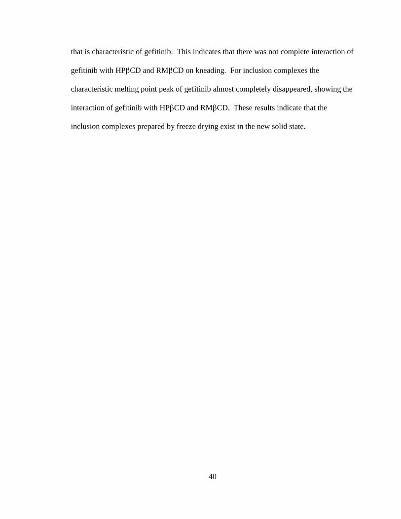
that is characteristic of gefitinib. This indicates that there was not complete interaction of
gefitinib with HP CD and RM CD on kneading. For inclusion complexes the
characteristic melting point peak of gefitinib almost completely disappeared, showing the
interaction of gefitinib with HP CD and RM CD. These results indicate that the
inclusion complexes prepared by freeze drying exist in the new solid state.

41
Gefitinib
CD
G e fit in ib - C D P H YS IC AL M IX T U R E
G efit in ib - C D K N E AD E D M IX TU R E
0 10 20 30 40 50 60 70
G efit in ib - C D C O MPLEX (FR EEZE D R IED )
2 theta
Figure 4-5: X-ray diffraction pattern of gefitinib, βCD, their physical mixture (1:1),
kneaded mixture (1:1), and inclusion complex (freeze dried, 1:1).

42
Gefitinib
HP CD
G efit in ib - H P C D P H YS IC AL MIX TU R E
G efit in ib - H P C D K N EAD ED MIXTU R E
0 1 0 2 0 3 0 4 0 5 0 6 0 7 0
G e fit in ib - H P C D C O M P L E X ( F R E E Z E D R IE D )
2 th e ta
Figure 4-6: X-ray diffraction analysis of gefitinib, HPβCD, their physical mixture (1:1),
kneaded mixture (1:1), and inclusion complex (freeze dried, 1:1).

43
Gefitinib
RM CD
Gefitinib - RM CD PHYSICAL MIXTURE
Gefitinib - RM CD KNEADED MIXTURE
0 1 0 2 0 3 0 4 0 5 0 6 0 7 0
G e fit in ib - R M C D C O M P L E X ( F R E E Z E D R IE D )
2 t h e t a
Figure 4-7: X-ray diffraction analysis of gefitinib, RMβCD, their physical mixture (1:1),
kneaded mixture (1:1), and inclusion complex (freeze dried, 1:1).

44
Gefitinib
Gefitinib – HPβCD kneaded mixture (1:1)
HPβCD
Gefitinib – HPβCD inclusion complex(1:1)
30 200100
He
at F
low
(W
/g)
Temperature oC
Figure 4-8: Differential scanning calorimetry analysis of gefitinib, HPβCD, their kneaded
mixture (1:1), and inclusion complex (freeze dried, 1:1).
45
Gefitinib
Gefitinib – RMβCD kneaded mixture (1:1)
RMβCD
Gefitinib – RMβCD inclusion complex(1:1)
30 200100
He
at F
low
(W
/g)
Temperature oC
Figure 4-9: Differential scanning calorimetry analysis of gefitinib, RMβCD, their
kneaded mixture (1:1), and inclusion complex (freeze dried, 1:1).

46
Dissolution Studies
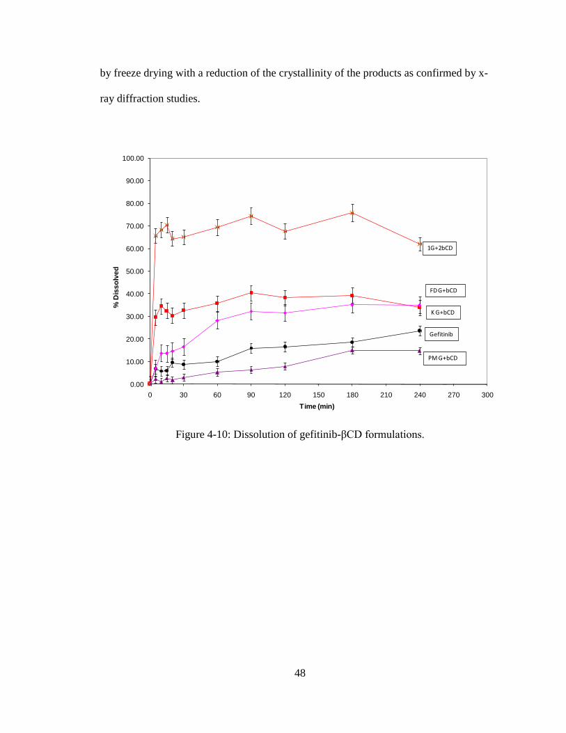
Dissolution studies on various gefitinib-cyclodextrin systems were conducted to
demonstrate the influence of the type of cyclodextrin, and the complexation method on
dissolution kinetics and the total amount of drug in solution. It is generally assumed that
the complexes show higher dissolution as compared with the pure drug.47
But the
objective is to achieve higher solubility that is characteristic of inclusion complexes. The
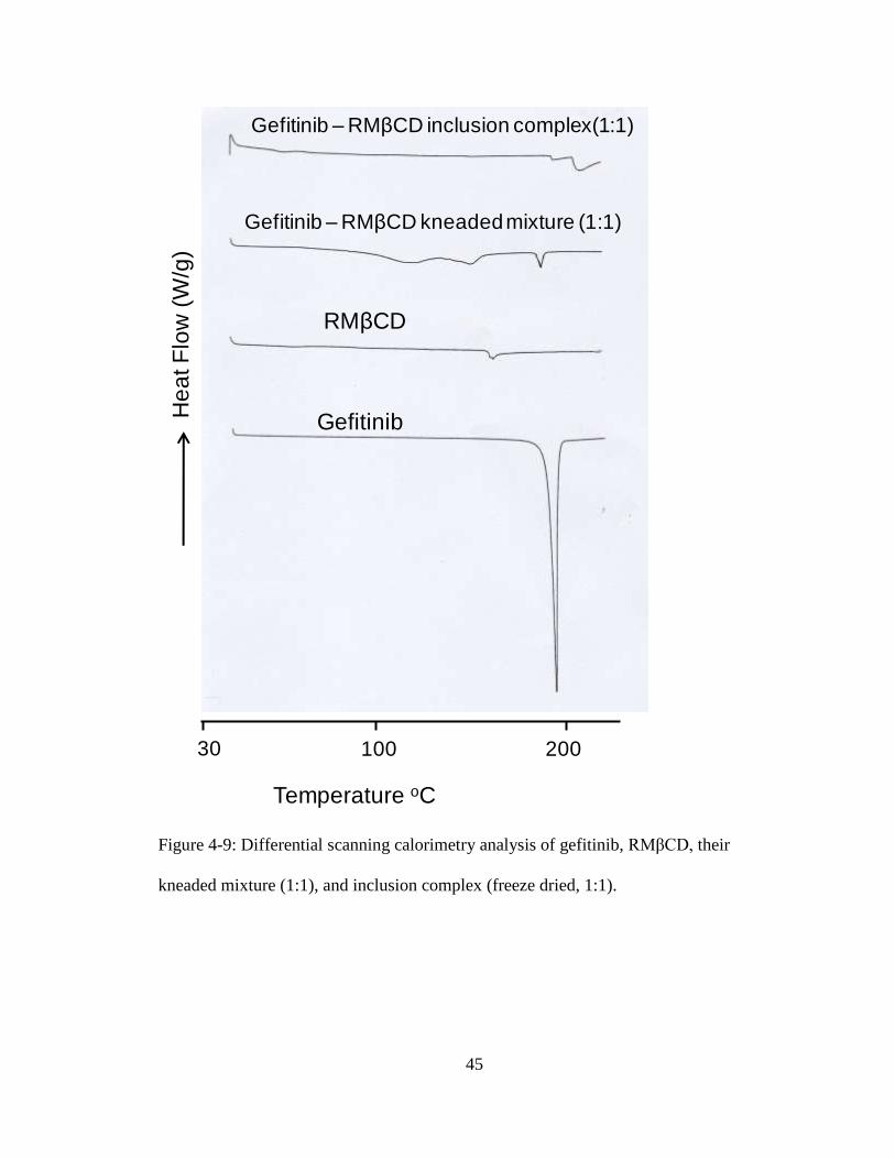
dissolution profiles of gefitinib and various binary systems of β-cyclodextrin are
presented in Figure 4-10. It is evident that the physical mixture prepared with β-
cyclodextrin did not show any improvement in the dissolution of gefitinib. The kneaded
mixture showed improvement in the dissolution of the drug. The concentration dissolved
from pure drug and kneaded mixtures at 1 hr were 10% and 30%, respectively. The βCD
inclusion complex (1:1) showed rapid dissolution; about 35% drug dissolved in 10 min,
but the total amount of drug dissolution was similar to kneaded mixture. The inclusion
complex (1:2) showed substantially higher dissolution (60% dissolution in 240 min)
compared to control (22% dissolution in 240 min).
The dissolution profiles of gefitinib and various binary systems of hydroxypropyl β-
cyclodextrin are presented in Figure 4-11. The physical mixtures prepared with
hydroxypropyl β-cyclodextrin did not show any improvement in the dissolution of
gefitinib. The kneaded mixture also showed little improvement in the dissolution of the
drug. The concentration dissolved from pure drug and kneaded mixture at 1 hr was 10%
and 20%, respectively. The HPβCD inclusion complex (1:1) showed rapid dissolution;
about 50% drug dissolved in 10 min. The total amount of drug dissolution in 240 min
was 50% which is two fold higher than the kneaded mixture. The HPβCD inclusion

47
complex (1:2) showed much higher dissolution (65% dissolution in 240 min) compared
to gefitinib alone (22% dissolution in 240 min).
The dissolution profiles of gefitinib and various binary systems of randomly
methylated β-cyclodextrin are presented in Figure 4-12. The physical mixtures prepared
with randomly methylated β-cyclodextrin did not show any improvement in the
dissolution of gefitinib. The kneaded mixture showed slight improvement in the
dissolution of the drug. The concentration dissolved from pure drug and kneaded
mixtures were 10% and 20%, respectively. The RMβCD inclusion complex (1:1)
showed rapid dissolution; about 40% drug dissolved in 10 min, and the total amount of
drug dissolution in 240 min was at ~40% which is two fold higher than the kneaded
mixture. The inclusion complex (1:2) showed substantially higher dissolution (70%
dissolution in 240 min) compared to gefitinib alone (22% dissolution in 240 min).
Two hydrophilic polymers, polyvinyl pyrrolidone and hydroxypropyl methyl
cellulose were individually blended at 10% and 25% w/w concentrations with gefitinib-
HPβCD inclusion complex (1:1). The dissolution profiles of these blends containing
PVP and HPMC as co-solubilizers are presented in Figures 4-13 and 4-14, respectively.
The addition of hydrophilic polymers markedly enhanced the dissolution of gefitinib.
The gefitinib-HPβCD (1:1) complex yielded 50% dissolution in 1 hr whereas 10% w/w
and 25% w/w PVP in association with the complex increased the dissolution to 80% and
90%, respectively. Similarly, addition of HPMC at 10% w/w and 25% w/w to the
complex increased the dissolution to 85% and 95%, respectively, in 1 hr.
The freeze dried complexes showed improved dissolution relative to the physical and
kneading complexes with all the CDs. This suggests formation of an inclusion complex
48
by freeze drying with a reduction of the crystallinity of the products as confirmed by x-
ray diffraction studies.
0.00
10.00
20.00
30.00
40.00
50.00
60.00
70.00
80.00
90.00
100.00
0 30 60 90 120 150 180 210 240 270 300
% D
iss
olv
ed
Time (min)
1G+2bCD
FD G+bCD
K G+bCD
Gefitinib
PM G+bCD
Figure 4-10: Dissolution of gefitinib-βCD formulations.

49
0.00
10.00
20.00
30.00
40.00
50.00
60.00
70.00
80.00
90.00
100.00
0 30 60 90 120 150 180 210 240 270 300
% D
iss
olv
ed
Time (min)
1G+2HPbCD
FD G+HPbCD
K G+HPbCD
Gefitinib
PM G+HPbCD
Figure 4-11: Dissolution of gefitinib-HPβCD formulations.
0.00
10.00
20.00
30.00
40.00
50.00
60.00
70.00
80.00
90.00
100.00
0 30 60 90 120 150 180 210 240 270 300
% D
iss
olv
ed
Time (min)
1G+2RMbCD
FD G+RMbCD
K G+RMbCD
Gefitinib
PM G+RMbCD
Figure 4-12: Dissolution of gefitinib-RMβCD formulations.

50
0.00
10.00
20.00
30.00
40.00
50.00
60.00
70.00
80.00
90.00
100.00
0 30 60 90 120 150 180 210 240 270 300
% D
iss
olv
ed
Time (min)
FD G+HPbCD
Gefitinib
Inclusion Complex + 25% PVP
Inclusion Complex + 10% PVP
Figure 4-13: Effect of PVP on the dissolution of gefitinib-HPβCD complex.
0.00
10.00
20.00
30.00
40.00
50.00
60.00
70.00
80.00
90.00
100.00
0 30 60 90 120 150 180 210 240 270 300
% D
iss
olv
ed
Time (min)
FD G+HPbCD
Gefitinib
Inclusion Complex + 10% HPMC
Inclusion Complex + 25% HPMC
Figure 4-14: Effect of HPMC on the dissolution of gefitinib-HPβCD complex.
51
Time (min)
Gefitinib and βCD Gefitinib and HPβCD
Physical Mix
Kneaded Freeze Dried
1:2 ratio
Physical Mix
Kneaded Freeze Dried
1:2 ratio
0 0.00 0.00 0.00 0.00 0.00 0.00 0.00 0.00
5 2.21 6.97 29.59 65.68 6.77 15.22 41.25 75.62
10 1.04 13.66 34.53 68.27 8.29 18.42 52.16 72.58
15 2.53 13.53 32.49 70.43 8.20 16.03 47.55 74.37
20 1.84 14.67 30.34 64.37 8.84 14.89 51.76 73.70
30 2.89 16.63 32.51 65.15 7.12 16.98 54.37 75.94
60 5.20 28.19 35.65 69.47 7.70 18.84 50.20 68.06
90 6.27 32.19 40.42 74.37 8.53 23.23 52.45 76.82
120 7.82 31.49 38.22 67.67 10.21 22.96 46.76 73.77
180 14.95 35.15 39.33 75.87 12.07 24.94 51.94 67.13
240 14.74 34.99 33.91 61.98 15.54 25.41 48.74 63.44
Time (min)
Gefitinib and RMβCD Gefitinib with polymer formulations
Gefitinib Physical Mix
Kneaded Freeze Dried
1:2 ratio
10% PVP
25% PVP
10% HPMC
25% HPMC
0 0.00 0.00 0.00 0.00 0.00 0.00 0.00 0.00 0.00
5 3.99 15.78 35.29 82.59 81.55 90.79 85.73 93.40 6.66
10 4.25 20.62 38.85 82.12 81.92 93.06 86.07 95.67 5.83
15 4.79 16.78 38.43 83.35 82.41 91.89 87.87 94.50 6.02
20 4.87 17.24 38.02 74.69 81.63 93.56 88.36 96.17 9.50
30 5.10 19.33 36.39 80.95 82.70 90.95 86.74 95.41 8.73
60 6.58 20.65 36.81 80.06 83.20 91.26 87.24 96.32 10.14
90 7.90 21.23 41.46 68.96 84.03 91.44 87.75 96.24 16.02
120 8.80 21.18 45.57 69.53 82.78 92.77 85.02 93.79 16.45
180 10.46 22.83 42.01 70.36 82.52 90.92 85.39 94.42 18.57
240 11.93 24.36 41.95 68.95 81.50 91.13 86.77 94.23 23.55
Table 4-2: Average percent dissolved for various gefitinib-cyclodextrin systems.

52
CONCLUSION
The association rate constants calculated from the phase solubility diagrams (735.9,
446.6 and 1021.9 M-1
) for βCD, HPβCD, and RMβCD, respectively indicate that gefitinib
can form a stable complex with all three CDs. The freeze dried formulations showed
substantial increases in the dissolution of gefitinib with all three of the cyclodextrins
compared to gefitinib alone; the most with HPβCD, followed by RMβCD, then βCD.
The kneaded mixtures showed some improvement, while the physical mixtures showed
no improvement in the dissolution of gefitinib over the drug itself. X-ray diffraction
analysis and differential scanning calorimetry (DSC) studies indicated the formation of
inclusion complexes in the solid state by various cyclodextrins. The cyclodextrin
derivatives HPβCD, and RMβCD formed amorphous complexes with gefitinib and
provided greater improvement in the solubility and dissolution of the drug.
The addition of hydrophilic polymers markedly enhanced the dissolution of gefitinib
from the CD complexes. The gefitinib-HPβCD (1:1) complex yielded 50% dissolution in
1 hr whereas the addition of 10% and 25% w/w PVP increased the dissolution to 80%
and 90%, respectively. Similarly, addition of HPMC at 10% w/w and 25% w/w to the
complex increased the dissolution of the complex to 85% and 95%, respectively in 1 hr.
In conclusion, gefitinib can form a stable inclusion complex with all three
cyclodextrins as demonstrated by the liquid and solid state complexation studies.

53
HPβCD showed the greatest improvement in the dissolution of gefitinib followed by
RMβCD and βCD, with further improvement upon the addition of PVP or HPMC.

54
REFERENCES
1. Iressa Label. U.S. Food and Drug Administration. Available at
http://www.fda.gov/cder/foi/label/2005/021399s008lbl.pdf. Accessed September 1,
2007.
2. Challa R, Ahuja A, Ali J, and Khar RK. Cyclodextrins in drug delivery: an updated
review. AAPS PharmSciTech. 2005;6:2:Article 43:E329-E357.
3. Natural Cyclodextrins. CTD Inc. Cyclodextrin Resource. Available at
http://cyclodex.com/NaturalCyclodextrins.html. Accessed September 1, 2007.
4. Loftsson, T and Masson, M. Cyclodextrins in topical drug formulations: theory and
practice. Int J Pharm. 2001;225:15-30.
5. Fromming, KH and Szejtli, J. Cyclodextrins in Pharmacy. Boston, MA; Kluwer
Academic Publishers; 1994.
6. Junco S, Casimiro T, Ribeiro N, da Ponte MN, and Marques, HC. A comparative
study of naproxen-beta cyclodextrin complexes prepared by conventional methods
and using supercritical carbon dioxide. J Incl Phenom. 2002;44:117-121.
7. Reddy MN, Rehana T, Ramakrishna S, Chowdary KPR, and Diwan PV. β-
Cyclodextrin complexes of celecoxib: molecular-modeling, characterization, and
dissolution studies. AAPS PharmSci. 2004;6:1:Article 7:1-9.
55
8. Nagarsenker MS, and Joshi MS. Celecoxib-cyclodextrin systems: characterization
and evaluation of in vitro and in vivo advantage. Drug Dev Ind Pharm.
2005;31:169-178.
9. Tozuka Y, Wongmekiat A, Sakata K, Moribe K, Oguchi T, and Yamamoto K. Co-
grinding with cyclodextrin as a nanoparticle preparation method of a poorly water
soluble drug. J Incl Phenom. 2004;50:67-71.
10. Rajendrakumar K, Pralhad T, and Madhusudan S. Comparative Study on Co-Ground
Products of Rofecoxib with ß-Cyclodextrin and Its Sulfobutyl Ether-7 Derivative in
Solution and in the Solid State. J Incl Phenom. 2004;49:259-266.
11. Rajendrakumar K, Madhusudan S, and Pralhad T. Cyclodextrin complexes of
valdecoxib: properties and anti-inflammatory activity in rat. EurJ Pharm Biopharm.
2005;60:39-46.
12. Ikeda Y, Kimura K, Hirayama F, Arima H, and Uekama K. Controlled release of a
water-soluble drug, captopril, by a combination of hydrophilic and hydrophobic
cyclodextrin derivatives. J Control Release. 2000;66:271-280.
13. Caira MR, de Vries EJC, and Nassimbeni LR. Cyclodextrin inclusion of p-
hydroxybenzoic acid esters. J Therm Anal Cal. 2003;73:647-651.
14. Caira MR, Dodds DR, and Nassimbeni LR. Polymorphism and cyclodextrin
inclusion of salbutamol laurate. J Therm Anal Cal. 2002;68:647-655.
15. Ginés JM, Arias MJ, Pérez-Martínez JI, Moyano JR, Morillo E, and Sánchez-Soto PJ.
Determination of the stoichiometry of 2,4-dichlorophenoxyacetic acid-β-cyclodextrin
complexes in solution and in solid state. Thermochim Acta. 1998;321:53-58.
56
16. Cirri M, Rangoni C, Maestrelli F, Corti G, and Mura P. Development of fast-
dissolving tablets of flurbiprofen-cyclodextrin complexes. Drug Dev Ind Pharm.
2005;31:697-707.
17. Bhandari BR, D’Arcy BR, and Padukka I. Encapsulation of Lemon Oil by Paste
Method Using -Cyclodextrin Encapsulation Efficiency and Profile of Oil Volatiles. J
Agric Food Chem. 1999;47:5194-5197.
18. Mora PC, Cirri M, Allolio B, Carli F, and Mura P. Enhancement of
dehydroepiandrosterone solubility and bioavailability by ternary complexation with
α-cyclodextrin and glycine. J Pharm Sci. 2003;92:2177-2184.
19. Gil A, Chamayou A, Leverd E, Bougaret J, Baron M, and Couarraze G. Evolution of
the interaction of a new chemical entity, eflucimibe, with -cyclodextrin during
kneading process. Eur J Pharm Sci. 2004;23:123-129.
20. Wongmekiat A, Tozuka Y, Oguchi T, and Yamamoto K. Formation of fine drug
particle by cogrinding with cyclodextrins Part II: The influence of moisture condition
during cogrinding process on fine particle formation. Int J Pharm. 2003;265:85-93.
21. Ammar HO, Salama HA, Ghorab M, and Mahmoud AA. Formulation and biological
evaluation of glimepiride cyclodextrin polymer systems. Int J Pharm.
2006;309:129-138.
22. Anguiano-Igea S, Otero-Espinar FJ, Vila-Jato JL, and Blanco-Méndez J.
Improvement of clofibrate dissolution by complexation with cyclodextrin. Int J
Pharm. 1996;135:161-166.
57
23. Veiga F, Teixeira-Dias JJC, Kedzierewicz F, Sousa A, and Maincent P. Inclusion
complexation of tolbutamide with ß-cyclodextrin and hydroxypropyl-ß-cyclodextrin.
Int J Pharm. 1996;129:63-71.
24. Mielcarek J, Czernielewska A, and Czarczyńska B. Inclusion complexes of
felodipine and amlodipine with methyl-ß-cyclodextrin. J Incl Phenom. 2006;54:17-
21.
25. Taneri F, Güneri T, Aigner Z, and Kata M. Influence of cyclodextrin complexation
on the physicochemical and biopharmaceutical properties of ketoconazole. J Incl
Phenom. 2003;47:15-23.
26. Veiga MD and Ahsan F. Influence of surfactants (present in the dissolution media)
on the release behavior of tolbutamide from its inclusion complex with ß-
cyclodextrin. Eur J Pharm Sci. 2000;9:291-299.
27. Veiga MD, Diaz PJ, and Ahsan F. Interactions of griseofulvin with cyclodextrins in
solid binary systems. J Pharm Sci. 1998;87:891-900.
28. Mura P, Faucci MT, Manderioli A, and Bramanti G. Influence of the preparation
method on the physicochemical properties of binary systems of econazole with
cyclodextrins. Int J Pharm. 1999;193:85-95.
29. Mura P, Faucci MT, Parrini PL, Furlanetto S, and Pinzauti S. Influence of the
preparation method on the physicochemical properties of ketoprofen cyclodextrin
binary systems. Int J Pharm. 1999;179:117-128.
30. Mura P, Zerrouk N, Faucci MT, Maestrelli F, Chemtob C. Comparative study of
ibuproxam complexation with amorphous ß-cyclodextrin derivatives in solution and
in the solid state. EurJ Pharm Biopharm. 2002;54:181-191.
58
31. Mura P, Bettinetti GP, Cirri M, Maestrelli F, Sorrenti M, and Catenacci L. Solid-state
characterization and dissolution properties of naproxen-arginine-hydroxypropyl-ß-
cyclodextrin ternary system. EurJ Pharm Biopharm. 2005;59:99-106.
32. Szafran B and Pawlaczyk J. Interaction between sulfaproxyline and ß-cyclodextrin in
the solution and solid states. J Incl Phenom. 1999;34:131-139.
33. Goyenechea N, Sánchez M, Vélaz I, Martín C, Martínez-Ohárriz C and Zornoza A.
Interactions of nabumetone with cyclodextrins in solution and in the solid state. J
Incl Phenom. 2002;44:283-287.
34. Agotonovic-Kustrin S, Glass BD, Brown ME, and Rotich MK. Modelling the
thermal behaviour of carboxylic acid derivatives with cylcodextrins in the solid-state.
J Therm Anal Cal. 2004;77:391-402.
35. Zingone G and Rubessa F. Preformulation study of the inclusion complex warfarin-
β-cyclodextrin. Int J Pharm. 2005;291:3-10.
36. Tenjarla S, Puranajoti P, Kasina R, and Mandal T. Preparation, characterization, and
evaluation of miconazole-cyclodextrin complexes for improved oral and topical
delivery. J of Pharm Sci. 1998;87:425-429.
37. Moyano JR, Arias-Blanco MJ, Ginés JM, and Giordano F. Solid-state
characterization and dissolution characteristics of gliclazide-ß-cyclodextrin inclusion
complexes. Int J Pharm. 1997;148:211-217.
38. Cirri M, Maestrelli F, Furlanetto S, and Mura P. Solid-state characterization of
glyburide-cyclodextrin co-ground products. J Therm Anal Cal. 2004;77:413-422.

59
39. Vertzoni M, Kartezini T, Reppas C, Archontaki H, and Valsami G. Solubilization
and quantification of lycopene in aqueous media in the form of cyclodextrin binary
systems. Int J Pharm. 2006;309:115-122.
40. Arias MJ, Moyano JR, Muñoz P, Ginés JM, Justo A, and Giordano F. Study of
omeprazole-γ-cyclodextrin complexation in the solid state. Drug Dev Ind Pharm.
2000;26:253-259.
41. Manolikar MK and Sawant MR. Study of solubility of isoproturon by its
complexation with β-cyclodextrin. Chemosphere. 2003;51:811-816.
42. Moyano JR, Arias-Blanco MJ, Ginés JM, Giordano F. Study of the complexation
behaviour of gliclazide with partially methylated β-cyclodextrin in solution and solid
state. Int J Pharm. 1997;157:239-243.
43. Taneri F, Güneri T, Aigner Z, Berkesi O, Kata M. Thermoanalytical studies on
complexes of clotrimazole with cyclodextrins. J Therm Anal Cal. 2004;76:471-479.
44. Sourbaji M, Pintye-Hódi K, Novák CS, Szabó-Révész P, Kása P Jr., and Erõs I. A
Study of Sulfadimidine-β-Cyclodextrin Mixtures. J Incl Phenom. 2000;37:299-307.
45. Higuchi T, and Connors KA. Phase solubility techniques. Adv in Analytical Chem
and Inst. 1965;4:117-212.
46. Pitha J, Szente I, and Szejtli J. Molecular encapsulation of drugs by cyclodextrins
and congeners. Controlled Drug Delivery. 1983;1:126-148.
47. Babu RJ, and Pandit JK. Enhancement of dissolution rate and hypoglycemic activity
of glibenclamide with β-cyclodextrin. STP Pharma Sci. 1995;5:196-201.

![87 Complexation Of P -Sulphonatocalix[4]Arene Complexation ...jnca.iau-saveh.ac.ir/Files/Journal/2014-05-21_12.15.39_e.pdf · 89 Complexation Of P -Sulphonatocalix[4]Arene of between](https://static.fdocuments.net/doc/165x107/5e14cb4e271e02747b0fae8f/87-complexation-of-p-sulphonatocalix4arene-complexation-jncaiau-savehacirfilesjournal2014-05-21121539epdf.jpg)