COMPARISON OF BIOACTIVITIES AND …...COMPARISON OF BIOACTIVITIES AND COMPOSITION OF CURCUMIN- FREE...
95
Clemson University TigerPrints All eses eses 12-2006 COMPARISON OF BIOACTIVITIES AND COMPOSITION OF CURCUMIN-FREE TURMERIC (CURCUMA LONGA L.) OILS FROM DIFFERENT SOURCES Yongxiang Yu Clemson University, [email protected] Follow this and additional works at: hp://tigerprints.clemson.edu/all_theses Part of the Food Science Commons is esis is brought to you for free and open access by the eses at TigerPrints. It has been accepted for inclusion in All eses by an authorized administrator of TigerPrints. For more information, please contact [email protected]. Recommended Citation Yu, Yongxiang, " COMPARISON OF BIOACTIVITIES AND COMPOSITION OF CURCUMIN-FREE TURMERIC (CURCUMA LONGA L.) OILS FROM DIFFERENT SOURCES" (2006). All eses. Paper 29.
Transcript of COMPARISON OF BIOACTIVITIES AND …...COMPARISON OF BIOACTIVITIES AND COMPOSITION OF CURCUMIN- FREE...

Clemson UniversityTigerPrints
All Theses Theses
12-2006
COMPARISON OF BIOACTIVITIES ANDCOMPOSITION OF CURCUMIN-FREETURMERIC (CURCUMA LONGA L.) OILSFROM DIFFERENT SOURCESYongxiang YuClemson University, [email protected]
Follow this and additional works at: http://tigerprints.clemson.edu/all_theses
Part of the Food Science Commons
This Thesis is brought to you for free and open access by the Theses at TigerPrints. It has been accepted for inclusion in All Theses by an authorizedadministrator of TigerPrints. For more information, please contact [email protected].
Recommended CitationYu, Yongxiang, " COMPARISON OF BIOACTIVITIES AND COMPOSITION OF CURCUMIN-FREE TURMERIC (CURCUMALONGA L.) OILS FROM DIFFERENT SOURCES" (2006). All Theses. Paper 29.

COMPARISON OF BIOACTIVITIES AND COMPOSITION OF CURCUMIN- FREE TURMERIC (CURCUMA LONGA L.) OILS FROM
DIFFERENT SOURCES
A Thesis Presented to
the Graduate School of Clemson University
In Partial Fulfillment of the Requirements for the Degree
Master of Science Food, Nutrition and Culinary Science
by Yongxiang Yu December 2006
Accepted by: Dr. Feng Chen, Committee Chair
Dr. Xi Wang Dr. Felix H Barron Dr. Jeff Adelberg

ABSTRACT
Composition, antioxidant capacities and cell inhibition properties of curcumin-
free turmeric (Curcuma longa L.) oils from different sources were evaluated by
chromatographic method, two different in vitro antioxidative activity assays (DPPH* free
radical scavenging assay and reducing power assay) and two different cancer cell lines
(Caco-2 and MCF-7). Turmeric oil A (TOA) contains zingiberene, turmerone, and ar-
turmerone, while turmeric oil B (TOB) contains 1-phellandrene and α-terpinolene as the
major compounds. The antioxidant tests showed that both turmeric oils possessed strong
free radical scavenging activities in the DPPH* free radical scavenging assay and high
reducing powers in the reducing power assay compared with standard antioxidants such
as BHT and commercial rosemary oil (RO). The free radical scavenging effect of 20
μL/mL TOA is comparable to that of 70 μL/mL TOB, comparable to that of 10 mM BHT
and better than that of 100 μL/mL RO (p<0.0001). The order of reducing powers is: 100
μL/mL TOA > 100 μL/mL TOB > 10 mM BHT > 100 μL/mL RO (p<0.0001). Among
the complex constituents in the crude TOA, ar-turmerone, turmerone, curlone and α-
terpineol were isolated and found with strong antioxidant activities. The anticancer
activity results showed that both turmeric oils possessed high inhibitive capacity against
cancer cell lines (ie. Caco-2 and MCF-7) at 20 μL/mL.

DEDICATION
I would like to dedicate this work to my husband, Shenghua Fan and my lovely daughter, Jiejie, with great thanks, love, and pride.

ACKNOWLEDGEMENTS
I would like to express my deepest appreciation and gratitude to my major
advisor, Dr. Feng Chen for providing me the opportunity to work with him and for his
encouragement, support, guidance, and trust throughout the course of my master
program.
I would like to appreciate my committee members Dr. Xi Wang (Department of
Genetics and Biochemistry), Dr. Jeff Adelberg (Department of Horticulture), and Dr.
Felix H. Barron (Department of Food Science and Human Nutrition), for their helpful
assistance and advice. Especially, I am thankful to Dr. Xi Wang for her guidance in my
cell culture research.
Thanks are extended to Dr. Ronald D. Galyean and Dr. Brandon Moore for
allowing me to use their instruments.
I also would like to thank Mr. Foster B. Wardlaw, Mrs. Elizabeth S. Halpin and
all staffs and faculties of Department of Food Science and Human Nutrition for their
friendship and help. Also, special thanks to my colleagues Dr. Hyun-jin Kim, Miss Yen-
hui Chen and Dr. (Hank) Huaping Zhang for their friendship and technical help.

TABLE OF CONTENTS
Page
TITLE PAGE.......................................................................................................... i ABSTRACT............................................................................................................ ii DEDICATION........................................................................................................ iii ACKNOWLEDGEMENTS.................................................................................... iv LIST OF TABLES.................................................................................................. viii LIST OF FIGURES ................................................................................................ ix CHAPTER 1. LITERATURE REVIEW ........................................................................ 1 Introduction........................................................................................ 1 Four Products of Turmeric................................................................. 2 Ground Turmeric ......................................................................... 2 Curry Powder ............................................................................... 3 Turmeric Oleoresin ...................................................................... 3 Turmeric Oil................................................................................. 3 Composition of Turmeric Oil............................................................. 4 Main Compounds in Turmeric........................................................... 4 Curcumin and Curcuminoids ....................................................... 4 Ar-turmerone and Turmerone ...................................................... 12 Other Bioactive Compounds........................................................ 14 Importance of the Project................................................................... 14 Reference ........................................................................................... 15
2. COMPOSITION AND BIOACTIVITIES OF CURCUMIN- FREE TURMERIC (Curcuma longa L.) OILS FROM DIFFERENT SOURCES ................................................................... 29
Abstract .............................................................................................. 29 Introduction........................................................................................ 30 Materials and Methods....................................................................... 31 Materials and Chemicals.............................................................. 31 HPLC Analysis ............................................................................ 32

vi
Table of Contents (Continued)
Page GC-MS Identification .................................................................. 33 Antioxidative Capacity ................................................................ 33 MTS Assay................................................................................... 35 Statistical Analysis............................................................................. 36 Results................................................................................................ 36 Confirming Absence of Curcumin or Curcuminoids in TOA and TOB ..................................................................... 36 Identification of Volatile Compounds in TOA and TOB................................................................................... 37 Comparing Antioxidant Activity of TOA and TOB.................... 37 Comparing Anti-cancer Activity of TOA and TOB .................... 38 Discussion.......................................................................................... 39 Conclusion ......................................................................................... 42 Reference ........................................................................................... 42
3. EVALUATION OF ANTIOXIDANT ACTIVITY OF CURCUMIN-FREE TURMERIC (Curcuma longa L.) AND IDENTIFICATION OF ITS ANTIOXIDANT CONSTITUENTS .................................................. 56
Abstract .............................................................................................. 56 Introduction........................................................................................ 57 Materials and Methods....................................................................... 59 Materials and Chemicals.............................................................. 59 HPLC Profile of Curcumin Standard and Crude TO ................................................................................. 59 Fractionation and Identification of Antioxidants From Crude TO....................................................................... 59 Antioxidative Capacity ................................................................ 61 Statistical Analysis............................................................................. 62 Results and Discussion ...................................................................... 62 Confirming No Curcumin or Curcuminoids in Crude TO ................................................................................. 62 Determination of Antioxidant Capacity of Crude TO ................................................................................. 63 Separation and Identification of Antioxidants in Crude TO ................................................................................. 64 Identification of Antioxidants in TO-I and TO-II Separated by Spherisorb Silica HPLC ..................................... 65 Reference ........................................................................................... 66

vii
Table of Contents (Continued)
Page
APPENDICES ........................................................................................................ 78 A: DPPH* Assay .................................................................................... 79 B: Reducing Power Assay ...................................................................... 81 C: Package Silica Gel Open Column...................................................... 82
LIST OF TABLES
Table Page 1.1 Turmeric (Curcuma longa L.) Oil Components ...................................... 26 2.1 Chemical Composition of TOB (1-4) and TOA (5-13) ........................... 54 2.2 IC50 Values of TOA and TOB Against Caco-2 and MCF-7 Cell Lines ............................................................................................. 55 3.1 Composition of Crude TO ....................................................................... 77
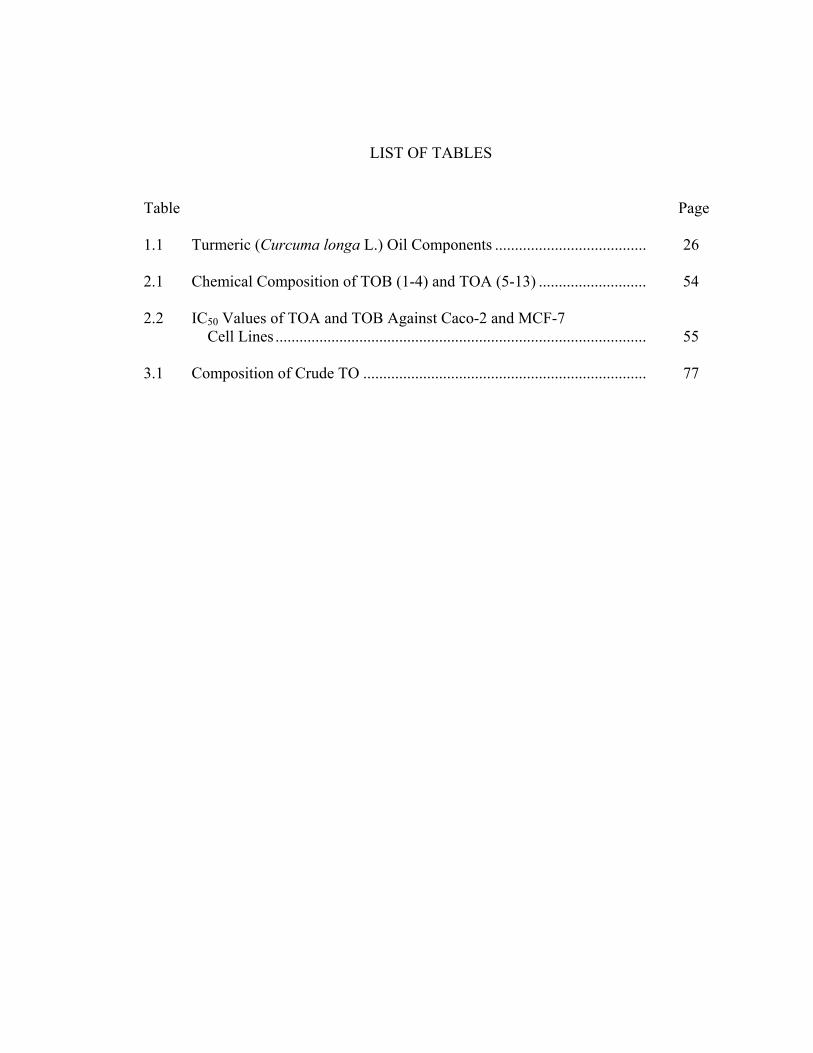
LIST OF FIGURES
Figure Page 1.1 The Leaves and Rhizomes of Turmeric .................................................. 23 1.2 Curcumin in Solution............................................................................... 24 1.3 Structures and Physical Characters of Turmerone and Ar-turmerone........................................................................................ 25 2.1 Structures of Curcumin and Curcuminoids.............................................. 48 2.2 HPLC Chromatogram of Curcuminoids (420nm) ................................... 49 2.3 Gas Chromatographic Profiles of TOA and TOB.................................... 50 2.4 Antioxidative Capacities of TOA, TOB and Standards........................... 51 2.5 Anticancer Ability of TOA and TOB ...................................................... 52 2.6 Inverted Micrographs of TOA and TOB Against MCF-7 Cells .............. 53 3.1 HPLC Chromatogram of Curcumin and Curcuminoids in Their Absorbance at 420nm................................................................ 69 3.2 Antioxidant Capacity of Crude TO ......................................................... 70 3.3 Free Radical Scavenging Activity of Fractions of Crude TO Separated by Silica Gel Open Column Chromatography .................. 71 3.4 Chromatograms of TO-I and TO-II and Their Free Radical Scavenging Activities ....................................................................... 72 3.5 Gas Chromatographic Profiles of Crude TO and Its Fractions................ 74 3.6 Structure of Turmeric Components that Have Antioxidant Activities ............................................................................................ 75 3.7 Free Radical Scavenging Assay and Reducing Power Assay of α-Terpineol .................................................................................... 76 4.1 Structure of DPPH* ................................................................................. 85

CHAPTER 1
LITERATURE REVIEW
Introduction
Turmeric (Curcuma longa L.) is a tropical herb indigenous to Southern Asia, and
probably originated on the slopes of the tropical forests of the west coast of South India.
Turmeric is a sterile triploid and has been propagated vegetatively for thousands of years.
Turmeric powder is used in food because of its spicy flavor and appealing bright yellow
color. It is used to brine pickles and to some extent in mayonnaise, relish, mustard, and
curry formulations; in non-alcoholic beverages such as orangeades and lemonades; in
breading of frozen fish sticks, etc. In all these cases, it functions predominantly as an
alternative of synthetic colors to decorate the products, as well as a flavoring ingredient
to enhance the food taste (Govindarajan, 1980).
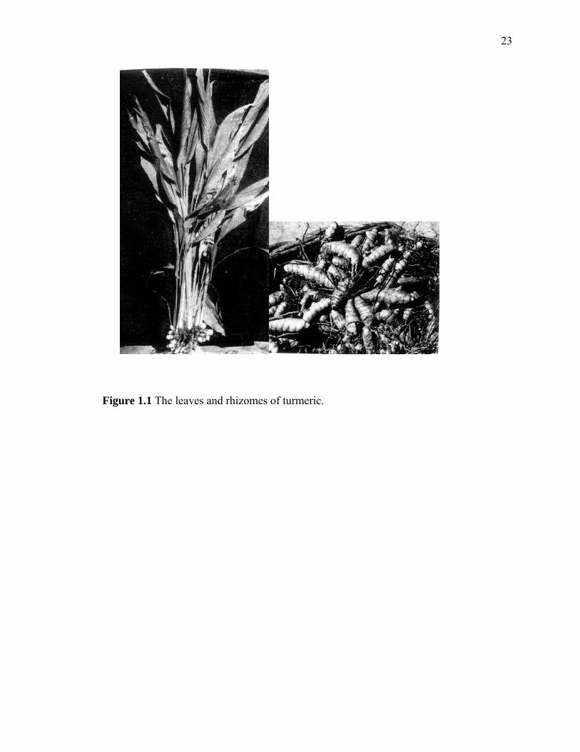
Turmeric is an erect perennial herb and grown as an annual crop. Its above-
ground morphology is mainly represented by an erect pseudo stem bearing leaves and
inflorescence. There may be 2-3 pseudostems (tillers) per plant. The height of the
pseudostem varies from 90-100 cm depending on varieties. Leaf number ranges from 7-
13 (Sasikumar, 2001). The underground rhizome that is commercially processed into the
spicy powder, consists of two distinct parts. The egg-shaped primary or mother rhizome
is an extension of the stem. Several long cylindrical multi-branched secondary rhizomes
grow downward from the primary rhizome (Govindarajan, 1980). Figure 1.1 shows the
leaves and rhizomes of turmeric.
2
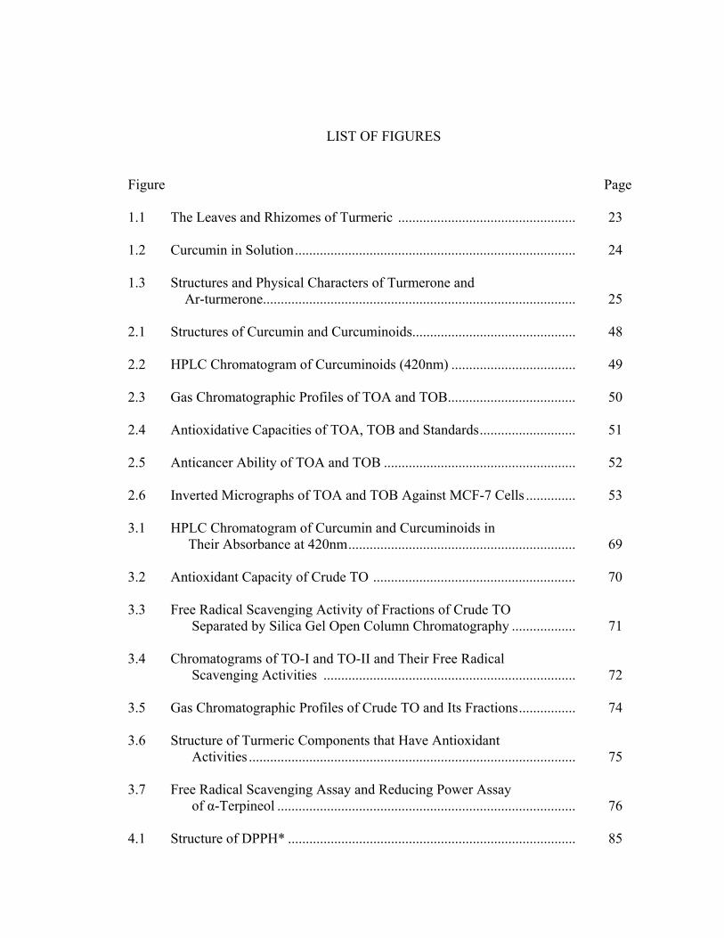
Turmeric, like ginger, belongs to the family Zingiberaceae that contains 49
genera and 1400 species. The taxonomic position of turmeric (Curcuma longa L.) is as
follows:
Kingdom Plantae
Subkingdom Tracheobionta---Vascular plants
Superdivision Spermatophyta---Seed plants
Division Magnoliophyta---Flowering plants
Class Liliopsida--Monocotyledons
Subclass Zingiberidae
Order Zingiberales
Family Zingiberaceae---Ginger family
Genus Curcuma
Species Curcuma longa L.
A wild ancestor of turmeric is called C. aromatica, while the domestic species is called
C. longa L. (Chattopadhyay, 2004). In addition, C. zedoaria Rosc and C. xanthorrhiza
Roxb are also minor crops grown for curcumin color (Sasikumar, 2001).
Four products of turmeric
Ground turmeric
Ground turmeric is made by milling the clean, dry fingers followed by disc-type
attrition mills to obtain 60-80 mesh powder. There is not much loss of quality from
oxidation of grinding turmeric (Sasikumar, 2001; Govindarajan, 1980).
3
Curry powder
Curry powder is a blend of a number of spices and herbs, in which turmeric
powder is the major component (about 40-50%) (Sasikumar, 2001) that provides
desirable color and background aroma (Govindarajan, 1980). In Asia, curry powder is a
spicy food ingredient used for seasoning dishes such as vegetables, meat, fish or eggs.
Turmeric oleoresin
Turmeric oleoresin is a mixture of curcumin, volatile oil, non-volatile fatty and
resinous material, and other active ingredients, which are extracted from ground turmeric
by solvents, used singly, in sequence or in combination (Govindarajan, 1980). For
example, acetone is a good solvent for oleoresin extraction (Sasikumar, 2001). Turmeric
oleoresin is in orange-red color and consists of an upper oily layer and a lower crystalline
layer. The content of curcumin determines the quality of turmeric oleoresin. Turmeric
oeloresin is the industrial starting material to produce pigment curcumin (Jayaprakasha,
2006).
Turmeric oil
Turmeric rhizome contains 3-5% volatile oil, which is obtained by steam
distillation of turmeric powder for about 8-10h (Sasikumar, 2001). Turmeric oil is in pale
yellow color with peppery and aromatic odor. Various sources of turmeric oils have been
reported with different chemical composition and content ascribed to the different
cultivars, different soil and climate, and age of plants that influenced the composition
(Lawrence, 2003; Cooray, 1998; Chatterjee, 2000; Hu, 1998). Because it is a byproduct

4
of curcumin industry (Saju, 1998) and has less commercial importance, the chemistry of
turmeric oil has not received much attention (Jayaprakasha, 2005).
Composition of turmeric oil
Nearly 100 chemicals have been reported in turmeric essential oils (Table 1.1)
(Raina, 2002; Jayaprakasha, 2001; Garg, 2002; Braga, 2003). Among these are terpenes
and oxygen derivative terpenoids that are believed to contribute “the character-impact”
turmeric flavor. Other minor aromas include short chain alcohols, ketones, and fatty
acids, which are degraded products of fatty acids.
Main compounds in turmeric
Curcumin and curcuminoids
Curcumin (1, 7-bis (4-hydroxy-3-methoxyphenyl)-1,6-heptadiene-3,5-dione) is
the most important compound in turmeric. It was first isolated in 1815 and its chemical
structure was determined in 1973 (Roughley, 1973). It is a yellowish crystalline, odorless
powder (mp 184-186˚C), poorly soluble in water, petroleum ether, and benzene; soluble
in ethyl alcohols, glacial acetic acid, and in propylene glycol; very soluble in acetone and
ethyl ether. Absorptive spectra of curcumin and curcuninoids are very similar, with their
maximum values at 429 and 424 nm, respectively (Govindarajan, 1980; Sharma, 2005).
In addition, curcumin is considered a non-nutritive and non-toxic chemical to mammals
even at very high doses (5-10%) by weight of diet (Weber, 2005; Samaha, 1997).

5
Extraction and separation of curcumin and curcuminoids Extraction
Many methods have been reported for extraction of curcumin and curcuminoids
(Chatterjee, 1999; Chowdhury, 2000; Surh, 2002). The most common method to extract
these compounds from turmeric powder involves sequential solvent extraction by, firstly,
using hexane to remove the non-polar volatiles and fatty compounds, and then followed
by alcohol or benzene extraction. After comparing the extraction efficiency of several
solvents, Gupta et al. confirmed that acetone was suitable for curcumin extraction since
this solvent can yield the highest recoveries of curcuminoids (Gupta, 1999). At the same
time, Chassagnez-Méndez et al. studied the feasibility of using supercritical fluid
extraction (SFE) method and confirmed that ethanol could increase the recovery of
curcumionoids in this method (Chassagnez-Méndez, 2000). Schieffer reported that
curcuminoids could be completely extracted by pressurized liquid extraction, which was
better than multiple ultrasonically-assisted extraction, although the latter was simpler
(Schieffer, 2002). Braga et al. also compared various techniques, including hydro-
distillation, low-pressure solvent extraction, Soxhlet extraction, and supercritical fluid
extraction using carbon dioxide and co-solvents (e.g., ethanol, isopropyl alcohol, and
their mixture in equal proportion) on the extraction of curcumin. It was found that the
largest yield (27%) was obtained by the Soxhlet extraction using ethanol, while the
lowest yield resulted from the hydrodistillation process (2.1%) (Braga, 2003).
Quantification by spectrophotometer Spectrophotometric methods are most often reported for the quantification of the
curcuminoids. Usually, the detective wavelength was set at 420-430 nm, at which
6
curcuminoids have their maximal spectrophotometric absorption (Ruby, 1995;
Deshpande, 1997; Braga, 2003; Leal, 2003; Manzan, 2003). A linear relationship
between the absorbance and curcumin concentration was obtained in the range of 0-15
μg/mL with a detective limit as low as 0.076 μg/mL (Tang, 2002). Although the
spectrophotometric method can quantify curcumin precisely within range mentioned
above, it is not able to quantify each curcuminoid individually.
Separation of curcuminoids by TLC Regarding the quantitative limitation of spectrophotometric method, high-
performance thin-layer chromatography was suggested as an alternative method for the
determination of individual curcuminoid in turmeric (Gupta, 1999). This method used
chloroform-methanol (95:5) as the developing solvent to separate curcuminoids that were
visualized at 430 nm. Quantitative linearity was found in the concentration range between
1 and 20 μg. At nearly the same time, Rasmussen et al. reported another simple but
efficient method using the dihydrogen phosphate impregnated silica gel TLC plate to
separate curcuminoids (Rasmussen, 2000). However, both TLC methods were restricted
by their lower resolution than HPLC.
Separation and quantification by HPLC Compared with the spectrophotometric and TLC methods, high performance
liquid chromatography (HPLC) coupled with mass spectrometer can provide more
powerful analytical capabilities in terms of quantitation and qualification. For example,
curcuminoids can be easily separated by reverse phase HPLC column under the following
condition: using the reverse phase Supelcosil LC-18 column, and mobile phase A: 1%
7
citric acid (pH adjusted to 3.0 with dilute NaOH) and B: acetonitrile (Hiserodt, 1996). A
gradient mobile phase was controlled at 1 mL/min from 50% acetonitrile with an initial
holding time of 10 min to 80% acetonitrile in 30 min. Although this method yielded
good resolution and desirable peak shape, some components in the mobile phase (citric
acid, NaOH) might clog the mass spectrometer interface leading to high back pressures,
and thus contaminate the MS ion source (Hiserodt, 1996; Schieffer, 2002). HPLC-PDA
(photo-diode array) was also used to determine curcuminoids and co-existing
sesquiterpenes (He, 1998) by mobile phase A: water (0.25% HOAc) and B: acetonitrile at
a flow rate of 0.2 mL/min. Jayaprakasha et al. used methanol as an additional mobile
phase, which included solvents A: methanol; B: 2% acetic acid; and C: acetonitrile.
Linearity was found in the concentration range between 0.0625 and 2.0 μg, with high
reproducibility and accuracy (Jayaprakasha, 2002). In these methods, curcuminoids and
sesquiterpenoids were detected at wavelengths at 426 nm and 240 nm, respectively
(Nishiyama, 2005). In addition to the gradient method, acetonitrile at isocratic flow rate
of 0.75 mL/min on LiChrosorb RP-8 column (Chowdhury, 2000) and ethanol/water
(96:4) on Spheri-5 amino column were tested (Manzan, 2003). Both have demonstrated
desirable resolution. Recently, Pak et al. reported the HPLC method could provide a
highly sensitive separation that could reliably determine the curcumin in plasma at a
concentration as low as 2.5 ng/mL (Pak, 2003).
Other methods Besides the methods mentioned above, capillary electrophoresis with
amperometric detection (CE-AD) pretreated by solid-phase extraction (SPE) was also
reported to quantify curcumin. CE-AD with SPE exhibited a low detection limit at 3x10⁻⁸
8
mol within a linear range of 7x10⁻⁴ to 3x10⁻⁶ mol/L for curcumin extracted in light
petroleum (Sun, 2002). Flow-injection analysis (FIA) with on-line UV and fluorescent
detector can provide detective limits at 30.0 ng/mL and 2.0 ng/mL, respectively (Inoue,
2001). The same research group also reported that LC/electrospray-MS could
successfully determine the trace amounts of curcuminoids in food samples with a
detective limit at 1.0 ng/mL (Inoue, 2003). Although these methods are much more
sensitive compared with the traditional TLC method, they are not efficient in separating
quantities of curcumin or curcuminoids larger than 10-20 mg. Therefore, Patel suggested
using pH-zone-refining high-speed countercurrent chromatography to separate curcumin
in large quantities (2g curcumin or 20g of turmeric powder) (Patel, 2000).
Antioxidant activity of curcumin The antioxidant activity of curcumin was found with equivalent activity to
butylated hydroxylanisole (BHA) and butylated hydroxyltoluene (BHT) (Sharma, 1976).
For example, 40 ppm curcumin could completely prevent aldehyde formation in
fermented cucumber tissue that was exposed to oxygen (Zhou, 2000). Like curcumin,
demethoxycurcumin and bisdemethoxycurcumin also exhibited antioxidant activity
(Chatterjee, 1999). In in vitro model systems, such as the phosphomolybdenum and
linoleic acid peroxidation, antioxidant capacities were in the order of curcumin >
demethoxycurcumin > bisdemethoxycurcumin (Jayaprakasha, 2006). Curcumin can act
as a scavenger of oxygen free radicals (Ruby, 1995; Rukkumani, 2004; Das, 2002). It can
protect hemoglobin from oxidation (Unnikrishnan, 1995). In an in vitro test, curcumin
could significantly inhibit the generation of reactive oxygen species (ROS) such as
superoxide anions and H2O2, and reactive nitric species (RNS), which play an important
9
role in inflammation (Joe, 1994). Also, curcumin exerted powerful inhibitory effect
against H2O2-induced damage in human keratinocytes and fibroblasts (Phan, 2001). Oral
administration of hydroalcoholic extract of C. longa decreased the susceptibility of LDL
to lipid peroxidation in a dose-dependent manner (Ramírez-Tortosa, 1999). Curcumin can
reduce the inflammatory response of ethanol by decreasing prostaglandin synthesis
(Rajakrishnan, 2001). Thus, curcumin helps maintain the membrane structure integrity
and function. It also protects against lead- and cadmium-induced lipid peroxidation in rat
brain homogenates and against lead-induced tissue damage in rat brain through metal
binding mechanism (Daniel, 2004). Administration of turmeric or curcumin to diabetic
rats reduced the blood sugar, hemoglobin (Hb) and glycosylated hemoglobin levels
significantly. Curcumin supplementation also reduced the oxidative stress encountered by
the diabetic rats (Arun, 2002). Dietary supplementation of curcumin (2%, w/v) to male
ddY mice for 30 days significantly increased the activities of glutathione peroxidase,
glutathione reductase, glucose-6-phosphate dehydrogenase and catalase as compared with
the same type mice fed normal diet. This may be one of the possible mechanisms of
cancer chemopreventive effects associated with curcumin in several animal tumor
bioassay systems (Iqbal, 2003). Since ROS have been implicated in the development of
various pathological conditions (Lee, 2004), curcumin has the potential to control these
diseases through potent antioxidant activity.
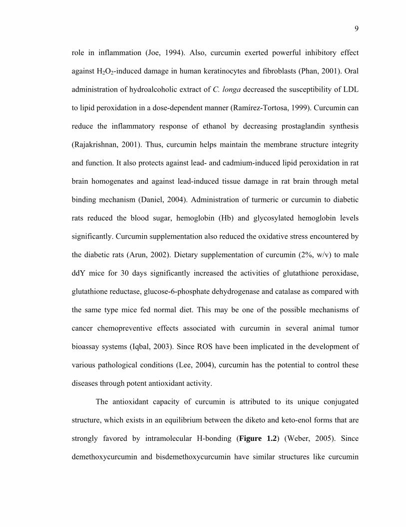
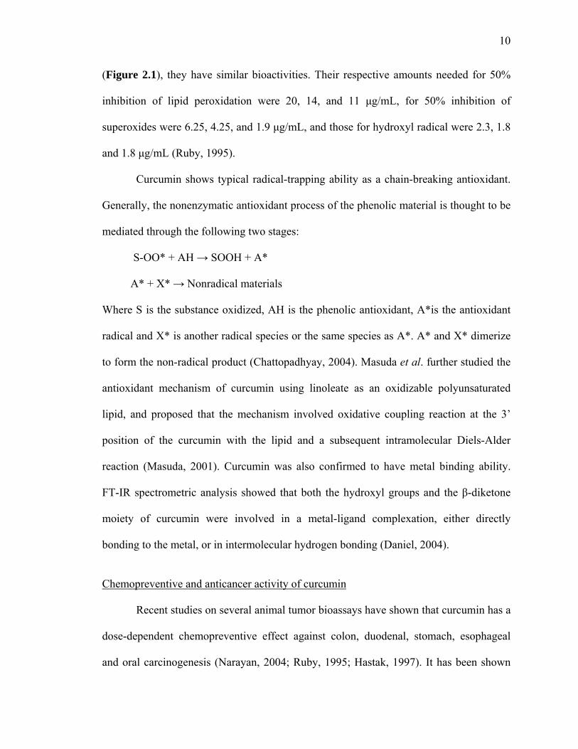
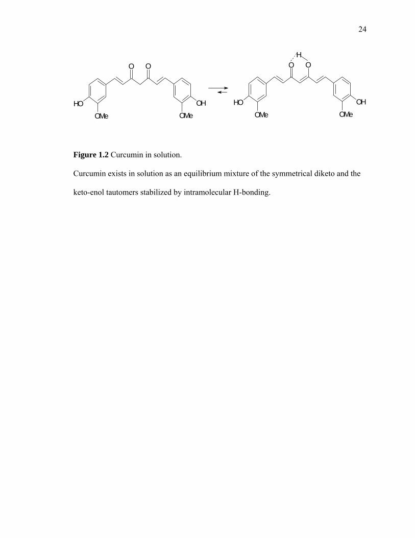
The antioxidant capacity of curcumin is attributed to its unique conjugated
structure, which exists in an equilibrium between the diketo and keto-enol forms that are
strongly favored by intramolecular H-bonding (Figure 1.2) (Weber, 2005). Since
demethoxycurcumin and bisdemethoxycurcumin have similar structures like curcumin
10
(Figure 2.1), they have similar bioactivities. Their respective amounts needed for 50%
inhibition of lipid peroxidation were 20, 14, and 11 μg/mL, for 50% inhibition of
superoxides were 6.25, 4.25, and 1.9 μg/mL, and those for hydroxyl radical were 2.3, 1.8
and 1.8 μg/mL (Ruby, 1995).
Curcumin shows typical radical-trapping ability as a chain-breaking antioxidant.
Generally, the nonenzymatic antioxidant process of the phenolic material is thought to be
mediated through the following two stages:
S-OO* + AH → SOOH + A*
A* + X* → Nonradical materials
Where S is the substance oxidized, AH is the phenolic antioxidant, A*is the antioxidant
radical and X* is another radical species or the same species as A*. A* and X* dimerize
to form the non-radical product (Chattopadhyay, 2004). Masuda et al. further studied the
antioxidant mechanism of curcumin using linoleate as an oxidizable polyunsaturated
lipid, and proposed that the mechanism involved oxidative coupling reaction at the 3’
position of the curcumin with the lipid and a subsequent intramolecular Diels-Alder
reaction (Masuda, 2001). Curcumin was also confirmed to have metal binding ability.
FT-IR spectrometric analysis showed that both the hydroxyl groups and the β-diketone
moiety of curcumin were involved in a metal-ligand complexation, either directly
bonding to the metal, or in intermolecular hydrogen bonding (Daniel, 2004).
Chemopreventive and anticancer activity of curcumin
Recent studies on several animal tumor bioassays have shown that curcumin has a
dose-dependent chemopreventive effect against colon, duodenal, stomach, esophageal
and oral carcinogenesis (Narayan, 2004; Ruby, 1995; Hastak, 1997). It has been shown

11
that administration of turmeric powder in the diet reduced tumors induced by
carcinogenic chemicals such as benzo[α]pyrene (BP) and 7, 12-dimethyl
benz[α]anthracene (DMBA) (Li, 2002). Curcumin can inhibit the growth of estrogen
positive human breast MCF-7 cells induced individually or by mixture of estrogenic
pesticides, such as endosulfane, DDT and chlordane or 17-beta estradiol (Verma, 1997).
Alcoholic extracts of turmeric (TE) and turmeric oleoresin (TOR) decreased the number
of micronucleated cells both in oral mucosal cells and in circulation lymphocytes
(Hastak, 1997).
Curcumin acts as a potent anticarcinogenic compound. Among various
mechanisms, induction of apoptosis plays an important role in its anticarcinogenic effect.
Apoptosis is an orchestrated series of events through which the cell precipitates its own
death. The stages of apoptosis include cell shrinkage, chromatin condensation, nuclear
segmentation and internucleosomal fragmentation of DNA, resulting in the generation of
apoptotic bodies (Aratanechemuge, 2002). The antiproliferative effect of curcumin is
mediated partly through inhibition of protein tyrosine kinase, c-myc mRNA expression
and bcl-2 mRNA expression (Chen, 1998). Nuclear factor (NF) - κB is known to control
cellular proliferation and apoptosis. Curcumin can also inhibit the cell proliferation and
induce apoptosis in human malignant astrocytoma cell lines and head and neck squamous
cell carcinoma (HNSCC) by inhibition of NF-κB activity (Nagai, 2005; LoTempio,
2005). For HNSCC, curcumin can induce cell apoptosis both in vitro and in vivo.
Curcumin caused lung cancer cell death by induction of apoptosis, which was
independent of p53 status of the cell lines (Pillai, 2004). Other research showed that
curcumin induced apoptosis in melanoma cell lines in a manner that was also
12
independent of p53 and the bcl-2 family (Bush, 2001). Moreover, recent research found
that curcumin had potent antiproliferative and proapoptotic effects in melanoma cells by
suppression of NF-κB and IKK activities but were independent of the B-Raf/MEK
(mitogen-activated)/ERK (extracellular signal-regulated protein kinase) and Akt pathway
(Siwak, 2005).
Other bioactivities of curcumin Curcumin has anti-inflammatory effects (Prasad, 2004). It can prevent rheumatoid
arthritis in animal model (Funk, 2006). Oral administration of 5 and 10 mg/kg curcumin
significantly reduced the duration of immobility in depressive-like behaviors (tail
suspension and forced swimming) in mice (Xu, 2005). Pretreatment with curcumin
significantly enhanced the rate of wound contraction, decreased mean wound healing
time, increased synthesis of collagen, hexosamine, DNA and nitric oxide, and improved
fibroblast and vascular densities (Jagetia, 2004).
Ar-turmerone and turmerone
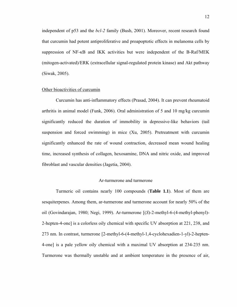
Turmeric oil contains nearly 100 compounds (Table 1.1). Most of them are
sesquiterpenes. Among them, ar-turmerone and turmerone account for nearly 50% of the
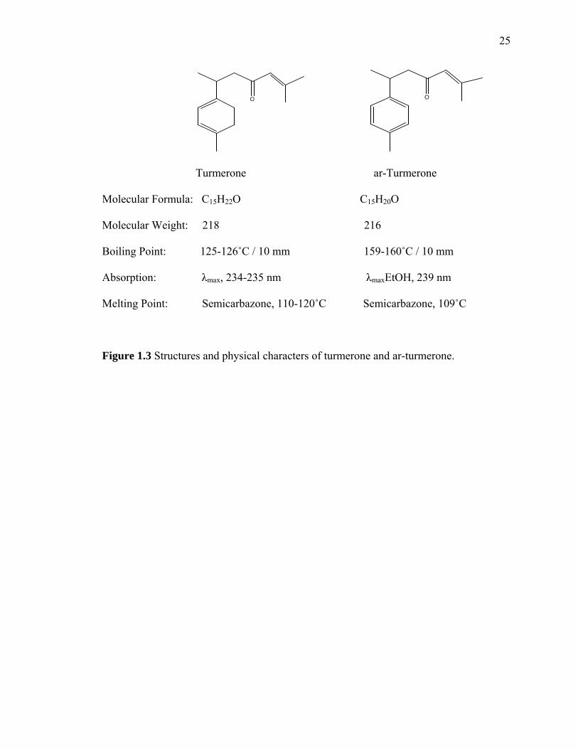
oil (Govindarajan, 1980; Negi, 1999). Ar-turmerone [(S)-2-methyl-6-(4-methyl-phenyl)-
2-hepten-4-one] is a colorless oily chemical with specific UV absorption at 221, 238, and
273 nm. In contrast, turmerone [2-methyl-6-(4-methyl-1,4-cyclohexadien-1-yl)-2-hepten-
4-one] is a pale yellow oily chemical with a maximal UV absorption at 234-235 nm.
Turmerone was thermally unstable and at ambient temperature in the presence of air,

13
yielding its dimmer, the more stable ar-turmerone (Su, 1982). Their structures and
physical characters are shown in Figure 1.3.
Extraction and separation Before 2000, little research was done on ar-turmerone and turmerone. The former
compound was identified by NMR after its extraction from turmeric powder by
petroleum ether and elution by chloroform in silica gel column (Su, 1982). However,
Manzan et al. found that petroleum ether extraction process produced less ar-turmerone
and turmerone than the steam distillation process (Manzan, 2003). Therefore, most of the
turmeric oil is now prepared by steam distillation. Recently, SFE with carbon dioxide as
solvent at 320K and 26MPa was also found to give a desirable production of turmeric oil
(Chang, 2006). The same author found the reverse-phase column Purospher RP-18 (5 μm,
125 mm x 4 mm) could successfully separate the ar-turmerone and α + β-turmerone with
following mobile phase with solvent A: 0.0025% TFA solution and solvent B:
acetonitrile at a flow rate of 1 mL/min in the following gradient program: the initial
solution of 80:20 (A:B) was held for 5 min, increased to 48% B in 5 min, to 60% in 10
min, held for 10 min, and to 100% in 10 min. The column temperature was maintained at
40˚C and UV detection was set at 254 nm.
Bioactivities of ar-turmerone and turmerone Early in 1992, Ferreira reported that ar-turmerone had antivenom effect (Ferreira,
1992). Later, ar-turmerone was reported with other biological activities such as anti-
mosquito effect (Roth, 1998), antibacterial activity (Negi, 1999) and antifungal activity
(Jayaprakasha, 2001). Turmeric oil rich in ar-turmerone, turmerone, and some other
14
oxygenated compounds showed antioxidant and antimutagenicity (Jayaprakasha, 2002).
Further research has focused on turmeric oil, ar-turmerone and turmerone. It was reported
that hexane extract from turmeric powder had antiproliferative activity, for which ar-
turmerone was a contributor (Aratanechemuge, 2002). Recent research also revealed that
ar-turmerone could induce the apoptotic activity in the K562, L1210, U937 and RBL-
2H3 cancer cell lines (Ji, 2004). In 2006, a new function was reported that ar-turmerone
had antiplatelet property. Its 50% inhibitory concentration (IC50) values for effectively
inhibiting platelet aggregation induced by collagen and arachidonic acid were 14.4 μM
and 43.6 μM, respectively (Lee, 2006).
Other bioactive compounds
Another important compound isolated from the aqueous extract of turmeric is a
protein, called turmeric anti-oxidant protein (TAP). It is a heat stable protein and has
antioxidant activity. Its maximal absorbance is 280 nm. The antioxidant activity may be
mediated through the protection of the –SH group of the enzyme (Selvam, 1995).
Importance of the project
According to the Food and Agriculture Organization of the United Nation, over
2400 tons of turmeric is imported annually in the USA for consumer use in recent years.
Since turmeric oil is the major by-product of curcumin production, it is important to
identify more bioactive chemicals in the curcumin-free turmeric oil and explore their
bioactivities. The current usage of turmeric oil as fuel (Saju, 1998) and for
aromatheraphy (Sasikumar, 2005) may not fully utilize this undervalued resource. Thus,
the specific objectives of this study were:

15
(1) To profile the composition of turmeric oil;
(2) To assay the bioactivities, such as antioxidant, anti-cancer activities of turmeric
oil;
(3) To separate and identify the individual bioactive compounds.
References
Aratanechemuge, Y.; Komiya, T.; Moteki, H.; Katsuzaki, H.; Imai, K.; Hibasami, H. Selective induction of apoptosis by ar-turmerone isolated from turmeric (Curcuma longa L.) in two human leukemia cell lines, but not in human stomach cancer cell line. International Journal of Molecular Medicine. 2002, 9, 481-484.
Arun, N.; Nalini, N. Efficacy of turmeric on blood sugar and polyol pathway in diabetic
albino rats. Plant Foods for Human Nutrition. 2002, 57, 41-52. Braga, M.E.M.; Leal, P.F.; Carvalho, J.E.; Meireles, M.A.A. Comparison of yield,
composition, and antioxidant activity of turmeric (Curcuma longa L.) extracts obtained using various techniques. Journal of Agricultural and Food Chemistry. 2003, 51, 6604-6611.
Bush, J.A.; Cheung, K.J.; Li, G. Curcumin induces apoptosis in human melanoma cells
through a Fas Receptor/Caspase-8 pathway independent of p53. Experimental Cell Research. 2001, 271, 305-314.
Chang, L.; Jong, T.; Huang, H.; Nien, Y.; Chang, C. Supercritical carbon dioxide
extraction of trumeric oil form Curcuma longa Linn and purification of turmerones. Separation and Purification Technology. 2006, 47, 119-125.
Chassagnez-Méndez, A.L. Machado, N.T.; Araujo, M.E.; Maia, J.G.; Meireles, M.A.A.
Supercritical CO2 extraction of curcumins and essential oil from the rhizomes of turmeric (Curcuma longa L.). Industrial & Engineering Chemistry Research. 2000, 39, 4729-4733.
Chatterjee, S.; Desai, S.R.P.; Thomas, P. Effect of γ-irradiation on antioxidant activity of
turmeric extracts. Food Research International. 1999, 32, 487-490. Chatterjee, S.; Variyar, P.S.; Gholap, A.S.; Padwal-Desai, S.R.; Bongirwar, D.R. Effect
of γ-irradiation on the volatile oil constituents of turmeric (Curcuma longa). Food Research International. 2000, 33(2), 103-106.

16
Chattopadhyay, I.; Biswas, K.; Bandyopadhyay, U.; Banerjee, R.K. Turmeric and curcumin: biological actions and medicinal applications. Current Science. 2004, 87(1), 44-53.
Chen, H.; Huang, H. Effect of curcumin on cell cycle progression and apoptosis in
vascular smooth muscle cells. British Journal of Pharmacology. 1998, 124, 1029-1040.
Chowdhury, H.; Walia, S.; Saxena, V.S. Isolation, characterization and insect growth
inhibitory activity of major turmeric constituents and their derivatives against Schistocerca gregaria (Forsk) and Dysdercus koenigii (Walk). Pest Management Science. 2000, 56, 1086-1092.
Cooray, N.F.; Jansz, E.R.; Ranatunga, J. Effect of maturity on some chemical
constutuents of turmeric (Curcuma longa L.). Journal of National Science Council of Sri Lanka. 1998, 16 (1), 39-51.
Daniel, S.; Limson, J.L.; Dairam, A.; Watkins, G.M.; Daya, S. Through metal binding,
curcumin protects against lead- and cadmium-induced lipid peroxidation in rat brain homogenates and against lead-induced tissue damage in rat brain. Journal of Inorganic Biochemistry. 2004, 98, 266-275.
Das. K.C.; Das, C.K. Curcumin (diferuloylmethane), a singlet oxygen (1O2) quencher.
Biochemical and Biophysical Research Communications. 2002, 295, 62-66. Deshpande, S.S.; Ingle, A.D.; Maru, G.B. Inhibitory effects of curcumin-free aqueous
turmeric extract on bezo[α]pyrene-induced forestomach papillomas in mice. Cancer Letters. 1997, 118, 79-85.
Ferreira, L.A.F.; Henriques, O.B.; Andreoni, A.A.S.; Vital, G.R.F.; Campos, M.M.C.;
Habermehl, G.G.; Moraes, V.L.G.D. Antivenom and biological effects of ar-turmerone isolated from Curcuma longa L. (Zingiberaceae). Toxicon. 1992, 30, 1211-1218.
Funk, J.L.; Oyarzo, J.N.; Frye, J.B.; Chen, G.; Lantz, R.C.; Jolad, S.D.; Sólyom, A.M.;
Timmermann, B.N. Turmeric extracts containing curcuminoids prevent experimental rheumatoid arthritis. Journal of Natural Products. 2006, 69, 351-355.
Garg, S.N.; Mengi, N.; Patra, N.K.; Charles, R; Kumar, S. Chemical examination of the
leaf essential oil of Curcuma longa L. from the north Indian plains. Flavour and Fragrance Journal. 2002, 17, 103-104.
Govindarajan, V.S. Turmeric---chemistry, technology, and quality. CRC Critical Reviews
in Food Science and Nutrition. 1980, 12, 199-301.
17
Gupta, A.P.; Gupta, M.M.; Kumar, S. Simultaneous determination of curcuminoids in curcuma samples using high performance thin layer chromatography. Journal of Liquid Chromatography & Related Technologies. 1999, 22, 1561-1569.
Hastak, K.; Lubri, N.; Jakhi, S.D.; More, C.; John, A.; Ghaisas, S.D.; Bhide, S.V. Effect
of turmeric oil and turmeric oleoresin in cytogenetic damage in patients suffering from oral submucous fibrosis. Cancer Letters. 1997, 116, 265-269.
He, X.G.; Lin, L.Z.; Lian, L.Z.; Lindenmaier, M. Liquid chromatography-electrospray
mass spectrometric analysis of curcuminoids and sesquiterpenoids in turmeric (Curcuma longa). Journal of Chromatography A. 1998, 818, 127-132.
Hiserodt, R.; Hartman, T.G.; Ho, C.T.; Rosen, R.T. Characterization of powdered
turmeric by liquid chromatography-mass spectrometry and gas chromatography-mass spectrometry. Journal of Chromatography A. 1996, 740, 51-63.
Hu, Y.; Du, Q.; Tang, Q. Determination of chemical constituents of the volatile oil from
Curcuma Longa by gas chromatography-mass spectrometry. Chinese Journal of Chromatography. 1998, 16 (6), 528-529.
Inoue, K.; Hamasaki, S.; Yoshimura. Y.; Yamada, M.; Nakamura, M.; Ito, Y.; Nakazawa,
H. Validation of LC/Electrospray-MS for determination of major curcuminoids in foods. Journal of Liquid Chromatography & Related Technologies. 2003, 26, 53-62.
Inoue, K.; Yoshimura, Y.; Nakazawa, H. Evaluation of the turmeric (Curcuma longa L.)
based on the flow-injection analysis with ultraviolet and fluorometric detections. Analytical Letters. 2001, 34 (10), 1711-1718.
Iqbal, M.; Sharma, S.D.; Okazaki, Y.; Fujisawa, M.; Okada, S. Dietary supplementation
of curcumin enhances antioxidant and phase II metabolizing enzymes in ddY male mice: possible role in protection against chemical carcinogenesis and toxicity. Pharmacology & Toxicology. 2003, 92, 33-38.
Jagetia, G.C.; Rajanikant, G.K. Role of curcumin, a naturally occurring phenolic
compound of turmeric in accelerating the repair of excision wound, in mice whole body exposed to various doses of γ-radiation. Journal of Surgical Research. 2004, 120, 127-138.
Jayaprakasha, G.K.; Jena, B.S.; Negi, P.S.; Sakariah, K.K.; Evaluation of antioxidant
activities and antimutagenicity of turmeric oil: a byproduct from curcumin production. Verlag der Zeitschrift für Naturforschung. 2002, 57c, 828-835.

18
Jayaprakasha, G.K.; Negi, P.S.; Anandharamakrishnan, C.; Sakariah, K.K. Chemical composition of turmeric oil – a byproduct from turmeric oleoresin industry and its inhibitory activity against different fungi. Verlag der Zeitschrift für Naturforschung. 2001, 56c, 40-44.
Jayaprakasha, G.K.; Rao, L.J.M.; Sakariah, K.K. Improved HPLC method for the
determination of curcumin, demethoxycurcumin, and bisdemethoxycurcumin. Journal of Agricultural and Food Chemistry. 2002, 50, 3668-3672.
Jayaprakasha, G.K.; Rao, L.J.M.; Sakariah, K.K. Chemistry and biological activities of C.
longa. Trends in Food Science & Technology. 2005, 16, 533-548. Jayaprakasha, G.K.; Rao, L.J.M.; Sakariah, K.K. Antioxidant activities of curcumin,
demethoxycurcumin and bisdemethoxycurcumin. Food Chemistry. 2006, 98, 720-724.
Ji, M.; Choi, J.; Lee, J.; Lee, Y. Induction of apoptosis by ar-turmerone on various cell
lines. International Journal of Molecular Medicine. 2004, 14, 253-256. Joe, B.; Lokesh, B.R. Role of capsaicin, curcumin and dietary n-3 fatty acids in lowering
the generation of reactive oxygen species in rat peritoneal macrophages. International Journal of Biochemistry, Biophysics and Molecular Biology. 1994, 1224, 255-263.
Lawrence, B.M. Turmeric oil. In Essential oils 1995-2000. Carol Stream: Allured
Publishing Corporation, 2003; pp. 375-380. Leal, P.F.; Braga, M.E.M. Sato, D.N.; Carvalho, J.E.; Marques, M.O.M.; Meireles,
M.A.A. Functional properties of spice extracts obtained via supercritical fluid extraction. Journal of Agricultural and Food Chemistry. 2003, 51, 2520-2525.
Lee, H.S. Antiplatelet property of Curcuma longa L. rhizome-derived ar-turmerone.
Bioresource Technology. 2006, 97, 1372-1376. Lee, J.; Koo, N.; Min, D.B. Reactive oxygen species, aging, and antioxidative
nutraceuticals. Comprehensive Reviews in Food Science and Food Safety. 2004, 3, 21-33.
Li, N.; Chen, X.; Liao, J.; Yang, G.; Wang, S.; Josephson, Y.; Han, C.; Chen, J.; Huang,
M.; Yang, C. Inhibition of 7, 12-dimethylbenz[α]anthracene (DMBA)-induced oral carcinogenesis in hamsters by tea and curcumin. Carcinogenesis. 2002, 23 (8), 1307-1313.

19
LoTempio, M.M.; Veena, M.S.; Steele, H.L.; Ramamurthy, B.; Ramalingam, T.S.; Cohen, A.N.; Chakrabarti, R.; Srivatsan, E.S.; Wang, M.B. Curcumin suppresses growth of head and neck squamous cell carcinoma. Clinical Cancer Research. 2005, 11, 6994-7002.
Manzan, A.C.C.M.; Toniolo, F.S.; Bredow, E.; Povh, N.P. Extraction of essential oil
pigments from Curcuma longa [L.] by steam distillation and extraction with volatile solvents. Journal of Agricultural and Food Chemistry. 2003, 51, 6802-6807.
Masuda, T.; Maekawa, T.; Hidaka, K.; Bando, H.; Takeda, Y.; Yamaguchi, H. Chemical
studies on antioxidant mechanism of curcumin: analysis of oxidative coupling products from curcumin and linoleate. Journal of Agricultural and Food Chemistry. 2001, 49(5), 2539-2547.
Nagai, S.; Kurimoto, M.; Washiyama, K.; Hirashima, Y.; Kumanishi, T.; Endo, S.
Inhibition of cellular proliferation and induction of apoptosis by curcumin in human malignant astrocytoma cell lines. Journal of Neuro-Oncology. 2005, 74, 105-111.
Narayan, S. Curcumin, a multi-functional chemopreventive agent, blocks growth of colon
cancer cells by targeting β-catenin-mediated transactivation and cell-cell adhesion pathways. Journal of Molecular Histology. 2004, 35, 301-307.
Negi, P.S.; Jayaprakasha, G.K.; Rao, L.J.M.; Sakariah, K.K. Antibacterial activity of
turmeric oil: A byproduct from curcumin manufacture. Journal of Agricultural and Food Chemistry. 1999, 47, 4297-4300.
Nishiyama, T.; Mae, T.; Kishida, H.; Tsukagawa, M.; Mimaki, Y.; Kuroda, M.; Sashida,
Y.; Takahashi, K.; Kawada, T.; Nakagawa, K.; Kitahara, M. Curcuminoids and sesquiterpenoids in turmeric (Curcuma longa L.) suppress an increase in blood glucose level in type 2 diabetic KK-Ay mice. Journal of Agricultural and Food Chemistry. 2005, 53, 959-963.
Pak, Y.; Patek, R.; Mayersohn, M. Sensitive and rapid isocratic liquid chromatography
method for the quantitation of curcumin in plasma. Journal of Chromatography B. 2003, 796, 339-346.
Pfeiffer, E.; Höhle, S.; Solyom, A.M.; Metzler, M. Studies on the stability of turmeric
constituents. Journal of Food Engineering. 2003, 56, 257-259. Phan, T.; See, P.; Lee, S.; Chan, S. Protective effects of curcumin against oxidative
damage on skin cells in vitro: its implication for wound healing. The Journal of Trauma Injury, Infection, and Critical Care. 2001, 51, 927-931.

20
Pillai, G.R.; Srivastava, A.S.; Hassanein, T.I.; Chauhan, D.P.; Carrier, E. Induction of apoptosis in human lung cancer cells by curcumin. Cancer Letters. 2004, 208, 163-170.
Prasad, N.S.; Raghavendra, R.; Lokesh, B.R.; Naidu, K.A. Spice phenolics inhibit human
PMNL 5-lipoxygenase. Prostaglandins, Leukotrienes and Essential Fatty Acids. 2004, 70, 521-528.
Priyadarsini, K.I.; Maity, D.K.; Naik, G.H.; Kumar, M.S; Unnikrishnan, M.K. Satav,
J.G.; Mohan, H. Role of phenolic O-H and methylene hydrogen on the free radical reactions and antioxidant activity of curcumin. Free Radical Biology and Medicine. 2003, 35, 475-484.
Raina, V.K.; Srivastava, S.K.; Jain, N.; Ahmad, A.; Syamasundar, K.V.; Aggarwal, K.K.
Essential oil composition of Curcuma longa L. cv. Roma from the plains of northern India. Flavour and Fragrance Journal. 2002, 17, 99-102.
Rajakrishnan, V.; Menon, V.P. Potential role of antioxidants during ethanol-induced
changes in the fatty acid composition and arachidonic acid metabolites in male Wistar rats. Cell Biology and Toxicology. 2001, 17, 11-22.
Ramírez-Tortosa, M.C.; Mesa, M.D.; Aguilera, M.C.; Quiles, J.L.; Baró, L.; Ramírez-
Tortosa. C.L.; Martinez-Victoria, E.; Gil, A. Oral administration of a turmeric extract inhibits LDL oxidation and has hypocholesterolemic effects in rabbits with experimental atherosclerosis. Atherosclerosis. 1999, 147, 371-378.
Rasmussen, H.B.; Christensen, S.B.; Kvist, L.P.; Karazmi, A. A simple and efficient
separation of the curcumins, the antiprotozoal constituents of Curcuma longa. Planta Medica. 2000, 66, 396-397.
Roth, G.N.; Chandra, A.; Nair, M.G. Novel bioactivities of Curcuma longa constituents.
Journal of Natural Products. 1998, 61, 542-545. Roughley, P.J.; Whiting, D.A. Experiments in the biosynthesis of curcumin. Journal of
the Chemical Society ---Perkin Transcations I. 1973, 20, 2379-2388. Ruby, A.J.; Kuttan, G.; Babu, K.D.; Rajasekharan, K.N.; Kuttan, R. Anti-tumor and
antioxidant activity of natural curcuminoids. Cancer letters. 1995, 94, 79-83. Rukkumani, R.; Aruna, K.; Varma, P.S.; Rajasekaran, K.N.; Menon, V.P.M. Comparative
effects of curcumin and an analog of curcumin on alcohol and PUFA induced oxidative stress. Journal of Pharmacy & Pharmaceutical Sciences. 2004, 7 (2), 274-283.

21
Saju, K.A.; Venugopal, M.N.; Mathew, M.J. Antifungal and insect-repellent activities of essential oil of turmeric (Curcuma longa L.). Current Science. 1998, 75 (7), 660-662.
Samaha, H.S.; Kelloff, G.J.; Steele, V.; Rao, C.V.; Reddy, B.S. Modulation of apoptosis
by sulindac, curcumin, phenylethyl-3-methylcaffeate, and 6-phenylhexyl isothiocyanate: apoptotic index as a biomarker in colon cancer chemoprevention and promotion. Cancer Research. 1997, 57, 1301-1305.
Sasikumar, B. Turmeric. In Handbook of Herbs and Spices I; Peter, K.V., Eds.;
Cambridge: Woodhead Publishing Limited, 2001; pp. 297-310. Sasikumar, B. Genetic resources of Curcuma: diversity, characterization and utilization.
Plant Genetic Resources. 2005, 3 (2), 230-251. Schieffer, G.W. Pressurized liquid extraction of curcuminoids and curcuminoid
degradation products from turmeric (Curcuma longa) with subsequent HPLC assays. Journal of Liquid Chromatography & Related Technologies. 2002, 25, 3033-3044.
Selvam, T.; Subramanian, L.; Gayathri, R.; Angayarkanni, N. The antioxidant activity of
turmeric (Curcuma longa). Journal of Ethnopharmacology. 1995, 47, 59-67. Sharma, O.P. Antioxidant activity of curcumin and related compounds. Biochemical
Pharmacology. 1976, 25, 1811-1812. Sharma, R.A. Gescher, A.J. Steward, W.P. Curcumin: the story so far. European Journal
of Cancer. 2005, 41, 1955-1968. Siwak, D.R.; Shishodia, S; Aggarwal, B.B.; Kurzrock, R. Curcumin-induced
antiproliferative and proapoptotic effects in melanoma cells are associated with suppression of IκB kinase and nuclear factor κB activity and are independent of the B-Raf/mitogen-activated/extracellular signal-regulated protein kinase pathway and the Akt pathway. Cancer. 2005, 104, 879-890.
Su, H.C.F.; Horvat, R.; Jilani, G. Isolation, purification, and characterization of insect
repellents from Curcuma longa L. Journal of Agricultural and Food Chemistry. 1982, 30, 290-292.
Sun, X.; Gao, C.; Cao, W.; Yang, X.; Wang, E. Capillary electrophoresis with
amperometric detection of curcumin in Chinese herbal medicine pretreated by solid-phase extraction. Journal of Chromatography A. 2002, 962, 117-125.
Surh, Y. Anti-tumor promoting potential of selected spice ingredients with antioxidative
and anti-inflammatory activities: a short review. Food and Chemical Toxicology. 2002, 40, 1091-1097.

22
Tang, B.; Ma, L.; Wang, H.; Zhang, G. Study on the supramolecular interaction of curcumin and β-cyclodextrin by spectrophotometry and its analytical application. Journal of Agricultural and Food Chemistry. 2002, 50, 1355-1361.
Unnikrishnan, M.K.; Rao, M.N.A. Inhibition of nitrite induced oxidation of hemoglobin
by curcuminoids. Die Pharmazie. 1995, 50, 490-492. Verma, S.P.; Salamone, E.; Goldin, B. Curcumin and genistein, plant natural products,
show synergistic inhibitory effects on the growth of human breast cancer MCF-7 cells induced by estrogenic pesticides. Biochemical and Biophysical Research Communications. 1997, 233, 692-696.
Weber, W.M.; Hunsaker, L.A.; Abcouwer, S.F.; Deck, L.M.; Jagt, D.L.V. Anti-oxidant
activities of curcumin and related enones. Bioorganic & Medicinal Chemistry. 2005, 13, 3811-3820.
Xu, Y.; Ku, B.; Yao, H.; Lin, Y.; Ma, X.; Zhang, Y.; Li, X. The effects of curcumin on
depressive-like behaviors in mice. European Journal of Pharmacology. 2005, 518, 40-46.
Zhou, A.; McFeeters, R.F.; Fleming, H.P. Inhibition of formation of oxidative volatile
components in fermented cucumbers by ascorbic acid and turmeric. Journal of Agricultural and Food Chemistry. 2000, 48, 4910-4912.
23
Figure 1.1 The leaves and rhizomes of turmeric.
24
OMeOH
O O
OMeOH OH
OMe
O
OHOMe
OH
Figure 1.2 Curcumin in solution. Curcumin exists in solution as an equilibrium mixture of the symmetrical diketo and the
keto-enol tautomers stabilized by intramolecular H-bonding.
25
O
O
Turmerone ar-Turmerone
Molecular Formula: C15H22O C15H20O
Molecular Weight: 218 216
Boiling Point: 125-126˚C / 10 mm 159-160˚C / 10 mm
Absorption: λmax, 234-235 nm λmaxEtOH, 239 nm
Melting Point: Semicarbazone, 110-120˚C Semicarbazone, 109˚C
Figure 1.3 Structures and physical characters of turmerone and ar-turmerone.

26
Table 1.1 Turmeric (Curcuma Longa L.) oil components. Chemicals KIa Sourcesb
(E)-α-atlantone 1743 1 (E)-Nerolidol 1550 1 (E)-β-Farnesene 1448 1 (E)-β-Ocimene 1042 1 (E)-γ-Atlantone 4 (Z)-α-Atlantone 4 (Z)-β-Ocimene 1030 1 (Z)-γ-Atlantone 4 1,8-Cineole 1023 1, 3, 4 10-epi-γ-Eudesmol 1617 1 1-Bisabolone 1714 1 1-epi-Cubenol 4 1-Hexen-3-ol 772 1 2-Decanol 1190 1 2-Nonanol 1092 1 3-Buten-2-ol 810 1 6S, 7R-Bisabolone 4 α-Bisabolol 1673 1 α-Cadinene 1436 1 α-cis-Bergamotene 2 α-Fenchol 1099 1 α-Guaiene 1451 1 α-Humelene 1445 1, 3 α-Longefoline 1399 1 α-Muurolene 1492 1 α-Patchouline 1456 1 α-Phellandrene 999 1 α-Pinene 935 1, 4 ar-Curcumene 1471 1, 3,4 ar-Turmerol 1559 1, 3, 4 ar-Turmerone 1640 1, 3, 4 α-Selinene 1496 1 α-Terpinene 1010 1, 4 α-Terpineol 1176 1 α-Thujene 925 1, 4 α-Turmerone 1650 1, 4 Borneol 1157 1, 3, 4 Camphene 946 1 Camphene hydrate 1142 1 Camphor 1130 1 Capric acid 1355 1 Carvacrol 1288 1

27
Table 1.1 Turmeric (Curcuma Longa L.) oil components (continued). Chemicals KIa Sourcesb
Carvone 1217 1 Caryophyllene oxide 1566 1 Cinnamyl cinnamate 2053 1 cis-3-Hexenol 849 1 cis-Carveol 1206 1 cis-Carvotanacetol 1199 1 cis-Carvyl acetate 1321 1 cis-Linalool oxide (furanoid) 1062 1 cis-Sabinol 1178 1, 3 cis-Sesquisabinene hydrate 1539 1 cis-β-Elemenone 1579 1 Curdione 1689 1 Curlone 2 Curzerene 1480 1 Elimicin 1524 1 epi-Curzerenone 1584 1 Furanodienone 1752 1 Geramacrene-B 1543 1 Geranial 1252 1 Geraniol 1242 1 Geranyl acetate 1359 1 Geranyl butyrate 1531 1 Geranyl formate 1278 1 Geranyl hexanoate 1722 1 Germacrene-D 1461 1 Germacrone 1669 1 Heptadeconic acid 2 Heptan-2-ol 887 1 Heptan-2-one 875 1 Hexadecanoic acid 2 Humilene epoxide II 1612 1 iso-Borneol 1148 1 iso-Bornyl acetate 1268 1 Linalool 1082 1, 3 Linalyl acetate 1247 1 Linalyl propionate 1314 1 Methyl eugenol 1366 1 Myrcene 984 1 Myrtenal 1173 1, 3 Myrtenol 1184 1 n-Heptyl salicylate 1789 1 n-Hexan-2-ol 787 1

28
Table 1.1 Turmeric (Curcuma Longa L.) oil components (continued). Chemicals KIa Sourcesb
p-Cymene 1014 1, 2, 3 p-Cymene-8-ol 3 Perillaketone 1230 1 p-Methyl acetophenone 1124 1 Sabinene 966 1 Sabinyl acetate 1289 1 Safrole 1262 1 T-cadinol 1624 1 Terpinen-4-ol 1166 1, 3 Terpinolene 1084 1 Tetradecane 1393 1 Thymol 1281 1 Thymol acetate 1348 1 trans-Linalool oxide (furanoid) 1077 1 trans-p-Menth-2-en-1-ol 1109 1 trans-Sesquisabinene hydrate 1602 1 Undecanol 1299 1 Undecanone 1273 1 Virdifloral 1591 1 γ-Elemene 1423 1 γ-Terpinene 1053 1 Zingiberene 1487 1, 3, 4 β-(Z)-Farnesene 2 β-Bisabolene 1501 1, 3, 4 β-Bisabolol 1659 1 β-Caryophyllene 1418 1, 3, 4 β-Curcumene 1510 1 β-Elemene 1385 1 β-Eudesmol 1629 1 β-Patchouline 1378 1 β-Pinene 974 1, 3 β-Sesquiphellandrene 1516 1, 3, 4 β-Turmerone 1681 1, 3 δ-3-Carene 1005 1 δ-Elemene 1331 1
a from source 1; b 1 (Raina, 2002), 2 (Jayaprakasha, 2001), 3 (Garg, 2002), 4 (Braga, 2003).
CHAPTER 2
COMPOSITION AND BIOACTIVITIES OF CURCUMIN-FREE TURMERIC
(Curcuma longa L.) OILS FROM DIFFERENT SOURCES
Abstract
Composition, antioxidant capacities and inhibitive anticancer activities of
curcumin-free turmeric (Curcuma longa L.) oils from different sources were evaluated by
GC-MS, two different in vitro antioxidative assays (i.e., DPPH* free radical scavenging
assay and reducing power assay), and MTS bioassays against two cancer cell lines (i.e.,
Caco-2 and MCF-7), respectively. The commercial turmeric oil A (TOA) was mainly
composed of zingiberene, ar-curcumene, turmerone, ar-turmerone and curlone; while
turmeric oil B (TOB) contained 1-phellandrene, cymene, 1,8-cineole, and α-terpinolene
as the major components. The antioxidative tests showed that both the two curcumin-free
turmeric oils possessed strong free radical scavenging activities and reducing powers
compared with the standard antioxidant products such as BHT and rosemary oil (RO).
The free radical scavenging effect increased with the increasing concentrations of TOA
or TOB and then leveled off as the concentration further increased. The free radical
scavenging effect can be saturated by 20 μL/mL TOA or 70 μL/mL TOB (90%) when
scavenging 0.25 mM DPPH* solution, which was comparable to 10 mM BHT (86%), and
was better than 100 μL/mL rosemary oil (68%) at the same reaction condition. The
reducing power is also dose-dependent for both TOA and TOB. There is a significant
difference between TOA, TOB, BHT and RO in reducing power (p<0.0001) in the order
of 100 μL/mL TOA > 100 μL/mL TOB > 10 mM BHT > 100 μL/mL RO. The anticancer

30
experiments showed that both TOA and TOB had antiproliferative activities against
human colon cancer cell (Caco-2) and human breast cancer cell (MCF-7). The IC50
values of TOA and TOB were 1.66 μL/mL and 21.5 μL/mL for Caco-2 cell line, 0.122
μL/mL and 1.219 μL/mL for MCF-7 cell line, respectively.
Introduction
Research on bioactive principles of essential oils extracted from various herbs and
spices has become increasingly popular. Essential oils have been discovered to have
many functional properties such as antimicrobial and anti-inflammatory activities
(Vardar-Ünlü, 2003; Hammer, 1999; Griffin, 1999; Güllüce, 2003). Besides these
activities, many essential oils have been qualified as natural antioxidants (Kim, 2004;
Ruberto, 2000; Lin, 2003; Domínguez, 2005; Zhang, 2006) and proposed as potential
substitutes of synthetic antioxidants in food preservation. Currently, many research
groups are focusing their investigation on the pharmacological actions of essential oils
from aromatic and medicinal plants. Many studies have shown that natural antioxidants
in various aromatic and medicinal plants are closely related to the reduction of chronic
diseases such as cancer, cardiovascular disease, diabetes, arthritis (Fridovich, 1999; Zhu,
2002; Covacci, 2001). Among them, turmeric is one of the most important studied
subjects because of its demonstrated broad spectrum of activities.
The main compounds in turmeric include curcumin (1, 7-bis (4-hydroxy-3-
methoxyphenyl)-1, 6-heptadien-3, 7-dione) and two curcuminoids, demethoxycurcumin
and bisdemethoxycurcumin (Figure 2.1). They contribute the yellow color to turmeric
and have received increasing attention because of their many bioactivities. Current
research shows that curcumin and curcuminoids have antioxidant, antivirus,

31
antimutagenicity, anti-arthritic and anti-thrombotic activities (Braga, 2003; Apisariyakul,
1995; Taher, 2003; Funk, 2006). Turmeric oil is usually obtained from turmeric powder
by steam distillation or from curcumin removed turmeric oleoresin (CRTO) that was
extracted with hexane and then concentrated. Turmeric oil accounts for 3-5% of the total
weight of raw turmeric rhizome. Turmeric oil is pale yellow with peppery and aromatic
odor. The reported major compounds in turmeric oil include some terpenic aromas such
as α-phellandrene, 1,8-cineol, zingiberene, ar-turmerone, turmerone, curlone, β-sesqui-
phellandrene, dehydro-zingerone, etc (Sasikumar, 2001). Because turmeric oil is the by-
product of curcumin industry and has long been considered with very little commercial
value (Saju, 1998), it was neglected in an extent of degree in its potent biological
activities, including its antioxidant activity. So it is our current interest to explore the
antioxidant activity and other bioactivities of curcumin-free turmeric oil.
Turmeric and turmeric oil from different sources may have different chemical
profiles with different bioactivities. However, the production of these bioactive
compounds is controlled by plant genotypes, postharvest processing (e.g., drying,
extraction, etc) and environmental conditions, such as temperature, humidity, light, soil,
etc. In this study, we compared two turmeric oils from two commercial sources for their
chemical profiles and antioxidant activities, as well as anti-cancer activities.
Materials and methods
Materials and chemicals
Two commercially available turmeric oils were designated as TOA (Aromaland
Co. USA) and TOB (Bianca Rosa Co. Canada). 2, 2-diphenyl-1-picrylhydrazyl (DPPH*),
butylated hydroxytoluene (BHT), β-caryophyllene, Minimum Essential Medium (Eagle),

32
MEM non-essential amino acids, Rosewell Park Memorial Institute 1640 (RPMI 1640)
medium, curcumin standard, rosemary oil, sodium bicarbonate, trypsin EDTA solution,
sodium pyruvate, α-terpinolene and sterile cell culture penicillin were purchased from
Sigma Chemical Co. (St. Louis, MO). Trichloroacetic acid and all solvents were obtained
from Fisher Scientific (Suwanee, GA). Potassium ferricyanide and ferric chloride were
obtained from J. T. Baker Chemical Co. (Phillipsburg. NJ). New born calf serum and
fetal bovine serum were purchased from Hyclone (Loga, Utah). 3-(4,5-dimethylthiazol-2-
yl)-5-(3-carboxymethoxyphenyl)-2-(4-sulfophenyl)-2H-tetrazolium, inner salt (MTS)
was provided by Promega (Madison, WI). Human breast carcinoma cell lines (MCF-7)
and human colon carcinoma (Caco-2) were obtained from the American Type Culture
Collection (Rockville, MD).
HPLC analysis
A Waters C18 reverse phase column (XterraTM, 4.6 x 150 mm, 5 μm) and a
photodiode-array (PDA) detector were equipped in a Shimadzu LC-10AT HPLC system
(Kyoto, Japan). UV spectra were recorded in the region of 200-800 nm. The mobile
phase consisted of solvents (A) water (0.25% acetic acid) and (B) acetonitrile was
programmed in the following gradient condition: 0-17 min, 40-60% B; 17-32 min, 60-
100% B; 32-38 min, 100% B; 38-40 min, 100-40% B; 41 min, stop. Total flow rate of the
mobile phase was controlled at 0.6 mL/min. Injection volume was 20 μL. Curcumin
standard (dissolved in ethanol, 0.15 mg/mL), TOA [dissolved in DCM/methanol (v/v
1:2), 100 μL/mL], TOB (dissolved in ethanol, 100 μL/mL) were analyzed by the same
method described above.

33
GC-MS identification
A Shimadzu GC-MS system consisting of a GC-17A with a QP5050 Mass
Spectrometer (Kyoto, Japan) was equipped with a DB-5 capillary column (60 × 0.25 mm,
thickness 0.25 µm; J&W Scientific, Folsom, CA, USA) for all chemical quantitative and
qualitative analyses in this research. The oven temperature was programmed from 60 to
280˚C at a ramp rate of 8˚C/min and held at 280˚C for 30 min. The injector and ion
source temperatures were set at 180 and 290˚C, respectively. The detector voltage was
70eV, and the scanning mass range was m/z 43-350. Helium was used as the carrier gas at
a column flow rate of 1.2 mL/min. Samples in volume of 2 µL was injected with a split
ratio of 1:1. Identification of compounds was based on comparison of their mass spectra
and retention indices (RIs) with those of the authentic standards. RIs were calculated
using series of n-alkanes (C8-C26). If standard compounds were not available, each
unknown compound was tentatively identified by comparing the mass spectrum with that
of the Wiley and NIST mass spectral databases and the previously published RIs obtained
under the same conditions (Negi, 1999; Chatterjee, 2000; Adams, 2001).
Antioxidative capacity
The antioxidant capacities of turmeric oils were determined by two methods: the
DPPH* free radical scavenging assay and the reducing power assay. BHT and rosemary
oil (RO) were used as standards for above in vitro bioassays in antioxidant comparison.
DPPH* free radical scavenging assay DPPH* free radical scavenging assay followed the procedure described by
Yamaguchi et al. (1998) with minor modification. In this antioxidant method, DPPH* is a
34
stable free radical and shows a characteristic absorption at 517 nm due to its odd electron.
As the odd electron of the radical becomes paired off in the presence of a hydrogen
donor, or a free radical scavenging antioxidant, the absorption strength decreased
resulting in decolorization that is stoichiometric with the number of electrons captured. In
this test, 0.4 mL of different concentrations of samples was mixed with 0.4 mL of 0.25
mM DPPH* solution. The mixtures were shaken vigorously and left in the dark at room
temperature for 30 min. A control consisted of 0.4 mL solvents instead of sample.
Because both TOA and TOB have colors, so we also add 0.4 mL turmeric oil (at each
concentration) to 0.4 mL solvent as blank, respectively. The absorbance of the mixtures
was measured spectrophotometrically at 517 nm. The scavenging effect of DPPH* free
radical was calculated by using the following formula:
Scavenging effect (%) = 100)nm517atcontrolofabsorbance
blanknm517atsampleofabsorbance1( ×−
− (1)
Reducing power assay The reducing power of two turmeric oils was determined by the method described
by Yen et al. (1995) and Chung et al. (2002) with minor modification. TOA, TOB and
RO were dissolved in acetone to prepare solutions in concentrations of 1, 5, 10, 20, 40,
70, 100 μL/mL. BHT was prepared in concentrations of 0.1, 0.5, 1, 2, 4, 7, 10 mM.
Solutions of 0.5 mL samples were mixed with 1 mL of 1% potassium ferricyanide
[K3Fe(CN)6]. The mixture was incubated at 50˚C for 20 min. Then 1 mL of
trichloroaceteic acid (10%) was added to the mixture and centrifuged at 3000 rpm for 10
min. The upper layer of the solution (1 mL) was mixed with distilled water (1 mL) and
35
FeCL3 (0.2 mL, 0.1%) to read the absorbance at 700 nm. Higher absorbance of the
reaction mixture indicated greater reducing power.
Triplicates were performed for each concentration of the tested samples and
standards in these two methods. The experiments were repeated three times on different
days.
MTS assay
Both MCF-7 and Caco-2 cells were plated at 10,000 cells/cm2 in 75 cm2 cell
culture flasks. The medium for MCF-7 was Minimum Essential Medium with 10%
newborn calf serum, 1% sodium pyruvate, 1% non-essential amino acid and 1%
penicillin. The medium for Caco-2 cells was formulated by RPMI with 10% fetal bovine
serum and 1% sodium pyruvate, and 1% penicillin. After 48h, cancer cells were fed with
fresh medium. The cells were determined with trypan blue. Exponentially growing cells
were harvested, counted, diluted and seeded into the 96-well tissue culture plate at 104
cells/well (100 μL). Then the plate was incubated in a 5% CO2 incubator at 37˚C for 24h.
The diluted TOA and TOB were added to obtain the final concentrations 0.02, 0.2, 2, 20
μL/mL, respectively. The solvent ethyl acetate was added as the control. The plates were
incubated in 5% CO2 incubator at 37˚C for 48h. Then, 20 μL of MTS reagent was added
into each well. These plates were incubated again in the CO2 incubator at 37˚C for 2h.
The MTS assay was based on the reduction of a soluble tetrazolium salt, by
mitochondrial dehydrogenase of viable tumor cells, into an insoluble colored formazan
product, which can be measured spectrophotometrically after dissolution. The enzymatic
activity and the number of formed formazan were proportional to the number of living
cells. This can generally be explained by cell inhibition or cell viability (Endrini, 2002).

36
Cell inhibition can be reflected by the spectrophotometrical absorbance recorded at 490
nm and calculated by the following formula:
Cell inhibition (%) = (1- %100)nm490atcontrolofabsorbancenm490atsampleofabsorbance
× (2)
The 50% inhibition concentration (IC50) was used to compare the inhibitive
activity of TOA and TOB. It was defined as the chemical concentration causing 50%
inhibition of cell growth. Triplicates were performed for each concentration of the tested
samples and the experiments were repeated three times on different days.
Statistical analysis
The data of the antioxidant activities and cell inhibitions of TOA, TOB and
standards were subjected to the analysis of variance (ANOVA). Treatment means were
separated by the least significant difference (LSD at p<0.05). Analyses were performed
using the statistical software SAS 9.1 operated on the Windows system (SAS Institute
Inc., Cary, NC).
Results
Confirming absence of curcumin or curcuminoids in TOA and TOB
Curcumin standard separated by the HPLC showed three separate peaks (Figure
2.2), i.e., (1): bisdemethoxycurcumin; (2): demethoxycurcumin; and (3): curcumin. Their
maximum absorbent wavelength was around 420 nm (Sasikumar, 2001). Since TOA and
TOB did not show absorbance after 300 nm, and there were no matching peaks with the
curcumin standards, it was concluded that there were no curcumin or curcuminoids in the
TOA or TOB, which excluded the possible interference from those chemicals on the
antioxidant and anticancer tests.

37
Identification of volatile compounds in TOA and TOB
GC-MS analyses of the chemical profiles of the turmeric oils indicated that the
two oils had quite different chemical compositions (Figure 2.3 A and B). There were 9
important volatile compounds with larger RIs (1400~1800) identified in TOA which
contained trans-caryophyllene, farnesene, ar-curcumene, zingiberene, β-bisabolene, β-
sesquiphellandrene, ar-turmerone, turmerone, and curlone (Figure 2.3 A, Table 2.1). In
contrast, there were only 4 major components with small RIs (less than 1100) found in
TOB which include 1-phellandrene, cymene, 1,8-cineole, and α-terpinolene (Figure 2.3
B, Table 2.1). 1-phellandrene was found to be the major compound (58%) in TOB
(Table 2.1).
Comparing antioxidant activity of TOA and TOB
Since the major compounds in two turmeric oils were remarkably different, it was
of our interest to investigate their antioxidant activities regarding their free radical
scavenging activity and the reducing power. The DPPH* free radical scavenging activity
of both TOA and TOB increased with the increase of concentrations and then leveled off
after 20 μL/mL TOA or 70 μL/mL TOB. 20 μL/mL TOA or 70 μL/mL TOB could
scavenge the free radicals at 90%, which was comparable to the scavenging effect of 10
mM BHT (86%) and higher than that of 100 μL/mL rosemary oil (68%) at the same
reaction condition (p<0.0001) (Figure 2.4 A).
In the reducing power assay, the presence of reductants (antioxidants) would
result in reducing Fe3+/ferricyanide complex to the ferrous form. The Fe2+ could be
monitored at 700 nm by measuring the formation of Perl’s Prussian blue. Figure 2.4 B
shows the reducing power of the turmeric oils, BHT and rosemary oil. The reducing

38
powers of TOA and TOB are in a dose-dependant manner that is similar to their
scavenging capacities in the DPPH* assay. Also, all the samples showed same ranking
orders in both DPPH* free radical scavenging assay and the reducing power assay. The
reducing power was in the order of 100 μL/mL TOA > 100 μL/mL TOB > 10 mM BHT
> 100 μL/mL RO (p<0.0001). These antioxidant tests demonstrated that both TOA and
TOB were strong electron donors and could react with and convert free radicals to more
stable products, and terminate the radical chain reactions (Yen, 1995).
Comparing anti-cancer activity of TOA and TOB
The anticarcinogenic properties of TOA and TOB were determined using the
MTS assay. TOA and TOB inhibited Caco-2 and MCF-7 cancer cells proliferation in a
dose dependent manner. At the concentration of 0.02 μL/mL, both TOA and TOB
showed no or very little effect on the two cancer cell lines. The range of 0.2-20 μL/mL of
TOA caused 23-69% cell inhibition against Caco-2 cells and 58-77% cell inhibition
against MCF-7 cells, while the same concentration range of TOB caused 11-53% cell
inhibition against Caco-2 cells and 10-80% cell inhibition against MCF-7 cells (Figure
2.5 A and B). The IC50 for Caco-2 cell and MCF-7 cell is 1.66 μL/mL (TOA), 21.5
μL/mL (TOB) and 0.122 μL/mL (TOA) and 1.219 μL/mL (TOB), respectively (Table
2.2). In case of Caco-2 cell line, at the range of 0.2-20 μL/mL, TOA exhibited higher
inhibitive activity than TOB at the same concentration (p<0.0001) (Figure 2.5 A). In case
of MCF-7, at the range of 0.2-2 μL/mL, TOA also exhibited higher inhibitive activity
than TOB at the same concentration. Figure 2.6 shows the inverted micrographs of the
effect of TOA and TOB against MCF-7 cells at the concentration of 2 μL/mL. But at the

39
highest concentration tested of 20 μL/mL, there is no significant difference between TOA
and TOB (p<0.0001) (Figure 2.5 B).
Discussion
Many researchers found that turmeric had strong antioxidant activity (Ruby,
1995; Rukkumani, 2004; Das, 2002; Zhou, 2000; Chatterjee, 1999) which was ascribed to
curcumin or curcuminoids. However, HPLC analysis of our samples confirmed that there
was absence of curcumin and curcuminoids. Therefore, we concluded that other
compounds besides curcumin and curcuminoids were responsible for these activities.
As we now know, BHT, the synthesized antioxidant as food additive, was found
to have carcinogenic side-effects (Ito, 1985) restricting its use (Mikova, 2001). The
essential oil of rosemary (Rosmarinus officinalis L.) has both a desirable aromatic flavor
and a strong antioxidant activity (Man, 2000). Because of these characters, rosemary oil
has been used in many food products as a desirable flavoring ingredient and preservative
(Özcan, 2003; Richheimer, 1996; Cuvelier, 1996; Man, 2000; Estévez, 2005; McCarthy,
2001; Sebranek, 2005). In this study, we found both TOA and TOB (100 μL/mL)
possessed strong DPPH* free radical scavenging capacity and high reducing power when
compared to the same concentration of rosemary oil and 10 mM BHT at the same
reaction conditions.
Although both turmeric oils were extracted from turmeric rhizome, they had
totally different chemical profiles. Sasikumar reported that turmeric oil obtained from
turmeric powder by steam distillation contained about 60% turmerone, 25% zingiberene
and small quantities of d-α-phellandrene, d-sabinene, cineole and forneol (Sasikumar,
2001). Other monoterpenes, sesquiterpenes, and their oxygenated derivatives were also

40
found in turmeric oil (Purseglove, 1981). Nevertheless, among turmeric oils from
different sources, sesquiterpenes, ar-turmerone and turmerone usually comprise more
than 50% of the oils. In TOA, ar-turmerone and turmerone accounts for about 40%,
which is similar to the previous reports (Lawrence, 2003). But TOB contained more
volatile chemicals such as 1-phellandrene (58%), cymene, 1,8-cineole, and α-terpinolene
instead of less volatile chemicals like turmerone or ar-turmerone. Hu et al. studied the
chemical constituents of turmeric rhizome essential oils of Chinese origin and found the
major compounds were α-curcumene (34%), 1-(3-cyclopentylpropyl)-2,4-dimethyl-
benzene (22%) and β-sesquiphellandrene (15%), but they could not detect ar-turmerone
and turmerone (Hu, 1998). Gopalam et al. compared the constituents in turmeric oil from
different cultivars and found there was significant quantitative variation in the main
compounds among different cultivars (Gopalam, 1987). Cooray et al. examined the effect
of maturity on the major components of the rhizome oil produced from a single turmeric
cultivar grown in Sri Lanka, and found that sesquiterpenes (ar-turmerone and turmerone)
increased while monoterpenes (1,8-cineole and α-phellandrene) decreased with maturity
(Cooray, 1988). Chane-Ming et al. examined the essential oils from rhizomes, leaves and
flowers of the same cultivar and found that major compounds in rhizomes were
turmerones (37%), terpinolene (15.8%) and zingiberene (11.8%), but the leaves and
flowers had similar chemical profile and were particularly rich in terpinolene with 76.8%
(leaves) and 67.4% (flowers) (Chane-Ming, 2002). Taking into account of the above
factors, the different compositions of our two tested turmeric oils (TOA and TOB) may
be from different cultivars, or from the different plant parts, or from different maturity, or
planted in different climate and soil, or extracted by different methods.

41
Although the chemical profiles of TOA and TOB are quite different, both of them
have shown potent antioxidant capacities and strong growth inhibition against cancer
cells. TOA showed a significantly higher antioxidant capacity than TOB in both in vitro
bioassays within the tested concentration range. TOB contained some volatile flavors like
α-terpinolene that was reported with a strong DPPH* free radical scavenging activity
(Kim, 2004). In contrast, TOA contained less volatile chemicals including sesquiterpenes
and oxygenated terpenics such as ar-turmerone, turmerone and curlone. These
oxygenated compounds were reported with a potent antioxidant activity in the β-
carotene-linoleate assay (Jayaprakasha, 2002). In addition, it was reported that ar-
turmeorne had an insect repellent activity (Su, 1982), antibacterial activity (Negi, 1999),
and antifungal activity (Jayaparakasha, 2001). Aratanechemuge et al. also reported that
ar-turmerone could inhibit the growth of leukemia cells by inducing apoptosis
(Aratanechemuge, 2002). Similarly in our study, oxygenated compounds-rich TOA also
exhibited strong anticancer capacities against the Caco-2 and MCF-7 cancer cell lines, for
which TOA at the concentration of 20 μL/mL showed inhibitive activity of 69% and
77%, respectively. In general, TOA showed a higher cell inhibition than TOB. The MCF-
7 cell line seemed more sensitive to TOA than to TOB based on their IC50 values that
were 0.122 μL/mL (TOA) and 1.219 μL/mL (TOB). However, due to the lack of
chemical standards of ar-turmerone, turmerone and curlone, we could not determine their
individual contribution and synergistic effects on their antioxidant activities in these two
bioassays. Their inhibitive capacities against the Caco-2 and MCF-7 cancer cell lines and
the molecular mechanism of how of ar-turmerone, turmerone and curlone inhibit cell
growth will be further investigated by our research group.
42
Conclusion
Since many researches have found some chronic diseases are closely related to the
existence of excessive free radicals (Grajek, 2005; Fridovich, 1999; McCord, 2000),
intake of exogenous phytochemical antioxidants that act as scavengers of reactive oxygen
species (ROS) and metal chelators may help to protect healthy human cells and reduce
oxidative damages. In this study, both curcumin-free TOA and TOB have shown potent
antioxidant and anticancer activities. In other words, such bioactive activities are
contributed by the terpenes and oxygenated terpenes rather than the more extensively
studied curcumin. In addition, since turmeric oil is considered non-toxic (Joshi, 2003)
and has aromatic flavors, it can be an alternative natural antioxidant to BHT and
rosemary oil for many food formulations, especially suitable for some ethnic foods.
References
Adams, R.P. Identification of essential oil components by gas chromatography /
quadrupole mass spectroscopy. Carol Stream, Illinois: Allured Publishing Corporation, 2001.
Apisariyakul, A.; Vanittanakom, N.; Buddhasukh, D. Antifungal activity of turmeric oil
extracted from Curcuma longa (Zingiberaceae). Journal of Ethnopharmacology. 1995, 49, 163-169.
Aratanechemuge, Y.; Komiya, T.; Moteki, H.; Katsuzaki, H.; Imai, K.; Hibasami, H.
Selective induction of apoptosis by ar-turmerone isolated from turmeric (Curcuma longa L.) in two human leukemia cell lines, but not in human stomach cancer cell line. International Journal of Molecular Medicine. 2002, 9, 481-484.
Braga, M.E.M.; Leal, P.F.; Carvalho, J.E.; Meireles, M.A.A. Comparison of yield,
composition, and antioxidant activity of turmeric (Curcuma longa L.) extracts obtained using various techniques. Journal of Agricultural and Food Chemistry. 2003, 51, 6604-6611.
Chane-Ming, J.; Vera, R.; Chalchat, J.; Cabassu, P. Chemical composition of essential
oils from rhizomes, leaves and flowers of Curcuma longa L. from Reunion Island. Journal of Essential Oil Research. 2002, 14, 249-251.

43
Chatterjee, S.; Desai, S.R.P.; Thomas, P. Effect of γ-radiation on antioxidant activity of turmeric (Curcuma longa L.) extracts. Food Research International. 1999, 32(7), 487-490.
Chatterjee, S.; Variyar, P.S.; Gholap, A.S.; Padwal-Desai, S.R.; Bongirwar, D.R. Effect
of γ-irradiation on the volatile oil constituents of turmeric (Curcuma longa). Food Research International. 2000, 33, 103-106.
Chung, Y.C.; Chang, C.T.; Chao, W.W.; Lin, C.F.; Chou, S.T. Antioxidative activity and
safety of the 50% ethanolic extract from red bean fermented by Bacillus subtilis IMR-NK1. Journal of Agricultural and Food Chemistry. 2002, 50, 2454-2458.
Cooray, N.F.; Jansz, E.R.; Ranatunga, J. Effect of maturity on some chemical
constituents of turmeric (Curcuma longa L.). Journal of National Science Council of Sri Lanka. 1998, 16 (1), 39-51.
Covacci, V.; Torsello, A.; Palozza, P.; Sgambato, A; Romano, G.; Boninsegan, A.;
Cittadini, A.; Wolf, F.I. DNA oxidative damage during differentiation of HL-60 human promyelocytic leukemia cells. Chemical Research in Toxicology. 2001, 14, 1492-1497.
Cuvelier, M.; Richard, H.; Berset, C. Antioxidative activity and phenolic composition of
pilot-plant and commercial extracts of sage and rosemary. Journal of the American Oil Chemists’ Society. 1996, 73, 645-652.
Das, K.C.; Das, C.K. Curcumin (diferuloylmethane), a singlet oxygen (1O2) quencher.
Biochemical and Biophysical Research Communications. 2002, 295, 62-66. Domínguez, M.; Nieto, A.; Marin, J.C.; Keck, A.; Jeffery, E.; Céspedes, C.L. Antioxidant
activities of extracts from Barkleyanthus salicifolius (Asteraceae) and Penstemon gentianoides (Scrophulariaceae). Journal of Agricultural and Food Chemistry. 2005, 53, 5889-5895.
Endrini, S.; Rahmat, A.; Ismail, P.; Hin, T.Y. Anticarcinogenic properties and antioxidant
activity of henna (Lawsonia inermis). Journal of Medical Science. 2002, 2, 194-197.
Estévez, M.; Ventanas, S.; Ramírez, R.; Cava, R. Influence of the addition of rosemary
essential oil on the volatiles pattern of porcine Frankfurters. Journal of Agricultural and Food Chemistry. 2005, 53, 8317-8324.
Fridovich, I. Fundamental aspects of reactive oxygen species, or what’s the matter with
oxygen? Annals of the New York Academy of Sciences. 1999, 893, 13-18.

44
Funk, J.L.; Oyarzo, J.N.; Frye, J.B.; Chen, G.; Lantz, R.C.; Jolad, S.D.; Sólyom, A.M.; Timmermann, B.N. Turmeric extracts containing curcuminoids prevent experimental rheumatoid arthritis. Journal of Natural Products. 2006, 69, 351-355.
Gopalam, A; Ratnambal, M.J. Gas chromatographic evaluation of turmeric essential oils.
Indian Perfumer. 1987, 31, 245-248. Grajek, W.; Olejnik, A.; Sip, A. Probiotics, prebiotics and antioxidants as functional
foods. The Journal of the Polish Biochemical Society and of the Committee of Biochemistry and Biophysics, Polish Academy of Sciences. 2005, 52, 665-671.
Griffin, S.G.; Wyllie, S.G.; Markham, J.L.; Leach, D.N. The role of structure and
molecular properties of terpenoids in determining their antimicrobial activity. Flavour and Fragrance Journal. 1999, 14, 322-332.
Güllüce, M.; Sökmen, M.; Daferera, D.; Ağar, G.; Özkan, H.; Kartal, N.; Polissiou, M.;
Sökmen, A.; Şahin, F. In vitro antibacterial, antifungal, and antioxidant activities of the essential oil and methanol extracts of herbal parts and callus cultures of Satureja hortensis L. Journal of Agricultural and Food Chemistry. 2003, 51, 3958-3965.
Hammer, K.A.; Carson, C.F.; Riley, T.V. Antimicrobial activity of essential oils and
other plant extracts. Journal of Applied Microbiology. 1999, 86, 985-990. Hu, Y.; Du, Q.; Tang, Q. Determination of chemical constituents of the volatile oil from
Curcuma longa by gas chromatography-mass spectrometry. Chinese Journal of Chromatography. 1998, 16 (6), 528-529.
Ito, N.; Fukushima, S.; Tsuda, H.; Shirai, T.; Hagiwara, A.; Imaida, K. Antioxidants:
carcinogenicity and modifying activity in tumorigenesis. In Food Toxicology: Real or Imaginary Problems? Gibson, G.G., Walker, R., Eds.; London and Philadelphia: Taylor & Francis, 1985; pp. 181-198.
Jayaprakasha, G.K.; Jena, B.S.; Negi, P.S.; Sakariah, K.K. Evaluation of antioxidant
activity and antimutagenicity of turmeric oil: a byproduct from curcumin production. Verlag der Zeitschrift für Naturforschung. 2002, 57c, 828-835.
Jayaprakasha, G.K.; Negi, P.S.; Anandharamakrishnan, C.; Sakariah, K.K. Chemical
composition of turmeric oil – a byproduct from turmeric oleoresin industry and its inhibitory activity against different fungi. Verlag der Zeitschrift für Naturforschung. 2001, 56c, 40-44.
45
Joshi, J.; Ghaisas, S.; Vaidya, A.; Vaidya, R.; Kamat, D.; Bhagwat, A.; Bhide, S. Early human safety study of turmeric oil (Curcuma longa oil) administered orally in healthy volunteers. Journal of the Association of Physicians of India. 2003, 51, 1055-1060.
Kim, H.J.; Chen, F.; Wu, C.Q.; Wang, X.; Chung, H.Y.; Jin, Z.Y. Evaluation of
antioxidant activity of Australian tea tree (Melaleuca alternifolia) oil and its components. Journal of Agricultural and Food Chemistry. 2004, 52, 2849-2854.
Lawrence, B.M. Turmeric oil. In Essential oils 1995-2000. Carol Stream: Allured
Publishing Corporation, 2003; pp. 375-380. Lin, C.; Wu, S.; Chang, C.; Ng, L. Antioxidant activity of Cinnamomum cassia.
Phytotherapy Research. 2003, 17, 726-730. Man, Y.B.C.; Jaswir, I. Effect of rosemary and sage extracts on frying performance of
refined, bleached and deodorized (RBD) palm olein during deep-fat frying. Food Chemistry. 2000, 69, 301-307.
McCarthy, T.L.; Kerry, J.P.; Kerry, J.F.; Lynch, P.B.; Buckley, D.J. Evaluation of the
antioxidant potential of natural food/plant extracts as compared with synthetic antioxidants and vitamin E in raw and cooked pork patties. Meat Science. 2001, 57, 45-52.
McCord, J.M. The evolution of free radicals and oxidative stress. The American Journal
of Medical Sciences. 2000, 108, 652-659. Mikova, K. The regulation of antioxidants in food. In Antioxidants in Food: Practical
Applications; Pokorny, J., Yanishlieva, N., Gordon, M., Eds; Cambridge: Woodhead publishing limited, 2001; pp. 276-277.
Negi, P.S.; Jayaprakasha, G.K.; Rao, L.J.M.; Sakariah, K.K. Antibacterial activity of
turmeric oil: A byproduct from curcumin manufacture. Journal of Agricultural and Food Chemistry. 1999, 47, 4297-4300.
Özcan, M. Antioxidant activities of rosemary, sage, and sumac extracts and their
combinations on stability of natural peanut oil. Journal of Medicinal Food. 2003, 6, 267-270.
Purseglove, J.W.; Brown, E.G.; Green, C.L.; Robbins, S.R.J. Turmeric. In Spices; New
York: Longman Group Limited, 1981; vol. 2, pp. 532-580. Richheimer, S.L.; Bernart, M.W.; King, G.A.; Kent, M.C.; Bailey, D.T. Antioxidant
activity of lipid-soluble phenolic diterpenes from rosemary. Journal of the American Oil Chemists’ Society. 1996, 73, 507-514.

46
Ruberto, G; Baratta, M.T. Antioxidant activity of selected essential oil components in two lipid model systems. Food Chemistry. 2000, 69, 167-174.
Ruby, A.J.; Kuttan, G.; Babu, K.D.; Rajasekharan, K.N.; Kuttan, R. Anti-tumor and
antioxidant activity of natural curcuminoids. Cancer letters. 1995, 94(1), 79-83. Rukkumani, R.; Aruna, K.; Varma, P.S.; Rajasekaran, K.N.; Menon, V.P.M. Comparative
effects of curcumin and an analog of curcumin on alcohol and PUFA induced oxidative stress. Journal of Pharmacy & Pharmaceutical Sciences. 2004, 7 (2), 274-283.
Saju, K.A.; Venugopal, M.N.; Mathew, M.J. Antifungal and insect-repellent activities of
essential oil of turmeric (Curcuma longa L.). Current Science. 1998, 75 (7), 660-662.
Sasikumar, B. Turmeric. In Handbook of Herbs and Spices I; Peter, K.V., Eds.;
Cambridge: Woodhead Publishing Limited, 2001; pp. 297-310. Sebranek, J.G.; Sewalt, V.J.H.; Robbins, K.L.; Houser, T.A. Comparison of a natural
rosemary extract and BHA/BHT for relative antioxidant effectiveness in pork sausage. Meat Science. 2005, 69, 289-296.
Su, H.C.F.; Horvat, R.; Jilani, G. Isolation, purification, and characterization of insect
repellents from Curcuma longa L. Journal of Agricultural and Food Chemistry. 1982, 30, 290-292.
Taher, M.M.; Lammering, G.; Hershey, C.; Valerie, K. Curcumin inhibits ultraviolet light
induced human immunodeficiency virus gene expression. Molecular and Cellular Biochemistry. 2003, 254, 287-297.
Vardar-Ünlü, G;, Candan, F.; Sökmen, A.; Daferera, D.; Polissiou, M.; Sökmen, M.;
Dönmez, E.; Tepe, B. Antimicrobial and antioxidant activity of the essential oil and methanol extracts of Thymus pectinatus Fisch. et Mey. Var. pectinatus (Lamiaceae). Journal of Agricultural and Food Chemistry. 2003, 51, 63-67.
Yamaguchi, T., Takamura, H., Matoba, T., Terao, J. HPLC method for evaluation of the
free radical-scavenging activity of foods by using 1, 1-diphenyl-2-picrylhydrazyl. Bioscience Biotechnology and Biochemistry. 1998, 62 (6), 1201-1204.
Yen, G.C.; Chen, H.Y. Antioxidant activity of various tea extracts in relation to their
antimutagenicity. Journal of Agricultural and Food Chemistry. 1995, 43, 27-32. Zhang, H.; Chen, F.; Wang, X.; Yao, H. Evaluation of antioxidant activity of parsley
(Petroselinum crispum) essential oil and identification of its antioxidant constituents. Food Research International. 2006, 39, 833-839.
47
Zhou, A.; McFeeters, R.F.; Fleming, H.P. Inhibition of formation of oxidative volatile components in fermented cucumbers by ascorbic acid and turmeric. Journal of Agricultural and Food Chemistry. 2000, 48, 4910-4912.
Zhu, Q.Y.; Hackman, R.M.; Ensunsa, J.L.; Holt, R.R.; Keen, C.L. Antioxidative activities
of oolong tea. Journal of Agricultural and Food Chemistry. 2002, 50, 6929-6934.

48
OH
O
OH
OHOMeMeO
curcumin
OH
O
OH
OHOMe
demethoxycurcumin
OH
O
OH
OH
bisdemethoxycurcumin Figure 2.1 Structures of curcumin and curcuminoids.

49
1
2
3
Figure 2.2 HPLC chromatogram of curcuminoids (420nm). 1. bisdemethoxycurcumin; 2. demethoxycurcumin; 3. curcumin.

50
TIC
0e3
100e3
200e3
300e3
9 10 11 12 13 14 15 16 17 18 19 20 21 22 230.0e6
1.0e6
2.0e6
3.0e6
4.0e6
5.0e6TIC
A
B1
2 3 4
5 6
7 8
9
10 11
12
13
Figure 2.3 Gas chromatographic profiles of TOA and TOB. A: TOA (250 ppm). B: TOB (250 ppm).

51
0
20
40
60
80
100
1 5 10 20 40 70 100TOA, TOB and RO concentration (μl/ml)
free
radi
cal s
cave
ngin
g ef
fect
(%)
0.1 0.5 1 2 4 7 10BHT concentration (mM)
TOA TOBROBHT
A
0
0.4
0.8
1.2
1.6
2
1 5 10 20 40 70 100TOA, TOB and RO concentration (μl/ml)
abso
rban
ce a
t 700
nm
0.1 0.5 1 2 4 7 10BHT concentration (mM)
TOATOBROBHT
B
Figure 2.4 Antioxidative capacities of TOA, TOB and standards. A: DPPH* assay, RO and BHT were used as standards. B: Reducing power assay, RO and BHT were used as standards.

52
Caco-2
0
20
40
60
80
100
0.02 0.2 2 20concentration (μl/ml)
cell
inhi
bitio
n (%
)TOA TOB
A
MCF-7
0
20
40
60
80
100
0.02 0.2 2 20concentration (μl/ml)
cell
inhi
bitio
n (%
)
TOA TOB
B
Figure 2.5 Anticancer ability of TOA and TOB. A: Anti Caco-2 ability. B: Anti MCF-7 ability.

53
A B C
Figure 2.6 Inverted micrographs of TOA and TOB against MCF-7 cells. A: Control. B: Treated with 2 μL/mL TOA. C: Treated with 2 μL/mL TOB.

54
Table 2.1 Chemical composition of TOB (1-4) and TOA (5-13). Peak No.
Compound RT (min)
RIa Composition (%)
Identification method
1 1-Phellandrene 9.9 1014 58.07 Standard, MS, KI 2 Cymene 10.242 1032 12.23 MS, KI 3 1,8-Cineole 10.442 1043 15.07 Standard, MS, KI 4 α-Terpinolene 11.425 1092 14.63 Standard, MS, KI
Subtotal amount of TOB 100 5 Trans-caryophyllene 17.65 1444 4.58 MS, KI 6 Farnesene 17.958 1463 2.93 MS, KI 7 Ar-curcumene 18.417 1491 16.44 MS, KI 8 Zingiberene 18.617 1505 16.51 MS, KI 9 β-bisabolene 18.842 1518 4.82 MS, KI 10 β-sesquiphellandrene 19.117 1537 14.35 MS, KI 11 Ar-turmerone 21.183 1675 16.84 MS, KI 12 Turmerone 21.267 1680 16.45 MS, KI 13 Curlone 21.767 1715 7.08 MS, KI
Subtotal amount of TOA 100 a RI was calculated using a series of n-alkanes (C8-C26).
MS: Mass spectra.
KI: Kovats indices.
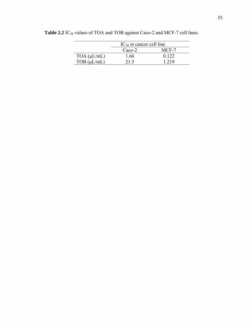
55
Table 2.2 IC50 values of TOA and TOB against Caco-2 and MCF-7 cell lines.
IC50 in cancer cell line Caco-2 MCF-7 TOA (μL/mL) 1.66 0.122 TOB (μL/mL) 21.5 1.219

CHAPTER 3
EVALUATION OF ANTIOXIDANT ACTIVITY OF CRCUMIN-FREE TURMERIC
(Curcuma longa L.) OIL AND IDENTIFICATION OF ITS ANTIOXIDANT
CONSTITUENTS
Abstract
Antioxidant capacity of curcumin-free turmeric (Curcuma longa L.) oil was
evaluated by two different in vitro assays: the DPPH* free radical scavenging assay and
reducing power assay. Results showed that the turmeric oil (TO) possessed strong free
radical scavenging activity and reducing power when compared to standard antioxidants
such as butylated hydroxytoluene (BHT) and α-tocopherol (VitE). An aliquot of 20
μL/mL TO showed 91% free radical scavenging activity in the DPPH* assay, which was
comparable to 10 mM BHT (86%) and 10 mM VitE (96%) at the same condition. In the
reducing power assay, the absorbance at 700 nm of 20 μL/mL TO was 1.085, which was
comparable to 10 mM BHT (1.164). Higher concentration of TO at 100 μL/mL reached
an absorbance at 1.537, which had no significant difference to 10 mM VitE (1.53).
Among the complex constituents in the crude TO, ar-turmerone, turmerone, curlone and
α-terpineol, were isolated and identified as the major components that have antioxidant
effect by using various chromatographic techniques including silica gel open column
chromatography, normal phase HPLC, and GC-MS. These results showed that TO and
some of its inherent components can be potentially alternative natural antioxidants.

57
Introduction
There is an increasing interest in research of antioxidant activities of
phytochemicals in diets due to an accumulative evidence that natural antioxidants can
protect human bodies against excessive reactive oxygen species (ROS), which are
considered the harmful by-products generated during normal cell aerobic respiration.
Intake of exogenous antioxidants may help to maintain an adequate antioxidant status and
the normal physiological function of a living system (Mimić-Oka, 1999). In food
processing, many foods are subject to various factors that lead to the quality
deterioration, i.e., lipid autoxidation. Therefore, natural and/or synthetic antioxidants are
often added as food additives to improve or secure food quality related to texture, color,
flavor and nutritional values, as well as the food shelf life. However, some synthetic
antioxidants such as butyl hydroxyanisole (BHA) and butyl hydroxytoluene (BHT) are
very effective, they are restricted in commercial applications because they may be
harmful to human health (Ito, 1985). For this reason, it is important to explore some other
naturally occurring non- or less-toxic antioxidants that can be used to prevent food
oxidative deterioration. Natural antioxidants also have important usage as nutraceutical or
cosmetic ingredients because they can form functional mixtures.
Turmeric (Curcuma longa L.) is a member of the Zingiberaceae family. It has
been used as an important food ingredient in India for thousands of years because of its
special aromatic flavors and attractive colors. Govindarajan (1980) reported that the dried
rhizome of turmeric contained 3-5% essential oil and 0.02-2.0% yellow curcuminoids.
Turmeric oil is usually obtained from turmeric powder by steam distillation for about 8-
10h. It has a pale yellow color with a peppery and aromatic odor. The major components

58
in the oil include α-phellandrene, 1,8-cineol, zingiberene, ar-curcumene, turmerone, β-
sesquiphellandrene, curlone and dehydrozingerone (Govindarajan, 1980). Turmeric is
highly esteemed by the local people and considered a traditional medicine in the
Ayurvedic system due to its medicinal properties. Many researches have also shown that
the curcuminoids have various biological activities, such as antioxidant (Daniel, 2004;
Ruby, 1995; Chatterjee, 1999; Rukkumani, 2004), anticancer (Nagai, 2005; Verma,
1997), and anti-arthritis (Funk, 2006) activities. Of the turmeric oil extracted from
curcumin removed turmeric oleoresin (CRTO), a strong antioxidant activity was ascribed
to the trace amounts of curcuminoids (Jayaprakasha, 2002). Despite numerous
investigations on biological activities of curcumin, few have involved the curcumin-free
turmeric oil that is considered a by-product with no commercial importance (Saju, 1998;
Jayaprakasha, 2005). Therefore, there are strong economic reasons to explore new
functionalities of other components in the curcumin-free turmeric oil. In this study, we
investigated the antioxidant capacity of curcumin-free turmeric oil using two
complimentary in vitro assays: 2, 2-diphenyl-1-picrylhydrazyl (DPPH*) free radical
scavenging assay and reducing power assay. The antioxidant activity of curcumin-free
TO was compared with that of commercial standard antioxidants, butylated
hydroxytoluene (BHT) and α-tocopherol (VitE). Moreover, some TO components
showing strong antioxidant activity were further separated from the crude TO by various
chromatographic techniques such as open-column silica gel chromatography and normal
phase HPLC, and identified by GC-MS.

59
Materials and methods
Materials and chemicals
The crude turmeric oil (TO) was purchased from Aromaland Company (Santa Fe,
NM, USA). 2,2-diphenyl-1-picrylhydrazyl (DPPH*) free radical, butylated hydroxyl-
toluene (BHT), α–tocopherol (VitE) and α-terpineol were obtained from Sigma Chemical
Co. (St. Louis, MO). Potassium ferricyanide and ferric chloride were obtained from J. T.
Baker Chemical Co. (Phillipsburg. NJ). Trichloroacetic acid and all solvents were
purchased from Fisher Scientific (Suwanee, GA).
HPLC profile of curcumin standard and crude TO
A Shimadzu LC-10AT HPLC system (Kyoto, Japan) was equipped with a Waters
C18 reverse phase column (XterraTM, 4.6 x 150 mm, 5 μm) and a SPD-M10A photodiode-
array (PDA) detector set at 420 nm. UV spectra were recorded in a region of 200-800
nm. Mobile phase was consisted of solvents: (A) water (0.25% acetic acid) and (B)
acetonitrile, and run in a gradient profile in: 0-17 min, 40-60% B; 17-32 min, 60-100% B;
32-38 min, 100% B; 38-40 min, 100-40% B; 41 min, stop. Total flow rate of the mobile
phase was 0.6 mL/min. Injection volume was 20 μL. Curcumin standard (dissolved in
ethanol, 0.15 mg/mL) and TO [dissolved in DCM/methanol (v/v 1:2), 100 μL/mL] were
analyzed by HPLC using the same aforementioned method.
Fractionation and Identification of Antioxidants from crude TO
Silica gel open column chromatography
A glass open column (25 x 2.5 cm) was packed with silica gel (70-230 mesh,
60Å). An aliquot of 1.5 mL crude TO was loaded and eluted by multistep solvent

60
gradients as follows: hexane, dichloromethane (DCM), ethyl acetate, acetonitrile, and
methanol. The flow rate of mobile phase was 4 mL/min. Each collected fraction was 4
mL. After the open-column separation, those TO fractions possessing strong DPPH* free
radical scavenging activities were pooled for further analyses.
Normal phase HPLC analysis A Spherisorb silica column (250 x 4.6 mm, 5 μm; Waters, Milford, MA) was
installed on a Shimadzu LC-10AT HPLC system (Kyoto, Japan) and equilibrated with
DCM. To separate components in the DCM fraction obtained from the open-column
chromatography, twenty microliters of the pooled fractions were further separated by the
HPLC normal phase column and eluted at a flow rate of 1 mL/min by isocratic DCM. For
separation of components in the ethyl acetate fraction, fifty microliters of pooled
fractions were separated by the same column and eluted at a flow rate of 1 mL/min by
linear gradient of methanol from 0 to 10% at 5-15 min to wash out polar compounds. The
eluant was collected into 1 mL fractions after passing through the PDA detector for
further analysis.
GC-MS identification A Shimadzu’s GC-MS system consisting of a GC-17A with a QP5050 Mass
Spectrometer (Kyoto, Japan) was equipped with a DB-5 capillary column (60 × 0.25 mm,
thickness 0.25 µm; J&W Scientific, Folsom, CA, USA) for all volatile chemicals’
quantitative and qualitative analyses in this research. The oven temperature was
programmed from 60 to 280˚C at a ramp rate of 8˚C/min and held at 280˚C for 30 min.
The injector and ion source temperatures were set at 180 and 290˚C, respectively. The
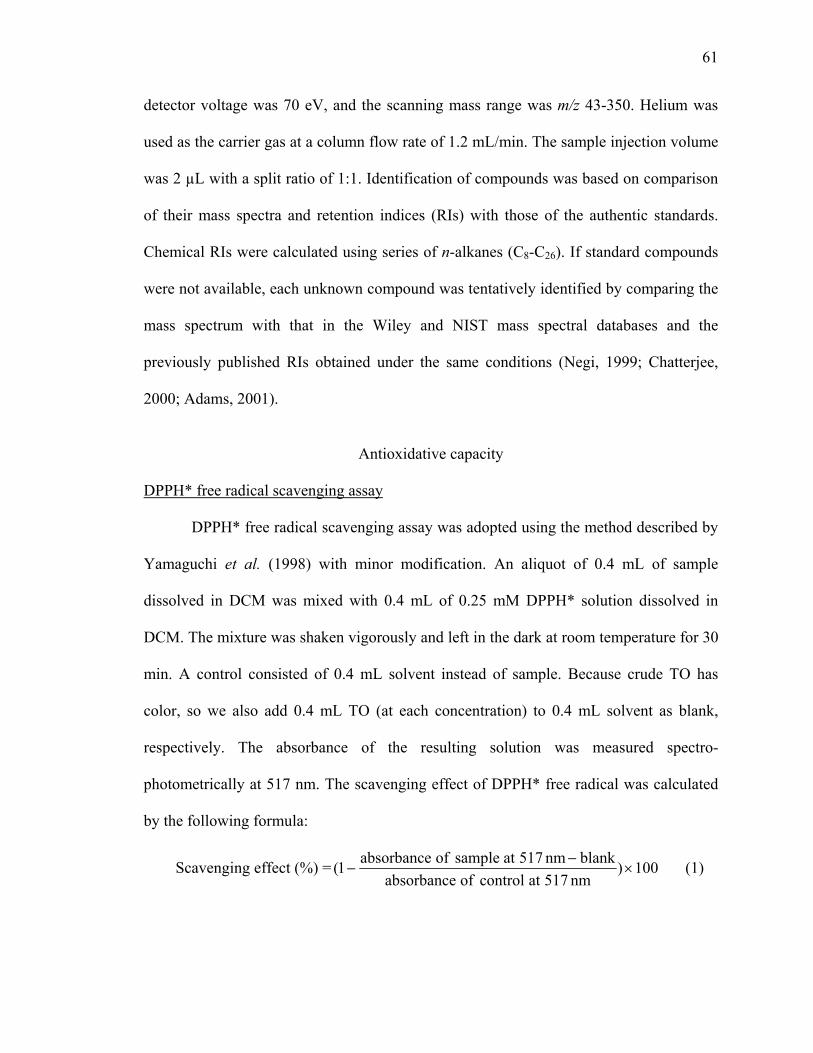
61
detector voltage was 70 eV, and the scanning mass range was m/z 43-350. Helium was
used as the carrier gas at a column flow rate of 1.2 mL/min. The sample injection volume
was 2 µL with a split ratio of 1:1. Identification of compounds was based on comparison
of their mass spectra and retention indices (RIs) with those of the authentic standards.
Chemical RIs were calculated using series of n-alkanes (C8-C26). If standard compounds
were not available, each unknown compound was tentatively identified by comparing the
mass spectrum with that in the Wiley and NIST mass spectral databases and the
previously published RIs obtained under the same conditions (Negi, 1999; Chatterjee,
2000; Adams, 2001).
Antioxidative capacity
DPPH* free radical scavenging assay
DPPH* free radical scavenging assay was adopted using the method described by
Yamaguchi et al. (1998) with minor modification. An aliquot of 0.4 mL of sample
dissolved in DCM was mixed with 0.4 mL of 0.25 mM DPPH* solution dissolved in
DCM. The mixture was shaken vigorously and left in the dark at room temperature for 30
min. A control consisted of 0.4 mL solvent instead of sample. Because crude TO has
color, so we also add 0.4 mL TO (at each concentration) to 0.4 mL solvent as blank,
respectively. The absorbance of the resulting solution was measured spectro-
photometrically at 517 nm. The scavenging effect of DPPH* free radical was calculated
by the following formula:
Scavenging effect (%) = 100)nm517atcontrolofabsorbance
blanknm517atsampleofabsorbance1( ×−
− (1)

62
Reducing power assay The reducing power of TO was determined by the method of Yen et al. (1995)
and Chung et al. (2002) with minor modification. Turmeric oil was dissolved in acetone
to prepare solutions in different concentrations. An aliquot of 0.5 mL of the sample was
mixed with 1 mL of 1% potassium ferricyanide [K3Fe(CN)6], then the mixture was
incubated at 50˚C for 20 min, followed by the addition of 1 mL of trichloroaceteic acid
(10%) to the mixture and centrifugation at 3000 rpm for 10 min. The upper layer of
solution (1 mL) was mixed with 1 mL distilled water and 0.2 mL FeCL3 (0.1%). The
absorbance of the mixture was measured at 700 nm. Higher absorbance of the reaction
mixture indicated a higher reducing power.
Triplicates were performed for each concentration of the tested samples and
standards in these two methods. The experiments were repeated three times on different
days.
Statistical analysis
The data of the antioxidant activities of TO, BHT and VitE were subjected to the
analysis of variance (ANOVA). Treatment means were separated by the least significant
difference (LSD at p<0.05). Analyses were performed using the statistical software SAS
9.1 operated on the Windows system (SAS Institute Inc., Cary, NC).
Results and discussion
Confirming no curcumin or curcuminoids in crude TO
Standards of curcumin and its derivatives were analyzed by HPLC resulting in
three peaks (Figure 3.1), including (1) bisdemethoxycurcumin; (2) demethoxycurcumin;
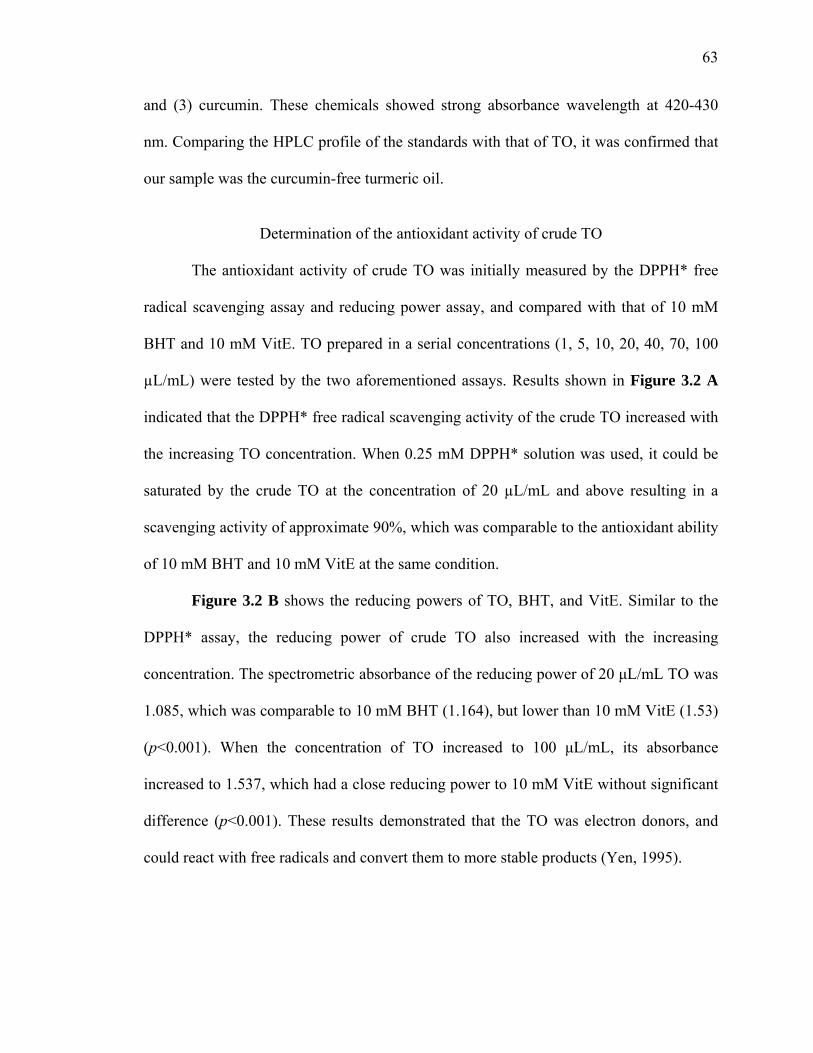
63
and (3) curcumin. These chemicals showed strong absorbance wavelength at 420-430
nm. Comparing the HPLC profile of the standards with that of TO, it was confirmed that
our sample was the curcumin-free turmeric oil.
Determination of the antioxidant activity of crude TO
The antioxidant activity of crude TO was initially measured by the DPPH* free
radical scavenging assay and reducing power assay, and compared with that of 10 mM
BHT and 10 mM VitE. TO prepared in a serial concentrations (1, 5, 10, 20, 40, 70, 100
µL/mL) were tested by the two aforementioned assays. Results shown in Figure 3.2 A
indicated that the DPPH* free radical scavenging activity of the crude TO increased with
the increasing TO concentration. When 0.25 mM DPPH* solution was used, it could be
saturated by the crude TO at the concentration of 20 µL/mL and above resulting in a
scavenging activity of approximate 90%, which was comparable to the antioxidant ability
of 10 mM BHT and 10 mM VitE at the same condition.
Figure 3.2 B shows the reducing powers of TO, BHT, and VitE. Similar to the
DPPH* assay, the reducing power of crude TO also increased with the increasing
concentration. The spectrometric absorbance of the reducing power of 20 μL/mL TO was
1.085, which was comparable to 10 mM BHT (1.164), but lower than 10 mM VitE (1.53)
(p<0.001). When the concentration of TO increased to 100 μL/mL, its absorbance
increased to 1.537, which had a close reducing power to 10 mM VitE without significant
difference (p<0.001). These results demonstrated that the TO was electron donors, and
could react with free radicals and convert them to more stable products (Yen, 1995).

64
Separation and identification of antioxidants in crude TO
Since the tested TO had shown strong antioxidant activities in two assays, its
inherent antioxidants were then subject to a series of sequential separations with silica gel
open column chromatography, and normal phase HPLC, and finally identified by GC-
MS.
When the crude TO was separated by the silica gel open column chromatography
using a stepwise solvent elution method with different solvents such as hexane, DCM,
ethyl acetate, acetonitrile and methanol, a total of 235 fractions were collected. As shown
in Figure 3.3, there were two strong antioxidant peaks corresponding to the DPPH*
assay, namely, TO-I and TO-II, respectively. TO-I contained the fractions 85-95 that
were eluted by DCM; while TO-II comprised the fractions 123-129 eluted by ethyl
acetate. The strength of antioxidant activities of both TO-I and TO-II were similar, with
the antioxidant activity values close to 85% in the DPPH* test. To further separate
antioxidants in these fractions, all fractions within TO-I and TO-II were pooled
respectively and separated by the Spherisorb silica HPLC column. HPLC separation
chromatograms and online antioxidant determination by the DPPH* assay are shown in
Figure 3.4. Major components of TO-I were eluted by DCM. TO-II was eluted by DCM
and methanol. In Figure 3.4 A, there were 3 peaks, named TO-I-1, TO-I-2 and TO-I-3,
respectively. However, only fraction TO-I-3 showed a high antioxidant activity (90%).
Figure 3.4 B showed there were two chromatographic peaks, denoted as TO-II-1 and
TO-II-2. The fraction TO-II-2 had a light yellow color and possessed a medium free
radical scavenging activity (60%). Since GC-MS analysis indicated that the TO-II-2
fraction still contained more than 10 compounds (Figure 3.5 C), it was further

65
concentrated and separated by DCM/methanol (v/v 94:6) using the same column, which
resulted into two peaks (Figure 3.4 C). However, only the second peak (TO-II-2-b)
showd the antioxidant activity. Therefore, both the fraction TO-I-3 and the fraction TO-
II-2-b were selected for further chemical identification by GC-MS.
Identification of antioxidants in TO-I and TO-II separated
by Spherisorb silica HPLC
Previous research (Raina, 2002) found that the crude TO contained nearly 100
compounds, including major chemicals such as ar-turmerone, turmerone, β-sesqui-
phellandrene, curcumene, etc. In our sample, the major components were ar-curcumene
(16.4%), zingiberene (16.5%), β-sesquiphellandrene (14.4%), ar-turmerone (16.8%) and
turmerone (16.5%) (Figure 3.5 A, Table 3.1), which was similar to the previous report.
Further chemical separation and identification of the compounds in the fractions TO-I-3
and TO-II-2-b revealed that there were only 3 compounds in the fraction TO-I-3 and
some more compounds in the fraction TO-II-2-b. As profiled in Figure 3.5, ar-turmerone,
turmerone, and curlone in the fraction TO-I-3 (Figure 3.5 B) and α-terpineol in the
fraction TO-II-2-b (Figure 3.5 D) were identified after comparing their RIs and mass
spectra with those in an essential oil library (Adams, 2001) and the Wiley and NIST mass
spectral databases, as well as with the standard compounds under the same experimental
conditions. Figure 3.6 shows the structures of the four identified compounds. This result
confirmed the previous suspect that the antioxidant activity of turmeric oil may be also
attributed to the chemicals such as ar-turmerone, turmerone, and some other oxygenated
compounds (Jayaprakasha, 2002). Although the mixture of ar-turmerone, turmerone and
curlone showed high antioxidant activity in our preliminary screening, their individual
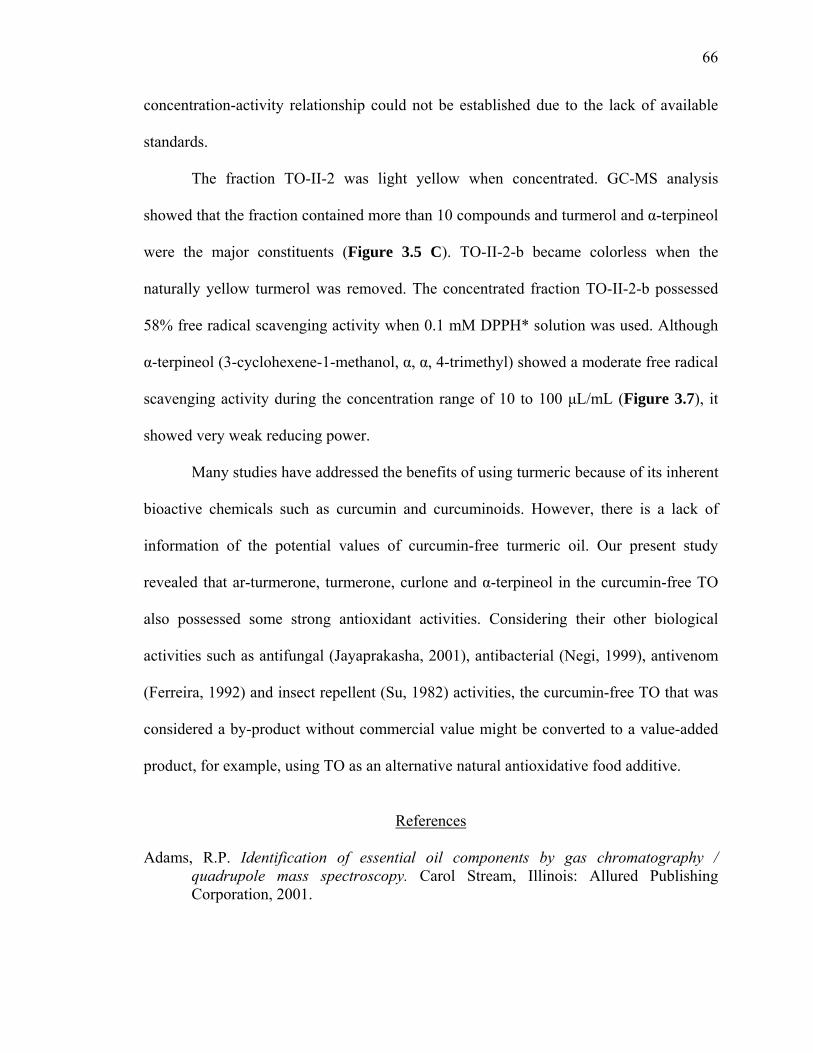
66
concentration-activity relationship could not be established due to the lack of available
standards.
The fraction TO-II-2 was light yellow when concentrated. GC-MS analysis
showed that the fraction contained more than 10 compounds and turmerol and α-terpineol
were the major constituents (Figure 3.5 C). TO-II-2-b became colorless when the
naturally yellow turmerol was removed. The concentrated fraction TO-II-2-b possessed
58% free radical scavenging activity when 0.1 mM DPPH* solution was used. Although
α-terpineol (3-cyclohexene-1-methanol, α, α, 4-trimethyl) showed a moderate free radical
scavenging activity during the concentration range of 10 to 100 μL/mL (Figure 3.7), it
showed very weak reducing power.
Many studies have addressed the benefits of using turmeric because of its inherent
bioactive chemicals such as curcumin and curcuminoids. However, there is a lack of
information of the potential values of curcumin-free turmeric oil. Our present study
revealed that ar-turmerone, turmerone, curlone and α-terpineol in the curcumin-free TO
also possessed some strong antioxidant activities. Considering their other biological
activities such as antifungal (Jayaprakasha, 2001), antibacterial (Negi, 1999), antivenom
(Ferreira, 1992) and insect repellent (Su, 1982) activities, the curcumin-free TO that was
considered a by-product without commercial value might be converted to a value-added
product, for example, using TO as an alternative natural antioxidative food additive.
References
Adams, R.P. Identification of essential oil components by gas chromatography /
quadrupole mass spectroscopy. Carol Stream, Illinois: Allured Publishing Corporation, 2001.

67
Chatterjee, S.; Desai, S.R.P.; Thomas, P. Effect of γ-irradiation on antioxidant activity of turmeric extracts. Food Research International. 1999, 32, 487-490.
Chatterjee, S.; Variyar, P.S.; Gholap, A.S.; Padwal-Desai, S.R.; Bongirwar, D.R. Effect
of γ-irradiation on the volatile oil constituents of turmeric (Curcuma longa). Food Research International. 2000, 33, 103-106.
Chung, Y.C.; Chang, C.T.; Chao, W.W.; Lin, C.F.; Chou, S.T. Antioxidative activity and
safety of the 50% ethanolic extract from red bean fermented by Bacillus subtilis IMR-NK1. Journal of Agricultural and Food Chemistry. 2002, 50, 2454-2458.
Daniel, S.; Limson, J.L.; Dairam, A.; Watkins, G.M.; Daya, S. Through metal binding,
curcumin protects against lead- and cadmium-induced lipid peroxidation in rat brain homogenates and against lead-induced tissue damage in rat brain. Journal of Inorganic Biochemistry. 2004, 98, 266-275.
Ferreira, L.A.F.; Henriques, O.B.; Andreoni, A.A.S.; Vital, G.R.F.; Campos, M.M.C.;
Habermehl, G.G.; Moraes, V.L.G.D. Antivenom and biological effects of ar-turmerone isolated from Curcuma longa (Zingiberaceae). Toxicon. 1992, 30 (10), 1211-1218.
Funk, J.L.; Oyarzo, J.N.; Frye, J.B.; Chen, G.; Lantz, R.C.; Jolad, S.D.; Sólyom, A.M.;
Timmermann, B.N. Turmeric extracts containing curcuminoids prevent experimental rheumatoid arthritis. Journal of Natural Products. 2006, 69, 351-355.
Govindarajan, V.S. Turmeric --- Chemistry, Technology, and Quality. CRC Critical
Reviews in Food Science and Nutrition. 1980, 12(3), 199-301. Ito, N.; Fukushima, S.; Tsuda, H.; Shirai, T.; Hagiwara, A.; Imaida, K. Antioxidants:
carcinogenicity and modifying activity in tumorigenesis. In Food Toxicology: Real or Imaginary Problems? Gibson, G.G., Walker, R., Eds.; London and Philadelphia: Taylor & Francis, 1985; pp. 181-198.
Jayaprakasha, G.K.; Jena, B.S.; Negi, P.S.; Sakariah, K.K. Evaluation of antioxidant
activity and antimutagenicity of turmeric oil: a byproduct from curcumin production. Verlag der Zeitschrift für Naturforschung. 2002, 57c, 828-835.
Jayaprakasha, G.K.; Negi, P.S.; Anandharamakrishnan, C.; Sakariah, K.K. Chemical
composition of turmeric oil – a byproduct from turmeric oleoresin industry and its inhibitory activity against different fungi. Verlag der Zeitschrift für Naturforschung. 2001, 56c, 40-44.
Jayaprakasha, G.K.; Rao, L.J.M.; Sakariah, K.K. Chemistry and biological activities of C.
longa. Trends in Food Science & Technology. 2005, 16, 533-548.
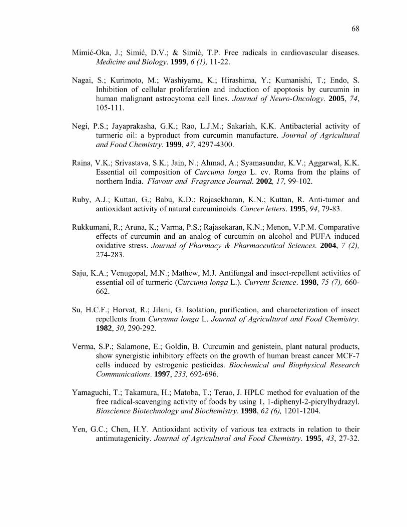
68
Mimić-Oka, J.; Simić, D.V.; & Simić, T.P. Free radicals in cardiovascular diseases. Medicine and Biology. 1999, 6 (1), 11-22.
Nagai, S.; Kurimoto, M.; Washiyama, K.; Hirashima, Y.; Kumanishi, T.; Endo, S.
Inhibition of cellular proliferation and induction of apoptosis by curcumin in human malignant astrocytoma cell lines. Journal of Neuro-Oncology. 2005, 74, 105-111.
Negi, P.S.; Jayaprakasha, G.K.; Rao, L.J.M.; Sakariah, K.K. Antibacterial activity of
turmeric oil: a byproduct from curcumin manufacture. Journal of Agricultural and Food Chemistry. 1999, 47, 4297-4300.
Raina, V.K.; Srivastava, S.K.; Jain, N.; Ahmad, A.; Syamasundar, K.V.; Aggarwal, K.K.
Essential oil composition of Curcuma longa L. cv. Roma from the plains of northern India. Flavour and Fragrance Journal. 2002, 17, 99-102.
Ruby, A.J.; Kuttan, G.; Babu, K.D.; Rajasekharan, K.N.; Kuttan, R. Anti-tumor and
antioxidant activity of natural curcuminoids. Cancer letters. 1995, 94, 79-83. Rukkumani, R.; Aruna, K.; Varma, P.S.; Rajasekaran, K.N.; Menon, V.P.M. Comparative
effects of curcumin and an analog of curcumin on alcohol and PUFA induced oxidative stress. Journal of Pharmacy & Pharmaceutical Sciences. 2004, 7 (2), 274-283.
Saju, K.A.; Venugopal, M.N.; Mathew, M.J. Antifungal and insect-repellent activities of
essential oil of turmeric (Curcuma longa L.). Current Science. 1998, 75 (7), 660-662.
Su, H.C.F.; Horvat, R.; Jilani, G. Isolation, purification, and characterization of insect
repellents from Curcuma longa L. Journal of Agricultural and Food Chemistry. 1982, 30, 290-292.
Verma, S.P.; Salamone, E.; Goldin, B. Curcumin and genistein, plant natural products,
show synergistic inhibitory effects on the growth of human breast cancer MCF-7 cells induced by estrogenic pesticides. Biochemical and Biophysical Research Communications. 1997, 233, 692-696.
Yamaguchi, T.; Takamura, H.; Matoba, T.; Terao, J. HPLC method for evaluation of the
free radical-scavenging activity of foods by using 1, 1-diphenyl-2-picrylhydrazyl. Bioscience Biotechnology and Biochemistry. 1998, 62 (6), 1201-1204.
Yen, G.C.; Chen, H.Y. Antioxidant activity of various tea extracts in relation to their
antimutagenicity. Journal of Agricultural and Food Chemistry. 1995, 43, 27-32.

69
1
2
3
Figure 3.1 HPLC chromatogram of curcumin and curcuminoids in their absorbance at 420nm. 1. bisdemethoxycurcumin; 2. demethoxycurcumin; 3. curcumin.

70
DPPH assay
0
20
40
60
80
100
1 5 10 20 40 70 100 10mMBHT
10mMVitETO concentration (μl/ml)
free
radi
cal s
cave
ngin
gef
fect
(%)
A
reducing power assay
0
0.5
1
1.5
2
1 5 10 20 40 70 100 10mMBHT
10mMVitETO concentration (μl/ml)
abso
rban
ce a
t 700
nm
B
Figure 3.2 Antioxidative capacity of crude TO. A: DPPH* assay, 10 mM and 10 mM VitE were used as standards.
B: Reducing power assay, 10 mM BHT and 10 mM VitE were used as standards.
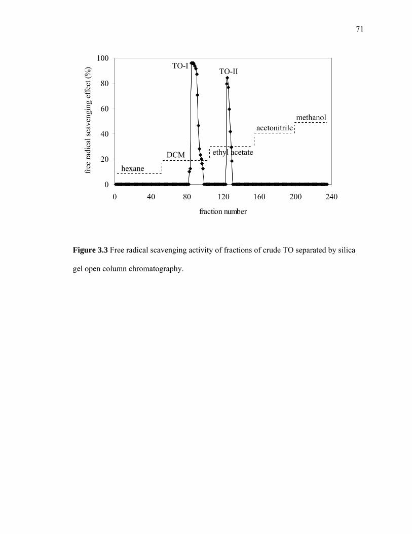
71
0
20
40
60
80
100
0 40 80 120 160 200 240
fraction number
free
radi
cal s
cave
ngin
g ef
fect
(%)
hexane
DCM ethyl acetate
acetonitrile methanol
TO-I TO-II
Figure 3.3 Free radical scavenging activity of fractions of crude TO separated by silica
gel open column chromatography.
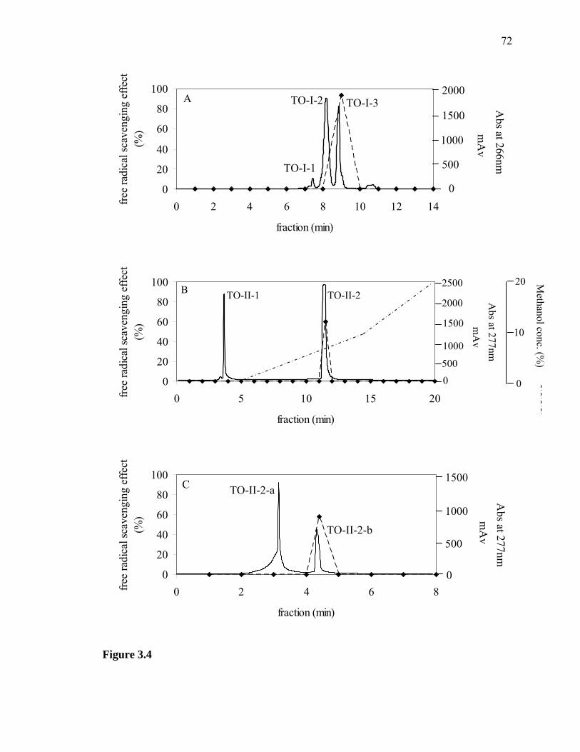
72
0
20
40
60
80
100
0 2 4 6 8 10 12 14
fraction (min)
free
radi
cal s
cave
ngin
g ef
fect
(%
)
0
500
1000
1500
2000 m
Av
Abs at 266nm
A
TO-I-1
TO-I-2 TO-I-3
0
20
40
60
80
100
0 5 10 15 20
fraction (min)
free
radi
cal s
cave
ngin
g ef
fect
(%
)
0 500
1000
1500
2000
2500
mA
v A
bs at 277nm
0
10
20 Methanol conc. (%
)
B TO-II-1 TO-II-2
0
20
40
60
80
100
0 2 4 6 8
fraction (min)
free
radi
cal s
cave
ngin
g ef
fect
(%
)
0
500
1000
1500
mA
v A
bs at 277nm
C TO-II-2-a
TO-II-2-b
Figure 3.4

73
Figure 3.4 (continued) Chromatograms of TO-I and TO-II and their free radical
scavenging activities.
A: TO-I separated by Spherisorb silica HPLC, isocratic method, mobile phase: DCM.
B: TO-II separated by Spherisorb silica HPLC, gradient method, mobile phase: a. DCM;
b. methanol.
C: TO-II-2 separated by Spherisorb silica HPLC, gradient method, mobile phase: DCM
/methanol (v/v = 94:6).
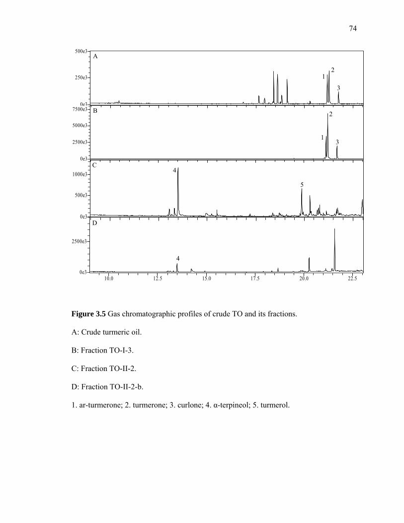
74
10.0 12.5 15.0 17.5 20.0 22.50e3
2500e3
0e3
2500e3
5000e3
7500e30e3
250e3
500e3
0e3
500e3
1000e3
A
B
C
D
1 2
3
1
2
3
4
5
4
Figure 3.5 Gas chromatographic profiles of crude TO and its fractions. A: Crude turmeric oil.
B: Fraction TO-I-3.
C: Fraction TO-II-2.
D: Fraction TO-II-2-b.
1. ar-turmerone; 2. turmerone; 3. curlone; 4. α-terpineol; 5. turmerol.
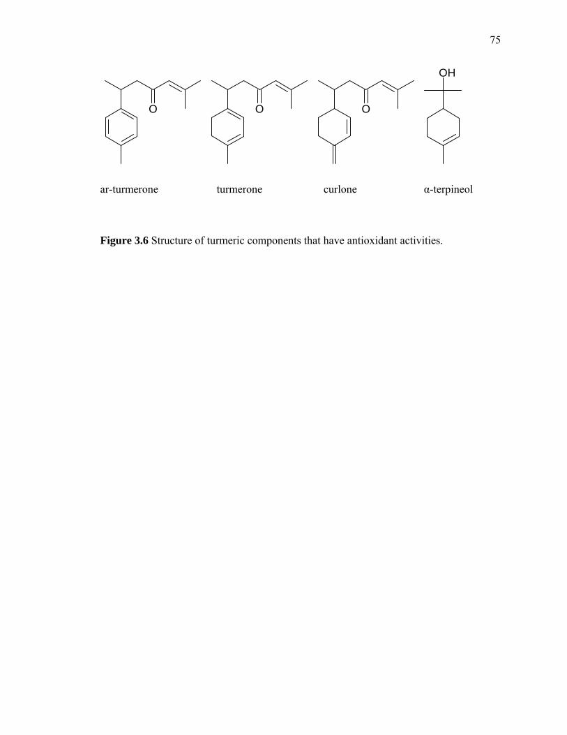
75
O O O
OH
ar-turmerone turmerone curlone α-terpineol
Figure 3.6 Structure of turmeric components that have antioxidant activities.

76
0
20
40
60
80
100
1 5 10 40 70 100
α-terpineol concentration (μl/ml)
free
radi
cal s
cave
ngin
g ef
fect
(%)
0
0.5
1
1.5
2
abso
rban
ce a
t 700
nm
DPPH assay
reducingpower assay
Figure 3.7 Free radical scavenging assay and reducing power assay of α–terpineol.

77
Table 3.1 Composition of crude TO.
Retention time (min)
Compound RIa Composition (%)
Identification method
17.650 Trans-caryophyllene 1444 4.58 MS, KI 17.958 Farnesene 1463 2.93 MS, KI 18.417 Ar-curcumene 1491 16.44 MS, KI 18.617 Zingiberene 1505 16.51 MS, KI 18.842 β-bisabolene 1518 4.82 MS, KI 19.117 β-sesquiphellandrene 1537 14.35 MS, KI 21.183 Ar-turmerone 1675 16.84 MS, KI 21.267 Turmerone 1680 16.45 MS, KI 21.767 Curlone 1715 7.08 MS, KI
a RI was calculated using a series of n-alkanes (C8-C26). MS: Mass spectra.
KI: Kovats indices.
APPENDICES
79
Appendix A
DPPH* assay
DPPH* (2,2-diphenyl-1-picrylhydrazyl, MW: 394.22) is a stable free radical and
shows a characteristic absorption at 517 nm due to its odd electron. It is purple color. As
the odd electron of the radical becomes paired off in the presence of a hydrogen donor, or
a free radical scavenging antioxidant, the absorption strength decreased resulting in
decolorization that is stoichiometric with respect to the number of electrons captured
(Figure 4.1). Although DPPH* is considered as a stable free radical, its solutions slowly
deteriorate. The solution need to be stored in glass container under nitrogen atmosphere,
and covered with aluminum foil. Under these conditions a standard solution may be
prepared and stored with a loss of free radical activity not exceeding 2-4% per week
(Blois, 1958).
Sample can be diluted to a series of concentration in solvents, then use the same
solvent to dissolve DPPH* and get the last concentration of 250 μM. Take one aliquot of
sample and add the same volume DPPH* to make the DPPH* concentration to 125 μM.
Then shake the mixture vigorously and left in the dark at room temperature for 30 min.
Then measure the absorbance at 517 nm using spectrometer. If the sample has color,
blank control should be set. The scavenging effect of DPPH* free radical was calculated
by this formula:
Scavenging effect (%) = 100)nm517atcontrolofabsorbance
blanknm517atsampleofabsorbance1( ×−
−
Regarding the solvent to be used, the method seems to work equally well with
methanol, ethanol and DCM, any of which seems not to interfere with the reaction. The

80
use of other solvent systems, such as almost neat extracts in water or acetone, seems to
give low values for the extent of reduction (Molyneux, 2004).
81
Appendix B
Reducing power assay
The mechanism of reducing power assay is the presence of reductants
(antioxidants) would result in reducing Fe3+/ferricyanide complex to the ferrous form.
The Fe2+ can be monitored by measuring the formation of Perl’s Prussian blue at 700 nm
(Yen, 1995; Chung, 2002). To accurately measure the total reducing power, the following
conditions must be met. (1) All, and only, antioxidants can reduce Fe3+ / ferricyanide
complex under the reaction conditions. (2) The reaction rate must be sufficiently fast
enough that the reaction can be completed in a short assay time. (3) The oxidized
antioxidant and its secondary reaction products should have no absorption at 700 nm, the
maximum absorption of Perl’s Prussian blue (Qu, 2002).
In this assay, sample needed to be diluted in solvent (solvent should be miscible
with water) in a series of concentration. 0.5 mL of sample mixed with 1 mL of 1%
potassium ferricyanide [K3Fe(CN)6, dissolved in distilled water], and the mixtures were
incubated at 50˚C for 20 min. Then 1mL of 10% trichloroaceteic acid (dissolved in
distilled water) was added to the mixture and centrifuged at 3000 rpm for 10 min. The
upper layer of solution (1mL) was mixed with distilled water (1mL) and 0.2 mL FeCL3
(0.1%). The mixture was shaken vigorously and the absorbance was measured at 700 nm.
Higher absorbance of the reaction mixture indicated higher reducing power.

82
Appendix C
Packaging silica gel open column
Preparing silica gel
Put suitable amount of silica gel into a beaker. Add some hexane into the beaker
and swirl gently to make a “slurry”. Remove air bubbles by sonication or vacuum pump.
Preparing open column
Clamp column securely and straightly to a ring stand, leaving enough space at the
bottom to allow for switching of the flasks or beakers you will use to collect the eluted
solvent. Attach a funnel to the top of the column and place a beaker underneath.
Packing open column
Pour the “slurry” into the column via the funnel and let it settle in the column.
Open the stopcock and let some of the solvent drip through. As the solvent collects in the
waste beaker below you can use it to resuspend the remaining silica that is stuck in your
“slurry” beaker and reapply to the top of the column. Repeat as necessary until almost all
of the silica has been transferred to the column and has settled down to form an even,
tightly packed cylinder, saturated with solvent. Close the stopcock when the column
seems to have packed down to about 2-3 cm from the top of the column. The solvent
level should just cover the top of the column. If the top surface of the packed silica
appears crooked, tap gently on the side of the column until it flattens out. Do not ever let
the column run dry. Avoid little cracks and channels. If your column runs dry or forms
channels, you need to pack it again.
83
Loading sample
If your sample is solid, you need to dissolve it first. The solvent you used is as
little as possible. Using a glass pipette, start to add your dissolved sample to the top of
the column gently. When you add the sample to the column, you want the amount of
solvent at the top of the column to be quite small. When there is a couple of millimeters
of solvent above top of saturated silica gel, open the stopcock and allow the sample to run
into the column, adding drops of the dissolved sample slowly and gently to the top via the
pipette so that the column will not run dry.
Separation
Once the sample is loaded onto the column, you can gently add 10 mL of fresh
solvent (you want to use it as mobile phase) to the column and start the separation. Add
more solvent or connect the column to the solvent pump. At all times make sure that
there is no danger of running dry.
84
References Blois, M.S. Antioxidant determinations by the use of a stable free radical. Nature. 1958,
181, 1199-1200. Chung, Y.C.; Chang, C.T. Chao, W.W.; Lin, C.F.; Chou, S.T. Antioxidative activity and
safety of the 50% ethanolic extract from red bean fermented by Bacillus subtilis IMR-NK1. Journal of Agricultural and Food Chemistry. 2002, 50, 2454-2458.
Molyneux, P. The use of the stable free radical diphenylpicrylhydrazyl (DPPH*) for
estimating antioxidant activity. Songklanakarin Journal of Science and Technology. 2004, 26 (2), 211-219.
Qu, B; Huang, D.; Hampsch-Woodill, M.; Flanagan, J.A.; Deemer, E.K. Analysis of
antioxidant activities of common vegetables employing oxygen radical absorbance capacity (ORAC) and ferric reducing antioxidant power (FRAP) assay: a comparative study. Journal of Agricultural and Food Chemistry. 2002, 50, 3122-3128.
Yen, G.C.; Chen, H.Y. Antioxidant activity of various tea extracts in relation to their
antimutagenicity. Journal of Agricultural and Food Chemistry. 1995, 43(1), 27-32.
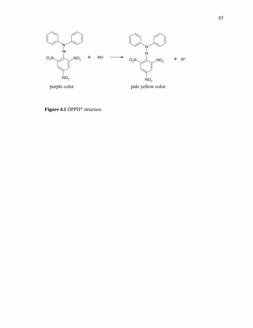
85
NN
NO2
NO2
O2N*
RH
NN
NO2
NO2
O2N R*+ +
purple color pale yellow color
Figure 4.1 DPPH* structure.