“COMPARATIVE STUDY BETWEEN RETROGRADE …repository-tnmgrmu.ac.in/3456/1/180400413sukumar.pdf ·...
69
“COMPARATIVE STUDY BETWEEN RETROGRADE URETHROGRAPHY AND MAGNETIC RESONANCE URETHROGRAPHY IN EVALUATING MALE URETHRAL STRICTURE DISEASE” Dissertation submitted for partial fulfilment of requirements of M.Ch DEGREE EXAMINATION BRANCH 1V – UROLOGY KILPAUK MEDICAL COLLEGE & HOSPITAL CHENNAI – 600 010 THE TAMIL NADU DR.M.G.R MEDICAL UNIVERSITY CHENNAI – 600 032 AUGUST-2013
Transcript of “COMPARATIVE STUDY BETWEEN RETROGRADE …repository-tnmgrmu.ac.in/3456/1/180400413sukumar.pdf ·...

“COMPARATIVE STUDY BETWEEN RETROGRADE
URETHROGRAPHY AND MAGNETIC RESONANCE
URETHROGRAPHY IN EVALUATING MALE URETHRAL
STRICTURE DISEASE”
Dissertation submitted for partial fulfilment of requirements of
M.Ch DEGREE EXAMINATION
BRANCH 1V – UROLOGY
KILPAUK MEDICAL COLLEGE
&
HOSPITAL
CHENNAI – 600 010
THE TAMIL NADU DR.M.G.R MEDICAL UNIVERSITY
CHENNAI – 600 032
AUGUST-2013

CERTIFICATE
This is to certify that Dr.R.Sukumar has been a post graduate
student during the period August 2010 to July 2013 at Department of
Urology, Govt Kilpauk Medical College, & Hospital, Chennai.
This Dissertation titled “COMPARATIVE STUDY BETWEEN
RETROGRADE URETHROGRAPHY AND MAGNETIC
RESONANCE URETHROGRAPHY IN EVALUATING MALE
URETHRAL STRICTURE DISEASE” is a bonafide work done by him
during the study period and is being submitted to the Tamilnadu Dr. M.G.R.
Medical University in a partial fulfilment of the MCh Branch IV Urology
Examination.
Prof.C.Ilamparuthi,M.S,MCh,DNB
Department of Urology,Govt.Royappettah Hospital
Govt Kilpauk Medical College
Chennai - 600 010.
Prof.P.Vairavel,D.G.O,M.S,MCh,
Professor & Head of the Department,
Department of Urology,
Govt Kilpauk Medical College &
Hospital
Chennai - 600 010.
Prof.P.Ramakrishnan, MD,DLO
Dean
Govt Kilpauk Medical College & Hospital
Chennai - 600 010

CERTIFICATE
This is to certify that Dr.R.Sukumar has been a post graduate student
during the period August 2010 to July 2013 at Department of Urology,
Govt Kilpauk Medical College, & Hospital, Chennai.
This Dissertation titled “COMPARATIVE STUDY BETWEEN
RETROGRADE URETHROGRAPHY AND MAGNETIC
RESONANCE URETHROGRAPHY IN EVALUATING MALE
URETHRAL STRICTURE DISEASE” is a bonafide work done by him
during the study period under my guidance.
Prof.K.Pitchaibalashanmugam,M.S, MCh
Dept.of Urology
Govt.Kilpauk Medical College and Hospital,
Chennai.1O

DECLARATION
I Dr.R.Sukumar solemnly declare that this dissertation entitled,
“COMPARATIVE STUDY BETWEEN RETROGRADE
URETHROGRAPHY AND MAGNETIC RESONANCE
URETHROGRAPHY IN EVALUATING MALE URETHRAL
STRICTURE DISEASE” is a bonafide work done by me at the Department
of Urology, Govt Kilpauk Medical College and Hospital during the period
2010 – 2013 under the supervision of Professor
P.Vairavel,D.G.O,M.S,MCh,Department of Urology, Govt Kilpauk Medical
College and Hospital.
This dissertation is submitted to The Tamil Nadu Dr.M.G.R Medical
University, towards a partial fulfilment of requirement for the award of MCh
Degree Branch IV Urology.
Place: Chennai
Date : 25.03.2013 Dr.R.SUKUMAR

ACKNOWLEDGEMENT
I express my sincere thanks to Prof.Dr.Ramakrishnan.M.D,D.L.O
Dean,Kilpauk Medical College for permitted me to conduct the study.I am
grateful to Prof.Dr.P.Vairavel DGO.,M.S.,M.Ch.,(Uro), for assigning this
topic and providing me with valuable guidance.
I thank Prof.Dr.K.Thiyagarajan, M.S. M.Ch. (Uro),
Prof.Dr.C.IlamparuthyM.S,MCh, Prof.Dr.V.Selvaraj M.S,MCh, and
Prof.Dr.PitchaiBala ShanmugamM.S,MCh for their excellent guidance.
I thank Prof.Dr.Babu PeterM.D (Radiodiagnosis), Institute of
Radiology, GRGG Hopital, Chennai for provided me with all the necessary
facilities and guidance.
My sincere thanks to Assistant Professors Dr.P.LeelaKrishna,
Dr.R.Jayaganesh, Dr.G.Sivashankar, Dr.A.Senthilvel, Dr.Srikala
Prasad for their excellent guidance.
I am indebted to all the patients in the study for their co-operation.
Last, I express my thanks to my family for their support and help.


TABLE OF CONTENTS
S.NO CONTENT PAGE NO
1 INTRODUCTION 1
2 AIM 3
3 REVIEW OF LITERATURE 4
4 IMAGING OF URETHRA 10
5 MATERIALS AND METHODS 14
6 RESULTS AND ANALYSIS 18
7 DISCUSSION 42
8 IMAGES 44
9 CONCLUSION 53
10 BIBLIOGRAPHY A-C
11 ANNEXURE I-PROFORMA D
12 ANNEXURE II- MASTER CHART E
13 ETHICAL COMMITIEE CERTIFICATE F
14 CONSENT FORM G

LIST OF ABBREVIATIONS
KMCH Kilpauk Medical College & Hospital
GRH Govt. Royapettah Hospital
RUG Retrograde Urethrography
MRU Magnetic Resonance Urethrography
PFUDD Pelvic Fracture Urethral Distraction Defect
Ant Anterior
Post Posterior
BNE Bladder Neck Elevation
BNI Bladder Neck Incision
VCUG Voiding CystoUrethroGraphy
SUG Sonourethrography
LOCM Low Osmolar Contrast Media
SURG Surgery
GUD Guided Urethral Dilatation
- 1 -
INTRODUCTION
Stricture urethra in male is a common urological problem causing
lower urinary tract symptoms. It can be divided in to anterior inflammatory
(or) iatrogenic urethral strictures and post traumatic bulbomembranous
distraction defects.
It can be caused by inflammation or trauma (pelvic injury, Iatrogenic,
post catheterization). Strictures usually present with obstructive voiding
symptoms or urinary tract infections and occasionally retention.
Various modalities have been devised to diagnose strictures.
Commonly used methods include retrograde urethrography (RUG), voiding
cystourethrography, Sonourethrography and Magnetic Resonance
urethrography (MRU). Role of imaging in strictures is significant in
determining the treatment protocols.The treatment options and approach
depends upon the site, length and associated spongiofibrosis and fistula.

- 2 -
The gold standard imaging technique is RUG which is readily
available, simple and cost effective. Disadvantages include its invasive
nature, contrast allergy, inaccurate estimation of stricture length and does
not show the spongiofibrosis.
Recent non invasive tests have been developed to circumvent the problems
of RUG, like Sonourethrography and Magnetic resonance urethrography that
do not require injection of contrast media. Though noninvasive each has its
own disadvantages. Sonourethrography which is not useful in the evaluation
of posterior urethra and the disadvantage of MR Urethrography is its cost
and availability.
This study is done to compare the clinical relevance of Retrograde
urethrography and Magnetic resonance urethrography in male urethral
stricture disease to guide further management.

- 3 -
AIM
To compare the clinical relevance of retrograde urethrography and
Magnetic resonance urethrography in male urethral stricture
disease for planning the surgical management

- 4 -
REVIEW OF LITERATURE
D.E.Andrich and A.R.Mundy1 described the stricture as narrowing of
urethral caliber due to scar following injury (or) infection. Nielsen and
Nordling2
on the basis of study done over 4000 male urethradescribed
stricture as urethral lumen less than 22Fr size,and found that stricture would
be symptomatic only if the lumen reduces to 18Fr.
The retrograde urethrogram was set as gold standard imaging modality for
the diagnosis of stricture urethra in 1910, by Cunningham.3But RUG was
not found to be useful in evaluation of posterior urethral strictures.
Gallentine ML, Morey AF concluded that in posterior urethral evaluation
retrograde urethrogram clubbed with voidingcystourethrography was very
informative to depict the proximal extent of the stricture when compare to
RUG alone.4
Nash PA and McAninch J5evaluated anterior urethral stricture based on
retrograde urethrogram and found that the actual stricture length determined
endoscopically correlated with penile urethral stricture better than that of
bulbar urethral stricture.

- 5 -
According to Syed Mamun Mahmud et al6, the sensitivity & specificity of
RUG for the diagnosis of urethral stricture was 91% &72% and by MRU
100%
In 1987,Friedburg&Wimmer studied the usefulness of Magnetic resonance
Urography and concluded that the MRU provided more findings than
conventional radiology and USG.7The findings were confirmed by Garcia-
Valtuille8in which MR Urography was found to be a safe diagnostic tool
with high quality images of urinary system than the other imagingmethods.
MM Oh et al 9 in their study of 25 patients concluded that MR
Urethrography was more effective in evaluation of obliterative posterior
urethral stricture than retrograde urethrography combined with voiding
cystourethrography.
Eaton J et al published the currentstatus of imaging of urethra in 2005.They
quoted that the MRI plays an important role in the diagnosis of various
urethral diseases like fistula,stricture,tumor and also traumatic urethral
stricture.10

- 6 -
Jeong-ah ryuet al. concluded that the imaging modalities using contrast
agents like RUG ,eventhough useful in imaging of urethral anatomical
abnormalities,have limited role in evaluation of adjacent structures. But the
Magnetic resonance imaging of urethra was very much useful to evaluate the
urethral abnormalities and its adjacent structures with high quality images.11
MR Urethrography was useful in the evaluation of internal organs, diagnosis
of fistula, sinus tract and periurethral abscess. In urethral trauma it was
helpful to know the site, length and severity. In urethral tumors Magnetic
resonance imaging can provide local staging of tumors.
Moon-Hae Choi and colleagues studied the role of MRI in fracture penis.
They concluded that it was very useful in showing tear in the
tunicaalbuginea, cavernosal hematoma and urethral rupture, because of its
multiplanar imaging capability.12
Comparative study between MR Urethrography and ascending
urethrography and sonourethrography was performed by MA El-Ghar et al.
The sensitivity, specificity for diagnosing anterior urethral stricture by RUG
was 91% & 90% and 89% & 91.7% for posterior urethra. By MRU it was
91.7% (ant &post). In Sonourethrography,the accuracy was 100% in anterior
- 7 -
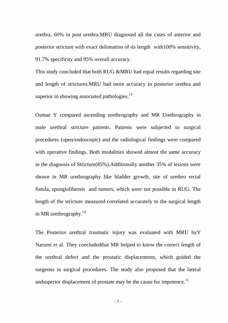
urethra, 60% in post urethra.MRU diagnosed all the cases of anterior and
posterior stricture with exact delineation of its length with100% sensitivity,
91.7% specificity and 95% overall accuracy.
This study concluded that both RUG &MRU had equal results regarding site
and length of strictures.MRU had more accuracy in posterior urethra and
superior in showing associated pathologies.13
Osman Y compared ascending urethrography and MR Urethrography in
male urethral stricture patients. Patients were subjected to surgical
procedures (open/endoscopic) and the radiological findings were compared
with operative findings. Both modalities showed almost the same accuracy
in the diagnosis of Stricture(85%).Additionally another 35% of lesions were
shown in MR urethrography like bladder growth, site of urethro rectal
fistula, spongiofibrosis and tumors, which were not possible in RUG. The
length of the stricture measured correlated accurately to the surgical length
in MR urethrography.14
The Posterior urethral traumatic injury was evaluated with MRU byY
Narumi et al. They concludedthat MR helped to know the correct length of
the urethral defect and the prostatic displacements, which guided the
surgeons in surgical procedures. The study also proposed that the lateral
andsuperior displacement of prostate may be the cause for impotence.15

- 8 -
Deukjae sung et al studied 12 patients with stricture disease and compared
MRU with RUG clubbed with VCUG in obliterative urethral stricture. They
concluded that the result of MRU gave the accurate length of the stricture
and linear correlation which is not correct in RUG clubbed with VCUG. 16
Bircan MK, Sahin H, Korkmaz K et al. performed a study to compare the
efficiency, complications of retrograde urethrography and urethroscopy in
38 male patients . 30 patients had the same findings in both procedures. In 8
patients the RGU did not correlate with urethroscopy. This study concluded
thatthe results of RGU were misleading, when it was not combined with
urethroscopy. 17
Pavlica P, Barozzi L, Menchi I et al used RUG,SUG and MR
Urethrography to study the anterior urethral anatomy and to diagnose
periurethral fibrosis, diverticula, and tumors. Additional information was
provided by MR urethrogram which was not possible in other modalities. 18
Koraitim MM,Reda IS and colleagues studied the usefulness of MRI in the
assessment of posterior urethral distraction defects in 21 male patients. The
MRI and urethrographic findings were compared and correlated with the

- 9 -
operative findings.Preoperative MRI provided correct length of the urethral
defect, type and degree of prostatic displacement, site and density of scar
tissue. 19
Choudhary S, Singh P et al27
, conducted the study to compare the
sonourethrography and retrograde urethrography in evaluation of anterior
urethral strictures. They concluded that, in the estimation of stricture length,
RGU showed a lower sensitivity (60-80%) when compared with
sonourethrography (73.3-100%). Spongiofibrosis was detected by
sonourethrography with a sensitivity of 77.3-83.3%. RGU and
sonourethrography were equally efficacious in detection of anterior urethral
strictures

- 10 -
IMAGING OF URETHRA
Urethra can be imaged with retrograde urethrogram, opposing urethrogram,
Sonourethrogram and MR Urethrogram. 20, 21
RETROGRADE URETHROGRAM
It is a simple, easy, cheap techniquereadily available, and no need for any
preparation.It was first popularized by Cunningham in 1910.
TECHNIQUE
It can be performed in a dynamic or static fashion. In dynamic RUG the film
is exposed while contrast is being injected. In static, image is obtained after
injection of contrast with clamping of the penis
The patient is placed in supine position, pelvis is oblique 45 degree with the
dependent thigh acutely flexed, penis is stretched moderately.Inject 20 cc of
contrast (LOCM 30% children & 60% adults) into the urethra.Xray will be
taken both before and after the contrast.It can be also performed by keeping
a 12 or 14 French Foley catheter, 2 - 3 cm into the urethra and filling the
balloon with 2-3 cc of water .21, 22, 23

- 11 -
IMAGE ANALYSIS
In normal the bulbar urethra is convex, symmetrical cone shaped, veru
appears as filling defect, no narrowing (or) irregularities. In stricture it is
narrowed,elongated, asymmetrical and irregular.
Entire urethra should be analyzed for narrowing, irregularities, fistula tracts
and site, number, length of the stricture.
Figure 1 - Normal retrograde urethrography
LIMITATIONS
Radiation exposure to testis, contrast allergy, Intravasation of contrast, if
high pressure is given during contrast injection
Does not give information about spongio (or) periurethral fibrosis.

- 12 -
Underestimation of length of the bulbar stricture (pelvis is aligned obliquely
with respect to the anterior–posterior x-ray beam).
The bulbarurethra is fixed in the same axis as the pelvis. As a result,
an‘‘end-on’’ view of bulbar strictures is observed, which reduces their
apparent length.22
Overestimation of posterior urethral distraction defect
OPPOSING URETHROGRAPHY
It is a combination of retrograde urethrography and voiding
cystourethrography. This technique is used to measure the length of
posterior urethra in distraction defect. In this technique 300-400ml of
contrast mixed with saline instilled into the bladder through suprapubic
catheter, 20ml of contrast instilled into the urethra and the patient is advised
to make attempt to void and the image is taken
LIMITATIONS
Effective antegrade imaging may not be possible in severe post-traumatic
distraction defect, if the bladder neck is not open during voiding film.
Overestimation of posterior urethral distraction defect

- 13 -
SONOURETHROGRAPHY
McAninch JW24
who was first popularized this technique to overcome the
limitations of conventional radiographic techniques
It can be done by injection of saline or jelly into the urethra and evaluate by
using 7.5 MHz transducer
It is a noninvasive, simple technique,useful in the assessment of length of
the stricture and the extent of spongiofibrosis in anterior urethra
DISADVANTAGE
It depends on the operator
It is not useful for the evaluation of posterior urethral distraction defect
MR URETHROGRAPHY 25
Bladder is filled with 150-300ml of saline,10ml of jelly is instilled into the
urethra and tied by gauze.MR imaging is done with T1,T2-weighted
sequences before and after contrast gadopentetate dimeglumine.Images are
obtained at axial,coronal,sagittal planes to delineate the length,site,number
of stricture and associated periurethral(or)spongiofibrosis.

- 14 -
MATERIALS AND METHODS
30 male patients who were attended the KMCH &GRH urology OPD
with symptoms of urethral stricture were taken up for study
STUDY PERIOD – April 2012- Feb 2013
STUDY DESIGN
It is a comparative, Prospective study
INCLUSION CRITERIA
All clinically suspected male urethral stricture patients
EXCLUSION CRITERIA
Acute urethritis
Previous optical internal urethrotomy
Previous urethroplasty
Cardiac pacemaker & implants ( in stricture urethra &PFUDD
patients)

- 15 -
STUDY PROCEDURES
METHODS
The patients were investigated by Retrograde urethrography and
opposing urethrogram(only for PFUDD) followed byMR
urethrography.
These patients were subjected to definitive endoscopic or open surgical
intervention under anesthesia.The radiological data were compared by
endoscopic (or) operative findings in all these patients
TECHNIQUE
RETROGRADE URETHROGRAM
In supine position with pelvis in 45 degree oblique position,with the
dependent thigh flexed. 20ml of contrast (60% Iohexol) mixed with normal
saline (10+10ml) was taken in a 20ml syringe with a small cannula, which
was injected in to the meatus, and x-rays were taken.
In cases of posterior urethral distraction defect , it was combined with
opposing urethrogram study to measure the defect

- 16 -
MR- URETHROGRAM
In supine position, 150–300 mL of normal saline was instilled slowly into
the bladder (if there was a SPC catheter); 10 mL of sterile jelly was injected
into the urethral meatus. The glans sulcus of the penis was gently tied by
gauze to avoid escape of the jelly. In the midsagittal plane of the pelvis, the
penile shaft was secured with the help of upward traction by tying gauze to
the abdomen. MR imaging was performed with the T1, T2-weighted
sequences before and 3 minutes after the administration of contrast
gadopentetate dimeglumine .The images at different axial, coronal, and
sagittal planes were obtained to delineate the entire length of the urethra,
with surrounding soft tissue.
IMAGE ANALYSIS
Image analysis was focused on the signal intensity, location, length, and
spongiofibrosis and associated pathology.
In anterior urethral stricture, length was measured along the long axis of the
fibrotic segment shown as low signal intensity on the sagittal T2-weighted
and contrast-enhanced T1-weighted images

- 17 -
In posterior distraction defect,length was estimated by the distance between
the proximal limit of the distended distal urethra and the prostatic apex on
the sagittal T2-weighted and contrast-enhanced T1-weighted images
PROSTATIC APEX DISPLACEMENT
Superior to inferior – measured between prostatic apex and inferior pubic
ramus (>1cm is significant)
Anteroposterior – distance between apex of prostate and urethra at penile
bulb
Lateral – distance between apex and bulbar urethra on coronal image
Length on RUG image was determined by measuring the distance between
the proximal end of the distal urethra and the distal end of the open proximal
urethra. The results of each imaging method were compared with either a
definitive urethroscopic procedure (or) description of the surgical findings
FACTORS ANALYSED
Site
Number
Length
Extent of spongiofibrosis
Associated pathology

- 18 -
RESULTS AND ANALYSIS
Total of 30 patients with stricture disease were included in the study.
Age group ranged from 19-58yrs.
10 cases were post inflammatory,14 were post instrumentation and 6
patients had post traumatic distraction defects.
AGE-GROUP -TABLE-1
AGE
Years Urethra
Anterior Posterior Total
20-30 Count 2 3 5
% within anterior/ post 8.30% 50.00% 16.70%
% of Total 6.70% 10.00% 16.70%
30-40 Count 10 1 11
% within anterior/ post 41.70% 16.70% 36.70%
% of Total 33.30% 3.30% 36.70%
>40yrs Count 12 2 14
% within anterior/ post 50.00% 33.30% 46.70%
% of Total 40.00% 6.70% 46.70%
Total Count 24 6 30
% within anterior/ post 100.00% 100.00% 100.00%
% of Total 80.00% 20.00% 100.00%
In total 30 cases most of them were above 40 yrs old (46.7%).the mean age
was 42.7 yrs.
- 19 -
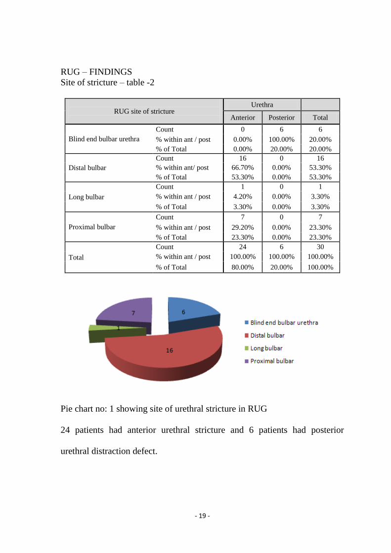
RUG – FINDINGS
Site of stricture – table -2
RUG site of stricture
Urethra
Anterior Posterior Total
Blind end bulbar urethra
Count 0 6 6
% within ant / post 0.00% 100.00% 20.00%
% of Total 0.00% 20.00% 20.00%
Distal bulbar
Count 16 0 16
% within ant/ post 66.70% 0.00% 53.30%
% of Total 53.30% 0.00% 53.30%
Long bulbar
Count 1 0 1
% within ant / post 4.20% 0.00% 3.30%
% of Total 3.30% 0.00% 3.30%
Proximal bulbar
Count 7 0 7
% within ant / post 29.20% 0.00% 23.30%
% of Total 23.30% 0.00% 23.30%
Total
Count 24 6 30
% within ant / post 100.00% 100.00% 100.00%
% of Total 80.00% 20.00% 100.00%
Pie chart no: 1 showing site of urethral stricture in RUG
24 patients had anterior urethral stricture and 6 patients had posterior
urethral distraction defect.

- 20 -
RUG – length of stricture –table -3
RUG length
Urethra
Anterior Posterior Total
<1.5cm
Count 19 0 19
% within anterior/ post 79.20% 0.00% 63.30%
% of Total 63.30% 0.00% 63.30%
>1.5cm
Count 5 6 11
% within anterior/ post 20.80% 100.00% 36.70%
% of Total 16.70% 20.00% 36.70%
Total
Count 24 6 30
% within anterior/ post 100.00% 100.00% 100.00%
% of Total 80.00% 20.00% 100.00%
Pie chart - 2 showing length group of the stricture in RUG
RUG showed 5 long anterior urethral stricture (> 1.5cm), 19 short anterior
stricture (<1.5cm), and 6 obliterative bulbar stricture (>1.5 cm) (pie chart –
2).In this 6 cases opposing urethrogram were done to delineate proximal
urethra, but the bladder neck was not opened and showed long defect.
In all cases it showed single stricture and in one case mild penile urethra
narrowing

- 21 -
MR Urethrography – Findings
Length of stricture –table – 4
MRU length
Urethra
Anterior Posterior Total
<1.5cm Count 16 0 16
% within ant / post 66.70% 0.00% 53.30%
% of Total 53.30% 0.00% 53.30%
>1.5cm Count 8 6 14
% within ant / post 33.30% 100.00% 46.70%
% of Total 26.70% 20.00% 46.70%
Total Count 24 6 30
% within ant / post 100.00% 100.00% 100.00%
% of Total 80.00% 20.00% 100.00%
Pie chart – 3 showing length group in MR Urethrography
MR urethrography showed 16 short anterior urethral strictures (<1.5cm), 8
long Anterior stricture (> 1.5cm), 6 long posterior urethral defect (> 1.5)

- 22 -
MRU - site of stricture – table -5
Site of stricture MRU Urethra
Anterior Posterior Total
bulbomembranous Count 0 6 6
% within ant/ post 0.00% 100.00% 20.00%
% of Total 0.00% 20.00% 20.00%
Distal bulbar Count 14 0 14
% within ant/ post 58.30% 0.00% 46.70%
% of Total 46.70% 0.00% 46.70%
Long bulbar Count 3 0 3
% within ant/ post 12.50% 0.00% 10.00%
% of Total 10.00% 0.00% 10.00%
Proximal bulbar Count 7 0 7
% within ant/ post 29.20% 0.00% 23.30%
% of Total 23.30% 0.00% 23.30%
Total Count 24 6 30
% within ant/ post 100.00% 100.00% 100.00%
% of Total 80.00% 20.00% 100.00%
Pie chart -4 showing site of stricture in MR urethrography
14 cases distal, 7 proximal bulbar, 3 long bulbar and 6 cases were
bulbomembranous junction.

- 23 -
MRU – other findings –table – 6
OTHER FINDINGS-MRU Urethra
Anterior Posterior Total
Nil Count 20 0 20
% within
anterior/post
83.30% 0.00% 66.70%
% of Total 66.70% 0.00% 66.70%
Spongiofibrosis,periurethralfibrosis
prostatic apex displacement
Count 4 6 10
% within
anterior/ post
16.70% 100.00% 33.30%
% of Total 13.30% 20.00% 33.30%
Total Count 24 6 30
% within
anterior/ post
100.00% 100.00% 100.00%
Pie chart-5 showing associated findings in MR Urethrography
In anterior urethra, 4 cases - showed spongiofibrosis, no spongiofibrosis in
20 cases. In posterior urethra, 3 cases – showed periurethral fibrosis, 3cases
posterior displacement of the prostate with periurethral fibrosis.

- 24 -
SURGICAL FINDINGS
Length of stricture –table -7
Surgery- length of stricture Urethra
Anterior Posterior Total
<1.5cm Count 16 0 16
% within anterior/ post 66.70% 0.00% 53.30%
% of Total 53.30% 0.00% 53.30%
>1.5cm Count 7 6 13
% within anterior/ post 29.20% 100.00% 43.30%
% of Total 23.30% 20.00% 43.30%
No stricture, BNE+
Mild catching in
bulbar urethra
Count 1 0 1
% within anterior/ post 4.20% 0.00% 3.30%
% of Total 3.30% 0.00% 3.30%
Total Count 24 6 30
% within anterior/ post 100.00% 100.00% 100.00%
% of Total 80.00% 20.00% 100.00%
Pie chart-6 showing surgical length
The surgical procedures were planned according to the findings of the RUG
and MRU. In surgery, 16 cases were <1.5cm, 7 cases of anterior urethra
were (>1.5cm) and 6 cases were posterior distraction defect (>1.5cm).

- 25 -
In one case there was no stricture,but mild catching in bulbar urethra &mild
BNE. Among the 19 short anterior stricture as showed by RUG,2 cases
were managed by augmentation urethroplasty,17 cases were treated by
VIU,In 5long anterior stricture 3 cases were managed by VIU, 2 cases by
augmentation and anastomotic urethroplasty
Among the 16 short anterior stricture as showed by MRU, all were treated
by VIU, in 8 long anterior stricture 4 treated by VIU and 4treated by open
repair. All the 6 cases of posterior distraction defect were treated by
anastomotic urethroplasty.
In two cases of short anterior stricture,there was a mild catching of bulbar
urethra & bladder neck elevation in one case for which bladder neck incision
was done.

- 26 -
Surgery –site of stricture –table -8
Surgery site of stricture Urethra
Anterior Posterior Total
bulbomembranous Count 0 6 6
% within
anterior/ post 0.00% 100.00% 20.00%
% of Total 0.00% 20.00% 20.00%
Distal bulbar Count 14 0 14
% within
anterior/ post 58.30% 0.00% 46.70%
% of Total 46.70% 0.00% 46.70%
Long bulbar Count 3 0 3
% within
anterior/ post 12.50% 0.00% 10.00%
% of Total 10.00% 0.00% 10.00%
Proximal bulbar Count 6 0 6
% within
anterior/ post 25.00% 0.00% 20.00%
% of Total 20.00% 0.00% 20.00%
No stricture Count 1 0 1
% within
anterior/ post 4.20% 0.00% 3.30%
% of Total 3.30% 0.00% 3.30%
Total Count 24 6 30
% within
anterior/ post 100.00% 100.00% 100.00%
% of Total 80.00% 20.00% 100.00%
In one case of distal bulbar stricture,there was only a mild narrowing,scope
passed with mild negotiation

- 27 -
Pie chart – 7 showing site of stricture in surgery
The above pie chart shows the site of the stricture in surgery.In anterior
urethral stricture 14 cases were distal bulbar (1 case only mild narrowing), 3
cases were long bulbar and 6 cases were proximal bulbar urethra and in one
case there was no stricture. But there was a mild catching in the bulbar
urethra and bladder neck elevation.In posterior urethra six cases were
obliterative stricture in bulbomembranous junction

- 28 -
TYPE OF SURGERY
Surgery –type –table-9
Surgery-type
Urethra
Anterior Posterior Total
anastomotic
urethroplasty
Count 1 6 7
% within ant/ post 4.20% 100.00% 23.30%
% of Total 3.30% 20.00% 23.30%
augmentation
urethroplasty
Count 3 0 3
% within ant/ post 12.50% 0.00% 10.00%
% of Total 10.00% 0.00% 10.00%
mild narrowing in the
distal bulbar urethra
scope passed with mild
negotiation
Count 1 0 1
% within ant/ post 4.20% 0.00% 3.30%
% of Total 3.30% 0.00% 3.30%
no stricture, BNI done,
mild catching in bulbar
urethra
Count 1 0 1
% within ant/ post 4.20% 0.00% 3.30%
% of Total 3.30% 0.00% 3.30%
OIU Count 15 0 15
% within ant/ post 62.50% 0.00% 50.00%
% of Total 50.00% 0.00% 50.00%
OIU & meatal dilatation Count 1 0 1
% within ant/ post 4.20% 0.00% 3.30%
% of Total 3.30% 0.00% 3.30%
OIU & guided urethral
dilatation
Count 2 0 2
% within ant/ post 8.30% 0.00% 6.70%
% of Total 6.70% 0.00% 6.70%
Total Count 24 6 30
% within ant/ post 100.00% 100.00% 100.00%
% of Total 80.00% 20.00% 100.00%

- 29 -
TYPE OF SURGERY
Pie chart – 8 showing the type of surgery.
Among the 24 anterior urethral stricture, endoscopic procedure (VIU) was
done for 20 patient’s .In these 20 cases, one case showed only mild
narrowing and the scope passed with little negotiation. In another case
showed mild catching of bulbar urethra with mild bladder neck elevation,
for which bladder neck incision was done. 4 cases of stricture of anterior
urethra (2cases <1.5cm & 2cases >1.5cm) was managed by open repair (3
augmentation urethroplasty,1anastomotic urethroplasty) 6 PFUDD cases
were managed by progressive perineal anastomotic urethroplasty.

- 30 -
Pie chart – 9 showing associated findings in surgery
During the procedure the following findings were noted
2 cases – mild narrowing & unhealthy penile urethra
1 case - mild meatal stenosis
4 cases – thick fibrosed urethra (spongiofibrosis)
6 cases – obliterated bulbar urethra with periurethral fibrosis
1 case – mild catch in the bulbar urethra and mild bladder neck elevation
16 cases – no associated findings other than stricture.

- 31 -
ANALYSIS
The following factors were analyzed.
1. Site
2. Number
3. Length
4. Associated findings
SITE –In anterior urethra the site of stricture in both RUG and MR
urethrogram were well co-related with the site in surgery.
In posterior urethra RUG showed only blind ending bulbar urethra and
closed bladder neck, but MRU showed the exact site in all the 6cases
NUMBER –In all cases both RUG and MRU showed single stricture.
LENGTH OF STRICTURE
In overall 30cases the mean length of the stricture by RUG, MRU,
SURGERY was 1.72, 1.56 and 1.56 cm
The mean length of stricture by RUG, MRU, SURG in anterior stricture
cases were 1.19,1.36,1.29 cm
The mean length of stricture by RUG, MRU, SURG in posterior distraction
defect cases were 4.08, 2.51, 2.5 cm

- 32 -
Table 10- shows the mean length of the stricture
Mean plot curve shows the mean length of the stricture by RUG, MRU, and
SURG
95% Confidence Interval for Mean
N Mean
Std.
Deviation Std. Error Lower Bound Upper Bound Minimum Maximum
RUG 30 1.726667 1.2376099 .2259556 1.264536 2.188798 .5000 4.5000
MRU 30 1.560000 .6387488 .1166190 1.321487 1.798513 .6000 2.9000
SURG 28 1.560714 .6505492 .1229422 1.308458 1.812971 .6000 3.0000
Total 88 1.617045 .8855444 .0943993 1.429417 1.804674 .5000 4.5000
- 33 -
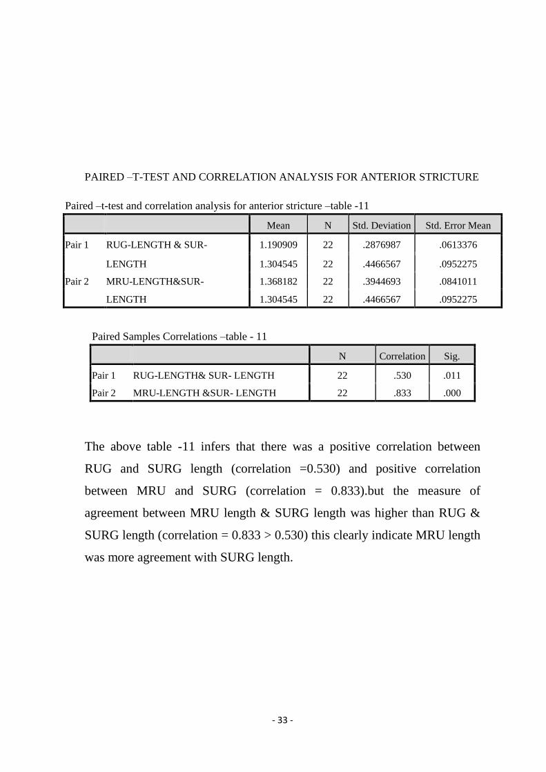
PAIRED –T-TEST AND CORRELATION ANALYSIS FOR ANTERIOR STRICTURE
Paired –t-test and correlation analysis for anterior stricture –table -11
Mean N Std. Deviation Std. Error Mean
Pair 1 RUG-LENGTH & SUR- 1.190909 22 .2876987 .0613376
LENGTH 1.304545 22 .4466567 .0952275
Pair 2 MRU-LENGTH&SUR- 1.368182 22 .3944693 .0841011
LENGTH 1.304545 22 .4466567 .0952275
Paired Samples Correlations –table - 11
N Correlation Sig.
Pair 1 RUG-LENGTH& SUR- LENGTH 22 .530 .011
Pair 2 MRU-LENGTH &SUR- LENGTH 22 .833 .000
The above table -11 infers that there was a positive correlation between
RUG and SURG length (correlation =0.530) and positive correlation
between MRU and SURG (correlation = 0.833).but the measure of
agreement between MRU length & SURG length was higher than RUG &
SURG length (correlation = 0.833 > 0.530) this clearly indicate MRU length
was more agreement with SURG length.

- 34 -
PAIRED –T-TEST AND CORRELATION ANALYSIS FOR POSTERIOR DEFECT
Paired –t-test and correlation analysis for posterior urethral defect –table - 12
Mean N Std. Deviation Std. Error
Mean
Pair 1 LENGTH-RUG 4.083333 6 .2316607 .0945751
SURG-LENGTH 2.500000 6 .3162278 .1290994
Pair 2 LENGTH-MR 2.550000 6 .2738613 .1118034
SURG-LENGTH 2.500000 6 .3162278 .1290994
Paired Samples Correlations table – 12
N Correlation Sig.
Pair 1 LENGTH-RUG & SURG-LENGTH 6 .491 .322
Pair 2 LENGTH-MR &SURG- LENGTH 6 .924 .000
In above table -12-In the posterior distraction defect it showed the measure
of agreement between MRU &SURG length was higher than RUG & SURG
length (correlation = 0.924>0.491 ) P =0.000<0.001 and statistically
significant

- 35 -
The line graphs show the comparison among RUG, MRU and Surgical
length. The MRU length was well correlated with surgical length
0
0.5
1
1.5
2
2.5
3
3.5
4
4.5
5
1 3 5 7 9 11 13 15 17 19 21 23 25 27 29
Stri
ctu
re le
ngt
h
RUG, MRU and Surgery Comparision
XRAY Length
MR Length
Surgery Length

- 36 -
The scatter diagram shows RUG length and MRU length was well co-related
or minimal difference in anterior urethral strictures but in Posterior urethra
there was gross difference in length between RUG and MRU.
0
0.5
1
1.5
2
2.5
3
0 1 2 3 4 5
MR
U le
ngt
h
RUG Length
RUG Vs MR Urethrography
Anterior
Posterior

- 37 -
The scatter diagram shows RUG length and surgical length was well co-
related or minimal difference in anterior urethral strictures but in Posterior
urethra there was gross difference in length between RUG and Surgical
length
0
0.5
1
1.5
2
2.5
3
3.5
0 1 2 3 4 5
Surg
ical
Le
ngt
h
RUG Length
RUG Vs Surgery
Anterior
Posterior

- 38 -
The scatter diagram shows SURG length and MRU was well co-related or
minimal difference in Anterior and Posterior urethral strictures.
0
0.5
1
1.5
2
2.5
3
3.5
0 0.5 1 1.5 2 2.5 3
Surg
ical
Le
nth
MRU Lennth
MR Urethrography Vs Surgery
Anterior
Posterior
- 39 -
ASSOCIATED FINDINGS
RUG showed mild penile urethral narrowing in one case which contributes
3.3%, but MRU showed additional findings in 33.3% of cases. It was
superior in delineating Spongiofibrosis (image1b, 2b), periurethral fibrosis
(image 3b, 4b), and prostatic apex displacement (image 4c).
In 3 cases RUG showed short stricture<1.5cm, but MR showed long
stricture>1.5cm with spongiofibrosis. Hence these patients(Two Cases) were
managed by urethroplasty(Image 1a & 1b). One case was managed
endoscopically in view of minimal spongiofibrosis although length was >1.5
cms
In 6 cases of posterior distraction defect, MR showed accurate length of
posterior urethra and associated periurethral fibrosis, prostatic apex
displacement. These findings helped to plan the operative approach (Images
3a, 3b, 4a, 4b &4c)

- 40 -
SENSITIVITY &SPECIFICITY BETWEEN RUG AND SURGERY
Crosstab – table-13
SURGERY TYPE
Endo Open
surgery Total
LEN-GROUP <1.5cm Count 17 2 19
% of Total 56.7% 6.7% 63.3%
>1.5cm Count 3 8 11
% of Total 10.0% 26.7% 36.7%
Total Count 20 10 30
% of Total 66.7% 33.3% 100.0%
Sensitivity 80%
Specificity 85%
Positive Predictive Value 72.73%
Negative Predictive Value 89.47%
Diagnostic Accuracy 83.33%
KAPPA MEASURE OF AGREEMENT IS = 0.634
This above table showed the accuracy of RUG for predicting the type of
surgery was about 83.33%

- 41 -
SENSITIVITY & SPECIFICITY BETWEEN MRU AND SURGERY
Crosstab – table-14
SURGERY TYPE
Endoscopy Open repair Total
LEN-MR-GROUP <1.5cm Count 16 0 16
% of Total 53.3% .0% 53.3%
>1.5cm Count 4 10 14
% of Total 13.3% 33.3% 46.7%
Total Count 20 10 30
% of Total 66.7% 33.3% 100.0%
Sensitivity 100%
Specificity 80%
Positive Predictive Value 71.43%
Negative Predictive Value 100%
Diagnostic Accuracy 90.1%
KAPPA AGREEMENT 0.727
This above table showed the accuracy of MRU for predicting the type of
surgery was about 90.1%
The sensitivity &specificity for diagnosing stricture by both RUG &MRU
was 100% &93.4% both in anterior and posterior urethra
Sensitivity 80% Specificity 85% Positive Predictive Value 72.73% Negative Predictive Value 89.47% Diagnostic Accuracy 83.33%
Crosstab – table-14
SURGERY TYPE
Endoscopy Open repair Total
LEN-MR-GROUP <1.5cm Count 16 0 16
% of Total 53.3% .0% 53.3%
>1.5cm Count 4 10 14
% of Total 13.3% 33.3% 46.7%
Total Count 20 10 30
% of Total 66.7% 33.3% 100.0%

- 42 -
DISCUSSION
The retrograde urethrogram was set as gold standard imaging modality for
the diagnosis of stricture urethra in 1910, by Cunningham3
because it is
easily available and a simple technique. But it has certain disadvantages like
over or under estimation, radiation effect and does not provide information
about spongiofibrosis.
To overcome this limitation, MRI was suggested, according to Garcia-
Valtuille,8 the treatment choice and route of approach depends upon the site,
length, spongiofibrosis and associated pathology. Endoscopic repair can be
effective for stricture < 1.5 cm without spongiofibrosis. The long stricture >
1.5cm with spongiofibrosis can be treated by open repair either anastomotic
or augmentation urethroplasty26
through perineal route, but complex
stricture needs transpubic approach.
This study showed the sensitivity &specificity for diagnosing stricture by
both RUG &MRU was 100% &93.4% both in anterior & posterior urethra.
whereas the study by Syed Mamun Mahmud et al 6 ,the sensitivity &
specificity of RUG for the diagnosis of urethral stricture was 91% &72%and
by MRU it was 100%.

- 43 -
The other study by MA El-Ghar et al 13
showed the sensitivity, specificity
for diagnosing anterior urethral stricture by RUG was 91% & 90% and 89%
& 91.7% for posterior urethra,by MRU it was 91.7% (ant &post). In
Sonourethrography the accuracy was 100% in ant urethra, 60% in post
urethra.
In this study the accuracy showed by RUG for planning surgery was 83%
and by MR urethrogram was 90.1%.But the study by Yasser osman14
for
both RUG and MR urethrogram the accuracy was 85%.
In this study MR urethrogram diagnosed all the cases of anterior and
posterior stricture with exact delineation of its length with 100% sensitivity,
93.4% specificity and 90.1% overall accuracy, which was well correlated the
study by MA El-Ghar et al 13
In this study in 6 cases of posterior distraction defect, RUG showed over
estimation of defect because of failure of bladder neck to relax. But MR
urethrogram showed accurate length and associated findings which helped to
plan the surgical approach similar to the study conducted by Sung DJ et al.16
In this small series of patients MR urethrography proved to be a promising
technique for evaluating male urethral stricture.It combined the advantages
of RUG and Sonourethrography with its few disadvantages of cost
effectiveness and its availability.

- 44 -
IMAGES
Anastomotic urethroplasty –
Proximal urethra mobilization.
Anastomotic urethroplasty –
Distal urethra mobilization.
After Anastomosis
The above picture shows per-operative pictures of Anastomotic
urethroplasty
Anastomotic urethroplasty –
distal urethral mobilization
Proximal urethral mobilization
- 45 -
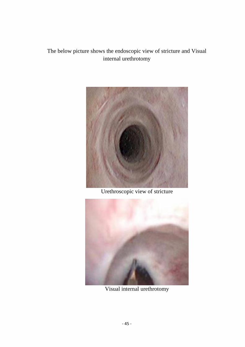
The below picture shows the endoscopic view of stricture and Visual
internal urethrotomy
Urethroscopic view of stricture
Visual internal urethrotomy

- 46 -
Buccal mucosal graft
The above picture shows the graft taken from the buccal mucosa for
augmentation urethroplasty.

- 47 -
Dorsal onlay graft
After augmentation
The above picture shows the per operative picture of augmentation
urethroplasty

- 48 -
Image-1a
Image-1a- RUG showing 1.1cm short bulbar stricture
Image-1b
Image-1b –MR urethrography for the same patient showing 2.1cm bulbar
stricture with hypointense spongiofibrosis
1.1cm
2.1cm

- 49 -
Image 2a
Image 2a –RUG showing long 1.7 cm bulbar stricture.
Image 2b
Image 2b –MR urethrography for the same patient showing 1.9cm bulbar
stricture With hypointense spongiofibrosis
1.7cm
1.9cm

- 50 -
Image 3a
Image 3a- RUG and opposing urethrogram showing 4 cm posterior urethral
defect, bladder neck not opened
Image 3b
Image 3b- MR urethrogram of above patient showing 2.2 cm bulbo
membranous defect with periurethral fibrosis
4 cm
2.2 cm

- 51 -
Image 4a
Image 4a-RUG and opposing urethrogram showing obliterative blind bulbar
urethra with 4.1 cm long posterior urethral defect, bladder neck not opened
Image 4b
Image 4b - MR urethrogram of above patient showing 2.2 cm bulbo
membranous defect with periurethral fibrosis.
4.1 cm
2.2 cm

- 52 -
Image 4c
Image 4c – MR coronal image showing displacement of the prostate apex.

- 53 -
CONCLUSION
In summary, our study has demonstrated that MR urethrography is a very
useful and promising noninvasive technique for the evaluation of male
anterior urethral stricture and posterior urethral distraction defect for
planning the surgical approach.
It was superior than RUG for the accurate assessment of length of stricture
and extent of spongiofibrosis in anterior urethral stricture.
In posterior urethral defect MR urethrography, correctly estimates the
length of the stricture, degree of prostatic displacement and delineates the
site & density of scar tissue, which helps to plan the surgical approach
This procedure is also well tolerated by patients, who are allergic to
iodinated contrast during RUG.

A

A
BIBLIOGRAPHY
1. Andrich DE, Mundy AR. Urethral strictures and their surgical
management. BJU 2000; 86:571-580.
2. Nielsen, Nordling J.UROLOGY1990; 35(1):18-24.
3. CunninhamJ.The diagnosis of stricture of the urethra by x-rays 1910; 5;
369-371.
4. GallentineML,MoreyAF.Imaging of the male urethra for stricture
disease.Urolclin north Am 2002:29; 361-72
5. Nash PA,McAninchJW,BruceJE,HanksDK.Sono-urethrography in the
evaluation of anterior urethral strictures J Urol.Jul;154(1);72-6
6. SyedMamun Mahmud et al- Is ascending urethrogram mandatory for all
urethral strictures? Jan-Dec 2002.
7. FriedburgHG,WimmerB,HennigUrologe A.1987 Nov; 26(6):309-16
8. Garcia-Valtuille, F Aascal et al – The British Journal of Radiology, July
2006.
9. Oh MM, Jin MH, Sung DJ, Yoon DK, Kim JJ, Moon du G.Magnetic
resonance urethrography to assess obliterative posterior urethral stricture:
comparison to conventional retrograde urethrography with
voidingcystourethrography. J Urol. 2010; 183(2):603-7.
10. J Eaton MS FRCS, J Richenberg et al – Imaging of urethra BJR 17, 139,
149, 2005
11. Jeong-ah Ryu – Imaging of male and female urethra - Department of
Radiology, Samsung medical centre.rsna.info/content/21/5/1169.
B
12. Moon-Hae Choi,MD,BohyunKim,Sung Won Lee MD- Department of
Radiology and Urology, Samsung medical centre -September 2000, 135-
710
13. MA El-Ghar – Department of Radiology and Urology, Mansoura
University, Egypt, EJR 2010 15 Jul 2009.
14. Y Osman-European Urology, MR –urethrography in comparison to
retrograde Urethrography in Diagnosis of male urethral strictures
50/3,587-594,September 2006.
15. YNarumi -1993 MR imaging of traumatic posterior urethral
injury.radiology.rsna.org/content/188/2/439.
16. DJ Sung et al. Obliterative Urethral Stricture: MR Urethrography verses
Conventional Retrograde Urethrography with voiding
Cystourethrography. Rsna 2006; 240(3):842-48.
17. Bircan MK, Sahin H, KorkmazK.IntUrolNephrol. 1996; 28(6):801-4
18. Pavlica P, Barozzi L, Menchi I. EurRadiol. 2003 Jul; 13(7):1583-96.
Epub 2002 Dec 19.
19. KoraitimMM,Reda IS- Role of MRI in assessment of posterior urethral
distraction defects; J Urol 2007 Aug; 28(4):258-73.
20. Campbell-Walsh Urology;9th
edi;vol1; Urinary tract imaging 4;116-119
21. ChambersRM, Baitera B – Anatomy of urethral stricture – BJU 1977;
49; 545-551
22. Michael L. Gallentine AF – Imaging of male urethra, UCNA vol29; 2; 2002,
361-372.
23. Pollack.M, Bruce L.McClennan, clinicalurography, 2edi, vol1; 8; 328-350
24. MoreyAF,McAninch JW – Role of sonourethrography in bulbar urethral
stricture.JUrol 1997; 158(4); 1376-1379.

C
25. Dixon CM. MR imaging of posterior urethral defect and pelvic injuries.JUrol
1992; 148; 1162-1165
26. PetersonAC, WebsterGD, Management of urethral stricture disease, BJU
2004; 94; 971-976.
27. Choudhary S, Singh P et al, comparison of sonourethrography and
retrograde urethrography in evaluation of anterior urethral strictures
JRadio 2004 Aug;59(8):736-42.

D
ANNEXURE I
PROFORMA
KMCH/GRH
Name : Date:
Age : Sex: Op No: IP No:
Phone No :
PRESENT ILLNESS:
LUTS – Obstructive/Irritative
Dysuria,frequency,urgency,hesitancy,intermittency,thin stream,strain to void
Fever
H/O pelvic trauma
H/O previous surgery/ instrumentation
Past H/O DM / HT/ COPD
O/E:
Fever, Pallor
BP
Pulse
Abdomen
o Bladder distended/not
o Genitalia
o DRE – Prostate size, consistency, nodule
INVESTIGATION
Urine –
o alb
o Sug
o Deposits
Urine c/s
Hemogram
Sr.creatinine , Urea
Blood sugar
Xray KUB
USG KUB
UROFLOMETRY
Retrograde urethrogram
MR urethrogram
Urethroscopy/Urethroplasty findings

E
ANNEXURE - II
MASTER CHART

F

G