INTRAVEANOUS UROGRAPHY (IVU) Enhancing Knowledge …hqe.moh.gov.my/radiologi/3. IVU.pdf ·...
39
INTRAVEANOUS UROGRAPHY (IVU) Enhancing Knowledge And Skills Of Radiographers “Update in Radiography 2015 KKM Radiographers” For Sabah & Sarawak By; Mohd Ari-Nadzahari Bin Abd Rahman Juru x-ray Hospital Queen Elizabeth
Transcript of INTRAVEANOUS UROGRAPHY (IVU) Enhancing Knowledge …hqe.moh.gov.my/radiologi/3. IVU.pdf ·...

INTRAVEANOUS UROGRAPHY (IVU)
Enhancing Knowledge And Skills Of Radiographers
“Update in Radiography 2015 KKM Radiographers” For
Sabah & Sarawak
By; Mohd Ari-Nadzahari Bin Abd Rahman
Juru x-ray Hospital Queen Elizabeth

Introduction
• KUB plain films only demonstrate very little of the urinary system, as the urinary system was blends in with other soft tissue structures in the abdominal cavity. That’s why we need contrast media to visualize the urinary system radiographically.
• So called excretory urogram. • Sometimes referred as IVP; (P; pyelogram, pyelo refers only
renal pelves). Not recommended to use this terms as this exam normally visualised more than just the renal pelves.
• Twofold purpose of IVU; – Visualize the collecting portion of the urinary system. – To assess the functional ability of the kidneys.

Anatomy of the urinary system
• Kidneys – Bean shaped, 11 cm long, 6 cm wide, 3cm thick and weigh
150 g. – Loosely attached within their fatty capsule (perirenal fat),
covered by a sheath of fibroelastic renal fascia, retroperitoneal organs.
– Positions; beside of the vertebral column, behind the peritoneum and below the diaphragm. extended from 11-12th t-spine to 3rd L-spine (halfway between the xiphoid process and the iliac crest), left kidney 1cm more superior the the right, slight protected by lower rib cage.
– Movement; move up and down with movement of the diaphragm, deep inhalation or standing drop the kidneys one lumbar or 5cm (2 inches),

Anatomy of the urinary system
• Kidneys

Anatomy of the urinary system
• Kidneys

Anatomy of the urinary system • Kidneys (zoom in)

Anatomy of the urinary system
• Ureters
– Convey urine from kidneys to bladder.
– Slow peristaltic wave and gravity forced to flow the urine.
– Length 28-32 cm, right is shorter than the left.
– Pass inferiorly - Lie on the anterior surface of each psoas major muscle - follow the curvature of vertebral column- enter the posterolateral portion of urinary bladder.
– Three constricted points; • Ureteropelvic UP junction.
• Pelvic brim.
• Ureterovesical UV junction.

Anatomy of the urinary system
Urinary bladder
• Musculomembranous sac.
• Capacity 350-500 mL.
Urethra

Diseases of urinary system
• Renal calculi
• Staghorn calculus
• Hydronephrosis
• Pyelonephritis
• Renal obstruction
• Polycystic kidney
• Bladder calculi
• Ectopic kidney
• Horseshoe kidney
• Etc.

Indications
• The following are accepted indications for IVU, as delineated by the American College of Radiology (ACR) guidelines published in 2010: – To evaluate the presence or continuing presence of suspected or
known ureteral obstruction. – To assess the integrity of the urinary tract status post trauma
particularly in situations in which cross-sectional imaging is unavailable or inappropriate.
– To assess the urinary tract for suspected congenital anomalies, particularly in situations in which cross-sectional imaging is unavailable or inappropriate.
– To assess the urinary tract for lesions that may explain hematuria or infection. In particular, IVU may be used to evaluate for an underlying parenchymal mass or may be used to evaluate for a lesion of the urinary tract in settings in which cross-sectional imaging is unavailable or inappropriate.

contraindications
The major contraindications; • Hypersensitivity to iodinated CM. • Anuria- absence of urine excretion • Multiple myeloma.-malignant condition of the plasma cells of the
bone marrow. • Pheochromocytoma- tumor that develops in cells in the center of
an adrenal gland. • Diabetes mellitus • Severe hepatic or renal disease. • Congestive hearth failure. • Sickle cell anemia. • Pt taking glucophage (metformin- diabetic medicine, ACR
recommends withheld 48h) • Renal failure, acute or chronic.

Patient preparations
• Bowel prep; – Light evening meal before the procedure – Laxative – NPO after midnight (min 8hours) – Enema morning of the examination – No chewing gum.
• If patient have H/O allergic cover with prednisolone • BUN and Creatinine result. (normal ; creatinine 0.6-1.5 mg/dl; BUN 8-
25mg/100ml) • IVU checklist and consent form should be filled up and sign properly. • Make sure female patient is not pregnant. • Explanations to patient regarding the process of the procedure is
crucial. • Change patient clothing with hospital clothing, make sure there is no
artifacts. • Prior to exam, ask patient to void.

Equipment and supplies preparation
• Correct type and amount CM drawn up in appropriate syringe.(maintain sterile!)
• Selection of sterile needle (branula-[blue or pink] or 19 gauge butterfly). • Alcohol sponges or wipes • Tourniquet. • Towel or sponge to support elbow. • Male gonadal shield. • Emesis basin (for pt vomit if reaction occur) • Lead markers; prelim, minute marker and post mict etc. • Emergency trolley • Emergency drugs (epinephrine and benadryl) • Ureteric compression devices. • Trendelenburg position device ???

Venipuncture Procedure VP
• Introduction than confirmation of pt ID. • Prelim film prior to VP. • Done by MO or nurse. Radiographer?
– Video how to set line.

Contrast media
• Recommended to use nonionic iodinated water soluble C.M. got various of brand. for example iopamiro, omnipaque and etc.

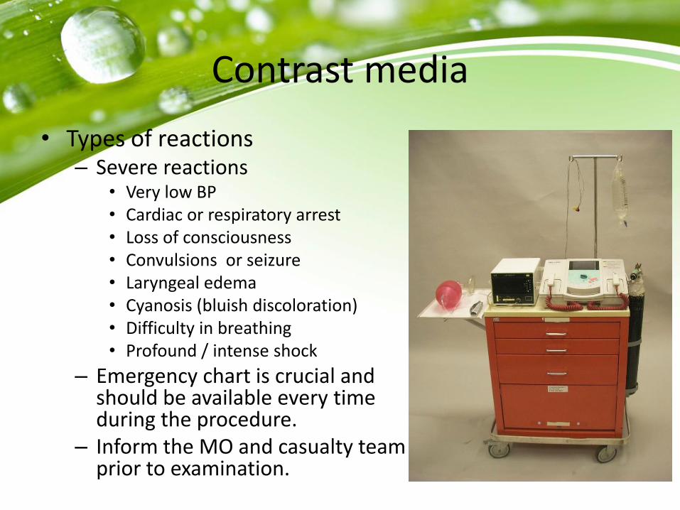
Contrast media
• Types of reactions
– Mild reactions
• Nausea and vomiting
• Small hives or urticaria
• Itching
• Sneezing
• Extravasation
• Vasovagal response

Contrast media
• Types of reactions
– Moderate reactions
• Excessive urticaria (hives)
• Tachycardia. Make sure monitoring device for vital sign is available.
• Giant hives
• Excessive vomitting
Contrast media
• Types of reactions – Severe reactions
• Very low BP • Cardiac or respiratory arrest • Loss of consciousness • Convulsions or seizure • Laryngeal edema • Cyanosis (bluish discoloration) • Difficulty in breathing • Profound / intense shock
– Emergency chart is crucial and should be available every time during the procedure.
– Inform the MO and casualty team prior to examination.

Radiation protection
• Use gonadal shielding appropriately without obscuring the radiographic information
• “Pregnancy “ rule should be followed.
• When bladder and lower ureters are not include then use gonad protection.

The chronology of IVU exam.
HQE1 protocol; 1. Preliminary or scout film 2. Immediate film (optional) 3. 5 minutes 4. 10 minutes with compression or Trendelenburg position 5. 15 minutes / release 6. Full bladder 7. Post micturition 8. Extra view;
– Delayed view ; 1-2 hours – Prone position – Posterior oblique view

The chronology of IVU exam.
• Preliminary or scout film – Verify patient preparation – Determine optimal exposure factor – Verify positioning and central ray – Detect any abnormality calcification
“Certain radiological examinations especially those that involve contrast studies, require adequate preparation like fasting, bowel preparation etc. the ever-obliging Radiologist who proceeds with the radiological examination, in an inadequately prepared patient, will risk missing out certain features and images that will hamper his ability in providing a clearer picture of the findings. The radiologist must learn to say no, as an inadequately prepared will not be an acceptable.” Dr N.K.S. tharmaseelan 2014

The chronology of IVU exam.
• Preliminary or scout film – 35 x 43 cm cassette (full
length) – Suprarenal region to a
level below the symphysis pubis.
– Kidney shadow demonstrate with minimal bowel gas appearance -good prep.
– Pt empty his/her bladder before start the procedure.

The chronology of IVU exam.
Immediate film (optional) • Nephrogram : produced primarily by filtered contrast material within the
nephron, with optimal visualization of the renal parenchyma 1–3 minutes after bolus injection
• Nephrogram phase/progression : – Vascular phase( cortical arteriogram)- CM in interlobular arteries & glomeruli – Cortical phase ( cortical nephrogram)- CM in cortical capillaries, peritubular spaces
and cortical tubular lumen – Parenchymal phase (generalised tubular nephrogram) – CM in loop of Henle and
collecting tubules – Excretory phase – CM within collecting system
• The renal parenchyma is optimally assessed during nephrographic phase 1) The entire renal contour should be assessed 2) There should be temporal symmetry of the nephrographic development (5). 3) The size of the kidneys is best seen during the nephrographic phase. 4) The position and axis of the kidney should also be assessed.

The chronology of IVU exam.
• Immediate film

The chronology of IVU exam.
• 5 minutes
– To assess temporal symmetry and progress of opacification.
– On the 5-minute image, the nephrogram should be receding as the collecting system becomes opacified.

The chronology of IVU exam.

The chronology of IVU exam.
• 10 minutes with compression or
Trendelenburg position. – An image collimated to the kidneys for evaluation
of the renal calices and collecting systems is obtained 10 minutes after the compression device is applied . This is especially important when low-osmolar contrast material is used because the osmotic diuresis and ultimate distention of the collecting system may not be as great as with high-osmolar materials.

The chronology of IVU exam.
• 10 minutes with compression or Trendelenburg position.

Contraindication to Ureteric compression
• Possible ureteric stones
• Abdominal mass
• AAA
• Recent abdominal surgery
• Severe abdominal pain
• Acute abdominal trauma

The chronology of IVU exam.
• 15 minutes / release
– At the release of compression, the bolus of contrast material urine entering the ureters provides optimal visualization throughout their length
– Segmental non-visualization of the ureter due to peristalsis can be overcome
– Image will be taken in 35x43cm cassette (full length)

The chronology of IVU exam.
• 15 minutes / release

The chronology of IVU exam.
• Full bladder
– If the bladder is sufficiently distended and opacified, it may be adequately evaluated on previously obtained images. If, however, there is specific concern for bladder disease, delayed images may be obtained to improve bladder distention, and oblique, prone, or postvoid images may be obtained to evaluate filling defects

The chronology of IVU exam.
• Full bladder

The chronology of IVU exam.
• Post micturition
– Early filling images followed by a postvoid examination may constitute the most sensitive imaging sequence for the evaluation of filling defects
– The postvoid image is most helpful in assessing residual volume when it is obtained immediately after voiding and demonstrates complete emptying of the bladder.

The chronology of IVU exam.
• Post micturition

The chronology of IVU exam.
• Extra view; – Delayed view ; 1-2 hours
• Requested if the filling of the involved ureter is slow.
– Prone position • To investigate pelviureteric and ureteric obstruction as the
heavy contrast mixed urine will more readily gravitate to the site of the obstruction.
• To displace the overlying bowel gas towards periphery
– Posterior oblique 30-35 deg view • Provide a different perspective of the kidney and project the
ureters away from the spine.

Posterior oblique images

After care
• Reassure that no late CM reaction.
• General psychological reassurance. Explain to the patient once more regarding action if late reaction occur.
• Treat and check the puncture site.
• Explain and make sure the patient understands how to receive the results.
• Escort to changing rooms and give a warm appreciation for their cooperation.

Discussion
• Any questions?


![Original Article - Endourology/Urolithiasis · follow-up of urolithiasis [1]. Prior to the use of computed tomography (CT), intravenous urography (IVU) was the Radiation dosing in](https://static.fdocuments.net/doc/165x107/5c8ae9ae09d3f232478d1cdb/original-article-endourologyurolithiasis-follow-up-of-urolithiasis-1-prior.jpg)