COMPARATIVE DNA EXTRACTION METHOD FROM ADHESIVE TAPE · 2010. 8. 2. · Electrical adhesive tape...
122
COMPARATIVE DNA EXTRACTION METHOD FROM ADHESIVE TAPE EVIDENCE FOR PERSON IDENTIFICATION By Natthalak Pakdeenarong A Thesis Submitted in Partial Fulfillment of the Requirements for the Degree MASTER OF SCIENCE Program of Forensic Science Graduate School SILPAKORN UNIVERSITY 2008
Transcript of COMPARATIVE DNA EXTRACTION METHOD FROM ADHESIVE TAPE · 2010. 8. 2. · Electrical adhesive tape...

COMPARATIVE DNA EXTRACTION METHOD FROM ADHESIVE TAPE
EVIDENCE FOR PERSON IDENTIFICATION
By
Natthalak Pakdeenarong
A Thesis Submitted in Partial Fulfillment of the Requirements for the Degree
MASTER OF SCIENCE
Program of Forensic Science
Graduate School
SILPAKORN UNIVERSITY
2008

COMPARATIVE DNA EXTRACTION METHOD FROM ADHESIVE TAPE
EVIDENCE FOR PERSON IDENTIFICATION
By
Natthalak Pakdeenarong
A Thesis Submitted in Partial Fulfillment of the Requirements for the Degree
MASTER OF SCIENCE
Program of Forensic Science
Graduate School
SILPAKORN UNIVERSITY
2008

������������������� ��� ������������������ � �������������������������!"�� #$�%
���&��'(%��))&
*��
��� �(�+�&��'(% �����(��)%
��������%��,��-� ��.�/��1�����3/�'����.&�� #�����44����3� ���.���(5��
�1��!��������3� ��%
��(5�����&�� �.����&��3�&����
�6���3/�'� 2551
&�1 ���:1����(5�����&�� �.����&��3�&����

The Graduate School, Silpakorn University has approved and accredited the Thesis
title of <Comparative DNA Extraction Method from Adhesive Tape Evidence for Person
Identification= submitted by Miss Natthalak Pakdeenarong as a partial fulfillment of the
requirements for the degree of Master of Science in Forensic Science.
??........................................................................
(Associate Professor Sirichai Chinatangkul, Ph.D.)
Dean of Graduate School
........../..................../..........
The Thesis Advisor
1. Associate Professor Pol. Col. Patchara Sinloyma, Ph.D.
2. Pol. Lt. Col. Krisda Ribroumsup
3. Assistant Professor Worawee Waiyawuth, M.D., Dr.Med.
The Thesis Examination Committee
.................................................... Chairman
(Prof. Clinic Emeritus Somchai Pholeamek, M.D., Dip.Am.Bd. of Path.)
............/......................../..............
.................................................... Member ...................................................... Member
(Asst. Prof. Sunee Kanyajit, Ph.D.) (Assoc. Prof. Pol. Col. Patchara Sinloyma, Ph.D.)
............/......................../.............. ............/......................../..............
.................................................... Member ...................................................... Member
(Pol. Lt. Col. Krisda Ribroumsup) (Asst. Prof. Worawee Waiyawuth, M.D., Dr.Med.)
............/......................../.............. ............/......................../..............

d
50312308 : MAJOR : FORENSIC SCIENCE KEY WORDS : ADHESIVE TAPE/ DNA EXTRACTION/ PERSON IDENTIFICATION NATTHALAK PAKDEENARONG : COMPARATIVE DNA EXTRACTION METHOD FROM ADHESIVE TAPE EVIDENCE FOR PERSON IDENTIFICATION. THESIS ADVISORS : ASSOC. PROF. POL. COL. PATCHARA SINLOYMA, Ph.D., POL. LT. COL. KRISDA RIBROUMSUP, AND ASST. PROF. WORAWEE WAIYAWUTH, M.D., Dr. Med. 105 pp.
Electrical adhesive tape always found in the crime scene, especially the explosion scene where use electrical adhesive tape for explosive assembling. User’s epithelial cells always leaving on the electrical adhesive tape, and can use for DNA extraction to apply in the field of person identification. One of the most important process is DNA extraction from several evidences, this research wants to study and compare the quantity of DNA extraction method from latent fingerprints on electrical adhesive tape evidence from ten volunteers for person identification. The volunteer’s touch the three trademarks of electrical adhesive tape which selling in the market with their fingers, Scotch® 3M 1710, Scotch® 3M Super 33+ and YAZAKI. Then, DNA extraction was performed by DNA IQ™ System, Chelex® 100 and Phenol: Chloroform Kit. The extracted DNA samples were quantified by Real-Time PCR to known PCR efficiency and all of data was analyzed by using SPSS program for windows. Chi-square test with the p-value lower than 0.05 was used to indicate statistical significance.
The results of this study indicated that the DNA extraction method were related to the efficiency in PCR process at the 0.05 level of significance (χ2 = 40.833; df = 2; p-value = 0.000). For Chelex® 100 extraction, extracted DNA which has PCR efficiency was observed less than expected results (Standardized residual = -3.3). While the extraction by DNA IQ™ System and Phenol: Chloroform Kit was observed to have PCR efficiency more than the expected results (Standardized residual = 1.6). DNA extraction method gives the highest amount of DNA obtained from electrical adhesive tape were Phenol: Chloroform Kit and DNA IQ™ System. While the extraction by Chelex® 100 gives the least amount of DNA. For electrical adhesive tape trademarks, the statistical analysis showed no significant differences between each electrical adhesive tape trademark and PCR efficiency.
This research showed that the electrical adhesive tape was valuable evidence in forensic science and method probable for DNA extraction on electrical adhesive tape were Phenol: Chloroform extraction and DNA IQ™ System.
Program of Forensic Science Graduate School, Silpakorn University Academic Year 2008 Student's signature ........................................ Thesis Advisors' signature 1. ............................. 2. .............................. 3. ..............................

e
50312308 : ��������������������
� \��\���� : �����/ �������������������/ ������ �������� �����
�� ������ !���"��#� : ������"��"����"�������������������$���%�# & '����(����
��)�!�������*+�'�,������ �������� �����. ����� "+��-����������� : ��. �.�.�..��# ��.����� ���
����, �.�.. �]�^� ��������� $�) _�.��.��. ���"� `���z �. 105 .�,�.
�������`{ ��/����(����"+��`�,��/���)� \�'���"�)����'��*3�"+ 3 ��#.�����$��!��'�, 4�
��/��%��.�-+#��#�����)����)���� ���������������_ ��.��#��#_ �,'�,#���������3� & $�)�����(� \���
������������������*+�� \�`�'�,������ �������� ����� ������������������� ��/���3����.�-+#"+�"�����\����
�%��������������������������(���� #�������"3�-# \�����-������"��"�������������������"+`�,���
���"�������������������$���%�# & '����(������)�!������������������ \���� 10 �� 4�'.,
���������'�,��3�����������`{����"+�"����%'�,�#���� 3 ���*+�#.������,� �*� Scotch® 3M 1710
Scotch® 3M Super 33+ $�) YAZAKI �����3�� \��������������������,����" DNA IQ™ System Chelex®
100 $�) Phenol: Chloroform Kit ����������������������,����" Real-Time PCR ��*+�'.,���(-#
���������('�����\�`����+������� \����������). �,�����,�4��$��� SPSS for windows $�)������). _�
�#�(���4�'�, Two-Way Chi-square
_�����������%����"��������������������"����������� ������������('�����\�`����+�
�������%�#�"���\�����#�(���"+�)������\���� 0.05 (χ2 = 40.833; df = 2; p-value = 0.000) 4���������,�
Phenol: Chloroform Kit $�) DNA IQ™ System '.,_��"���������('����� \�`����+������������%�"+���
(Standardized residual = 1.6) �%����������,� Chelex® 100 '.,_��"���������('����� \�`����+��������,�
��%�"+��� (Standardized residual = -3.3) ���"�������������������������(������)�!�����"+'.,������
���������������"+��� �*� Phenol: Chloroform Kit $�) DNA IQ™ System ���"�������������������������(�
�����)�!�����"+'.,�������������������,�"+��� �*� Chelex® 100 �-�������������� �).�%�#
���*+�#.������,�������������('����� \�`����+����������%� ���*+�#.������,�`�%�"����������� ���
���������('�����\�`����+�������
��������"3$��#'.,�. ���%�����������`{��/����(����"+�"����%��#������������ $�)
�����( \��������������������������(������)�!����������`{`�,������"��������,� Phenol:
Chloroform $�) DNA IQ™ System �%�#�"��)����!��
�������������������� ���6������� �.������������� �7����-��� 2551
���*��*+�����-���........................................
���*��*+������ "+��-����������� 1. ................................ 2. ................................ 3. .......................................

f
ACKNOWLEDGMENTS
I am grateful to declare the deepest sense of thankfulness to my major advisor, Assoc.
Prof. Pol. Col. Dr. Patchara Sinloyma, my co-advisor, Pol. Lt. Col. Krisda Ribroumsup and Asst.
Prof. Dr. Worawee Waiyawuth, Their find and valuable advice made my study very successful
and help me accomplish the thesis within the time. The success of this thesis can be attributed to
the extensive support and assistance from them.
I would like to use this opportunity to express my deepest sense of thankfulness to
Prof. Clinic Emeritus Somchai Pholeamek and Asst. Prof. Dr. Sunee Kanyajit, for performed
thesis examiner.
I wish to special thank Lect. Wilaiwan Keerativutisest and Dr. Chandee Rabablerd,
who has never say no when asked for help, and Pol. Lt. Col. Dr. Somwadee Chaiwate for their
valuable information and advice.
I am particularly indebted to the volunteers who participated in this experiment. If I
don�t have their participation, the success of my research will not occurred.
Thanks are due to the Central Institute of Forensic Science (CIFS) and my chief
executive officer, for kindness in providing access to the instruments employed.
I would like to thank a number of people, among them are Mr. Jeerapan Machaopa,
Ms. Aree Lempan, Mrs. Sajee Imtang, Ms. Thitima Sanpachudayan and all colleague at CIFS, for
their valuable suggestions and sincere help in so many ways.
Finally, I would like to thank my family for their assistance, entirely care and love. I
would also thank all the teachers for their any usefulness knowledge.
Natthalak Pakdeenarong

g
TABLE OF CONTENTS
Page
English Abstract ....................................................................................................................... d
Thai Abstract ............................................................................................................................ e
Acknowledgment ...................................................................................................................... f
List of Tables ............................................................................................................................ j
List of Figures ........................................................................................................................... m
List of Abbreviations ................................................................................................................ o
Chapter
1 Introduction ................................................................................................................... 1
Background and the importance of the problem ................................................... 1
Research objectives............................................................................................... 3
Research hypothesis .............................................................................................. 3
Scope of research .................................................................................................. 3
Conceptual framework .......................................................................................... 4
Research usefulness .............................................................................................. 4
2 Literature review........................................................................................................... 5
Explosives and explosions .................................................................................... 6
Adhesive tape ........................................................................................................ 6
Epithelial cells....................................................................................................... 7
Biological evidence collection .............................................................................. 8
DNA ...................................................................................................................... 9
Nuclear DNA .................................................................................................... 9
Mitochondrial DNA (mtDNA).......................................................................... 9
DNA extraction ..................................................................................................... 9
Conventional DNA extraction methods ............................................................ 10
Organic (Phenol-Chloroform) extraction...................................................... 10
Inorganic methods......................................................................................... 10
Chelex®extraction.......................................................................................... 10

h
Chapter Page
Commercial DNA extraction kits ...................................................................... 11
QIAamp extraction........................................................................................ 11
DNA IQTM system ......................................................................................... 11
FTA® paper ................................................................................................... 12
PCR inhibition ...................................................................................................... 12
Tandem repeats ..................................................................................................... 13
Variable number tandem repeats � VNTRs ...................................................... 13
Short tandem repeats � STRs ............................................................................ 13
Single nucleotide polymorphisms � SNPs ........................................................ 14
Relate articles ........................................................................................................ 14
3 Materials and methods.................................................................................................. 19
Supplies ................................................................................................................. 19
Volunteers ............................................................................................................. 19
Adhesive tape sample preparation ........................................................................ 20
Epithelial cells collection ...................................................................................... 20
Reference sample .............................................................................................. 20
Epithelial cells collection from the volunteers.................................................. 20
DNA extraction ..................................................................................................... 21
DNA IQ™ System ............................................................................................ 21
Chelex® 100 extraction ...................................................................................... 21
Phenol-Chloroform extraction .......................................................................... 22
DNA quantitation .................................................................................................. 22
DNA Standard and sample preparations for Real-Time PCR .......................... 22
Real-Time PCR ................................................................................................. 23
Results recording and interpretation ................................................................. 23
DNA typing by Multiplex Polymerase Chain Reaction ....................................... 23
Reaction preparation ......................................................................................... 24
DNA amplification by Multiplex PCR ............................................................. 25

i
Chapter Page
Capillary electrophoresis and genotyping............................................................. 25
Sample preparation ........................................................................................... 25
Capillary electrophoresis................................................................................... 26
Data analysis ......................................................................................................... 26
Statistical analysis ................................................................................................. 26
4 Results ............................................................................................................................ 27
Different methods of DNA extraction .................................................................. 27
The quantitation of DNA extracted from electrical adhesive tape........................ 28
Statistical analysis ................................................................................................. 29
Hypothesis I ...................................................................................................... 29
Hypothesis II ..................................................................................................... 31
DNA profiles of collected samples ....................................................................... 31
5 Conclusions, discussions and recommendations .............................................................. 57
Conclusions ........................................................................................................... 57
Discussions ........................................................................................................... 59
Recommendations ................................................................................................. 62
Bibliography.............................................................................................................................. 64
Appendix................................................................................................................................... 67
Appendix A Standard curve of Real-Time PCR result from extracted DNA
of the electrical adhesive tape quantitation ...................................... 68
Appendix B Concentration of extracted DNA from electrical adhesive tape....... 71
Appendix C STR profile of the reference DNA sample from the volunteers....... 77
Appendix D STR profile of the DNA from each extraction method .................... 88
Appendix E Suggested protocol ........................................................................... 101
Biography.................................................................................................................................. 105

j
LIST OF TABLES
Tables Page
1 Amounts of template DNA from collected samples in PCR amplification ............. . 24
2 DNA yield from 10 volunteers after extraction by DNA IQ™ System,
Chelex® 100 and Phenol: Chloroform Kit......................................................... 27
3 Statistical test showed the effect of DNA extraction method on PCR efficiency...... 29
4 Standardized residual of DNA extraction methods.................................................... 30
5 Statistical test showed the effect of electrical adhesive tape trademarks
on PCR efficiency............................................................................................ 31
6 Comparative reference DNA profiles and sample DNA profiles of
volunteer no. 1, 4 and 5................................................................................... 33
7 Comparative reference DNA profiles and sample DNA profiles of
volunteer no. 6, 7 and 9................................................................................... 34
8 STR profile of the DNA from buccal swab for reference sample of
volunteer no.1 ................................................................................................. 35
9 STR profile of the DNA from buccal swab for reference sample of
volunteer no.2 ................................................................................................. 36
10 STR profile of the DNA from buccal swab for reference sample of
volunteer no.3 ................................................................................................. 37
11 STR profile of the DNA from buccal swab for reference sample of
volunteer no.4 ................................................................................................. 38
12 STR profile of the DNA from buccal swab for reference sample of
volunteer no.5 ................................................................................................. 39
13 STR profile of the DNA from buccal swab for reference sample of
volunteer no.6 ................................................................................................. 40
14 STR profile of the DNA from buccal swab for reference sample of
volunteer no.7 ................................................................................................. 41

k
Tables Page
15 STR profile of the DNA from buccal swab for reference sample of
volunteer no.8 ................................................................................................. 42
16 STR profile of the DNA from buccal swab for reference sample of
volunteer no.9 ................................................................................................. 43
17 STR profile of the DNA from buccal swab for reference sample of
volunteer no.10 ............................................................................................... 44
18 STR profile of the DNA from DNA IQ™ System extraction (ReSI) collected
by Scotch® 3M 1710 Vinyl Electrical Tape of volunteer no.5....................... 45
19 STR profile of the DNA from DNA IQ™ System extraction (ReSI) collected
by Scotch® 3M 1710 Vinyl Electrical Tape of volunteer no.9 ....................... 46
20 STR profile of the DNA from DNA IQ™ System extraction (ReSI) collected by
Scotch® 3M Super 33+ All Weather Vinyl Electrical Tape of volunteer no.9 47
21 STR profile of the DNA from DNA IQ™ System extraction (ReSI) collected
by YAZAKI P.V.C. Tape of volunteer no.6 ................................................... 48
22 STR profile of the DNA from Chelex® 100 extraction (ReSC) collected by
Scotch® 3M Super 33+ All Weather Vinyl Electrical Tape of volunteer no.5 49
23 STR profile of the DNA from Chelex® 100 extraction (ReSC) collected by
Scotch® 3M Super 33+ All Weather Vinyl Electrical Tape of volunteer no.9 50
24 STR profile of the DNA from Phenol: Chloroform extraction (ReSP) collected
by Scotch® 3M 1710 Vinyl Electrical Tape of volunteer no.1 ....................... 51
25 STR profile of the DNA from Phenol: Chloroform extraction (ReSP) collected
by Scotch® 3M 1710 Vinyl Electrical Tape of volunteer no.5 ....................... 52
26 STR profile of the DNA from Phenol: Chloroform extraction (ReSP) collected by
Scotch® 3M Super 33+ All Weather Vinyl Electrical Tape of volunteer no.4 53
27 STR profile of the DNA from Phenol: Chloroform extraction (ReSP) collected
by Scotch® 3M 1710 Vinyl Electrical Tape of volunteer no.7 ....................... 54

l
Tables Page
28 STR profile of the DNA from Phenol: Chloroform extraction (ReSP) collected
by YAZAKI P.V.C. Tape of volunteer no.5 ................................................... 55
29 STR profile of the DNA from Phenol: Chloroform extraction (ReSP) collected
by YAZAKI P.V.C. Tape of volunteer no.7 ................................................... 56
30 Concentration of extracted DNA from electrical adhesive tape .............................. 72

m
LIST OF FIGURES
Figures Page
1 A conceptual framework for the research ................................................................ 4
2 A diagram showing typical positions of allelic ladder markers, PCR positive
and negative control, electrophoresis reagent blank and DNA samples on
a 96-Well plate. ............................................................................................... 25
3 Amounts of DNA on electrical adhesive tape samples from 10 volunteers after
extraction by DNA IQ™ System, Chelex® 100 and Phenol: Chloroform Kit. 28
4 Representative electophoregram from volunteer no.5. ............................................ 32
5 Standard curve of Real-Time PCR result from extracted DNA of the electrical
adhesive tape quantitation, slope is -2.84, intercept is 29.557 and R2 value
is 0.999 ............................................................................................................ 69
6 Standard curve of Real-Time PCR result from extracted DNA of the electrical
adhesive tape quantitation, slope is -2.58, intercept is 29.033 and R2 value
is 0.997 ............................................................................................................ 70
7 STR profile of the reference DNA sample from the volunteer no.1........................ 78
8 STR profile of the reference DNA sample from the volunteer no.2........................ 79
9 STR profile of the reference DNA sample from the volunteer no.3........................ 80
10 STR profile of the reference DNA sample from the volunteer no.4........................ 81
11 STR profile of the reference DNA sample from the volunteer no.5........................ 82
12 STR profile of the reference DNA sample from the volunteer no.6........................ 83
13 STR profile of the reference DNA sample from the volunteer no.7........................ 84
14 STR profile of the reference DNA sample from the volunteer no.8........................ 85
15 STR profile of the reference DNA sample from the volunteer no.9........................ 86
16 STR profile of the reference DNA sample from the volunteer no.10...................... 87
17 STR profile of the DNA from DNA IQ™ System extraction (ReSI) collected by
Scotch® 3M 1710 Vinyl Electrical Tape of volunteer no.5 ............................ 89

n
Figures Page
18 STR profile of the DNA from DNA IQ™ System extraction (ReSI) collected by
Scotch® 3M 1710 Vinyl Electrical Tape of volunteer no.5 ............................ 90
19 STR profile of the DNA from DNA IQ™ System extraction (ReSI) collected by
Scotch® 3M Super 33+ All Weather Vinyl Electrical Tape of volunteer no.9 91
20 STR profile of the DNA from DNA IQ™ System extraction (ReSI) collected by
YAZAKI P.V.C. Tape of volunteer no.6 ........................................................ 92
21 STR profile of the DNA from Chelex® 100 extraction (ReSC) collected by
Scotch® 3M Super 33+ All Weather Vinyl Electrical Tape of volunteer no.5 93
22 STR profile of the DNA from Chelex® 100 extraction (ReSC) collected by
Scotch® 3M Super 33+ All Weather Vinyl Electrical Tape of volunteer no.9 94
23 STR profile of the DNA from Phenol: Chloroform extraction (ReSP) collected
by Scotch® 3M 1710 Vinyl Electrical Tape of volunteer no.1 ....................... 95
24 STR profile of the DNA from Phenol: Chloroform extraction (ReSP) collected
by Scotch® 3M 1710 Vinyl Electrical Tape of volunteer no.5 ....................... 96
25 STR profile of the DNA from Phenol: Chloroform extraction (ReSP) collected by
Scotch® 3M Super 33+ All Weather Vinyl Electrical Tape of volunteer no.4 97
26 STR profile of the DNA from Phenol: Chloroform extraction (ReSP) collected
by Scotch® 3M 1710 Vinyl Electrical Tape of volunteer no.7 ....................... 98
27 STR profile of the DNA from Phenol: Chloroform extraction (ReSP) collected
by YAZAKI P.V.C. Tape of volunteer no.5 ................................................... 99
28 STR profile of the DNA from Phenol: Chloroform extraction (ReSP) collected
by YAZAKI P.V.C. Tape of volunteer no.7 ................................................... 100

o
LIST OF ABBREVIATIONS
Abbreviation Term
bp Base pair
cm Centrimetre
CT Threshold cycle
°C Degree celcius
DEAE Diethylaminoethyl
DNA Deoxyribonucleic acid
EDTA Ethylenediamine tetraacetic acid
HCl Hydrochloric acid
IPC Internal PCR control
kbp Kilo base pair
LCN Low copy number
mg Milligram
MgCl2 Magnesium cloride
min Minute
ml Millilitre
mM Millimolar
mtDNA Mitochondrial DNA
Na2EDTA Disodium ethylenediamine tetraacetate
ng Nanogram
PCR Polymerase chain reaction
pg Picogram
RFLP Restriction fragment length polymorphism
rpm Revolutions per minute
SDS Sodium dodecyl sulfate
sec Second
SNPs Single nucleotide polymorphisms

p
Abbreviation Term
STR Short tandem repeat
TBE Tris (hydroxymehyl) aminomethan � boric EDTA
TE Tris (hydroxymehyl) aminomethan � EDTA
VNTR Variable number tandem repeat
µl Microlitre

CHAPTER I
INTRODUCTION
1. Background and the importance of the problem
Unrest situation in the 3 deep-south provinces of Thailand is one of the most important
problems being solved by the Royal Thai Government. Solving this problem requires co-
operations from several government bodies including the Royal Thai Police, Ministry of Defense,
Ministry of Interior and Ministry of Justice. Major violence problems are bomb trapping, set-up
attack, or face-to-face fighting between terrorists and government officers. These cases have to be
investigated to find the facts, then proceeding to seek for the real guilty person consequently.
The explosive means <any substance when touching the heat, force of impact or rub against,
can transmute from originally become large amount of gas that cause a lot of pressure and heats=.1
From studying on the explosion cases of the unrest situation in the 3 deep-south provinces, found
that, there are many different methods to assemble the explosives. An important component being
used is electrical adhesive tape. It has been used to adjoin the explosive material together with the
electronics circuit that makes it workable.
Adhesive tape has been used in forensic science, for analysis as a genetic material from the
crime scene. There are many kinds of adhesive tape such as paper tape; plastic tape and cloth
tape, which have been found in many typical crime scenes. Especially for the electrical adhesive
tapes, that has been found as an explosive component in the explosion scenes, always. These
tapes are used for assembling the explosive. Therefore, the epithelial cells of the one who had
used the adhesive tapes had always been left on it. Then, we can use the DNA extraction to
identify the person who had used it. The DNA analysis for person identification in forensic
science has been developed continuously, from detecting of the blood group heredity such as,
ABO blood group, MNSs blood group, and Rh blood group.
1Atthapon Chamsuwannavong et al., Forensic science for investigation I, 4th ed. (Bangkok:
TCG Printing, 2003), 270. 1

2
ABO blood group is the first blood group system discovered and is most important blood
group in biological and medical. The person, who has a kind of antigen in his/her erythrocyte,
will not have the antibody for that kind of antigen in his/her plasma or serum. Therefore, we can
test to identify all kind of blood groups.
MNSs blood group shows the resemble characters of having two gene loci in the close
location. There are MN and Ss locus, which each of them is the co-dominant. Each kind of
antigen can be tested by reacting with the same antibody, which are anti M, anti N, anti S and anti
s.
Rh blood group have the autosomal dominant hereditary relay, the Rh+ character prominent
is similar to the Rh- one. The Rh blood group has special properties distinct from the ABO and
the other one. It has no naturally occurring antibody. The person who has the Rh- will not has anti
Rh in his/her body, if he/she has never been immunized by receiving the Rh+ blood inside the
body before.2
Later on, it had been found that the blood group heredity detecting techniques have it own
limitation on discrimination power. It requires many evidence samples to react between antigen
and antibody. Therefore, the genetic material has been used in the forensic science from then up
to now.
The genetic material or DNA, the shorten form of Deoxyribonucleic acid, is the material
derived from all kind of living creatures. It is exist in nucleus and mitochondria of the cells such
as blood cells, skin cells, buccal cells, and hair roots. The DNA stores the hereditary information
for reproducing of molecular material such as all kinds of proteins both in high-level and low-
level life forms. Further than that, it also controls the growth of cells, division of cells, and
changing of tissues. Therefore, the DNA of high and low level life forms is the code or blueprint
of construction.
Forensic DNA typing for person identification was published in a Nature journal in 1985 by
Alex J. Jeffreys. It has been developing continuously until it becomes to the variety of readymade
kits now. The most important process to collect genetic information from the evidences is the
DNA extraction from several forms of evidences that cannot be expected. This makes the
2 Wijarn panich et al., Human genetics, 2nd ed. (Bangkok : Pikanes printing, 2004), 271-272,
288, 292.

3
difficulty in DNA typing. Therefore, the theories to be used for DNA extraction from the
evidences must be the most appropriate. There are many chemical extraction methods and
procedures for getting the most DNA quantity from evidences, without the contamination caused
failure in DNA typing.
For the reason aforementioned, the researchers want to study and to compare the quantity of
DNA extracted from various methods for person identification purpose, which are collected from
the latent fingerprints on electrical adhesive tape evidence.
2. Research objectives
2.1 To determine the relationships among DNA extraction methods and its efficiency in PCR
processes, for the latent fingerprints left on electrical adhesive tape evidence.
2.2 To determine the relationships among various brands of electrical adhesive tape and its
efficiency in PCR processes.
2.3 To study problems and to develop recommendations, for the DNA extraction collected
from latent fingerprints left on the electrical adhesive tape evidence.
3. Research hypotheses
3.1 The DNA extraction methods relate to the efficiency in PCR process.
3.2 The trademarks of electrical adhesive tape relate to the efficiency in PCR process.
4. Scope of research
To study the quantity of extracted DNA from ten volunteers by using nanogram per
microlitre (ng/µl) as analyse unit, by controlling of the volunteers� finger touching on the three
trademarks of electrical adhesive tapes available in the market, for three samples per one
trademark, per volunteer.

4
5. Conceptual framework
A conceptual framework for the research, which provides a guideline for conducting the
research. The details are illustrated in Figure 1
Figure 1 A conceptual framework for the research.
6. Research usefulness
6.1 To know the effective methodology guidelines of DNA extraction from electrical
adhesive tape evidences.
6.2 To know the problems and recommendations about DNA extractions from the latent
fingerprints left on electrical adhesive tape evidences.
6.3 To use the DNA profiles that extraction from electrical adhesive tapes to be DNA
database and link to the suspect for solve the unrest situation in the 3 deep-south
provinces of Thailand.
DNA extraction method
Electrical adhesive
tape trademark
Efficiency in
PCR process
Independent Variable
Dependent Variable

5
CHAPTER II
LITERATURE REVIEW
Forensic science combines and applies every fields of science altogether in favor to the
justice processes. Therefore, the scopes of forensic science are wide spread, starting from proving
of a small case of stealing up to the serious crime cases, such as rape, sexual assault and
homicide. However, the forensic scientists should certainly aim to use the science knowledge to
prove and to present the analysis result clearly and transparently. Forensic sciences comprise of
many fields such as Forensic Odontology, Forensic Osteology, Forensic Pathology, Forensic
Entomology, Forensic Toxicology, Forensic Medicine and Forensic DNA typing.
�DNA fingerprinting� or DNA typing (profiling) was first described in 1985 by an English
geneticist named Alec Jeffreys. By developing a technique to examine the length variation of
these DNA repeat sequences, Dr. Jeffreys created the ability to perform human identity tests.1
The applications of human identity are crime solving, accident victims, soldier in war, paternity
testing, inheritance claims, missing persons investigations, and convicted felons database.
DNA is present in every nucleated cell and is therefore present in biological materials left at
crime scenes. It has been successfully isolated and analyzed from a variety of biological
materials,2 For example: buccal cells, blood, tissues, bones, hair roots and epithelial cells.
Some epithelial cells always leavings on the object, including the floor, pen, table, glass,
clothes and electrical adhesive tape, was used to DNA extraction for apply in the field of person
identification.
Electrical adhesive tapes were used in many ways. Therefore, it has always been found in the
crime scenes, especially for the explosion scenes in the unrest situation of three deep-south
provinces of Thailand. The terrorists have used the electrical adhesive tape to assemble the
explosives. These explosions cause varied damage to the country including international
1J.M. Butler, Forensic DNA Typing: Biology and Technology behind STR markers,
(Harcourt Place London : Academic Press, 2001), 2-3.
2Ibid., 171. 5

6
relationship, economy, politics, social, religion, personnel and education. So, there are many set-
ups of co-operations between several government bodies including the Royal Thai Police,
Ministry of Defense, Ministry of Interior, and Ministry of Justice, for solving the unrest.
1. Explosives and explosions
An explosive is a material capable of rapid conversion from either a solid or liquid to a gas
with resultant heat, pressure, and loud noise.3 The effects of an explosion can all be explained by
understanding what happens when an explosion or detonation takes place. Solid or liquid fuels
combine with oxygen to from gaseous products such as carbon dioxide and other products that are
converted to gases from the heat of the combustion. These very hot gases expand rapidly away
from the origin of the explosion (the bomb seat). These rapidly moving gases create three primary
effects: blast pressure, fragmentation, and thermal or heat effects.4
Collecting physical evidence at the scene consists of the search for and recovery of items
that may lead to information about the nature and type of explosive and the identity of the
suspect. The area should be searched for the fusing mechanism of the bomb. Items such as timing
mechanisms, batteries, pieces of wire, safety fuse, blasting cap debris, and the like may yield
information about the way in which the bomb was set to detonate.
The investigator should not forget to search for other evidence besides the bomb debris.
Items such as fingerprints, tire tracks, tool marks, electrical adhesive tape, and the like are
valuable and must not be overlooked.5
2. Adhesive tape
Electrical, adhesive, masking, and cellophane tape are sometimes recovered at crime scenes.
The tape may have been used to bind a victim or to tape two objects together. If a roll of tape is
3Barry A.J. Fisher, Techniques of crime scene investigation, 5th ed. (Florida : CRC Press,
2000), 315.
4Max M. Houck and Jay A. Siegel, Fundamental of Forensic Science, (Oxford : Elsevier
Limited, 2006), 492-493.
5Barry A.J. Fisher, Techniques of crime scene investigation, 327-328.
7
located in the suspect�s belongings, it may be possible to piece together the portion of tape from
the scene and that found in the suspect�s possession. If the tear at the end of the tape is ragged
enough, a conclusive statement about a common source may be made. If the cut on the tape is
very sharp, such as may be made by scissors, only a statement about class characteristics can be
made.6
Adhesive tape has been used in forensic science, for analysis as a genetic material from the
crime scene. Especially for the electrical adhesive tapes, that has been found as an explosive
component in the explosion scenes, always. These tapes are used for assembling the explosive.
Therefore, the epithelial cells of the one who had used the adhesive tapes had always been left on
it. Then, we can use the DNA extraction to identify the person who had used it.
The electrical adhesive tape is very sticky, an adhesive tape comprising of a layer of
adhesive material, a cured butyl rubber based composition formed by compounding a portion of a
butyl rubber copolymer.7 Therefore, extraction of the DNA on electrical adhesive tape is difficult.
3. Epithelial cells
Epithelium is a tissue composed of a layer of cells. Structurally, epithelium lines the outside
(skin) can be divided into two regions, the outer epidermis and the thicker inner dermis. The
epidermis is composed of several layers of cells that are often described as being a keratinized
stratified squamous epithelium.8
Functions of epithelial cells include protection, homeostasis, excretion, temperature
regulation, vitamin D production, sensory perception, psychosocial function, and wound healing.9
6Barry A.J. Fisher, Techniques of crime scene investigation, 5th ed. (Florida : CRC Press,
2000), 212.
7Adhesive tape compositions. [Online], accessed 25 September 2008. Available from
http://www.patentstorm.us/patents/4588637.html
8Stephen R.B. et al., Cell Biology (A Short Course). 2nd ed. (New Jersey: John Wiley &
Sons; 2004).
9Christina Marino, Skin Physiology, Irritants, Dry Skin and Moisturizers [Online], accessed
12 September 2008. Available from http://www.lni.wa.gov/Safety/Research/Dermatitis/ 1-888-
66-SHARP

8
Epithelial cells can be deposited when an object is touched.10 The amount of cellular material
transferred depends upon the amount of time the skin is in contact with the object; the amount of
pressure applied; and the presence of fluid such as sweat to mediate the transfer. Some people
transfer their skin cells more readily than others; these people are classified as good shedders.11 In
most cases the number of cells is very low and the success rate of DNA profiling is limited.
Screening methods, for example using the reagent ninhydrin, which detects the presence of amino
acids, can be helpful in identifying samples that are likely to contain epithelial cells.12
4. Biological evidence collection
The methods used for collection will vary depending on the type of sample. Dry stains and
contact marks on large immovable items are normally collected using a sterile swab that has been
moistened with distilled water.13 Lifting from the surface using high quality adhesive tape is an
alternative method for collecting epithelial cells.14 Liquid blood can also be applied to FTA®
paper that is impregnated with chemicals to prevent the action of microbial agents and stabilize
the DNA.
Smaller moveable objects, such as weapons, which might contain biological material are
packaged at the scene of crime and examined in the controlled environment of the forensic
laboratory. The same range of swabbing, scraping and lifting techniques as used in the field can
10van Oorschot, R.A.H., and M.K. Jones, <DNA fingerprints from fingerprints,= Nature
(1997) : 387,767�767.
11G.N. Rutty et al., <The effectiveness of protective clothing in the reduction of potential
DNA contamination of the scene of crime,= International Journal of Legal Medicine (2003) : 117,
170�174.
12K. Anslinger et al., <Ninhydrin treatment as a screening method for the suitability of swabs
taken from contact stains for DNA analysis,= International Journal of Legal Medicine (2004) :
118, 122�124.
13M. Benecke, <Forensic DNA samples�collection and handling,= Encyclopedia of Medical
Genomics and Proteomics (2005) : 500�504.
14D. Hall and M. Fairley, <A single approach to the recovery of DNA and firearm discharge
residue evidence,= Science and Justice (2004) : 15�19.

9
be employed to collect the biological material.15 Clothing taken from suspects and victims
presents an important source of biological evidence. This is also analysed in the forensic biology
laboratory where stains and contact areas can be recorded and then cut out or swabbed.
5. DNA
5.1 Nuclear DNA
Nuclear DNA is located in the nucleus and found in all body cells, except for red blood cells.
Each person has the same DNA in every part of the body, including buccal cells, blood, tissues,
and bones. Each person receives half of his or her DNA from his/her father and another half from
his/her mother. The DNA of each person is unique to that person, except for identical twins,
whose DNA are exactly the same.16-17
5.2 Mitochondrial DNA (mtDNA)
Mitochondrial DNA is found in the mitochondrial and is passed from a mother to her
children. In short, mtDNA shows maternal lineage.18
6. DNA extraction
DNA can be extracted from a wide rang of samples such as whole blood, blood stains,
semen, hair roots, bones, saliva and skin epithelial cells. The DNA extraction can be disruption of
the cellular membranes, resulting in cell lysis, protein denaturation, and the separation of DNA
from the denatured protein and other cellular components. There are many methods available for
extracting DNA. The choice of which method to use depends on a number of factors, including
15L. Kobilinsky, Liotti T.F., and Oeser-Sweat J., DNA: Forensic and Legal Applications,
(New Jersey : John Wiley & Sons, 2005).
16Langford A. et al., Practical Skills in Forensic Science, (Pearson Practice Hall, 2005).
17J.M. Butler, Forensic DNA Typing: Biology and Technology behind STR markers,
(Harcourt Place London : Academic Press, 2001), 17-20.
18Chulalongkorn University and Royal Thai Police, National guideline of forensic science
services for disaster victim identification in Thailand, (Bangkok : Chulalongkorn Univerersity,
2007), 185.

10
the sample type and quantity. Difference extraction method are use to isolate DNA from each
difference samples.
6.1 Conventional DNA extraction methods
6.1.1 Organic (Phenol-Chloroform) extraction. The phenol�chloroform method has
been widely used in molecular biology. Organic extraction involves the serial addition of several
chemicals. Sodium dodecylsulfate (SDS) and proteinase K are added to break open the cell walls
and to break down the proteins. Next a phenol/chloroform mixture is added to separate the
proteins from the DNA. The DNA is more soluble in the aqueous portion of the organic�aqueous
mixture. When centrifuged, the unwanted proteins and cellular debris are separated away from the
aqueous phase and double stranded DNA molecules can be cleanly transferred for analysis. The
method produces clean DNA but has some drawbacks: in addition to the toxic nature of phenol,
multiple tube changes are required and the process is labour intensive.19-20
6.1.2 Inorganic methods. Inorganic methods are based on the use of either sodium
chloride or lithium chloride to �salt out� proteins from mixture of DNA and protein. Procedures
using salt have been largely used to extract DNA from blood, cytological samples, and soils
material, and proved to be less laborious and toxic than the phenol-chloroform technique while
the salting-out extraction method is as efficient as the phenol-chloroform extraction.
6.1.3 Chelex® extraction. The Chelex® 100 method was one of the first extraction
techniques adopted by the forensic community. Chelex® 100 is a resin that is composed of
styrene-divinylbenzene copolymers containing paired iminodiacetate ions.21 The resin has a very
high affinity for polyvalent metal ions, such as magnesium (Mg2+); it chelates the polyvalent
metal ions and effectively removes them from solution. The extraction procedure is very simple.
In most protocols, biological samples such as bloodstains are added to a 5% Chelex suspension
and boiled for several minutes to break open the cells and release the DNA.
19J.M. Butler, Forensic DNA Typing: Biology and Technology behind STR markers,
(Harcourt Place London : Academic Press, 2001), 44.
20William Goodwin, Adrian Linacre and Sibte Hadi, An Introduction to Forensic Genetics,
(England : John Wiley & Sons Ltd, 2007), 29-30.
21P.S. Walsh et al., <Chelex-100 as a medium for simple extraction of DNA for PCR-based
typing from forensic material,= Biotechniques 10 (1991) : 506�513.

11
The cellular material is incubated with the Chelex® 100 suspension at 56 ˚C for up to 30
minutes. Proteinase K is often added at this point. This incubation is followed by 8�10 minutes at
100 ˚C to ensure that all the cells have ruptured and that the protein is denatured. The tube is then
simply centrifuged to pellet the Chelex® 100 resin and the denatured protein at the bottom of the
tube, leaving the aqueous solution containing the DNA to be used in PCR.
The major advantages of this method are: it is quick, simple, the cost is very low, avoids
the use of harmful chemicals and does not involve the movement of liquid between tubes. The
DNA extract produced using this method is relatively crude but sufficiently clean in most cases to
generate a DNA profile.
6.2 Commercial DNA extraction kits
6.2.1 QIAamp extraction. QIAamp® (QIAGEN, CA, USA) DNA extraction kits are
widely used for DNA extraction from forensic samples.22 QIAamp membrane is a silica column-
based membrane with a DEAE anion-exchange resin that interacts with the negatively charged
phosphate of the DNA backbone to remove the DNA from other cellular components after the
cells are broken open. Impurities are washed from the column with medium ionic strength
buffers. The DNA remains bound until eluted with a high-salt buffer. The clean DNA is collected
from the bottom end of the column in a drop of liquid.23
6.2.2 DNA IQTM system. A novel approach to quantification is used in the commercially
available DNA IQTM Isolation System (Promega Corporation). The isolation method is based on
salting-out and binding to silica: a very specific amount of silica coated beads is added to the
extraction and these bind a maximum amount of DNA; therefore, when the DNA is eluted from
the beads the maximum concentration is known. It has the advantage of combining the extraction
and quantification steps but has the disadvantage of not being human specific.24
22Kathryn Sinclair and Victoria M. McKechnie, <DNA extraction from stamps and envelope
flaps using QIAmp and QIAshredder,= Journal of Forensic Science. 1 (2000) : 229-230.
23McGillivray B., <The role of Victorian emergency nurses in the collection and preservation
of forensic evidence: a review of the literature,= Accident and Emergency Nursing 13, 2 (2005) :
95-100.
24William Goodwin, Adrian Linacre and Sibte Hadi, An Introduction to Forensic Genetics,
(England : John Wiley & Sons Ltd, 2007), 36.

12
6.2.3 FTA® paper. FTA® paper is an absorbent cellulose-based paper that contains four
chemical substances to protect DNA molecules from nuclease degradation and preserve the paper
from bacterial growth. As a result, DNA FTA® paper is stable at room temperature over a period
of several years.25
FTA® paper as a method for sample collection and storage, particularly from buccal swabs
and fresh blood samples. Once a sample is applied to the FTA® paper it is stable at room
temperature for several years. Cellular material lyses on contact with the FTA® paper and the
DNA becomes bound to the paper. To analyse the DNA sample, the first step is to take a small
region of the card, normally a 2-mm diameter circle, place it into a 1.5-ml tube and the non-DNA
components are simply washed off, leaving only DNA on the card. The small circle of FTA®
paper is then added directly to a PCR. The FTA paper extractions are very simple to perform and
do not require multiple tube changes, thus reducing the possibility of sample mixing.26
7. PCR inhibition
Outdoor crimes may leave body fluids such as blood and semen on soil, sand, wood, or leaf
litter that contain substances which may co-extract with the perpetrator�s DNA and prevent PCR
amplification. Textile dyes, leather, and wood from interior crime scenes may also contain DNA
polymerase inhibitors.27 Inhibitors can interfere with the cell lysis necessary for DNA extraction,
interfere by nucleic acid degradation or capture and inhibit polymerase activity thus preventing
enzymatic amplification of the target DNA.
Samples containing PCR inhibitors often produce partial profile results that look similar to a
degraded DNA sample. Thus, failure to amplify the larger STR loci for a sample can be either due
to degraded DNA where there are not enough intact copies of the DNA template or due to the
presence of a sufficient level of PCR inhibitor that reduces the activity of the polymerase.
25J.M. Butler, Forensic DNA Typing: Biology and Technology behind STR markers,
(Harcourt Place London : Academic Press, 2001), 45.
26William Goodwin, Adrian Linacre and Sibte Hadi, An Introduction to Forensic Genetics,
(England : John Wiley & Sons Ltd, 2007), 29-30.
27Bessetti J., An introduction to PCR inhibition. Promega Corporation. March, (2007).

13
Reduced size STR amplicons can aid in recovery of information from a sample that is inhibited
since smaller PCR products may be amplified more efficiently than larger ones.28
8. Tandem repeats
Two important categories of tandem repeat have been used widely in forensic genetics:
minisatellites, also referred to as variable number tandem repeats (VNTRs); and microsatellites,
also referred to as short tandem repeats (STRs).
8.1 Variable number tandem repeats � VNTRs
VNTRs were the first polymorphisms used in DNA profiling and they were successfully
used in forensic casework for several years.29
Each VNTR contain a define sequence of nucleotides about 9-50 bp long repeated a number
of times in a tandem fashion. The longer of the minisatellite can vary from several 100 bp to over
20 kbp. The technique used to examine the VNTRs was called restriction fragment length
polymorphism (RFLP).30
The use of VNTRs was, however, limited by the type of sample that could be successfully
analysed because a large amount of high molecular weight DNA was required. Interpreting
VNTR profiles could also be problematic. Their use in forensic genetics has now been replaced
by short tandem repeats (STRs).
8.2 Short tandem repeats � STRs
STRs are currently the most commonly analysed genetic polymorphism in forensic genetics.
STR loci are spread throughout the genome including the 22 autosomal chromosomes and the X
and Y sex chromosomes. They have a core unit of between 1 and 6 bp and the repeats typically
range from 50 to 300 bp. The majority of the loci that are used in forensic genetics are
28J.M. Butler, Forensic DNA Typing: Biology and Technology behind STR markers,
(Harcourt Place London : Academic Press, 2001), 150.
29A.J. Jeffreys et al., <Positive identification of an immigration test-case using human DNA
fingerprints,= Nature 317 (1985) : 818�819.
30Keiji T. and Alec J.J., <Human tandem repeat sequences in forensic DNA typing= Legal
Medicine 7(2005) : 244-250.

14
tetranucleotide repeats, which have a four base pair repeat motif.
STRs satisfy all the requirements for a forensic marker. They are robust, leading to
successful analysis of a wide range of biological material, the results generated in different
laboratories are easily compared, highly discriminatory, very sensitive, requiring only a few cells
for a successful analysis and there is a large number of STRs throughout the genome that do not
appear to be under any selective pressure.31
8.3 Single nucleotide polymorphisms � SNPs
The simplest type of polymorphism is the SNP; single base differences in the sequence of
the DNA. SNPs are formed when errors (mutations) occur as the cell undergoes DNA replication
during meiosis. SNPs normally have just two alleles, for example one allele with a guanine and
one with an adenine, and therefore are not highly polymorphic and do not fit with the ideal
properties of DNA polymorphisms for forensic analysis. However, SNPs are so abundant
throughout the genome that it is theoretically possible to type hundreds of them. This will make
the combined power of discrimination very high. It is estimated that to achieve the same
discriminatory power that is achieved using 10 STRs, 50 � 80 SNPs would have to be analysed.32
With current technology, this is much more difficult than analysing 10 STR loci.33
9. Relate articles
In 2002, Schulz and Reichert tried to assess the potential use of latent fingerprints as a DNA
source for STR typing. Magnetic powder, soot powder and scotch tape were used for visualization
and archiving fingerprints in Germany were tested for their PCR inhibitory characteristics.
Fingerprints were placed on clean glass surfaces, visualized and tested for their usefulness as a
DNA source. The scotch tape was removed from the evidence cards and fingerprints were cut out
31William Goodwin, Adrian Linacre and Sibte Hadi, An Introduction to Forensic Genetics,
(England : John Wiley & Sons Ltd, 2007), 12-13.
32P. Gill, <Anassessment of the utility of single nucleotide polymorphisms (SNPs) for
forensic Purposes,= International Journal of Legal Medicine 114 (2001) : 204�210.
33M. Krawczak, <Informativity assessment for biallelic single nucleotide polymorphisms,=
Electrophoresis 20 (1999) : 1676�1681.

15
from the scotch tape, the sample was cut into small pieces and DNA isolation was carried out
using the InViSorbTM Forensic Kit I (InViTek GmbH, Berlin). Obtained DNA was quantified and
tested in an STR system at locus FGA. The results showed that 48 scotch tape-archived
fingerprints, nine could be successfully amplified and typed. Four of these were visualized with
soot and five with magnetic powder. It proved possible to type fingerprints removed from the
surface with scotch tape and showed that magnetic powder, soot powder and scotch tape not
disturbed DNA amplification.34
In 2003, Pesaresi et al. recovered that DNA can be successfully extracted from fingerprints
and analysed using short tandem repeat (STR) profiling. The fingerprints of 11 persons were
applied on the following clean substrates, glass, metal and wood. Fingerprints were prepared in
two different ways: pressure at a standard time of 30 s, and tangential contact with a rolling
friction effect on the skin. In addition, dactyloscopic powders were investigated for their
influence on DNA. DNA was extracted with phenol�chloroform, quantified using the dot-blot
procedure and concentrated using the Microcon-30 procedure (Amicon, Beverley, MA, USA).
Using the AmpFlSTR Profiler Plus kit. PCR products were electrophoresed on an ABI Prism 310
Genetic Analyzer. The results show that it is possible to type DNA from biological material left
by simple skin contact on several kinds of substrate. Unambiguous profiles were obtained from
fingerprints left by subjects with clean hands, and mixed profiles were often found in DNA of
fingerprints from subjects with unwashed hands.35
In 2003, latent fingerprint on paper was used in STR genotyping and mtDNA sequencing by
Balogh et al. In them work, commercial office printer paper was used as starting material. In
order to evaluate the performance of latent fingerprint analysis in a criminal case, experiments
with varying conditions were carried out to improve our understanding of low copy number
(LCN) DNA typing. After optimising the extraction methods to achieve increased sensitivity, the
examination of touched paper can routinely yield the STR profile of the individual who has
34M.M. Schulz and W. Reichert, <Archived or directly swabbed latent fingerprints as a DNA
source for STR typing,= Forensic Science International, 127 (2002) : 128�130.
35M. Pesaresi et al., <Qualitative and quantitative analysis of DNA recovered from
fingerprints,= International Congress Series, 1239 (2003) : 947�951.

16
touched it. In the touching period experiment, paper was touched by the four donors for seven
different handling time periods ranging from 1 to 60 s. Complete STR profile indicating that the
proportion of successfully detected DNA profiles is not dependent on touching period. The
experiment time of day was introduced to determine whether there is a difference between
fingerprints prepared in the morning, during the course of a day at noon, after sports and in the
evening. The results show that the time of day experiment shows no significant differences
between fingerprints deposited in the morning without prior hand washing and during the normal
course of a day. For these results, a fingerprint can therefore be considered as a potential source
of DNA for genetic identification.36
In 2003, Richard and Howard tried to use hydrophilic adhesive tape for collection of
evidence for forensic DNA analysis. In this study, the authors have developed a method that
employed a hydrophilic adhesive tape (HAT) for collecting DNA evidence. The HAT method
was used to remove surface cells from relatively hairless areas on the body. The area examined
were ankle, arm, behind the ear, between fingers and back of the neck. The HAT was then
dissolved in the extraction buffer. DNA typing was performed at vWA, TH01, F13A1, and FES
loci using the short tandem repeat (STR) analysis. The results show that the samples collected
from ear give the best results with a success rate of 100%. All subjects tested by this method had
known STR genotypes established from buccal swabs. The authors� results suggest that the HAT
method can be used as a less invasive method for collecting biological evidence for forensic DNA
analysis. In addition, this collection method should reduce the risk of DNA degradation due to the
moisture, which is encountered using conventional collecting methods.37
In 2006, Prinz et al. developed methodologies were tested on control samples as well on
fingerprints deposited on a variety of substrates such as credit cards, keys, and pens. All samples
were amplified in triplicate to confirm the presence of each allele and to detect drop-ins. Overall
the modifications implemented produced reproducible results for DNA titrated to 20 pg. For DNA
36M. Kinga Balogh et al., <STR genotyping and mtDNA sequencing of latent fingerprint on
paper,= Forensic Science International, 137 (2003) : 188�195.
37Richard C. Li and Howard A. Harris, <Using hydrophilic adhesive tape for collection of
evidence for forensic DNA analysis,= Journal of Forensic Sciences, 48 (2003) : 1�4.

17
dilutions, 25 pg routinely resulted in full profiles, and 12.5 pg determined 76.9% of the database
loci tested. Similarly, for the touched objects, 75.8% of the 20-pg to 100-pg samples yielded
database-eligible profiles; the remaining samples either were mixtures or contained an insufficient
number of allelic calls. The three-amplification approach was crucial and produced more
complete profiles with confidence in the allelic assignments. DNA amounts below 20 pg did
show partial profiles with correct allelic determinations that could have been compared in a
specific case but were often too incomplete for database entry.38
In 2007, Aree Lempan tried to investigated DNA recovery from forensic clothing samples by
tape-lift. The three difference DNA extraction methods was Chelex® 100, ChargeSwitch®
Forensic DNA Purification Kit and QIAamp® DNA Mini Kit. The results showed that DNA
extraction by ChargeSwitch® Forensic DNA Purification Kit was the most effective method for
extraction of DNA from tape-lifting. Using 3100 Genetic Analyzer (Applied Biosystems) for
DNA typing, DNA profiles of the volunteers� epithelial cells were matched to those collected
from corresponding clothing samples. These results demonstrated that DNA typing from tape-lift
was possible.39
In 2008, Lagoa et al. compared the application of autosomic STR, Y-STR and mini STR
markers on fingerprints genetic analysis, increasing the number of PCR cycles as a strategy to
accomplish more sensitivity. 180 fingerprints left on slides were arranged in 60 groups of 3. Each
group was swabbed with sterile swab moistened and dry swab. DNA was extracted with phenol:
chloroform: isoamyl alcohol, and concentrated with Microcon® spin columns. Samples were
analyzed with AmpFlSTR® IdentifilerTM (Applied Biosystems) kit, AmpFlSTR® Y filerTM
(Applied Biosystems) kit and two mini STR multiplex using, respectively 34, 36 and 36 cycles.
PCR products were separated by 3100 AB Prism Genetic Analyzer. The number of detected
alleles increased when miniSTR were used; however, contamination level was higher.
AmpFlSTR® Y filerTM kit revealed high sensitivity to DNA degradation and it seems less efficient
38Mechthild Prinz et al., <Maximization of STR DNA typing success for touched objects,=
International Congress Series, 1288 (2006) : 651�653.
39Aree Lempan, <DNA recovery from forensic clothing samples by tape-lift,= (M.Sc.
dissertation, Mahidol University, 2007), 37-41.

18
than AmpFlSTR® IdentifilerTM kit. They concluded that mini STR are the best choice, though
strict guidelines and caution are required when interpreting mini STR low copy number (LCN)
profiles.40
In 2008, Sewell et al. tried to investigated the various factors affecting DNA profiling from
DNA recovered from fingerprints deposited on paper before and after fingerprint enhancement
treatments. QIAmp DNA mini kit was compared to DNeasy® plant mini kit (both from
QIAGEN®, UK) for efficiency in recovering DNA from both saliva and fingerprints on paper.
The DNeasy® plant mini kit (QIAGEN®) was found to improve DNA recovery from paper by
over 150% compared with the QIAamp® mini kit. Furthermore, this study found that whilst
certain paper types, such as newspaper, magazine and filter paper allowed for the good recovery
of DNA, common office paper and white card, strongly interfered with the recovery of DNA
resulting in poor quality profiles.41
Based on related literature, a fingerprint can therefore be considered as a potential source of
DNA for genetic identification and DNA from fingerprint can be successfully extracted and
analysed using short tandem repeat (STR) profiling. In addition, fingerprints deposited is not
dependent on touching period and time of day.
Adhesive tape has been used in forensic science, for analysis as a genetic material from the
crime scene. Extracted DNA from adhesive tape could be successfully amplified and typed. It
proved possible to type fingerprint removed from the surface with adhesive tape and showed that
adhesive composition in adhesive tape not disturbed DNA amplification.
40A.M. Lagoa, T. Magalha˜es and M.F. Pinheiro, <Autosomic STR, Y-STR and miniSTR
markers evaluation for genetic analysis of fingerprints,= Forensic Science International: Genetics
Supplement Series, (2008) : n.pag.
41Jonathan Sewell et al., <Recovery of DNA and fingerprints from touched documents,=
Forensic Science International: Genetics, 2 (2008) : 281�285.

19
CHAPTER III
MATERIALS AND METHODS
1. Supplies
Scotch® 3M Super 33+ All Weather Vinyl Electrical Tape from Tesco Lotus Department
Store (Bangkok, Thailand), Scotch® 3M 1710 Vinyl Electrical Tape and YAZAKI P.V.C. Tape
from a shop (Nontaburi, Thailand), Chelex® 100 resin (Bio-RAD, CA, USA), Phenol: Chloroform
Kit (PIERCE, CA, USA), Microcon® 100 (Millipore, MA, USA). Chemicals and reagents
supplied from Promega Corporation (Medison, WI, USA) as: DNA IQ™ System, Proteinase K,
TE buffer and Hypure water. Following Supplies were obtained from Applied Biosystems (Foster
City, CA, USA): 10X TBE buffer, Hi-Di formamide, Quantifler™ Human DNA Quantification
Kit, AmpFlSTR® Identifiler™ PCR Amplification Kit, GenerScan™ -500 LIZ® Size Standard,
AmpFlSTR® Identifiler™ Allelic Ladder, Optical Adhesive Cover, Plate Septa 96-Well,
MicroAmp® Caps (8 caps/strip), MicroAmp® 96 Well Tray for Tubes with Caps, MicroAmp® 96-
Well Tray-Retainer Set, MicroAmp® Optical 96-Well Reaction Plate, ABI PRISM® 7000
Sequence Detection (SDS), GeneAmp® PCR System 9700, ABI PRISM® 3100 genetic analyzer.
Instruments as the Vortex mixer (Labnet VX100), Microcentrifuge (PORVALL® pico), Heat
block (FINEPCR SLBI28) and Autoclave (Astell AMA 240) were used in this study.
2. Volunteers
Ten volunteers (five males and five females) already had DNA profiles for the staff DNA
profiles database and worked at the Forensic DNA Service Center, one service center of the
Central Institute of Forensic science (CIFS), were asked for helpful in this study. The volunteers
were stayed in office and laboratory. Their activities such as did the laboratory, document and
computer.
19

20
3. Adhesive tape sample preparation
The three trademarks commercial electrical adhesive tape were used as starting material for
all experiments: Scotch® 3M 1710 Vinyl Electrical Tape, Scotch® 3M Super 33+ All Weather
Vinyl Electrical Tape and YAZAKI P.V.C. Tape. These trademarks commercial electrical
adhesive tape available in the market, the cost was not expensive and always found at the crime
scene. Every time before using, each electrical adhesive tape was cleaned with 80% ethanol and
aqua bidest, to remove to foreign DNA, before storing in the sterile clean glass plate.
4. Epithelial cells collection
4.1 Reference sample
The volunteers already had DNA profiles for the staff DNA profiles database. Their
epithelial cells were collected by buccal swab method and extracted by Chelex® 100 extraction
method. The quantity of extracted DNA was quantified by Real-Time PCR, using ABI PRISM®
7000 Sequence Detection System (7000 SDS) and Quantifiler™ Human DNA Quantification kit
(Applied Biosystems, CA, USA). After quantitative, the DNA was amplified by using GeneAmp®
PCR System 9700 and AmpFlSTR® Identifiler™ PCR Amplification Kit (Applied Biosystems).
The capillary electrophoresis and STRs typing were performed with Data Collection Software
Version 2.0 on ABI PRISM® 3100 Genetic Analyzer (Applied Biosystems).
4.2 Epithelial cells collection from the volunteers
A total 90 fingerprints from ten different person who working in the office and laboratory
were applied to the three trademark electrical adhesive types: Scotch® 3M 1710 Vinyl Electrical
Tape, Scotch® 3M Super 33+ All Weather Vinyl Electrical Tape and YAZAKI P.V.C. Tape.
These electrical adhesive tapes were cut into 10 cm long, then, each volunteer was used their
thumbs and index fingers to touch on upper and lower end of the tape. For the orderliness, the
fingerprints information were carried out by pressing for 10 sec. Subsequently, the tape was
folded over itself and stored in a plastic zip locked bag at room temperature until the process of
DNA extraction. Three sets of specimens, each electrical adhesive tape trademark was used for
three extraction methods, were collected in the evening without washing the hand and each was
used for quantitative and qualitative analysis.

21
5. DNA extraction
Before DNA extraction, each electrical adhesive tape was removed from the plastic zip
locked bag. The tape was cut into small pieces and placed into a sterile 50 ml centrifuge tube.
Negative control sample was made with electrical adhesive tape without touching by any person.
5.1 DNA IQ™ System
The extraction procedure was performed following DNA IQ™ System-Database Protocal.
For extraction, Four ml of prepared Lysis Buffer were added into a sterile 50 ml centrifuge tube
which containing the electrical adhesive tape sample and mixed by the vortex for 10 sec. The
sample was incubated in the Lysis Buffer overnight at room temperature. Then, the sample was
mixed by vortex 10 sec and incubated at 95˚C for 30 min. The tube was removed from the heat
source and transferred the solution to a new labeled sterile 15 ml centrifuge tube. Subsequently,
seven µl of DNA IQ resin were added, followed by pulse-vortex and incubated at room
temperature for 5 min. Then, the tube was placed in the MagneSphere® Technology Magnetic
Separation Stand (Promega, WI, USA). The supernatant was carefully removed and discarded
without removing the tube from magnetic stand. 100 µl of prepared Lysis Buffer were added and
carefully removed again. After that, 100 µl of 1X Wash Buffer were added to the tube and pipette
up and down gently to re-suspend the magnetic beads. The vortex tube was placed in the
magnetic stand. The supernatant was carefully removed and discarded. 1X Wash Buffer were
added and repeated three times, then, opened the lid of the tube for air-dry at room temperature
for 5 min. 15 µl of Elution Buffer were added and incubated at 65˚C for 5 min. Then, the tube
was removed from the source, vortexed and placed in magnetic stand. Subsequently, specific
volume -15 µl of the supernatant containing the DNA- were removed to a new sterile 1.5 ml
microcentrifuge tube and stored at 4˚C in refrigerator.
5.2 Chelex® 100 extraction
The extraction procedure was performed previously described. Four ml of sterile double
distilled water were added into a sterile 50 ml centrifuge tube which containing the electrical
adhesive tape sample followed by pulse-vortex for 10 sec. The tube was then incubated at room
temperature and shook overnight followed by pulse - vortex for 10 sec and centrifuged at 13,000
rpm for 5 min. After that, the supernatant was removed and discarded, but obtained about 30 µl.
Subsequently, 200 µl of 5% Chelex stock suspension were added into each tube, followed by 20
µl of 20 mg/ ml of Proteinase K and the tube was pulse-vortex for 10 sec. The tube was incubated

22
at 56˚C for 30 min, after vortexed at maximum speed for 10 sec the tube was incubated at 100˚C
for 8 min. Finally the tube was vortexed at maximum speed for 10 sec and centrifuged at 13,000
rpm for 5 min. 200 µl of supernatant was pipetted into a new sterile 1.5 ml microcentrifuge tube.
The Microcon™ YM-100 (Millipore, MA, USA) was used to concentrate samples up to 20 µl and
stored at 4˚C in refrigerator.
5.3 Phenol-Chloroform extraction
The extraction procedure was performed using Phenol: Chloroform Kit (PIERCE, CA,
USA). Four ml of Extraction buffer (contains 10 mM TRIS-HCl, 100 mM NaCl, 60 mM DTT, 50
mM EDTA, 20% Sodium dodecyl sulfate (20% SDS) and 20 mg/ml Proteinase K) were added
into a sterile 50 ml centrifuge tube which containing the electrical adhesive tape sample followed
by pulse-vortex for 10 sec. The tube was then incubated overnight in 56˚C water bath followed
by pulse-vortex for 10 sec, then, the tube was removed from water bath and spin briefly before
opening. Two ml of phenol/chloroform/isoamyl-alcohol were added to the tube and vortexed for
30 sec to achieve a milky emulsion, then, the electrical adhesive tape was removed and
subsequently centrifuged at 10,000 rpm for 5 min. Upper aqueous layer were removed into new
labeled 2.0 ml microcentrifuge tube, then, an equal volume of phenol/chloroform/isoamyl-alcohol
was added to the tube and vortexed for 30 sec. The tube was centrifuged at 10,000 rpm for 5 min
and removed upper aqueous layer into new labeled 2.0 ml microcentrifuge tube. The Microcon™
YM-100 (Millipore) was used to concentrate sample up to 20 µl and stored at 4˚C in refrigerator.
6. DNA quantitation
For quantitation the extracted DNA, QuantifilerTM Human DNA Quantification Kit (Applied
Biosystems, CA, USA) was used. The kit contained QuantifilerTM PCR Reaction Mix, contained
AmpliTag Gold® DNA Polymerase, dNTPs, dUTP, passive reference and buffer components;
QuantifilerTM Primer Mix, contained forward and reverse primer, probe and internal PCR control
(IPC) system which included primer, probe and template; and QuantifilerTM DNA Standard.
6.1 DNA Standard and sample preparations for Real-Time PCR
The standard curve was developed by preparing four folds dilution series. For dilution, 200
ng/µl QuantifilerTM Human DNA Standard was diluted by sterile TE buffer, containing 10 mM
Tris-HCI (ph 8.0) and 0.1 mM Na2EDTA, to concentration 25, 6.25, 0.5625, 0.3905, 0.0975 and
0.0245 ng/µl.

23
The master mix containing 10.5 ul of QuantifilerTM Primer Mix and 12.5 µl of QuantifilerTM
PCR Reaction Mix per sample was pipetted to the tube followed by briefly the vortexed and
centrifuged. Subsequently, 23 µl of the master mix were pipetted into each well of the 96-Well
Optical Reaction Plate (Applied Biosystems). After created a plate document � contained sample
type information, primer and probe used and sample location on the plate � 2 µl of each
concentration of QuantifilerTM Human DNA Standard was loaded into each well in duplications,
then, 2 µl of the extracted DNA were loaded. Therefore, the total volume in each well was 25 µl.
The bubbles were checked that there are no in each well before the plate was sealed with Optical
Adhesive Cover and then a compression pad (Applied Biosystems) was placed over the plate.
Finally, this plate was placed in the Real-Time PCR instrument.
6.2 Real-Time PCR
The Real-Time PCR data collection and analysis were performed by using ABI PRISM 7000
Sequence Detection System (7000 SDS) and SDS Software Version 1.0 (Applied Biosystems).
The instrument was turned on for 15 min before running and the Real-Time PCR condition were
set to three steps that were initial denaturation at 95˚C for 10 min, denaturation at 95˚C for 15
min, annealing and extension at 60˚C for 1 min was repeated 40 times.
6.3 Results recording and interpretation
After analysis Real-Time PCR results, the standard curve�s slope, intercept, R2 and CT value
of the IPC in each well were recorded and referred to PCR efficiency. The human DNA quantity
in each sample was recorded in concentration ng/µl. After that, assign number <1= to the DNA
samples that having the concentration equal or above 0.1 ng/µl in order to be referred as
<effective sample for PCR=, and assign number <0= to the DNA samples that having the
concentration less than 0.1 ng/µl (Low Copy Number (LCN) DNA concentration) in order to be
referred as <ineffective sample for PCR=. These numbers were used for statistical analysis.
7. DNA typing by Multiplex Polymerase Chain Reaction
After quantitation, the twelve <effective= extracted DNA samples (given number <1=) by
methods of DNA IQ™ System, Chelex® 100 and Phenol: Chloroform Kit of all electrical
adhesive tape trademarks were amplified and genotyped. The DNA amplification was performed
with AmpFlSTR® Identifiler™ PCR Amplification Kit (Applied Biosystems).

24
7.1 Reaction preparation
A master mix containing 10.0 µl of AmpFlSTR® PCR Reaction Mix, 5.0 µl of AmpFlSTR®
Identifiler™ Primer Set and 0.5 µl of Amplitaq Gold® DNA Polymerase per sample was
performed.
After vortexing, this master mix was aliquoted into PCR tube followed by the DNA template
for each sample was added. The <effective= extracted DNA samples (given number <1=) were
added to the PCR tubes which containing 15 µl of master mix. The PCR total volume was 25 µl
(Table 1).
Table 1 Amounts of template DNA from collected samples in PCR amplification
DNA samples DNA concentration
(ng/µl)
DNA template
Volume (µl)
dH2O
(µl)
Total PCR
Template (ng)
Reaction
Volume (µl)
ReSI105
ReSI109
ReSI209
ReSI306
ReSC205
ReSC209
ReSP101
ReSP105
ReSP204
ReSP207
ReSP305
ReSP307
3.290
2.970
2.340
1.845
0.234
1.440
1.015
0.630
0.429
1.425
0.590
3.365
0.608
0.673
0.855
1.084
8.547
1.389
1.970
3.175
4.662
1.404
3.390
0.594
9.392
9.327
9.145
8.916
1.453
8.611
8.030
6.825
5.338
8.596
6.610
9.406
2.00
2.00
2.00
2.00
2.00
2.00
2.00
2.00
2.00
2.00
2.00
2.00
25
25
25
25
25
25
25
25
25
25
25
25
<ReSI=, <ReSC= and <ReSP= indicated that the adhesive tape extracted by DNA IQ™ System,
Chelex® 100 extraction and Phenol-Chloroform extraction method, respectively; the number <1=,
<2= and <3= after the alphabet indicated the adhesive tape�s trademark Scotch® 3M 1710 Vinyl
Electrical Tape, Scotch® 3M super 33+ All the weather Vinyl Electrical Tape and YAZAKI
P.V.C. Tape, respectively; the next two number indicated to the volunteer number.

25
7.2 DNA amplification by Multiplex PCR
DNA samples were amplified by using GeneAmp® PCR System 9700 (Applied Biosystems).
The condition was followed: initial denaturation at 25˚C for 11 min; 28 cycles of denaturation at
94˚C, annealing at 59˚C and extension at 72˚C (one min at each temperature); and the final
extension at 60˚C for 60 min. After the final extension step, the temperature was then brought
down to 4˚C. The PCR product tubes were covered with foil and stored at -20˚C until
genotyping.
8. Capillary electrophoresis and genotyping
8.1 Sample preparation
A master mix containing 0.5 µl of GeneScan™ -500 LIZ Size Standard (Applied
Biosystems) and 11.5 µl of Hi-Di formamide per sample was prepared. After vortexing, 12 µl of
master mix were aliquoted into each well of 96-Well Optical Reaction Plate (Applied
Biosystems). For each run (Figure 2), 3.5 µl of AmpFlSTR® Identifiler™ Allelic Ladder were
loaded into the first well, which corresponded to position A1. 1.5 µl of PCR positive (A2) and
PCR negative control (B1) were loaded next well. Only the master mix was loaded in well B2 for
a blank of electrophoresis. After that, 2 µl of PCR product from each sample was loaded into its
well. The plate was sealed with Plate Septa 96-Well (Applied Biosystems) before placed on
GeneAmp® PCR System 9700 (Applied Biosystems) and heated at 95˚C for 5 min before the
temperature was rapidly down to 4˚C and held for 3 min. Then, the plate was placed on plate
base and covered with plate retainer.
1 2 3 4 5 6 7 8 9 10 11 12
L P L P L P L P L P L P
N R 13 14 27 28 41 42 55 56 69 70
1 2 15 16 29 30 43 44 57 58 71 72
3 4 17 18 31 32 45 46 59 60 73 74
5 6 19 20 33 34 47 48 61 62 75 76
7 8 21 22 35 36 49 50 63 64 77 78
9 10 23 24 37 38 51 52 65 66 79 80
A
B
C
D
E
F
G
H 11 12 25 26 39 40 53 54 67 68 81 82
Figure 2 A diagram showing typical positions of allelic ladder markers, PCR positive and
negative control, electrophoresis reagent blank and DNA samples on a 96-Well plate.

26
8.2 Capillary electrophoresis
The capillary electrophoresis and STRs typing was performed with Data Collection Solfware
Version 2.0 on ABI PRISM® 3100 Genetic Analyzer. Before operating the instrument, 1X TBE
butter (Applied Biosystems) and distilled water were changed every time.
9. Data analysis
The STRs fragment were analysed with GeneMapper™ ID Software Version 3.1 on ABI
PRISM® 3100 Genetic Analyzer (Applied Biosystems). The peak amplitude threshold (PAT) was
set at 100 RFU. DNA profiles of AmpFlSTR® Identifiler™ Allelic Ladder, size standard and
positive control were checked in the process of DNA analysis compared with the reference profile
for each run. No peak should be present for negative control analyzed. Then, DNA profiles of the
volunteers on electrical adhesive tape were analyzed and matched with reference volunteers DNA
profiles.
10. Statistical analysis
The effective and ineffective DNA sample for PCR were analyzed by Two-way (Test of
Independence) Chi-Square, separately for DNA extraction method and electrical adhesive tape
trademarks. In this study, Test of Independence Chi-Square was used to examine the relationship
between subjects� scores on two qualitative or categorical variables. A value of p < 0.05 was
considered statistically significant. SPSS program for windows was used for all analyses.

27
CHAPTER IV
RESULTS
1. Different methods of DNA extraction
Extractions of DNA samples from the electrical adhesive tapes lead to conclusion that the
DNA could be extracted from the skin epithelial cells left on electrical adhesive tape. Electrical
adhesive tapes were collected from 10 volunteers by using 3 trademarks of the commercial
electrical adhesive tapes, later on extracted by DNA IQ™ System, Chelex® 100 and Phenol:
Chloroform Kit, as previously described in section 3.5. Each scheme of the extracted DNA was
quantitated as described in section 3.6.
The quantitation results of extracted DNA lead to the conclusion that different extraction
method yields different quantity of DNA. Among three DNA extraction methods; Phenol:
Chloroform kit (260.344 ng/µl), and DNA IQ™ System (259.458 ng/µl) were better than Chelex®
100 (54.634 ng/µl). No different DNA yields from Phenol: Chloroform kit and DNA IQ™
System was significantly demonstrated (Table 2 and Figure 3).
Table 2 DNA yield from 10 volunteers after extraction by DNA IQ™ System, Chelex® 100 and
Phenol: Chloroform Kit.
DNA yield (ng/µl) DNA Samples
DNA IQ™ System Chelex® 100 Phenol: Chloroform kit
Volunteer no. 1 0.2594 0.0282 0.4827
Volunteer no. 2 0.2302 0.0210 0.1480
Volunteer no. 3 0.1018 0.0156 0.0917
Volunteer no. 4 0.2373 0.0258 0.3632
Volunteer no. 5 1.4315 0.1562 0.3867
Volunteer no. 6 0.8902 0.0185 1.3490
Volunteer no. 7 0.2987 0.0497 0.5905
Volunteer no. 8 0.1277 0.0723 0.3062
Volunteer no. 9 1.8663 0.4969 0.3917
Volunteer no.10 0.3227 0.0263 0.2295
27

28
Figure 3 Amounts of DNA on electrical adhesive tape samples from 10 volunteers after
extraction by DNA IQ™ System, Chelex® 100 and Phenol: Chloroform Kit.
2. The quantitation of DNA extracted from electrical adhesive tape
All the extracted DNA from each trademark sample of the electrical adhesive tape was
quantified as described in section 3.6 previously. After the quantitations by Real-Time PCR
method, the results were found as standard curve (Appendix A) and CT result was shown that R2
value were 0.999 and 0.997 that were grater than 0.99. The slope of standard curves was between
-2.84 and -2.58, which were in the range of -2.9 to 3.3.
Then, measure the concentration of each DNA extraction method, by each adhesive tape
trademark sample, in nanogram per microlitre (ng/µl). Among three DNA extraction methods;
Phenol: Chloroform kit (260.344 ng/µl), and DNA IQ™ System (259.458 ng/µl) were better than
Chelex® 100 (54.634 ng/µl). No different DNA yields from Phenol: Chloroform kit and DNA
IQ™ System was significantly demonstrated as shown in Figure 3.
After that, assign number <1= to the DNA samples that having the concentration equal or
above 0.1 ng/µl in order to be referred as <effective sample for PCR=, and assign number <0= to
Comparison of extracted DNA from volunteers' epithelial cells
on electrical adhesive tape
54.634
260.344259.458
0
50
100
150
200
250
300
DNA IQTM System Chelex 100® Resin Phenol: Chloroform Kit
DNA extraction method
Tota
l DN
A (n
g)
DNA IQTM
System Chelex®
100 Resin

29
the DNA samples that having the concentration less than 0.1 ng/µl in order to be referred as
<ineffective sample for PCR= (Appendix B). The summary results were as following; extracted
DNA samples from both DNA IQ™ System and Phenol: Chloroform Kit, were <effective= at 25
out of 30, or 83.33 %. The extracted DNA samples from Chelex® 100 were <effective= at 4 out of
30, or 13.33 %. And, the extracted DNA samples from each electrical adhesive tape trademark
were <effective= at 18 out of 30, or 60.00 %.
3. Statistical analysis
All data was analyzed by SPSS program for windows. The statistical test methods are two-
ways Chi-square test with p-value less than 0.05. These are to indicate the statistical significance
relationship between independent and dependent variables. In this study, there are two
hypotheses.
3.1 Hypothesis I
Research Hypothesis: The DNA extraction methods relate to the efficiency in PCR
process.
Statistical Hypothesis: H0: r = 0
H1: r = 0
Analysis of association between DNA extraction method and PCR efficiency was found
that it had statistically significance (χ2 = 40.833; df = 2; p-value = 0.000). These statistically
significance indicated that the PCR efficiency depended on DNA extraction methods (Table 3 and
Table 4).
Table 3 Statistical test showed the effect of DNA extraction method on PCR efficiency
40.833a 2 .000
43.514 2 .000
.000 1 1.000
90
Pearson Chi-Square
Likelihood Ratio
Linear-by-Linear
Association
N of Valid Cases
Value df Asymp. Sig. (2-sided)
0 cells (.0%) have expected count less than 5. The minimum expected count is 12.00.a.

30
Table 4 Standardized residual of DNA extraction methods
We found that the Chelex® 100 DNA extraction method had less PCR efficiency than
expectation (Standardize residual=-3.3). And there was no PCR efficiency more than expectation,
too (Standardize residual=4.0). In the other hand, the DNA extraction methods of DNA IQ™
System and Phenol: Chloroform Kit had PCR efficiency more than expectation (Standardize
residual=1.6), and not less than expectation (Standardize residual=-2.0).
5 25 30
12.0 18.0 30.0
-7.0 7.0
-2.0 1.6
26 4 30
12.0 18.0 30.0
14.0 -14.0
4.0 -3.3
5 25 30
12.0 18.0 30.0
-7.0 7.0
-2.0 1.6
36 54 90
36.0 54.0 90.0
Count
Expected Count
Residual
Std. Residual
Count
Expected Count
Residual
Std. Residual
Count
Expected Count
Residual
Std. Residual
Count
Expected Count
DNA IQ
System
Chelex 100
Phenol:
Chloroform Kit
DNA extraction
method
Total
Have not PCR
efficiency
Have PCR
efficiency
Analysis results
Total

31
3.2 Hypothesis II
Research Hypothesis: The trademarks of electrical adhesive tape relate to the
efficiency in PCR process.
Statistical Hypothesis: H0: r = 0
H1: r = 0
For the PCR efficiency analysis results of each trademark of the electrical adhesive tapes
were shown as no significant differences among all of them (Scotch® 3M 1710 Vinyl Electrical
Tape, Scotch® 3M Super 33+ All Weather Vinyl Electrical Tape and YAZAKI P.V.C. Tape) and
the PCR efficiency (χ2 = 0.000; df = 2; p-value = 1.000). This statistical analysis is shown in
Table 5.
Table 5 Statistical test showed the effect of electrical adhesive tape trademarks on PCR efficiency
4. DNA profiles of collected samples
After measuring the DNA quantity extracted from the Real-Time PCR method, the twelve
effective samples of extracted DNA by all 3 methods and all 3 trademarks were amplified and
analyzed as previously described in section 3.7 and 3.8, respectively. The electrophoregrams
(Appendix D) were analyzed to compare with the DNA profile reference of each volunteer kept in
laboratory (Appendix C). DNA profiles of Allelic ladder, size standard, positive control and
negative control were checked every time for DNA samples analysis.
The DNA profile reference collected from volunteers� buccal swabs, were showed in Table
8-17. And, the volunteers� DNA profiles extracted from the touched electrical adhesive tape, were
.000a 2 1.000
.000 2 1.000
.000 1 1.000
90
Pearson Chi-Square
Likelihood Ratio
Linear-by-Linear
Association
N of Valid Cases
Value df Asymp. Sig. (2-sided)
0 cells (.0%) have expected count less than 5. The minimum expected count is 12.00.a.

32
showed in Table 18-29. The full DNA profile described as allele presents in every locus of 15
STR loci. The homologous allele shows one allele on each locus while the heterozygous allele
shows two alleles at a locus.
Representative electrophoregram was showed in Figure 4, an alleles was presented at all loci
as full DNA profile. The blue, green, black and red peak indicated that labeled the amplified
DNA product with 6-FAM™, VIC™, NED™ and PET™, respectively. The homologous alleles
were examined at D19S433, TPOX and AMEL loci while the heterozygous alleles were showed
at D8S1179, DbS11, D7S820, CSF1PO, D3S1358, TH01, D13S539, D2S1338, vWA, D18S51,
D5S818 and FGA loci.
Figure 4 Representative electophoregram from volunteer no.5.
The full STR profile of the volunteers no.1, 4, 5, 6, 7 and 9 (Table 18-29) showed that alleles
were obtained at all loci and matches with the DNA profiles derived from buccal swabs as the
reference sample of each volunteer (Table 6 and 7).

33
Table 6 Comparative reference DNA profiles and sample DNA profiles of volunteer no. 1, 4
and 5.
Marker
Reference
DNA Profile
of volunteer
no.1
DNA Profile
from sample
of volunteer
no.1
Reference
DNA Profile
of volunteer
no.4
DNA Profile
from sample
of volunteer
no.4
Reference
DNA Profile
of volunteer
no.5
DNA Profile
from sample
of volunteer
no.5
D8S1179 11,13 11,13 10,10 10,10 13,16 13,16
D21S11 32.2,32.2 32.2,32.2 28,29 28,29 31,32.2 31,32.2
D7S820 11,12 11,12 11,11 11,11 10,12 10,12
CSF1PO 11,11 11,11 12,12 12,12 11,13 11,13
D3S1358 16,16 16,16 15,16 15,16 16,18 16,18
TH01 6,10 6,10 7,7 7,7 7,9 7,9
D13S317 8,11 8,11 8,13 8,13 10,12 10,12
D16S539 9,11 9,11 11,12 11,12 11,12 11,12
D2S1338 17,24 17,24 24,24 24,24 20,22 20,22
D19S433 14,14 14,14 13,14.2 13,14.2 13,13 13,13
vWA 14,16 14,16 14,17 14,17 17,18 17,18
TPOX 8,9 8,9 8,8 8,8 8,8 8,8
D18S51 13,14 13,14 12,15 12,15 16,21 16,21
AMEL X,X X,X X,X X,X X,X X,X
D5S818 9,13 9,13 10,11 10,11 10,12 10,12
FGA 22,25 22,25 22,23 22,23 19,21 19,21

34
Table 7 Comparative reference DNA profiles and sample DNA profiles of volunteer no. 6, 7
and 9.
Marker
Reference
DNA Profile
of volunteer
no.6
DNA Profile
from sample
of volunteer
no.6
Reference
DNA Profile
of volunteer
no.7
DNA Profile
from sample
of volunteer
no.7
Reference
DNA Profile
of volunteer
no.9
DNA Profile
from sample
of volunteer
no.9
D8S1179 16,17 16,17 11,15 11,15 13,14 13,14
D21S11 29,32.2 29,32.2 30,34.2 30,34.2 29,29 29,29
D7S820 8,10 8,10 8,11 8,11 10,11 10,11
CSF1PO 12,13 12,13 12,12 12,12 10,14 10,14
D3S1358 15,15 15,15 17,18 17,18 16,18 16,18
TH01 6,9.3 6,9.3 8,9.3 8,9.3 6,10 6,10
D13S317 13,14 13,14 8,11 8,11 8,8 8,8
D16S539 9,11 9,11 11,12 11,12 11,11 11,11
D2S1338 18,18 18,18 17,24 17,24 18,23 18,23
D19S433 13,14.2 13,14.2 13,13.2 13,13.2 14.2,16.2 14.2,16.2
vWA 17,18 17,18 14,19 14,19 17,17 17,17
TPOX 8,9 8,9 8,8 8,8 8,8 8,8
D18S51 13,15 13,15 13,18 13,18 12,14 12,14
AMEL X,Y X,Y X,Y X,Y X,Y X,Y
D5S818 12,13 12,13 10,10 10,10 13,13 13,13
FGA 25,25 25,25 22,22 22,22 21,22 21,22

35
Table 8 STR profile of the DNA from buccal swab for reference sample of volunteer no.1
Sample Name: -
Gender: Male � Female
Marker Allele 1 Allele 2
D8S1179 11 13
D21S11 32.2 32.2
D7S820 11 12
CSF1PO 11 11
D3S1358 16 16
TH01 6 10
D13S317 8 11
D16S539 9 11
D2S1338 17 24
D19S433 14 14
vWA 14 16
TPOX 8 9
D18S51 13 14
AMEL X X
D5S818 9 13
FGA 22 25
Source : Ministry of Justice, Central Institute of Forensic Science, Forensic DNA Service Center,
CIFS STAFF Reference DNA Profile (FM-DNA-051) (Bangkok : Forensic DNA Service Center,
2008), n. pag.

36
Table 9 STR profile of the DNA from buccal swab for reference sample of volunteer no.2
Sample Name: -
Gender: Male � Female
Marker Allele 1 Allele 2
D8S1179 15 15
D21S11 29 30
D7S820 11 12
CSF1PO 10 11
D3S1358 15 16
TH01 7 9
D13S317 8 11
D16S539 13 13
D2S1338 19 24
D19S433 13 14
vWA 16 16
TPOX 8 11
D18S51 15 15
AMEL X X
D5S818 10 11
FGA 23 23
Source : Ministry of Justice, Central Institute of Forensic Science, Forensic DNA Service Center,
CIFS STAFF Reference DNA Profile (FM-DNA-051) (Bangkok : Forensic DNA Service Center,
2008), n. pag.

37
Table 10 STR profile of the DNA from buccal swab for reference sample of volunteer no.3
Sample Name: -
Gender: Male � Female
Marker Allele 1 Allele 2
D8S1179 12 13
D21S11 29 30
D7S820 8 11
CSF1PO 12 13
D3S1358 16 16
TH01 6 10
D13S317 9 11
D16S539 12 12
D2S1338 23 24
D19S433 14 15
vWA 16 18
TPOX 8 9
D18S51 13 13
AMEL X X
D5S818 12 14
FGA 23 23
Source : Ministry of Justice, Central Institute of Forensic Science, Forensic DNA Service Center,
CIFS STAFF Reference DNA Profile (FM-DNA-051) (Bangkok : Forensic DNA Service Center,
2008), n. pag.

38
Table 11 STR profile of the DNA from buccal swab for reference sample of volunteer no.4
Sample Name: -
Gender: Male � Female
Marker Allele 1 Allele 2
D8S1179 10 10
D21S11 28 29
D7S820 11 11
CSF1PO 12 12
D3S1358 15 16
TH01 7 7
D13S317 8 13
D16S539 11 12
D2S1338 24 24
D19S433 13 14.2
vWA 14 17
TPOX 8 8
D18S51 12 15
AMEL X X
D5S818 10 11
FGA 22 23
Source : Ministry of Justice, Central Institute of Forensic Science, Forensic DNA Service Center,
CIFS STAFF Reference DNA Profile (FM-DNA-051) (Bangkok : Forensic DNA Service Center,
2008), n. pag.

39
Table 12 STR profile of the DNA from buccal swab for reference sample of volunteer no.5
Sample Name: -
Gender: Male � Female
Marker Allele 1 Allele 2
D8S1179 13 16
D21S11 31 32.2
D7S820 10 12
CSF1PO 11 13
D3S1358 16 18
TH01 7 9
D13S317 10 12
D16S539 11 12
D2S1338 20 22
D19S433 13 13
vWA 17 18
TPOX 8 8
D18S51 16 21
AMEL X X
D5S818 10 12
FGA 19 21
Source : Ministry of Justice, Central Institute of Forensic Science, Forensic DNA Service Center,
CIFS STAFF Reference DNA Profile (FM-DNA-051) (Bangkok : Forensic DNA Service Center,
2008), n. pag.

40
Table 13 STR profile of the DNA from buccal swab for reference sample of volunteer no.6
Sample Name: -
Gender: � Male Female
Marker Allele 1 Allele 2
D8S1179 16 17
D21S11 29 32.2
D7S820 8 10
CSF1PO 12 13
D3S1358 15 15
TH01 6 9.3
D13S317 13 14
D16S539 9 11
D2S1338 18 18
D19S433 13 14.2
vWA 17 18
TPOX 8 9
D18S51 13 15
AMEL X Y
D5S818 12 13
FGA 25 25
Source : Ministry of Justice, Central Institute of Forensic Science, Forensic DNA Service Center,
CIFS STAFF Reference DNA Profile (FM-DNA-051) (Bangkok : Forensic DNA Service Center,
2008), n. pag.

41
Table 14 STR profile of the DNA from buccal swab for reference sample of volunteer no.7
Sample Name: -
Gender: � Male Female
Marker Allele 1 Allele 2
D8S1179 11 15
D21S11 30 34.2
D7S820 8 11
CSF1PO 12 12
D3S1358 17 18
TH01 8 9.3
D13S317 8 11
D16S539 11 12
D2S1338 17 24
D19S433 13 13.2
vWA 14 19
TPOX 8 8
D18S51 13 18
AMEL X Y
D5S818 10 10
FGA 22 22
Source : Ministry of Justice, Central Institute of Forensic Science, Forensic DNA Service Center,
CIFS STAFF Reference DNA Profile (FM-DNA-051) (Bangkok : Forensic DNA Service Center,
2008), n. pag.

42
Table 15 STR profile of the DNA from buccal swab for reference sample of volunteer no.8
Sample Name: -
Gender: � Male Female
Marker Allele 1 Allele 2
D8S1179 10 16
D21S11 31.2 33.2
D7S820 8 12
CSF1PO 11 12
D3S1358 17 18
TH01 9 9
D13S317 8 13
D16S539 12 12
D2S1338 18 19
D19S433 14 16
vWA 17 17
TPOX 8 11
D18S51 13 18
AMEL X Y
D5S818 13 15
FGA 22 23.2
Source : Ministry of Justice, Central Institute of Forensic Science, Forensic DNA Service Center,
CIFS STAFF Reference DNA Profile (FM-DNA-051) (Bangkok : Forensic DNA Service Center,
2008), n. pag.

43
Table 16 STR profile of the DNA from buccal swab for reference sample of volunteer no.9
Sample Name: -
Gender: � Male Female
Marker Allele 1 Allele 2
D8S1179 13 14
D21S11 29 29
D7S820 10 11
CSF1PO 10 14
D3S1358 16 18
TH01 6 10
D13S317 8 8
D16S539 11 11
D2S1338 18 23
D19S433 14.2 16.2
vWA 17 17
TPOX 8 8
D18S51 12 14
AMEL X Y
D5S818 13 13
FGA 21 22
Source : Ministry of Justice, Central Institute of Forensic Science, Forensic DNA Service Center,
CIFS STAFF Reference DNA Profile (FM-DNA-051) (Bangkok : Forensic DNA Service Center,
2008), n. pag.

44
Table 17 STR profile of the DNA from buccal swab for reference sample of volunteer no.10
Sample Name: -
Gender: � Male Female
Marker Allele 1 Allele 2
D8S1179 12 15
D21S11 30 32.2
D7S820 12 12
CSF1PO 11 12
D3S1358 15 16
TH01 8 8
D13S317 8 8
D16S539 10 12
D2S1338 20 24
D19S433 12 13
vWA 17 19
TPOX 8 11
D18S51 13 14
AMEL X Y
D5S818 11 12
FGA 22 22.2
Source : Ministry of Justice, Central Institute of Forensic Science, Forensic DNA Service Center,
CIFS STAFF Reference DNA Profile (FM-DNA-051) (Bangkok : Forensic DNA Service Center,
2008), n. pag.

45
Table 18 STR profile of the DNA from DNA IQ™ System extraction (ReSI) collected by
Scotch® 3M 1710 Vinyl Electrical Tape of volunteer no.5
Sample Name: ReSI105
Gender: Male � Female
Marker Allele 1 Allele 2
D8S1179 13 16
D21S11 31 32.2
D7S820 10 12
CSF1PO 11 13
D3S1358 16 18
TH01 7 9
D13S317 10 12
D16S539 11 12
D2S1338 20 22
D19S433 13 13
vWA 17 18
TPOX 8 8
D18S51 16 21
AMEL X X
D5S818 10 12
FGA 19 21

46
Table 19 STR profile of the DNA from DNA IQ™ System extraction (ReSI) collected by
Scotch® 3M 1710 Vinyl Electrical Tape of volunteer no.9
Sample Name: ReSI109
Gender: � Male Female
Marker Allele 1 Allele 2
D8S1179 13 14
D21S11 29 29
D7S820 10 11
CSF1PO 10 14
D3S1358 16 18
TH01 6 10
D13S317 8 8
D16S539 11 11
D2S1338 18 23
D19S433 14.2 16.2
vWA 17 17
TPOX 8 8
D18S51 12 14
AMEL X Y
D5S818 13 13
FGA 21 22

47
Table 20 STR profile of the DNA from DNA IQ™ System extraction (ReSI) collected by
Scotch® 3M Super 33+ All Weather Vinyl Electrical Tape of volunteer no.9
Sample Name: ReSI209
Gender: � Male Female
Marker Allele 1 Allele 2
D8S1179 13 14
D21S11 29 29
D7S820 10 11
CSF1PO 10 14
D3S1358 16 18
TH01 6 10
D13S317 8 8
D16S539 11 11
D2S1338 18 23
D19S433 14.2 16.2
vWA 17 17
TPOX 8 8
D18S51 12 14
AMEL X Y
D5S818 13 13
FGA 21 22

48
Table 21 STR profile of the DNA from DNA IQ™ System extraction (ReSI) collected by
YAZAKI P.V.C. Tape of volunteer no.6
Sample Name: ReSI306
Gender: � Male Female
Marker Allele 1 Allele 2
D8S1179 16 17
D21S11 29 32.2
D7S820 8 10
CSF1PO 12 13
D3S1358 15 15
TH01 6 9.3
D13S317 13 14
D16S539 9 11
D2S1338 18 18
D19S433 13 14.2
vWA 17 18
TPOX 8 9
D18S51 13 15
AMEL X Y
D5S818 12 13
FGA 25 25

49
Table 22 STR profile of the DNA from Chelex® 100 extraction (ReSC) collected by Scotch® 3M
Super 33+ All Weather Vinyl Electrical Tape of volunteer no.5
Sample Name: ReSC205
Gender: Male � Female
Marker Allele 1 Allele 2
D8S1179 13 16
D21S11 31 32.2
D7S820 10 12
CSF1PO 11 13
D3S1358 16 18
TH01 7 9
D13S317 10 12
D16S539 11 12
D2S1338 20 22
D19S433 13 13
vWA 17 18
TPOX 8 8
D18S51 16 21
AMEL X X
D5S818 10 12
FGA 19 21

50
Table 23 STR profile of the DNA from Chelex® 100 extraction (ReSC) collected by Scotch® 3M
Super 33+ All Weather Vinyl Electrical Tape of volunteer no.9
Sample Name: ReSC209
Gender: � Male Female
Marker Allele 1 Allele 2
D8S1179 13 14
D21S11 29 29
D7S820 10 11
CSF1PO 10 14
D3S1358 16 18
TH01 6 10
D13S317 8 8
D16S539 11 11
D2S1338 18 23
D19S433 14.2 16.2
vWA 17 17
TPOX 8 8
D18S51 12 14
AMEL X Y
D5S818 13 13
FGA 21 22

51
Table 24 STR profile of the DNA from Phenol: Chloroform extraction (ReSP) collected by
Scotch® 3M 1710 Vinyl Electrical Tape of volunteer no.1
Sample Name: ReSP101
Gender: Male � Female
Marker Allele 1 Allele 2
D8S1179 11 13
D21S11 32.2 32.2
D7S820 11 12
CSF1PO 11 11
D3S1358 16 16
TH01 6 10
D13S317 8 11
D16S539 9 11
D2S1338 17 24
D19S433 14 14
vWA 14 16
TPOX 8 9
D18S51 13 14
AMEL X X
D5S818 9 13
FGA 22 25

52
Table 25 STR profile of the DNA from Phenol: Chloroform extraction (ReSP) collected by
Scotch® 3M 1710 Vinyl Electrical Tape of volunteer no.5
Sample Name: ReSP105
Gender: Male � Female
Marker Allele 1 Allele 2
D8S1179 13 16
D21S11 31 32.2
D7S820 10 12
CSF1PO 11 13
D3S1358 16 18
TH01 7 9
D13S317 10 12
D16S539 11 12
D2S1338 20 22
D19S433 13 13
vWA 17 18
TPOX 8 8
D18S51 16 21
AMEL X X
D5S818 10 12
FGA 19 21

53
Table 26 STR profile of the DNA from Phenol: Chloroform extraction (ReSP) collected by
Scotch® 3M Super 33+ All Weather Vinyl Electrical Tape of volunteer no.4
Sample Name: ReSP204
Gender: Male � Female
Marker Allele 1 Allele 2
D8S1179 10 10
D21S11 28 29
D7S820 11 11
CSF1PO 12 12
D3S1358 15 16
TH01 7 7
D13S317 8 13
D16S539 11 12
D2S1338 24 24
D19S433 13 14.2
vWA 14 17
TPOX 8 8
D18S51 12 15
AMEL X X
D5S818 10 11
FGA 22 23

54
Table 27 STR profile of the DNA from Phenol: Chloroform extraction (ReSP) collected by
Scotch® 3M Super 33+ All Weather Vinyl Electrical Tape of volunteer no.7
Sample Name: ReSP207
Gender: � Male Female
Marker Allele 1 Allele 2
D8S1179 11 15
D21S11 30 34.2
D7S820 8 11
CSF1PO 12 12
D3S1358 17 18
TH01 8 9.3
D13S317 8 11
D16S539 11 12
D2S1338 17 24
D19S433 13 13.2
vWA 14 19
TPOX 8 8
D18S51 13 18
AMEL X Y
D5S818 10 10
FGA 22 22

55
Table 28 STR profile of the DNA from Phenol: Chloroform extraction (ReSP) collected by
YAZAKI P.V.C. Tape of volunteer no.5
Sample Name: ReSP305
Gender: Male � Female
Marker Allele 1 Allele 2
D8S1179 13 16
D21S11 31 32.2
D7S820 10 12
CSF1PO 11 13
D3S1358 16 18
TH01 7 9
D13S317 10 12
D16S539 11 12
D2S1338 20 22
D19S433 13 13
vWA 17 18
TPOX 8 8
D18S51 16 21
AMEL X X
D5S818 10 12
FGA 19 21

56
Table 29 STR profile of the DNA from Phenol: Chloroform extraction (ReSP) collected by
YAZAKI P.V.C. Tape of volunteer no.7
Sample Name: ReSP307
Gender: � Male Female
Marker Allele 1 Allele 2
D8S1179 11 15
D21S11 30 34.2
D7S820 8 11
CSF1PO 12 12
D3S1358 17 18
TH01 8 9.3
D13S317 8 11
D16S539 11 12
D2S1338 17 24
D19S433 13 13.2
vWA 14 19
TPOX 8 8
D18S51 13 18
AMEL X Y
D5S818 10 10
FGA 22 22

57
CHAPTER V
CONCLUSIONS, DISCUSSIONS AND RECOMMENDATIONS
5.1 CONCLUSIONS
In this study, there are three DNA extraction methods to be assessed and to be determined
which one is best suited to recover DNA from electrical adhesive tape, DNA IQ™ System,
Chelex® 100 and Phenol: Chloroform kit. DNA recovery from commercial three electrical
adhesive tape trademarks, Scotch® 3M 1710 Vinyl Electrical Tape, Scotch® 3M Super 33+ All
Weather Vinyl Electrical Tape and YAZAKI P.V.C. Tape, were compared in the same ways.
Twelve extracted DNA samples were chosen to create the profile and to compare the quality as
well as quantity of the DNA extracted.
The main objectives of this study were to determine the relationships among DNA extraction
methods and its efficiency in PCR processes, determine the relationships among various
trademarks of electrical adhesive tape and its efficiency in PCR processes, and study problems
and to develop recommendations, for the DNA extraction from electrical adhesive tape evidence.
Electrical adhesive tapes were collected from 10 volunteers by using 3 trademarks of the
commercial electrical adhesive tapes. These electrical adhesive tapes were cut into 10 cm long,
then, each volunteer was used their thumbs and index fingers to touch on upper and lower end of
the tape. The fingerprints information were carried out by pressing for 10 sec. Subsequently, the
tape was folded over itself and stored in a plastic zip locked bag, later on extracted by DNA IQ™
System, Chelex® 100 and Phenol: Chloroform Kit.
After DNA extraction, the extracted DNA samples were quantified by Real-Time PCR by
measuring concentration in ng/µl. All the three DNA extraction methods had been evaluated in
this study to identify the quantity difference of all electrical adhesive tape. Among three DNA
extraction methods; Phenol: Chloroform kit (260.344 ng/µl), and DNA IQ™ System (259.458
ng/µl) were better than Chelex® 100 (54.634 ng/µl). No different DNA yields from Phenol:
Chloroform kit and DNA IQ™ System was significantly demonstrated.
57

58
After that, the DNA samples with concentration equal to 0.1 ng/µl or above, were given the
number <1=, in order to be referred as <effective= PCR. And, the DNA samples with
concentration lower than 0.1 ng/µl -defined as low copy number (LCN) DNA samples- were
given the number <0=, in order to referred as <ineffective= PCR (Appendix B).
Here is the summary result. The extracted DNA from both DNA IQ™ System and Phenol:
Chloroform Kit were recovered with PCR efficiency at the rate of 83.33 %. The extracted DNA
from Chelex® 100 was recovered with PCR efficiency at the rate of 13.33 %. The extracted DNA
from each trademark of the electrical adhesive tapes were recovered with PCR efficiency at the
rate of 60.00 %. And all the extracted DNA from this study were recovered with PCR efficiency
at 54 out of 90 samples, or equal to 60.00 %.
This study used Chi-Square test as a tool to test the association between the dependent and
independent variables. The criteria of statistical significance was set at the p-value for lower than
0.05. There was a statistically significant association between DNA extraction methods and PCR
efficiency (χ2 = 40.833; p-value = 0.000). It was concluded that DNA extraction method affected
the PCR efficiency. Extraction by DNA IQ™ System and Phenol: Chloroform Kit were observed
to have PCR efficiency more than expected (Standardized residual = 1.6). For Chelex® 100
extraction, the extracted DNA ad PCR efficiency less than expected (Standardized residual =
-3.3).
The statistical analysis results by trademarks of the electrical adhesive tape, showed no
statistical significance of the association between different trademarks of the electrical adhesive
tape and the PCR efficiency on extraction of DNA samples.
Other than the quantitative analysis results, the twelve <effective= extracted DNA samples
(given number <1=) by methods of DNA IQ™ System, Chelex® 100 and Phenol: Chloroform Kit
of all electrical adhesive tape trademarks were amplified and genotyped. The electrophoregrams
were generated after amplified PCR fragments of DNA samples were separated and analyzed for
matching with their reference DNA profiles of each volunteer kept in the laboratory. The full
DNA profile of each sample was presented and matched with their reference DNA profiles. There
is no mixed DNA profile in this study.

59
5.2 DISCUSSIONS
Adhesive tape is always recovered at crime scenes, especially in the explosion scenes.
Almost every scene, all kinds of adhesive tape may have been used to bind a victim or to adjoin
two objects together. Characteristic of the both end of tape can identify some facts related to the
case such as the smooth and neat edge cuts may tell that it had been cut by scissor or knife, while
the rough and pulling force may tell us that the tape had been pulled until it is broke.
In the explosion scenes, electrical adhesive tape were used to adjoin between explosive and
electrical circuit in explosive assembly, caused the explosive workable. Usually, the electrical
adhesive tape is torn by grasping it between the index fingers and the thumbs of both hands with
thumbs touching. Pulling the hands apart stretches the tape until it was broke, cause the epithelial
cells on latent fingerprints of the user always leavings on the electrical adhesive tape.
The explosion creates three kinds of effect: blast pressure, fragmentation, and thermal or
heat effects. Blast pressure is the most serious damaged, but thermal or temperature effects are
generally the least damaging effects. Heat effect can destroy the DNA a little bit, or cannot do
that at all. So, user�s epithelial cells were found on electrical adhesive tape and applicable for
DNA extraction to identify the person.
In order to create the DNA profiles for person identification, each testing procedure
including sample recovery, extraction, amplification and separation. Every step is all-important.
One of the most important procedure is DNA extraction. The DNA extraction methods have been
developed to separate proteins and other cellular materials from the DNA molecules. Nowadays,
there are many methods of DNA extraction and various procedures for get the most DNA
quantity from the evidence without contamination that cause unsuccessful DNA typing.
In this study, there are three DNA extraction methods to be assessed and to be determined
which one is best suited to recover DNA from electrical adhesive tape, DNA IQ™ System,
Chelex® 100 and Phenol: Chloroform kit. DNA recovery from commercial three electrical
adhesive tape trademarks, Scotch® 3M 1710 Vinyl Electrical Tape, Scotch® 3M Super 33+ All
Weather Vinyl Electrical Tape and YAZAKI P.V.C. Tape, were compared in the same ways.
In this experiment, extracted DNA by DNA IQ™ System gave high amount of DNA
(259.458 ng). The DNA IQ™ System uses high efficiency lysis buffer to lyses cells and
paramagnetic resin to capture a consistent amount of DNA. The solution impurities can easily be
removed by drawing the liquid off the beads and washed multiple times to more thoroughly clean

60
the DNA. These extraction methods take less time, easy, and high yield of extracted DNA. For all
extractions, the electrical adhesive tape was cut into small pieces and extraction was carried out
on the whole electrical adhesive tape, caused to use more lysis buffer in the cell lysis process.
Therefore, DNA IQ™ System cost was so expensive than other extraction methods.
Extracted DNA by Phenol: Chloroform Kit was yielded high amount of DNA (260.344 ng),
due to an adhesive compositions of the electrical adhesive tape called butyl rubber copolymer.
When the Phenol: Chloroform was added into the sample tubes, butyl rubber copolymer was
melted by chloroform and cause the user�s epithelial cells get free from electrical adhesive tape
more than other DNA extraction methods. Phenol: chloroform mixture was added to separate the
proteins from DNA. The organic extraction method works well for recovery of high molecular
weight DNA, removed more contaminants and yielded highest purity of extracted DNA. But, it is
time-consuming and involves with hazardous chemicals. The extracted DNA with Phenol:
Chloroform Kit had been concentrated by Microcon™ YM-100 (Appendix E) at last.
In this study, the extracted DNA by Chelex® 100 was yielded the lowest amount of DNA
(54.634 ng). The electrical adhesive tape samples were incubated in the sterile double distilled
water, so epithelial cells could not get free from the electrical adhesive tape. The solution
impurities were not removed by this method. The major advantages of this method are quick,
simple, very low cost, and not involved with hazardous chemicals. The extracted DNA by
Chelex® 100 has been concentrated with Microcon™ YM-100, but the concentration did not yield
good results. Chelex® 100 yields rather crude extracted DNA, the remainder impurities might
possible to blocked the pores on Microcon™ YM-100.
The statistical analysis results by trademarks of the electrical adhesive tape, showed no
statistical significance of the association between different trademarks of the electrical adhesive
tape and the PCR efficiency on extraction of DNA samples. Comparison of the sticky on each
electrical adhesive tape trademark, from high to low, were Scotch® 3M 1710 Vinyl Electrical
Tape, Scotch® 3M Super 33+ All Weather Vinyl Electrical Tape and YAZAKI P.V.C. Tape,
respectively. Difference in sticky properties of each trademark might yield different amount of
epithelial cells left on the tape, more sticky electrical adhesive tape might yielded more epithelial
cells, but it also cannot get free from the tape, because it has more sticky properties. On the
contrary, electrical adhesive tape that has a little sticky properties might yielded less epithelial
cells, but can get free from the tape because it has a little more sticky properties. So, the statistical

61
analysis results concluded that there was no statistical significance in association with the
trademarks of different electrical adhesive tapes.
Other than the quantitative analysis results, the twelve <effective= extracted DNA samples
were amplified and genotyped. The electrophoregrams were generated after amplified PCR
fragments of DNA samples were separated and analyzed for matching with their reference DNA
profiles of each volunteer kept in the laboratory. The full DNA profile of each sample was
presented and matched with their reference DNA profiles. Mixed DNA profile may occur from
secondary transfer, the DNA may be transferred from object back to hand. There is no mixed
DNA profile in this study.
In order to identify the samples recovered from crime scene, the PCR inhibitor is one of an
important concern. DNA extraction is an importance step that can remove contaminants and
various ions from explosions. The experimental results obtained in this study show that it is
possible to type DNA from electrical adhesive tape.
In this study, the adhesive components could not be the reason for PCR inhibition. This
finding is consistent with prior research by Schulz and Reichert (2002), which tried to assess the
potential use of latent fingerprints as a DNA source for STR typing and proved possible to type
fingerprints removed from the surface with scotch tape and showed that magnetic powder, soot
powder and scotch tape not disturbed DNA amplification.
It is also consistent with a study conducted by Li and Harris (2003), which used hydrophilic
adhesive tape for collection of evidence for forensic DNA analysis and recovered that DNA
profiles can be obtained using hydrophilic adhesive tape without an inhibitory effect on DNA
amplification. In addition, Lempan (2007) tried to investigated DNA recovery from forensic
clothing samples by tape-lift. These results demonstrated that DNA typing from tape-lift was
possible and adhesive components could not inhibitory effect on DNA amplification.
From the results, it can be applied to the forensic cases as; the most suitable method to
extract DNA from electrical adhesive tape evidence are using DNA IQ™ System and Phenol:
Chloroform extraction method. DNA IQ™ System is the method given high amount of DNA, but
more expensive than other DNA extraction method. While, Phenol: Chloroform extraction
method is the method given high amount and best quality of DNA. But it involves of using the
hazardous chemicals. Therefore, finding of the uses of other chemicals to melt the adhesive
compositions of electrical adhesive tape, is interesting and suggested for the further studies.

62
In this study, the majority of DNA lost could be caused by epithelial cells were not get free
from electrical adhesive tape. In the crime scenes, it is possible to discover both spread out and
folded electrical adhesive tape. The collecting of biological evidence is so importance to DNA
typing. In this study, the electrical adhesive tapes were folded over itself, and then stored in a
plastic zip locked bag at room temperature until the time to process for DNA extraction. When
spread the tape out, the epithelial cells may be packed or being covered by adhesive or glue
components and cannot get free from electrical adhesive tape. Therefore, the other collecting
biological evidence method is suggested. The spread electrical adhesive tape should be collected
in an evidence box in order to avoid packing of the electrical adhesive tape and to get more
epithelial cells free from the adhesive of the electrical adhesive tapes. This could be used to
generate further hypotheses in the further studies to understand and to evaluate of the collecting
methods for electrical adhesive tape evidence, too.
This study demonstrate the DNA extraction method using the Phenol: Chloroform and DNA
IQ™ System as the effective DNA extraction method of the epithelial cells from electrical
adhesive tape. At the crime scenes in the three borderland provinces in the South of Thailand,
these electrical adhesive tapes are very important forensic evidences that can be linked to the
suspect. These will benefit the further justice processes.
5.3 RECOMMENDATIONS
5.3.1 Phenol: Chloroform extraction method involves of using the hazardous chemicals.
Therefore, finding of the uses of other chemicals to melt the adhesive compositions of electrical
adhesive tape, is interesting and suggested for the further studies.
5.3.2 In this study, the electrical adhesive tapes were folded over itself until the time to
process for DNA extraction. When spread the tape out, the epithelial cells may be packed or being
covered by adhesive or glue components. Therefore, the spread electrical adhesive tape should be
collected in an evidence box in order to avoid packing of the electrical adhesive tape. This could
be used to generate further hypotheses in the further studies to understand and to evaluate of the
collecting methods for electrical adhesive tape evidence.
5.3.3 For all extractions, the electrical adhesive tape was cut into small pieces and extraction
was carried out on the whole electrical adhesive tape, caused to use more lysis buffer in the cell
lysis process. For this reason, DNA IQ™ System cost was so expensive than other extraction

63
methods. Therefore, finding of the uses of other sampling techniques is interesting and suggested.
The electrical adhesive tape can be use swabbing techniques in order to avoid using more lysis
buffer in the cell lysis process. This could be used to generate further hypotheses in the further
studies to understand and to evaluate of the sampling techniques for electrical adhesive tape
evidence.
5.3.4 In this study, electrical adhesive tapes were collected by using 3 trademarks of the
commercial electrical adhesive tapes. Therefore, finding of the uses of other electrical adhesive
tapes trademarks is interesting and suggested for the further studies.
5.3.5 In this study, electrical adhesive tapes were collected from 10 volunteers. Therefore,
using more numbers of the volunteer is interesting and suggested for the further studies.
5.3.6 In this study, all electrical adhesive tapes were black. For all DNA extractions, the
color of electrical adhesive tape was not extracted. Therefore, finding of the uses of other colors
of electrical adhesive tape is interesting and suggested. Difference in colors of each trademark
might difference in PCR inhibition. This could be used to generate further hypotheses in the
further studies to understand and to evaluate of the colors for electrical adhesive tape evidence.
5.3.7 For the most benefit, latent fingerprints on electrical adhesive tape can be detect before
the DNA typing. Because, the DNA typing is possible after treatment with classical latent
fingerprint techniques. It is interesting and suggested for the further studies.

64
BIBLIOGRAPHY
THAI
Central Institute of Forensic Science, Forensic DNA Service Center. <CIFS STAFF Reference
DNA Profile (FM-DNA-051).= Paper Presented at Forensic DNA Service Center, Bangkok.
12 February 2008.
Chamsuwannavong, Atthapon et al. Forensic science for investigation I. 4th ed. Bangkok : TCG
Printing, 2003.
Panich, Wijarn et al. Human genetics. 2nd ed. Bangkok : Pikanes printing, 2004.
ENGLISH
Adhesive tape compositions. [Online], accessed 25 September 2008. Available from
http://www.patentstorm.us/patents/4588637.html
Anslinger, K. et al. <Ninhydrin treatment as a screening method for the suitability of swabs taken
from contact stains for DNA analysis.= International Journal of Legal Medicine. 118 (2004) :
122�124.
Balogh, M. Kinga et al. <STR genotyping and mtDNA sequencing of latent fingerprint on paper.=
Forensic Science International. 137 (2003) : 188�195.
Benecke, M. <Forensic DNA samples�collection and handling.= Encyclopedia of Medical
Genomics and Proteomics. (2005) : 500�504.
Butler, J.M. Forensic DNA Typing: Biology and Technology behind STR markers. Harcourt
Place London : Academic Press, 2001.
Chulalongkorn University and Royal Thai Police. National guideline of forensic science services
for disaster victim identification in Thailand. Bangkok : Chulalongkorn Univerersity, 2007.
Fisher, Barry A.J. Techniques of crime scene investigation. 5th ed. Florida : CRC Press, 2000.
Gill, P. <Anassessment of the utility of single nucleotide polymorphisms (SNPs) for forensic
Purposes.= International Journal of Legal Medicine. 114 (2001) : 204�210.
Goodwin, W., Linacre, A. and Hadi, S. An Introduction to Forensic Genetics. England : John
Wiley & Sons Ltd, 2007.
Hall, D. and Fairley, M. <A single approach to the recovery of DNA and firearm discharge
residue evidence.= Science and Justice. (2004) : 15�19.

65
BIBLIOGRAPHY
Houck, Max M. and Siegel, Jay A. Fundamental of Forensic Science. Oxford : Elsevier Limited,
2006.
J., Bessetti. An introduction to PCR inhibition. Promega Corporation. March (2007).
Jeffreys, A.J. et al. <Positive identification of an immigration test-case using human DNA
fingerprints.= Nature. 317 (1985) : 818�819.
Kobilinsky, L., Liotti, T.F., and J., Oeser-Sweat. DNA: Forensic and Legal Applications. New
Jersey : John Wiley & Sons, 2005.
Krawczak, M. <Informativity assessment for biallelic single nucleotide polymorphisms.=
Electrophoresis. 20 (1999) : 1676�1681.
Lagoa, A.M., Magalha˜es, T. and Pinheiro, M.F. <Autosomic STR, Y-STR and miniSTR markers
evaluation for genetic analysis of fingerprints.= Forensic Science International: Genetics
Supplement Series. (2008) : n. pag.
Langford, A. et al. Practical Skills in Forensic Science. Pearson Practice Hall, 2005.
Lempan, Aree. <DNA recovery from forensic clothing samples by tape-lift.= M.Sc. dissertation,
Mahidol University, 2007.
Li, R.C. and Harris, H.A., <Using hydrophilic adhesive tape for collection of evidence for
forensic DNA analysis,= Journal of Forensic Sciences, 48 (2003) : 1�4.
Marino, Christina. Skin Physiology, Irritants, Dry Skin and Moisturizers. [Online], accessed 12
September 2008. Available from http://www.lni.wa.gov/Safety/Research/Dermatitis/ 1-888-
66-SHARP
McGillivray, B. <The role of Victorian emergency nurses in the collection and preservation of
forensic evidence: a review of the literature.= Accident and Emergency Nursing. 13, 2 (2005)
: 95-100.
Oorschot, van, R.A.H., and Jones, M.K. <DNA fingerprints from fingerprints.= Nature. 387
(1997) : 767.
Pesaresi, M. et al. <Qualitative and quantitative analysis of DNA recovered from fingerprints.=
International Congress Series. 1239 (2003) : 947�951.
Prinz, Mechthild et al. <Maximization of STR DNA typing success for touched objects.=
International Congress Series. 1288 (2006) : 651�653.

66
BIBLIOGRAPHY
Rutty, G.N. et al. <The effectiveness of protective clothing in the reduction of potential DNA
contamination of the scene of crime.= International Journal of Legal Medicine. 117 (2003) :
170�174.
Schulz, M.M. and Reichert, W. <Archived or directly swabbed latent fingerprints as a DNA
source for STR typing.= Forensic Science International. 127 (2002) : 128�130.
Sewell, Jonathan et al. <Recovery of DNA and fingerprints from touched documents.= Forensic
Science International: Genetics. 2 (2008) : 281�285.
Sinclair, K. and McKechnie, V.M. <DNA extraction from stamps and envelope flaps using
QIAmp and QIAshredder.= Journal of Forensic Science. 1 (2000) : 229-230.
Stephen, R.B. et al. Cell Biology (A Short Course). 2nd ed. New Jersey: John Wiley&Sons, 2004.
T., Keiji and J.J., Alec. <Human tandem repeat sequences in forensic DNA typing.= Legal
Medicine. 7 (2005) : 244-250.
Walsh, P.S. et al. <Chelex-100 as a medium for simple extraction of DNA for PCR-based typing
from forensic material.= Biotechniques. 10 (1991) : 506�513.

67
APPENDIX

68
APPENDIX A
STANDARD CURVE OF REAL-TIME PCR RESULT FROM EXTRACTED DNA
OF THE ELECTRICAL ADHESSIVE TAPE QUANTITATION

69
Figure 5 Standard curve of Real-Time PCR result from extracted DNA of the electrical adhesive
tape quantitation, slope is -2.84, intercept is 29.557 and R2 value is 0.999

70
Figure 6 Standard curve of Real-Time PCR result from extracted DNA of the electrical adhesive
tape quantitation, slope is -2.58, intercept is 29.033 and R2 value is 0.997

71
APPENDIX B
CONCENTRATION OF EXTRACTED DNA FROM ELECTRICAL ADHESSIVE TAPE

72
Table 30 Concentration of extracted DNA from electrical adhesive tape
DNA samples DNA concentration
(ng/µl)
PCR efficiency
(�0�= Ineffective, �1�= Effective)
ReSI101 0.1080 1
ReSI102 0.1135 1
ReSI103 0.0815 0
ReSI104 0.2215 1
ReSI105 3.2900 1
ReSI106 0.2105 1
ReSI107 0.3670 1
ReSI108 0.1895 1
ReSI109 2.9700 1
ReSI110 0.5400 1
ReSI201 0.0002 0
ReSI202 0.5150 1
ReSI203 0.0660 0
ReSI204 0.3235 1
ReSI205 0.7200 1
ReSI206 0.6150 1
ReSI207 0.1860 1
ReSI208 0.1260 1
ReSI209 2.3400 1
ReSI210 0.1220 1
<ReSI= indicated that the adhesive tape extracted by DNA IQ™ System, the number <1= and <2=
after the alphabet indicated the adhesive tape�s trademark Scotch® 3M 1710 Vinyl Electrical Tape
and Scotch® 3M super 33+ All the weather Vinyl Electrical Tape, and the next two number
indicated that the volunteer number.

73
Table 30 Concentration of extracted DNA from electrical adhesive tape (continued)
DNA samples DNA concentration
(ng/µl)
PCR efficiency
(�0�= Ineffective, �1�= Effective)
ReSI301 0.6700 1
ReSI302 0.0620 0
ReSI303 0.1580 1
ReSI304 0.1670 1
ReSI305 0.2845 1
ReSI306 1.8450 1
ReSI307 0.3430 1
ReSI308 0.0675 0
ReSI309 0.2890 1
ReSI310 0.3060 1
ReSC101 0.0123 0
ReSC102 0.0014 0
ReSC103 0.0046 0
ReSC104 0.0179 0
ReSC105 0.0630 0
ReSC106 0.0394 0
ReSC107 0.0830 0
ReSC108 0.0322 0
ReSC109 0.0206 0
ReSC110 0.0216 0
<ReSI= and <ReSC= indicated that the adhesive tape extracted by DNA IQ™ System and Chelex®
100 extraction, respectively; the number <1= and <3= after the alphabet indicated the adhesive
tape�s trademark Scotch® 3M 1710 Vinyl Electrical Tape and YAZAKI P.V.C. Tape,
respectively; the next two number indicated that the volunteer number.

74
Table 30 Concentration of extracted DNA from electrical adhesive tape (continued)
DNA samples DNA concentration
(ng/µl)
PCR efficiency
(�0�= Ineffective, �1�= Effective)
ReSC201 0.0448 0
ReSC202 0.0310 0
ReSC203 0.0328 0
ReSC204 0.0397 0
ReSC205 0.2340 1
ReSC206 0.0160 0
ReSC207 0.0313 0
ReSC208 0.0393 0
ReSC209 1.4400 1
ReSC210 0.0058 0
ReSC301 0.0274 0
ReSC302 0.0306 0
ReSC303 0.0094 0
ReSC304 0.0199 0
ReSC305 0.1715 1
ReSC306 0.0002 0
ReSC307 0.0349 0
ReSC308 0.1455 1
ReSC309 0.0301 0
ReSC310 0.0515 0
<ReSC= indicated that the adhesive tape extracted by Chelex® 100 extraction, the number <2= and
<3= after the alphabet indicated the adhesive tape�s trademark Scotch® 3M super 33+ All the
weather Vinyl Electrical Tape and YAZAKI P.V.C. Tape, respectively; the next two number
indicated that the volunteer number.

75
Table 30 Concentration of extracted DNA from electrical adhesive tape (continued)
DNA samples DNA concentration
(ng/µl)
PCR efficiency
(�0�= Ineffective, �1�= Effective)
ReSP101 1.0150 1
ReSP102 0.1805 1
ReSP103 0.2030 1
ReSP104 0.0705 0
ReSP105 0.6300 1
ReSP106 0.4545 1
ReSP107 0.1405 1
ReSP108 0.5100 1
ReSP109 0.3230 1
ReSP110 0.3045 1
ReSP201 0.1005 1
ReSP202 0.0840 0
ReSP203 0.0491 0
ReSP204 0.4290 1
ReSP205 0.2195 1
ReSP206 0.2275 1
ReSP207 1.4250 1
ReSP208 0.2520 1
ReSP209 0.4775 1
ReSP210 0.2925 1
<ReSP= indicated that the adhesive tape extracted by Phenol-Chloroform extraction method, the
number <1= and <2= after the alphabet indicated the adhesive tape�s trademark Scotch® 3M 1710
Vinyl Electrical Tape and Scotch® 3M super 33+ All the weather Vinyl Electrical Tape,
respectively; the next two number indicated that the volunteer number.

76
Table 30 Concentration of extracted DNA from electrical adhesive tape (continued)
DNA samples DNA concentration
(ng/µl)
PCR efficiency
(�0�= Ineffective, �1�= Effective)
ReSP301 0.3325 1
ReSP302 0.1795 1
ReSP303 0.0231 0
ReSP304 0.3105 1
ReSP305 0.5900 1
ReSP306 0.2060 1
ReSP307 3.3650 1
ReSP308 0.1565 1
ReSP309 0.3745 1
ReSP310 0.0915 0
<ReSP= indicated that the adhesive tape extracted by Phenol-Chloroform extraction method, the
number <3= after the alphabet indicated the adhesive tape�s trademark YAZAKI P.V.C. Tape, the
next two number indicated that the volunteer number.

77
APPENDIX C
STR PROFILE OF THE REFERENCE DNA SAMPLE FROM THE VOLUNTEERS

78
Figure 7 STR profile of the reference DNA sample from the volunteer no.1
Source : Ministry of Justice, Central Institute of Forensic Science, Forensic DNA Service Center,
CIFS STAFF Reference DNA Profile (FM-DNA-051) (Bangkok : Forensic DNA Service Center,
2008), n. pag.

79
Figure 8 STR profile of the reference DNA sample from the volunteer no.2
Source : Ministry of Justice, Central Institute of Forensic Science, Forensic DNA Service Center,
CIFS STAFF Reference DNA Profile (FM-DNA-051) (Bangkok : Forensic DNA Service Center,
2008), n. pag.

80
Figure 9 STR profile of the reference DNA sample from the volunteer no.3
Source : Ministry of Justice, Central Institute of Forensic Science, Forensic DNA Service Center,
CIFS STAFF Reference DNA Profile (FM-DNA-051) (Bangkok : Forensic DNA Service Center,
2008), n. pag.
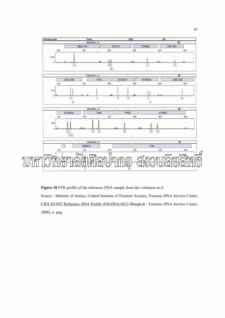
81
Figure 10 STR profile of the reference DNA sample from the volunteer no.4
Source : Ministry of Justice, Central Institute of Forensic Science, Forensic DNA Service Center,
CIFS STAFF Reference DNA Profile (FM-DNA-051) (Bangkok : Forensic DNA Service Center,
2008), n. pag.

82
Figure 11 STR profile of the reference DNA sample from the volunteer no.5
Source : Ministry of Justice, Central Institute of Forensic Science, Forensic DNA Service Center,
CIFS STAFF Reference DNA Profile (FM-DNA-051) (Bangkok : Forensic DNA Service Center,
2008), n. pag.

83
Figure 12 STR profile of the reference DNA sample from the volunteer no.6
Source : Ministry of Justice, Central Institute of Forensic Science, Forensic DNA Service Center,
CIFS STAFF Reference DNA Profile (FM-DNA-051) (Bangkok : Forensic DNA Service Center,
2008), n. pag.

84
Figure 13 STR profile of the reference DNA sample from the volunteer no.7
Source : Ministry of Justice, Central Institute of Forensic Science, Forensic DNA Service Center,
CIFS STAFF Reference DNA Profile (FM-DNA-051) (Bangkok : Forensic DNA Service Center,
2008), n. pag.

85
Figure 14 STR profile of the reference DNA sample from the volunteer no.8
Source : Ministry of Justice, Central Institute of Forensic Science, Forensic DNA Service Center,
CIFS STAFF Reference DNA Profile (FM-DNA-051) (Bangkok : Forensic DNA Service Center,
2008), n. pag.

86
Figure 15 STR profile of the reference DNA sample from the volunteer no.9
Source : Ministry of Justice, Central Institute of Forensic Science, Forensic DNA Service Center,
CIFS STAFF Reference DNA Profile (FM-DNA-051) (Bangkok : Forensic DNA Service Center,
2008), n. pag.

87
Figure 16 STR profile of the reference DNA sample from the volunteer no.10
Source : Ministry of Justice, Central Institute of Forensic Science, Forensic DNA Service Center,
CIFS STAFF Reference DNA Profile (FM-DNA-051) (Bangkok : Forensic DNA Service Center,
2008), n. pag.

88
APPENDIX D
STR PROFILE OF THE DNA FROM EACH EXTRACTION METHOD

89
Figure 17 STR profile of the DNA from DNA IQ™ System extraction (ReSI) collected by
Scotch® 3M 1710 Vinyl Electrical Tape of volunteer no.5

90
Figure 18 STR profile of the DNA from DNA IQ™ System extraction (ReSI) collected by
Scotch® 3M 1710 Vinyl Electrical Tape of volunteer no.9

91
Figure 19 STR profile of the DNA from DNA IQ™ System extraction (ReSI) collected by
Scotch® 3M Super 33+ All Weather Vinyl Electrical Tape of volunteer no.9

92
Figure 20 STR profile of the DNA from DNA IQ™ System extraction (ReSI) collected by
YAZAKI P.V.C. Tape of volunteer no.6

93
Figure 21 STR profile of the DNA from Chelex® 100 extraction (ReSC) collected by Scotch®
3M Super 33+ All Weather Vinyl Electrical Tape of volunteer no.5

94
Figure 22 STR profile of the DNA from Chelex® 100 extraction (ReSC) collected by Scotch®
3M Super 33+ All Weather Vinyl Electrical Tape of volunteer no.9

95
Figure 23 STR profile of the DNA from Phenol: Chloroform extraction (ReSP) collected by
Scotch® 3M 1710 Vinyl Electrical Tape of volunteer no.1

96
Figure 24 STR profile of the DNA from Phenol: Chloroform extraction (ReSP) collected by
Scotch® 3M 1710 Vinyl Electrical Tape of volunteer no.5

97
Figure 25 STR profile of the DNA from Phenol: Chloroform extraction (ReSP) collected by
Scotch® 3M Super 33+ All Weather Vinyl Electrical Tape of volunteer no.4

98
Figure 26 STR profile of the DNA from Phenol: Chloroform extraction (ReSP) collected by
Scotch® 3M Super 33+ All Weather Vinyl Electrical Tape of volunteer no.7

99
Figure 27 STR profile of the DNA from Phenol: Chloroform extraction (ReSP) collected by
YAZAKI P.V.C. Tape of volunteer no.5

100
Figure 28 STR profile of the DNA from Phenol: Chloroform extraction (ReSP) collected by
YAZAKI P.V.C. Tape of volunteer no.7

101
APPENDIX E
SUGGESTED PROTOCOL

102
Suggested protocol for DNA extraction from electrical adhesive tape by DNA IQ™ System
1. Place the electrical adhesive tape in sterile 50 ml centrifuge tube
2. Add 4 ml of prepared Lysis Buffer. Close the lid, and incubate the tube overnight room
temperature.
3. Vortex tube for 10 seconds at high speed, and incubate at 95˚C for 30 minutes.
4. Remove the tube from the heat source, and transfer the solution to a new 15 ml centrifuge
tube
5. Vortex the stock resin bottle for 10 seconds at high speed or until resin is thoroughly
mixed. Add 7 µl of DNA IQ. Resin to the sample. Keep the stock resin resuspended
while dispensing to obtain uniform results.
6. Vortex the sample/Lysis Buffer/resin mixture for 3 seconds at high speed. Incubate at
room temperature for 5 minutes. Vortex mixture for 3 seconds once every minute during
this 5 minute incubation.
7. Vortex tube for 2 seconds at high speed. Place tube in the magnetic stand. Separation will
occur instantly.
Note: If resin does not form a distinct pellet on the side of the tube, vortex the tube and
quickly place back in the stand.
8. Discard the solution until indicate volume 1 ml, vortex for 2 seconds and place the tube
in the magnetic stand. Then remove all of the solution to a new 1.5 ml microcentrifuge
tube and place tube in the magnetic stand.
9. Carefully remove and discard all of the solution without disturbing the resin pellet on the
side of the tube.
Note: If some resin is drawn up in tip, gently expel resin back into tube to allow re-
separation.
10. Add 100 µl of prepared Lysis Butter. Remove the tube from the magnetic stand, and
vortex for 2 seconds at high speed.
11. Return tube to the magnetic stand, and discard all Lysis Buffer.
12. Add 100 µl of prepared 1X Wash Buffer. Remove tube from the magnetic stand, and
vortex for 2 seconds at high speed.
13. Return tube to the magnetic stand, and discard all Wash Buffer.

103
14. Repeat Steps 12 and 13 two more times for a total of three washes. Be sure that all of the
solution has been removed after the last wash.
15. With the tube in the magnetic stand and the lid open, air-dry the resin for 5 minutes.
Note: Do not dry for more than 20 minutes, as this may inhibit removal of DNA.
16. Add 15 µl of Elution Buffer.
17. Close the lid and vortex the tube for 2 seconds at high speed. Incubate the tube at 65˚C
for 5 minutes.
18. Remove the tube from the heat source, and vortex for 2 seconds at high speed.
Immediately place the tube on the magnetic stand.
Note: Tubes must remain hot until placed in the magnetic stand or yield will decrease.
19. Carefully transfer the DNA-containing solution to a container of choice.
Note: DNA can be stored at 4˚C for short-term storage or at-20 or-70˚C for long-term
storage.

104
Suggested protocol for DNA concentration by Microcon™ YM-100
1. Assemble and label a fresh microcon unit for each sample. Add 50 µl of TE buffer (0.1
mM EDTA) the filter side of the membrane (upper side).
Note: The maximum volume Microcon 100�s (blue retanate cup) can accept is 500 µl.
2. Add sample to corresponding microcon retanate cup.
3. Centrifuge microcon with lid closed for 5 minutes at 8,000 rpm until entire volume is
filtered.
4. Add 100 µl TE buffer and 200 µl of extract DNA to filter side of cup, and centrifuge for
5 minutes at 8,000 rpm until the entire volume is filtered. Discard filtrate, return cup to
microcon tube.
5. Label new microcon tube for each sample. Carefully loosen cup in original microcon
tube but do not remove. Add 20 µl of TE buffer to the filter side of cup of original
microcon unit. Vortex tube with new labeled microcon tube held over top, and centrifuge
for 5 minutes at 10,000 rpm.
6. Discard retenate cup, retain filtrate, and store appropriately.
7. Pooling samples. If necessary to pool samples, prepare new microcon unit and add 50 µl
of TE buffer to the filter side of the membrane (upper side). Pool the 20 µl volumes from
several tubes of the same sample into microcon retanate cup. Centrifuge microcon for at
least 5 minutes at 10,000 rpm with lid closed until the entire volume is filtered. Discard
filtrate, return cup to microcon tube. Elute DNA from membrane following Steps 5-6.

105
BIOGRAPHY
Name Miss Natthalak Pakdeenarong
Home Address 2448/2, Mukhamontri, Muang,
Nakhonratchasima, 30000
Office Address 499, Sukprapruti building, 16th floor
Prachachuen, Bangsue, Bangkok, 10800
Education Background
2006 Bachelor of Science, Department of Biology,
Srinakharinwirot University
2008 Master of Science, Program of Forensic Science,
Graduate School, Silpakorn University
Working Experience
2008 - present Staff in the project of convicted felons DNA database,
Forensic DNA Service Center,
Central Institute of Forensic Science,
Ministry of Justice