cobas TaqScreen DPX - QualTex Laboratories
28
The Document Revision Information Section is located at the end of this document. 05509238001-02EN 1 Doc Rev. 2.0 cobas ® TaqScreen DPX Test for use on the cobas s 201 system cobas ® TaqScreen DPX Test 96 Tests P/N: 05509211 190 cobas ® TaqScreen DPX Control Kit 12 Sets P/N: 05509190 190 cobas ® TaqScreen Wash Reagent 5.1 L P/N: 04404220 190 INDICATION FOR USE The cobas ® TaqScreen DPX Test, for use on the cobas s 201 system, is an in vitro nucleic acid amplification test for the direct quantitation of human parvovirus B19 (B19V) DNA (genotypes 1, 2, and 3) and the direct qualitative detection of hepatitis A virus (HAV) RNA (genotypes I, II, and III) in human plasma. SUMMARY AND EXPLANATION OF THE TEST The cobas ® TaqScreen DPX Test incorporates the use of multiple dyes which allows for the simultaneous detection of individual targets without the use of discriminatory tests. Based on the Test Definition File (TDF) installed, the cobas ® TaqScreen DPX Test will provide either both the quantitative B19V and qualitative HAV results, or only the quantitative B19V result. B19V is a small, non-enveloped, single-stranded DNA virus belonging to the genus Erythrovirus of the family Parvoviridae 1 and is grouped into genotypes 1, 2 and 3 (originally known as B19, A6 and V9, respectively) 1 . B19V has a world wide distribution and serological studies have indicated that at least 50% of adults have circulating B19V IgG, indicating a past infection 2, 3 . The virus is associated with clinical illness, the manifestation and severity of which are dependent on the immunological and hematological status of the individual 4 . In immunocompetent individuals, the infection is often asymptomatic or may result in a mild illness including erythema infectiosium (fifth disease) in children or arthropathy in adults. However, B19V may cause severe disease such as transient aplastic anemia in individuals with hematological disorders and hydrops, congenital anemia or fetal death in pregnant women 4 . The prevalence of B19V in blood and plasma donors can vary from 0.003-0.6% depending on whether the collection is done during an epidemic or non-epidemic period 5, 6 . Although B19V is normally transmitted by the respiratory route, transmission by plasma products can occur due to the size of plasma pools, the incidence of acute, inapparent B19V infections, the high levels of virus in a viremic donation (up to 10 12 IU/mL), and the resistance of B19V to most of the commonly used viral inactivation/removal steps, such as solvent/detergent (S/D) treatment or pasteurization 7 . The presence of B19V DNA in plasma pools as well as plasma products have been reported 8-10 and there are many reports in the literature of transmission of B19V by administration of plasma products, especially coagulation factors 7, 11-13 . Hepatitis A virus (HAV) is a small, non-enveloped, RNA virus which belongs to the Hepatovirus group of the Picornaviridae family 14 . HAV, which has a global distribution, is transmitted by the oral-fecal route, primarily by close personal contact. Epidemics are common in developing countries where the infection is acquired early in life and results in a large proportion of the population having protective antibodies to HAV 15 , while in industrialized countries, the decline in the incidence rate of the virus has led to a shift towards infection in adulthood 16 . HAV infections in humans range from asymptomatic infections, mainly seen in young children, to fulminant hepatitis, which in some cases may lead to death. In Northern Europe, Japan, Canada and the USA, the prevalence in the general population is very low (about 0.01%) and outbreaks are associated mainly with risk groups, such as travelers to endemic regions 17 . HAV can be grouped into different genotypes (I – VI), with genotypes I, II, and III being found in humans 18, 19 . Although infectious HAV can be found in blood during the serology window period 20 , the risk of transfusion transmission of HAV is very low. Nevertheless, like B19V, HAV is not easily inactivated by S/D treatment or pasteurization, and there have been some reports on the transmission of HAV through plasma products, mainly coagulation factors 21-23 . DPX CTL TS WR DPX FOR REFERENCE USE ONLY
Transcript of cobas TaqScreen DPX - QualTex Laboratories
The Document Revision Information Section is located at the end of this document. 05509238001-02EN 1 Doc Rev. 2.0
cobas® TaqScreen DPX Test for use on the cobas s 201 system
cobas® TaqScreen DPX Test 96 Tests P/N: 05509211 190
cobas® TaqScreen DPX Control Kit 12 Sets P/N: 05509190 190
cobas® TaqScreen Wash Reagent 5.1 L P/N: 04404220 190
INDICATION FOR USE
The cobas® TaqScreen DPX Test, for use on the cobas s 201 system, is an in vitro nucleic acid amplification test for the direct quantitation of human parvovirus B19 (B19V) DNA (genotypes 1, 2, and 3) and the direct qualitative detection of hepatitis A virus (HAV) RNA (genotypes I, II, and III) in human plasma.
SUMMARY AND EXPLANATION OF THE TEST
The cobas® TaqScreen DPX Test incorporates the use of multiple dyes which allows for the simultaneous detection of individual targets without the use of discriminatory tests. Based on the Test Definition File (TDF) installed, the cobas® TaqScreen DPX Test will provide either both the quantitative B19V and qualitative HAV results, or only the quantitative B19V result.
B19V is a small, non-enveloped, single-stranded DNA virus belonging to the genus Erythrovirus of the family Parvoviridae1 and is grouped into genotypes 1, 2 and 3 (originally known as B19, A6 and V9, respectively)1 . B19V has a world wide distribution and serological studies have indicated that at least 50% of adults have circulating B19V IgG, indicating a past infection2, 3. The virus is associated with clinical illness, the manifestation and severity of which are dependent on the immunological and hematological status of the individual4. In immunocompetent individuals, the infection is often asymptomatic or may result in a mild illness including erythema infectiosium (fifth disease) in children or arthropathy in adults. However, B19V may cause severe disease such as transient aplastic anemia in individuals with hematological disorders and hydrops, congenital anemia or fetal death in pregnant women4. The prevalence of B19V in blood and plasma donors can vary from 0.003-0.6% depending on whether the collection is done during an epidemic or non-epidemic period5, 6.
Although B19V is normally transmitted by the respiratory route, transmission by plasma products can occur due to the size of plasma pools, the incidence of acute, inapparent B19V infections, the high levels of virus in a viremic donation (up to 1012 IU/mL), and the resistance of B19V to most of the commonly used viral inactivation/removal steps, such as solvent/detergent (S/D) treatment or pasteurization7. The presence of B19V DNA in plasma pools as well as plasma products have been reported8-10 and there are many reports in the literature of transmission of B19V by administration of plasma products, especially coagulation factors7, 11-13.
Hepatitis A virus (HAV) is a small, non-enveloped, RNA virus which belongs to the Hepatovirus group of the Picornaviridae family14. HAV, which has a global distribution, is transmitted by the oral-fecal route, primarily by close personal contact. Epidemics are common in developing countries where the infection is acquired early in life and results in a large proportion of the population having protective antibodies to HAV15, while in industrialized countries, the decline in the incidence rate of the virus has led to a shift towards infection in adulthood16. HAV infections in humans range from asymptomatic infections, mainly seen in young children, to fulminant hepatitis, which in some cases may lead to death. In Northern Europe, Japan, Canada and the USA, the prevalence in the general population is very low (about 0.01%) and outbreaks are associated mainly with risk groups, such as travelers to endemic regions17. HAV can be grouped into different genotypes (I – VI), with genotypes I, II, and III being found in humans18, 19. Although infectious HAV can be found in blood during the serology window period20, the risk of transfusion transmission of HAV is very low. Nevertheless, like B19V, HAV is not easily inactivated by S/D treatment or pasteurization, and there have been some reports on the transmission of HAV through plasma products, mainly coagulation factors21-23.
DPX CTL
TS WR
DPX
FOR REFERENCE U
SE ONLY
05509238001-02EN 2 Doc Rev. 2.0
The cobas® TaqScreen DPX Test is a duplex test for the simultaneous detection of B19V and HAV in individual samples or pooled plasma samples of human origin. The use of multi-dye technology enables the identification of each viral target without the need for further discriminatory testing. In addition, the test provides a quantitative value (in IU/mL) for the B19V target through the use of a Quantitation Standard (QS), directly traceable to the WHO B19V International Standard24. The QS together with an Internal Control (IC) (for the HAV target) is co-extracted and co-amplified with each sample. The cobas® TaqScreen DPX Test uses a generic nucleic acid preparation technique on the COBAS® AmpliPrep Instrument. B19V DNA, HAV RNA, the QS and IC are amplified and detected in a single tube using automated, real time PCR on the COBAS® TaqMan® Analyzer. Discrimination of the viral target, the QS and the IC is achieved by the use of fluorescently labeled probes which are detected in separate channels of the COBAS® TaqMan® Analyzer. The test incorporates the AmpErase (uracil-N-glycosylase enzyme) to reduce potential contamination by previously amplified material (amplicon).
PRINCIPLES OF THE PROCEDURE
The cobas® TaqScreen DPX Test for use on the cobas s 201 system is based on 4 major processes:
1. Automated Specimen Pooling and Control Pipetting using the Hamilton MICROLAB® STAR/STARlet IVD Pipettor (optional)
2. Automated Specimen Preparation using the COBAS® AmpliPrep Instrument
3. Automated Amplification of Nucleic Acid and Real Time Automated Detection of PCR products using the COBAS® TaqMan® Analyzer
4. Automated Data Management using the Pooling and Data Management (PDM) Software
Automated Specimen Pooling and Pipetting using the Hamilton MICROLAB® STAR/STARlet IVD Pipettor
The Hamilton MICROLAB® STAR/STARlet IVD Pipettor automates pipetting of individual donor specimens, pooling of multiple donor specimens, and pipetting of Test Controls. The cobas s 201 system resolves reactive pools into individual component specimen results. The cobas s 201 system is designed to process specimens in batches. A batch is defined as a collection of specimens and controls that are pipetted, extracted, amplified and detected together. When the pipetting of a batch is completed on the Hamilton MICROLAB® STAR/STARlet IVD Pipettor, the entire sample rack is transferred into the COBAS® AmpliPrep Instrument for the next step of the process.
Automated Specimen Preparation using the COBAS® AmpliPrep Instrument
Nucleic acids from the specimens, and added Armored HAV RNA IC molecules and lambda-encapsulated B19V DNA QS molecules (which serve as specimen preparation and amplification/ detection/quantitation process controls) are simultaneously processed. The cobas® TaqScreen DPX Test contains reagents that accomplish five sequential steps on the COBAS® AmpliPrep Instrument. The Proteinase Solution digests proteins to promote lysis, inactivate nucleases, and facilitate the release of RNA and DNA from viral particles. Addition of Lysis Reagent to the specimen results in viral lysis and nuclease inactivation by denaturation of proteins. RNA and DNA are released and simultaneously protected from nucleases. The released nucleic acids bind to the silica surface of the added Magnetic Glass Particles. This is mainly due to the net positive charge on the glass particle surface and net negative charge of the nucleic acids due to the chaotropic salt concentration and ionic strength of the Lysis reaction. Wash Reagent removes unbound substances and impurities such as denatured proteins, cellular debris and potential PCR inhibitors (such as hemoglobin, etc.) and reduces the salt concentration. Purified nucleic acids are released from the Magnetic Glass Particles at elevated temperature with Elution Buffer.
Automated Amplification and Detection of Nucleic Acid using the COBAS® TaqMan® Analyzer
After isolation of the purified nucleic acids from human plasma during automated specimen preparation, cobas® TaqScreen DPX Master Mix (MMX) is used for the amplification and detection of HAV RNA and IC RNA, and amplification and quantitation of B19V DNA and QS DNA. Once activated by the addition of manganese acetate, the cobas® TaqScreen DPX Master Mix permits reverse transcription (for RNA targets), followed by PCR amplification of highly conserved regions of HAV RNA, IC RNA, B19V DNA, and QS DNA using specific primers. The amplicon are detected by hybridization of specific oligonucleotide probes. Amplification, hybridization and detection occur simultaneously.
FOR REFERENCE U
SE ONLY
05509238001-02EN 3 Doc Rev. 2.0
Reverse Transcription and PCR Amplification
Reverse transcription and amplification reactions are performed with a thermostable recombinant enzyme, Z05 DNA Polymerase. In the presence of manganese (Mn2+), the Z05 DNA Polymerase has reverse transcriptase and DNA polymerase activities. This allows both reverse transcription and PCR amplification to occur in the same reaction mixture.
During PCR amplification, the intermittent high temperature during the cycling denatures the target and IC /QS amplicon to form single stranded DNA. Z05 DNA Polymerase extends the annealed primers along the target templates to produce double-stranded DNA (amplicon). This process is repeated for multiple cycles, with each cycle doubling the amount of amplicon. Amplification occurs only in the region of the target genomes between the primers; the entire genomes are not amplified.
Prevention of Carry-over Contamination
Amplicon carry-over contamination is prevented by the use of AmpErase (uracil-N-glycosylase) and deoxyuridine triphosphate (dUTP). Deoxyuridine is not present in naturally occurring DNA, but is always present in amplicon because of the use of a deoxyuridine triphosphate/thymidine triphosphate mixture as one of the dNTPs in the Master Mix reagent; therefore, only amplicon contains deoxyuridine. AmpErase recognizes and catalyzes the destruction of DNA strands containing deoxyuridine25 by opening the deoxyribose chain at the C1-position, thereby rendering the DNA non-amplifiable. DNA containing deoxythymidine or RNA containing ribouridine26, 27 is not affected. During the initial reverse transcription step, AmpErase catalyzes the cleavage of carry-over amplicon at the deoxyuridine residues. AmpErase is inactive for a prolonged period of time once exposed to temperatures above 55ºC and therefore does not destroy newly formed target amplicon.
Detection of PCR Products28, 29
The cobas® TaqScreen DPX MMX contains detection probes which are specific for HAV, IC, B19V or QS nucleic acid. Each detection probe is labeled with 1) one of four fluorescent dyes which act as a reporter and 2) another dye which acts as a quencher. Each specific reporter dye is associated with a corresponding target, each measured at a defined wavelength. A single type of quencher dye is used in all probes. This system permits detection of all the amplified targets at different wavelengths.
Before PCR amplification begins, the probes are intact and the reporter dye fluorescence is suppressed by the quencher dye due to Förster-type energy transfer. During PCR amplification, the probes hybridize to specific single stranded DNA sequences and are cleaved by the 5' to 3' nuclease activity of the Z05 DNA Polymerase at the same time that amplification is occurring. Once the reporter and quencher dyes are separated by this cleavage, the fluorescent activity of the reporter dye is unmasked. With each PCR cycle, increasing amounts of cleaved probes are generated and the cumulative signal of the reporter dye is concomitantly increased.
Real time detection of PCR products is accomplished by measuring the fluorescence of released reporter dyes representing the viral targets, IC and the QS independently.
Data Management using the PDM Software
The Roche PDM software allows the user to review and report results. The Roche PDM software assigns test results for the cobas® TaqScreen DPX Test as non-reactive, reactive, or invalid for the HAV target, and < cutoff, ≥ cutoff, or invalid for the B19V target. In addition to retrieving and examining PCR results, the Roche PDM software allows the operator to print reports, search for results, accept donor results, and optionally transmit results to an LIS.
FOR REFERENCE U
SE ONLY

05509238001-02EN 4 Doc Rev. 2.0
MATERIALS PROVIDED BY ROCHE
Three kits are required and provided for the detection of HAV RNA and quantitation of B19V DNA in plasma specimens: 1) cobas® TaqScreen DPX Test, 2) cobas® TaqScreen DPX Control Kit, and 3) cobas® TaqScreen Wash Reagent. Material Safety Data Sheets (MSDS) are available on request from your local Roche office.
cobas® TaqScreen DPX Test 96 Tests (P/N: 05509211 190)
DPX CS1 (DPX Magnetic Glass Particles Reagent Cassette)
DPX CS2 (DPX Lysis Reagent Cassette)
DPX CS3 (DPX Multi-Reagent Cassette)
DPX CS4 (DPX Test-Specific Reagent Cassette)
cobas® TaqScreen DPX Control Kit 12 Sets (P/N: 05509190 190)
DPX D(+)C (HAV Positive and B19V Low Positive Dual Control)
DPX H(+)C (B19V High Positive Control)
DPX (–) C (cobas® TaqScreen DPX Negative Control)
cobas® TaqScreen Wash Reagent 5.1 L (P/N: 04404220 190)
TS WR (cobas® TaqScreen Wash Reagent)
DPX CTL
TS WR
DPX
FOR REFERENCE U
SE ONLY
05509238001-02EN 5 Doc Rev. 2.0
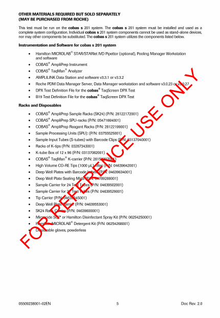
OTHER MATERIALS REQUIRED BUT SOLD SEPARATELY (MAY BE PURCHASED FROM ROCHE)
This test must be run on the cobas s 201 system. The cobas s 201 system must be installed and used as a complete system configuration. Individual cobas s 201 system components cannot be used as stand-alone devices, nor may other components be substituted. The cobas s 201 system utilizes the components listed below.
Instrumentation and Software for cobas s 201 system
• Hamilton MICROLAB® STAR/STARlet IVD Pipettor (optional), Pooling Manager Workstation and software
• COBAS® AmpliPrep Instrument • COBAS® TaqMan® Analyzer • AMPLILINK Data Station and software v3.3.1 or v3.3.2 • Roche PDM Data Manager Server, Data Manager workstation and software v3.0.23 or v3.0.27 • DPX Test Definition File for the cobas® TaqScreen DPX Test • B19 Test Definition File for the cobas® TaqScreen DPX Test
Racks and Disposables
• COBAS® AmpliPrep Sample Racks (SK24) (P/N: 28122172001) • COBAS® AmpliPrep SPU-racks (P/N: 05471664001) • COBAS® AmpliPrep Reagent Racks (P/N: 28122199001) • Sample Processing Units (SPU): (P/N: 03755525001) • Sample Input Tubes (S-tubes) with Barcode Clips (P/N: 03137040001) • Racks of K-tips (P/N: 03287343001) • K-tube Box of 12 x 96 (P/N: 03137082001) • COBAS® TaqMan® K-carrier (P/N: 28150397001) • High Volume CO-RE Tips (1000 μL), filter (P/N: 04639642001) • Deep Well Plates with Barcode Labels (P/N: 04639634001) • Deep Well Plate Sealing Mats (P/N: 04789288001) • Sample Carrier for 24 Test Tubes (P/N: 04639502001) • Sample Carrier for 32 Test Tubes (P/N: 04639529001) • Tip Carrier (P/N: 04639545001) • Deep Well Plate Carrier (P/N: 04639553001) • SK24 Rack Carrier (P/N: 04639600001) • Microcide SQ™ or Hamilton Disinfectant Spray Kit (P/N: 06254250001) • Hamilton MICROLAB® Detergent Kit (P/N: 06254268001) • Disposable gloves, powderless
FOR REFERENCE U
SE ONLY
05509238001-02EN 6 Doc Rev. 2.0
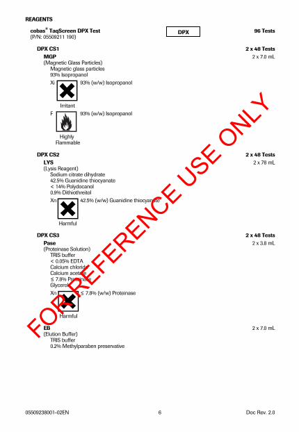
REAGENTS
cobas® TaqScreen DPX Test 96 Tests(P/N: 05509211 190)
DPX CS1 2 x 48 TestsMGP 2 x 7.0 mL(Magnetic Glass Particles)
Magnetic glass particles 93% Isopropanol Xi 93% (w/w) Isopropanol
Irritant F 93% (w/w) Isopropanol
Highly Flammable
DPX CS2 2 x 48 TestsLYS 2 x 78 mL(Lysis Reagent)
Sodium citrate dihydrate 42.5% Guanidine thiocyanate < 14% Polydocanol 0.9% Dithiothreitol Xn 42.5% (w/w) Guanidine thiocyanate
Harmful
DPX CS3 2 x 48 TestsPase 2 x 3.8 mL(Proteinase Solution)
TRIS buffer < 0.05% EDTA Calcium chloride Calcium acetate ≤ 7.8% Proteinase Glycerol Xn ≤ 7.8% (w/w) Proteinase
Harmful EB 2 x 7.0 mL(Elution Buffer)
TRIS buffer 0.2% Methylparaben preservative
DPX
FOR REFERENCE U
SE ONLY
05509238001-02EN 7 Doc Rev. 2.0
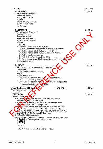
DPX CS4 2 x 48 Tests
DPX MMX-R1 2 x 3.0 mL (DPX Master Mix Reagent 1)
Potassium acetate Manganese acetate Glycerol 14.4% Dimethyl sulfoxide 0.08% Sodium azide Acetic Acid
DPX MMX-R2 2 x 2.5 mL (DPX Master Mix Reagent 2)
Tricine buffer Potassium acetate Potassium hydroxide 4.1% Dimethyl sulfoxide Glycerol Tween 20 < 0.09% dATP, dGTP, dCTP, dUTP, dTTP < 0.01% Upstream and downstream B19V and HAV primers < 0.01% Fluorescent-labeled B19V and HAV probes < 0.01% Fluorescent-labeled B19V QS and HAV IC probes < 0.01% Oligonucleotide aptamer < 0.01% Z05 DNA Polymerase (microbial) < 0.01% AmpErase (uracil-N-glycosylase) enzyme (microbial) 0.08% Sodium azide
DPX IC/QS 2 x 13 mL (DPX Internal Control and Quantitation Standard)
TRIS buffer ≤ 0.002% Poly rA RNA (synthetic) EDTA 0.05% Sodium azide < 0.001% Non-infectious, synthetic IC RNA encapsulated
in MS2 bacteriophage coat protein < 0.001% Non-infectious, synthetic QS B19V DNA encapsulated
in Lambda bacteriophage coat protein cobas® TaqScreen DPX Control Kit 12 Sets (P/N: 05509190 190)
DPX D(+)C 12 x 1.6 mL (DPX Dual Positive Control)
< 0.001% Non-infectious, synthetic HAV RNA encapsulated in MS2 bacteriophage coat protein
< 0.001% Non-infectious, synthetic B19V DNA encapsulated in bacteriophage Lambda coat protein
Negative Human Plasma, non-reactive by US FDA licensed tests for B19V IgG and IgM Ab, HBsAg, HBV core Ab, HCV Ab, HIV-1/2 Ab, and HIV-1 RNA. Non-reactive by NAT for HAV RNA, and B19V DNA ≤ 5 IU/mL by PCR methods.
0.1% ProClin® 300 preservative Xi (3:1) mixture of 5-Chloro-2-methyl-2H-isothiazol-3-one and 2-Methyl-2H-isothiazol-3-one
Irritant R43: May cause sensitization by skin contact.
DPX CTL
FOR REFERENCE U
SE ONLY
05509238001-02EN 8 Doc Rev. 2.0
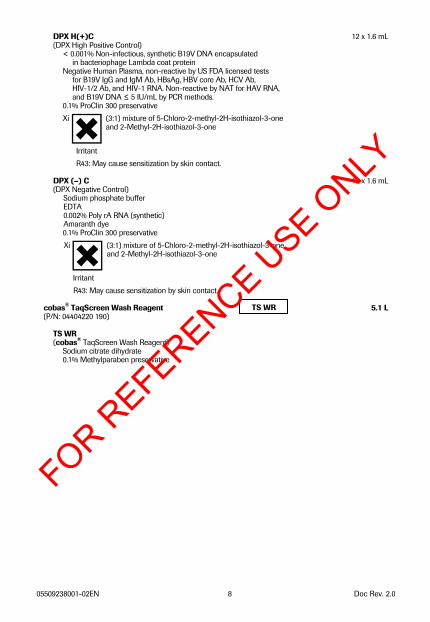
DPX H(+)C 12 x 1.6 mL (DPX High Positive Control)
< 0.001% Non-infectious, synthetic B19V DNA encapsulated in bacteriophage Lambda coat protein
Negative Human Plasma, non-reactive by US FDA licensed tests for B19V IgG and IgM Ab, HBsAg, HBV core Ab, HCV Ab, HIV-1/2 Ab, and HIV-1 RNA. Non-reactive by NAT for HAV RNA, and B19V DNA ≤ 5 IU/mL by PCR methods.
0.1% ProClin 300 preservative Xi (3:1) mixture of 5-Chloro-2-methyl-2H-isothiazol-3-one and 2-Methyl-2H-isothiazol-3-one
Irritant R43: May cause sensitization by skin contact.
DPX (–) C 12 x 1.6 mL (DPX Negative Control)
Sodium phosphate buffer EDTA 0.002% Poly rA RNA (synthetic) Amaranth dye 0.1% ProClin 300 preservative Xi (3:1) mixture of 5-Chloro-2-methyl-2H-isothiazol-3-one
and 2-Methyl-2H-isothiazol-3-one
Irritant R43: May cause sensitization by skin contact.
cobas® TaqScreen Wash Reagent 5.1 L (P/N: 04404220 190)
TS WR (cobas® TaqScreen Wash Reagent)
Sodium citrate dihydrate 0.1% Methylparaben preservative
TS WR
FOR REFERENCE U
SE ONLY
05509238001-02EN 9 Doc Rev. 2.0
STORAGE AND HANDLING REQUIREMENTS
A. Room temperature is defined as 15 to 30°C.
B. Do not freeze reagents or controls.
C. Store DPX CS1, DPX CS2, DPX CS3, and DPX CS4 at 2 to 8°C. Unused reagents are stable until the expiration date indicated.
D. After initial use, reagents are stable for 30 days at 2 to 8°C or until the expiration date, whichever comes first.
E. Reagents can be used for up to 6 instrument runs, and up to a maximum of 48 cumulative hours in the COBAS® AmpliPrep Instrument. Reagents must be stored at 2 to 8°C between uses. The AMPLILINK software monitors the cumulative hours of the reagent cassettes on the COBAS® AmpliPrep Instrument, and blocks the cassettes from being used once the 48 cumulative hours are reached.
F. The AMPLILINK software does not monitor the number of instrument runs the cassettes have been used for. It is the user’s responsibility to discard the reagent cassettes once 6 instrument runs are reached.
G. Store DPX D(+)C, DPX H(+)C, and DPX (–) C at 2 to 8°C. The controls are stable until the expiration date indicated. Once opened, any unused portion must be discarded.
H. Store TS WR at 15 to 30°C. Unopened TS WR is stable until the expiration date indicated. Once opened, this reagent is stable for 30 days at 15 to 30°C or until the expiration date, whichever comes first.
PRECAUTIONS
A. Specimens may be infectious. Use universal precautions when performing the test.30, 31 Only personnel proficient in the use of the cobas® TaqScreen DPX Test and trained in handling infectious materials should perform this procedure. Thoroughly clean and disinfect all laboratory work surfaces with a freshly prepared solution of 0.5% sodium hypochlorite in distilled or deionized water (diluted bleach). Follow by wiping the surface with 70% Ethanol.
B. CAUTION: DPX D(+)C and DPX H(+)C contain human plasma derived from human blood. The plasma used has been tested and found non-reactive for IgG and IgM antibodies to B19V, HBsAg, HBV core Ab, HCV Ab, HIV-1/2 Ab; non reactive for HIV-1 RNA by NAT. The plasma used has also been tested by the cobas® TaqScreen DPX Test, and shown to be non-reactive for HAV RNA, and to contain ≤ 5 IU/mL B19V DNA. No known test method can offer complete assurance that products derived from human blood will not transmit infectious agents. All human blood-sourced materials should be considered potentially infectious and should be handled with universal precautions. If spillage occurs, immediately disinfect with a freshly prepared solution of 0.5% sodium hypochlorite (dilute bleach) or follow appropriate site procedures.
C. Use routine laboratory precautions. Do not pipette by mouth. Do not eat, drink or smoke in designated work areas. Wear disposable gloves, laboratory coats and eye protection when handling specimens and kit reagents. Wash hands thoroughly after handling specimens and kit reagents.
D. DPX MMX-R1, DPX MMX-R2, and DPX IC/QS contain sodium azide as a preservative. If solutions containing azide are disposed of in a plumbing system, they should be diluted and flushed with generous amounts of running water. These precautions are recommended to avoid accumulation of deposits in metal piping in which explosive conditions could develop.
E. Heparin has been shown to inhibit PCR. Do not use heparinized plasma with this procedure.
F. The use of sterile disposable pipettes and nuclease-free pipette tips is recommended. False positive results may occur if cross contamination of specimens is not prevented during specimen handling and processing.
G. Use only supplied or specified required disposables to ensure optimal test performance.
H. Handle all materials containing specimens or controls according to Good Laboratory Practices in order to prevent cross-contamination of specimens or controls.
I. Before use, visually inspect each reagent cassette, control tube and Wash Reagent to ensure that there are no signs of leakage. If there is any evidence of leakage, do not use that material for testing.
FOR REFERENCE U
SE ONLY
05509238001-02EN 10 Doc Rev. 2.0
Whole Blood(2°C - 30°C)
Plasma(2°C - 8°C)
Whole Blood(2°C - 8°C)
0
10
20
30
0 1 2 3 4 5 6 7 8 9 10 11 12 13 14
Days
Tem
pera
ture
(°C
)
J. Dispose of all materials that have come in contact with specimens and reagents in accordance with country, federal, state, and local regulations.
K. Do not use a cobas® TaqScreen DPX Test kit, cobas® TaqScreen DPX Control Kit, or cobas® TaqScreen Wash Reagent after its expiration date. Do not interchange, mix, or combine reagents from different kits or different lots. Do not load mixed reagent lots on the COBAS® AmpliPrep Instrument.
L. Material Safety Data Sheets (MSDS) are available on request from your local Roche office.
M. Avoid contact of reagents with skin, eyes or mucous membranes. If contact does occur, immediately wash with large amounts of water, otherwise, burns can occur. If these reagents are spilled, dilute with water before wiping dry.
N. Do not allow LYS, which contains guanidine thiocyanate, to contact sodium hypochlorite (bleach) solution. This mixture can produce a highly toxic gas.
O. Closely follow procedures and guidelines provided to ensure that the test is performed correctly. Any deviation from the procedures and guidelines may affect optimal test performance.
P. The use of excessively hemolyzed specimens should be avoided.
Q. Red blood cell contamination of plasma specimens (>5 %) may inhibit the cobas® TaqScreen DPX Test.
R. Do not use any component with damaged barcode labels at any phase of testing.
CONTROL AND REAGENT PREPARATION
A. Equilibrate the cobas® TaqScreen DPX Control Kit to room temperature for 30 minutes prior to loading onto the Hamilton MICROLAB® STAR/STARlet IVD Pipettor prior to use. Equilibrate cobas® TaqScreen DPX Test reagents in the COBAS® AmpliPrep Instrument for 30 minutes prior to use.
SPECIMEN COLLECTION, STORAGE, and POOLING
Note: Handle all specimens as if they are infectious agents.
Specimens
A. Specimens collected using EDTA, CPD, CPDA-1, CP2D, ACD-A, 4% Citrate, and BD Vacutainer Plasma Preparation Tube (PPT) may be used with the cobas® TaqScreen DPX Test. Follow the sample tube manufacturer’s instructions.
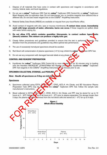
B. Blood collected in EDTA, CPD, CPDA-1, CP2D, ACD-A, 4% Citrate, and PPT may be stored for up to 72 hours at 2 – 30°C followed by up to 48 hours at 2 – 8°C prior to plasma separation. For storage longer than 5 days, the plasma should be removed from the red blood cells and stored at 2 – 8°C for up to 9 days.
Figure 1 Whole Blood Stability
FOR REFERENCE U
SE ONLY
05509238001-02EN 11 Doc Rev. 2.0
C. Apheresis plasma in EDTA, CPD, CPDA-1, CP2D, ACD-A, or 4% sodium citrate may be stored for up to 10 days at 2 – 30°C, followed by storage for up to 46 days at 2 – 8°C.
D. The following plasma volume guidelines are based on pipetting from 13 x 100 mm glass or plastic donor tubes on the Hamilton MICROLAB® STAR/STARlet IVD Pipettor. The listed volumes are for plasma on top of packed red blood cells, and are for use when running the cobas® TaqScreen DPX Test.
Table 1 Volume Guideline for Hamilton MICROLAB® STAR/STARlet IVD Pipettor
Pool Type Minimum Plasma Volume Primary Pool * 3 mL Repeat Pool 1.5 mL Resolution Pool 2 mL
*Includes creation of Deep Well Plate (Library Plate)
E. Do not freeze whole blood.
F. Covered Deep Well Plates may be stored at ≤ -18°C for up to 150 days, followed by 2-8°C for up to 30 days.
G. Plasma in EDTA, CPD, CPD-A, CP2D, ACD-A, and 4% sodium citrate may be stored for up to 6 months at ≤ -18ºC, followed by up to 15 days at 2 – 8°C. No adverse effect on test performance was observed when plasma specimens were subjected to 3 freeze/thaw cycles.
H. Equilibrate pooled or individual donor specimens to room temperature before using.
I. The user must validate other collection and storage conditions. If specimens are to be shipped, they should be packaged and labeled in compliance with applicable federal and international regulations covering the transport of specimens and etiologic agents.32
SPECIMEN POOLING AND PIPETTING
A. The cobas s 201 system utilizes the Hamilton MICROLAB® STAR/STARlet IVD Pipettor for all pipetting and pooling activities. The Hamilton MICROLAB® STAR/STARlet IVD Pipettor performs barcode scanning and pooling operations from specimens to form pools.
B. PDM Software version 3 or higher allows a complete cobas s 201 system to be installed without a Hamilton MICROLAB® STAR/STARlet IVD Pipettor by requiring the manual entry of the specimen barcode ID, SK24 rack and position barcode IDs, and the s-clip barcode ID. Refer to the cobas s 201 System Operator's Manual for instructions on manually entering this information.
C. If pools are detected by the cobas® TaqScreen DPX Test as being reactive for HAV or ≥ cutoff for B19V, the Hamilton MICROLAB® STAR/STARlet IVD Pipettor is used to pipette from either Deep Well Plates or original specimen tubes to create subdivided pools for secondary testing.
FOR REFERENCE U
SE ONLY
05509238001-02EN 12 Doc Rev. 2.0
PROCEDURAL NOTES
A. Equipment
1. Prepare the cobas s 201 system for use according to instructions in the cobas s 201 System Operator's Manual.
2. Perform recommended maintenance on instruments to ensure proper functioning.
B. Reagents
1. The cobas® TaqScreen DPX Test reagents must be equilibrated for 30 minutes in the COBAS® AmpliPrep Instrument prior to use. The cobas® TaqScreen DPX Control Kit and cobas® TaqScreen Wash Reagent must be at room temperature before use. See Storage and Handling Requirements Section for reagent storage conditions.
2. Each cobas® TaqScreen DPX Test kit contains sufficient material for processing a total of 96 tests which are recommended to be run in batches consisting of up to 24 tests per SK24 rack. One Negative Control and one of each Positive Control must be processed with each batch or SK24 rack. Controls are processed in the same way as specimens with the cobas® TaqScreen DPX Test.
3. All controls are single use only.
4. The system will prevent the use of reagents from different lots, reagents which have exceeded the allowed hours on instruments, reagents which have expired or mixed cassettes from a set of four cassettes previously used on the system. Do not load mixed reagent lots on the COBAS® AmpliPrep Instrument.
C. Specimen Processing
1. Avoid contaminating gloves when handling specimens and controls.
2. Care should be used to avoid contamination of specimens and Negative Controls with Positive Control material.
INSTRUCTIONS FOR USE
The cobas s 201 system includes four major processes: Specimen and Control Pipetting on the Hamilton MICROLAB® STAR/STARlet IVD Pipettor, Specimen preparation on the COBAS® AmpliPrep Instrument using the cobas® TaqScreen DPX Test, Amplification/Detection on the COBAS® TaqMan® Analyzer, and Data Management.
Each cobas® TaqScreen DPX Test kit contains eight cassettes: two DPX CS1 cassettes with Magnetic Glass Particles, two DPX CS2 cassettes with Lysis Reagent, two DPX CS3 cassettes with Proteinase, and Elution Buffer, and two DPX CS4 cassettes with IC, MMX Reagent 1, and MMX Reagent 2. This test kit is to be used in conjunction with the cobas® TaqScreen DPX Control Kit and the cobas® TaqScreen Wash Reagent.
Note: Do not open the cassettes.
Note: Do not pool reagents from different lots or from different bottles of the same lot.
Note: Do not mix cassettes from different lots on the COBAS® AmpliPrep Instrument.
Note: Do not separate control tubes from adapters.
Perform all required maintenance as described in the cobas s 201 system Operator’s Manual.
The cobas® TaqScreen DPX Test offers an alternate workflow that preserves specimens in a Library Plate (LP) so that testing can be performed at a later date. The DPX LP workflow requires three pooling runs. During the first pooling run (Library Plate Run), single-specimen aliquots are pipetted into a Library Plate which is then frozen. After the Quarantine Interval (configured at installation) has elapsed, the Library Plate can be thawed and used to prepare 12-specimen intermediate pools (Intermediate Plate Run). During the third pooling run (Batch Run), aliquots are pipetted from the Intermediate Plate wells into S-tubes to create the Primary Pools.
Refer to the cobas s 201 system Operator’s Manual for detailed instructions for use.
FOR REFERENCE U
SE ONLY
05509238001-02EN 13 Doc Rev. 2.0
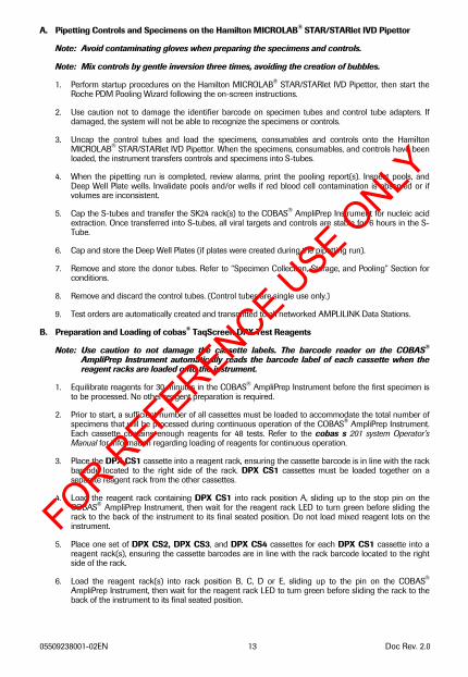
A. Pipetting Controls and Specimens on the Hamilton MICROLAB® STAR/STARlet IVD Pipettor
Note: Avoid contaminating gloves when preparing the specimens and controls.
Note: Mix controls by gentle inversion three times, avoiding the creation of bubbles.
1. Perform startup procedures on the Hamilton MICROLAB® STAR/STARlet IVD Pipettor, then start the Roche PDM Pooling Wizard following the on-screen instructions.
2. Use caution not to damage the identifier barcode on specimen tubes and control tube adapters. If damaged, the system will not be able to recognize the specimens or controls.
3. Uncap the control tubes and load the specimens, consumables and controls onto the Hamilton MICROLAB® STAR/STARlet IVD Pipettor. When the specimens, consumables, and controls have been loaded, the instrument transfers controls and specimens into S-tubes.
4. When the pipetting run is completed, review alarms, print the pooling report(s). Inspect pools, and Deep Well Plate wells. Invalidate pools and/or wells if red blood cell contamination is observed or if volumes are inconsistent.
5. Cap the S-tubes and transfer the SK24 rack(s) to the COBAS® AmpliPrep Instrument for nucleic acid extraction. Once transferred into S-tubes, all viral targets and controls are stable for 6 hours in the S-Tube.
6. Cap and store the Deep Well Plates (if plates were created during the pipetting run).
7. Remove and store the donor tubes. Refer to “Specimen Collection, Storage, and Pooling” Section for conditions.
8. Remove and discard the control tubes. (Control tubes are single use only.)
9. Test orders are automatically created and transmitted to all networked AMPLILINK Data Stations.
B. Preparation and Loading of cobas® TaqScreen DPX Test Reagents
Note: Use caution to not damage the cassette labels. The barcode reader on the COBAS® AmpliPrep Instrument automatically reads the barcode label of each cassette when the reagent racks are loaded onto the instrument.
1. Equilibrate reagents for 30 minutes in the COBAS® AmpliPrep Instrument before the first specimen is to be processed. No other reagent preparation is required.
2. Prior to start, a sufficient number of all cassettes must be loaded to accommodate the total number of specimens that will be processed during continuous operation of the COBAS® AmpliPrep Instrument. Each cassette contains enough reagents for 48 tests. Refer to the cobas s 201 system Operator’s Manual for information regarding loading of reagents for continuous operation.
3. Place the DPX CS1 cassette into a reagent rack, ensuring the cassette barcode is in line with the rack barcode located to the right side of the rack. DPX CS1 cassettes must be loaded together on a separate reagent rack from the other cassettes.
4. Load the reagent rack containing DPX CS1 into rack position A, sliding up to the stop pin on the COBAS® AmpliPrep Instrument, then wait for the reagent rack LED to turn green before sliding the rack to the back of the instrument to its final seated position. Do not load mixed reagent lots on the instrument.
5. Place one set of DPX CS2, DPX CS3, and DPX CS4 cassettes for each DPX CS1 cassette into a reagent rack(s), ensuring the cassette barcodes are in line with the rack barcode located to the right side of the rack.
6. Load the reagent rack(s) into rack position B, C, D or E, sliding up to the pin on the COBAS® AmpliPrep Instrument, then wait for the reagent rack LED to turn green before sliding the rack to the back of the instrument to its final seated position.
FOR REFERENCE U
SE ONLY
05509238001-02EN 14 Doc Rev. 2.0
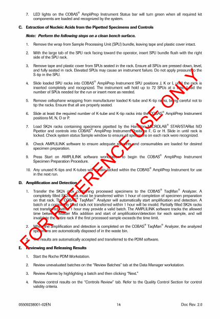
7. LED lights on the COBAS® AmpliPrep Instrument Status bar will turn green when all required kit components are loaded and recognized by the system.
C. Extraction of Nucleic Acids from the Pipetted Specimens and Controls
Note: Perform the following steps on a clean bench surface.
1. Remove the wrap from Sample Processing Unit (SPU) bundle, leaving tape and plastic cover intact.
2. With the large tab of the SPU rack facing toward the operator, insert SPU bundle flush with the right side of the SPU rack.
3. Remove tape and plastic cover from SPUs seated in the rack. Ensure all SPUs are pressed down, level, and fully seated in rack. Elevated SPUs may cause an instrument failure. Do not apply pressure to the S-tip in the SPU.
4. Slide loaded SPU racks into COBAS® AmpliPrep Instrument SPU positions J, K or L until the rack is inserted completely and recognized. The instrument will hold up to 72 SPUs at a time. Load the number of SPUs needed for the run or insert more as needed.
5. Remove cellophane wrapping from manufacturer loaded K-tube and K-tip racks, being careful not to tip the racks. Ensure that all are properly seated.
6. Slide at least the required number of K-tube and K-tip racks into the COBAS® AmpliPrep Instrument positions M, N, O or P.
7. Load SK24 racks containing specimens pipetted by the Hamilton MICROLAB® STAR/STARlet IVD Pipettor and controls into COBAS® AmpliPrep Instrument Positions F, G or H. Slide in until rack is locked. Check system status Sample window to ensure all specimens on each rack were recognized.
8. Check AMPLILINK software to ensure adequate reagents and consumables are loaded for desired specimen preparation.
9. Press Start on AMPLILINK software workstation to begin the COBAS® AmpliPrep Instrument Specimen Preparation Procedure.
10. Any unused K-tips and K-tubes will remain locked within the COBAS® AmpliPrep Instrument for use in the next run.
D. Amplification and Detection
1. Transfer the SK24 rack containing processed specimens to the COBAS® TaqMan® Analyzer. A completely filled SK24 rack must be transferred within 1 hour of completion of specimen preparation on that rack. The COBAS® TaqMan® Analyzer will automatically start amplification and detection. A batch of a completely filled rack not transferred within 1 hour will be invalid. Partially filled SK24 racks not transferred within 1 hour may provide a valid batch. The AMPLILINK software tracks the allowed time between Master Mix addition and start of amplification/detection for each sample, and will invalidate the entire rack if the first processed sample exceeds the time limit.
2. When the amplification and detection is completed on the COBAS® TaqMan® Analyzer, the analyzed specimens are automatically disposed of in the waste bin.
3. The results are automatically accepted and transferred to the PDM software.
E. Reviewing and Releasing Results
1. Start the Roche PDM Workstation.
2. Review unevaluated batches on the “Review Batches” tab at the Data Manager workstation.
3. Review Alarms by highlighting a batch and then clicking “Next.”
4. Review control results on the “Controls Review” tab. Refer to the Quality Control Section for control validity criteria.
FOR REFERENCE U
SE ONLY
05509238001-02EN 15 Doc Rev. 2.0
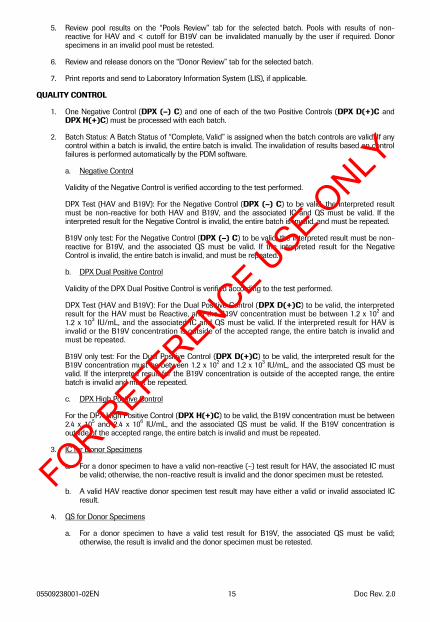
5. Review pool results on the “Pools Review” tab for the selected batch. Pools with results of non-reactive for HAV and < cutoff for B19V can be invalidated manually by the user if required. Donor specimens in an invalid pool must be retested.
6. Review and release donors on the “Donor Review” tab for the selected batch.
7. Print reports and send to Laboratory Information System (LIS), if applicable.
QUALITY CONTROL
1. One Negative Control (DPX (–) C) and one of each of the two Positive Controls (DPX D(+)C and DPX H(+)C) must be processed with each batch.
2. Batch Status: A Batch Status of “Complete, Valid” is assigned when the batch controls are valid. If any control within a batch is invalid, the entire batch is invalid. The invalidation of results based on control failures is performed automatically by the PDM software.
a. Negative Control
Validity of the Negative Control is verified according to the test performed.
DPX Test (HAV and B19V): For the Negative Control (DPX (–) C) to be valid, the interpreted result must be non-reactive for both HAV and B19V, and the associated IC and QS must be valid. If the interpreted result for the Negative Control is invalid, the entire batch is invalid, and must be repeated.
B19V only test: For the Negative Control (DPX (–) C) to be valid, the interpreted result must be non-reactive for B19V, and the associated QS must be valid. If the interpreted result for the Negative Control is invalid, the entire batch is invalid, and must be repeated.
b. DPX Dual Positive Control
Validity of the DPX Dual Positive Control is verified according to the test performed.
DPX Test (HAV and B19V): For the Dual Positive Control (DPX D(+)C) to be valid, the interpreted result for the HAV must be Reactive, and the B19V concentration must be between 1.2 x 10² and 1.2 x 10³ IU/mL, and the associated IC and QS must be valid. If the interpreted result for HAV is invalid or the B19V concentration is outside of the accepted range, the entire batch is invalid and must be repeated.
B19V only test: For the Dual Positive Control (DPX D(+)C) to be valid, the interpreted result for the B19V concentration must be between 1.2 x 10² and 1.2 x 10³ IU/mL, and the associated QS must be valid. If the interpreted result for the B19V concentration is outside of the accepted range, the entire batch is invalid and must be repeated.
c. DPX High Positive Control
For the DPX High Positive Control (DPX H(+)C) to be valid, the B19V concentration must be between 2.4 x 10⁵ and 2.4 x 10⁶ IU/mL, and the associated QS must be valid. If the B19V concentration is outside of the accepted range, the entire batch is invalid and must be repeated.
3. IC for Donor Specimens
a. For a donor specimen to have a valid non-reactive (–) test result for HAV, the associated IC must be valid; otherwise, the non-reactive result is invalid and the donor specimen must be retested.
b. A valid HAV reactive donor specimen test result may have either a valid or invalid associated IC result.
4. QS for Donor Specimens
a. For a donor specimen to have a valid test result for B19V, the associated QS must be valid; otherwise, the result is invalid and the donor specimen must be retested.
FOR REFERENCE U
SE ONLY
05509238001-02EN 16 Doc Rev. 2.0
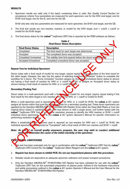
RESULTS
1. Specimen results are valid only if the batch containing them is valid. See Quality Control Section for acceptance criteria. Four parameters are measured for each specimen, one for the HAV viral target, one for B19V viral target, one for the IC, and one for the QS.
B19V only test: only two parameters are measured for each specimen, the B19V viral target, and the QS.
2. The final test results are non-reactive, reactive, or invalid for the HAV target, and < cutoff, ≥ cutoff, or invalid for the B19V target.
3. The final donor status for the cobas® TaqScreen DPX Test is reported by the PDM software as follows:
Table 2 Final Donor Status Description
Final Donor Status Description Completed The final result for each target was determined. Accepted The completed donor was accepted. Completed Unresolved The viability time limit expired before the donor was accepted. Accepted Unresolved Completed unresolved donor was accepted.
Repeat Test for Individual Specimen
Donor tubes with a final result of invalid for one target, require repeat testing regardless of the final result for the other target. However, the user has the option of selecting the “Force Unresolve” button to complete the workflow for a donor. The “Force Unresolve” function assigns an Accepted Unresolved donor status to donors not having a final result of reactive for HAV or ≥ cutoff for B19V, or assigns an Accepted donor status to donors having a final result of reactive for HAV, or ≥ cutoff for B19V.
Secondary Pooling Test
Donor tubes in a multi-specimen pool with a final result of invalid for one target, require repeat testing if the final result for the other target is non-reactive or invalid for HAV, or < cutoff or invalid for B19V.
When a multi-specimen pool is reported as reactive for HAV, or ≥ cutoff for B19V, the cobas s 201 system assigns all donors within that pool the pooling request for a secondary pooling test. These donor specimens are pipetted by the Hamilton MICROLAB® STAR/STARlet IVD Pipettor (from either the Deep Well Plates or from the original donor tubes) in subdivided pools with less donors or with a single donor, and further tested with the cobas® TaqScreen DPX Test as part of the resolution process to identify the HAV reactive or B19V ≥ cutoff individual donor specimens. Refer to the cobas s 201 system Operator's Manual for specific information on performing resolution testing.
When a subdivided, multi-specimen pool is reported as non-reactive for HAV and < cutoff for B19V, the individual specimen(s) are reported as "Completed” with a final result of HAV non-reactive and B19V < cutoff.
Note: As part of an overall quality assurance program, the user may wish to conduct additional testing to determine the cause of the initial reactivity of the specimen.
PROCEDURAL LIMITATIONS
1. This test has been evaluated only for use in combination with the cobas® TaqScreen DPX Test kit, cobas® TaqScreen DPX Control Kit, the cobas® TaqScreen Wash Reagent and the cobas s 201 system.
2. Heparin has been shown to inhibit PCR. Do not use heparinized plasma with this procedure.
3. Reliable results are dependent on adequate specimen collection and proper transport procedures.
4. Only the Hamilton MICROLAB® STAR/STARlet IVD Pipettor has been validated for use with the cobas® TaqScreen DPX Test, for the automated preparation of plasma pools. Adhere to the hardware instructions and safety precautions outlined in the cobas s 201 system Operator’s Manual and the User Manual for the Hamilton MICROLAB® STAR/STARlet IVD Pipettor.
FOR REFERENCE U
SE ONLY
05509238001-02EN 17 Doc Rev. 2.0
5. Detection of HAV RNA and quantitation of B19V DNA is dependent on the number of virus particles present in the specimen and may be affected by specimen collection methods, patient factors (i.e., age, presence of symptoms), and/or stage of infection and pool size.
6. Though rare, mutations within the highly conserved regions of the viral genomes covered by the cobas® TaqScreen DPX Test primers and/or probes may result in failure to detect HAV or correctly quantitate B19V.
7. Due to inherent differences between technologies, it is recommended that, prior to switching from one technology to the next; users perform method correlation studies in their laboratory to qualify technology differences. Users should follow their own specific policies/procedures.
PERFORMANCE CHARACTERISTICS
Clinical Reproducibility Study
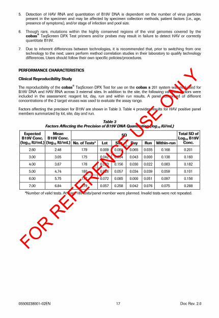
The reproducibility of the cobas® TaqScreen DPX Test for use on the cobas s 201 system was evaluated for B19V DNA and HAV RNA across 3 external sites. In addition to the site, the following variance factors were included in the assessment: reagent lot, day, run and within run results. A panel comprised of different concentrations of the 2 target viruses was used to evaluate the assay range.
Factors affecting the precision for B19V are shown in Table 3. Table 4 provides results for HAV positive panel members summarized by lot, site, day and run.
Table 3 Factors Affecting the Precision of B19V DNA Quantitation (log10 IU/mL)
SD Expected B19V Conc.
(log10 IU/mL)
Mean B19V Conc.
(log10 IU/mL) No. of Tests* Lot Site Day Run Within-run
Total SD of Log10 B19V
Conc.
2.60 2.48 179 0.009 0.081 0.065 0.035 0.168 0.201
3.00 3.05 175 0.042 0.054 0.043 0.000 0.138 0.160
4.00 3.87 178 0.023 0.156 0.030 0.022 0.083 0.182
5.00 4.74 180 0.026 0.057 0.034 0.039 0.059 0.101
6.00 5.75 177 0.072 0.085 0.000 0.051 0.097 0.156
7.00 6.84 179 0.057 0.258 0.042 0.076 0.075 0.288
*Number of valid tests. At least 180 tests/panel member were planned. Invalid tests were not repeated.
FOR REFERENCE U
SE ONLY
05509238001-02EN 18 Doc Rev. 2.0
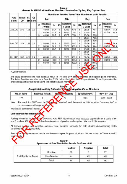
Table 4 Results for HAV Positive Panel Members Summarized by Lot, Site, Day and Run
Number of Positive Tests/Total Number of Valid Results HAV
Conc. Mean
Ct* Ct SD
Ct CV%
Lot Site Day Run
ID Reactive/ Valid
% ID Reactive/ Valid
% ID Reactive/ Valid
% ID Reactive/ Valid
%
0.5xLOD 37.9 1.35 3.6 1 49/60 81.7 1 40/59 67.8 1 25/35 71.4 1 62/87 71.3 2 40/56 71.4 2 46/60 76.7 2 27/35 77.1 2 67/89 75.3 3 40/60 66.7 3 43/57 75.4 3 23/36 63.9 4 25/34 73.5 5 29/36 80.6
1.0xLOD 37.4 1.26 3.4 1 59/60 98.3 1 56/60 93.3 1 35/36 97.2 1 84/89 94.4 2 56/59 94.9 2 60/60 100.0 2 34/36 94.4 2 89/90 98.9 3 58/60 96.7 3 57/59 96.6 3 34/36 94.4 4 35/35 100.0 5 35/36 97.2
3.0x LOD 35.9 0.87 2.4 1 60/60 100.0 1 60/60 100.0 1 36/36 100.0 1 88/88 100.0 2 58/58 100.0 2 60/60 100.0 2 35/35 100.0 2 90/90 100.0 3 60/60 100.0 3 58/58 100.0 3 36/36 100.0 4 35/35 100.0 5 36/36 100.0
*Cycle threshold
The study generated one false Reactive result in 177 valid DPX tests performed on negative panel members. The false Reactive was due to a Reactive B19V below the lower limit of quantitation. Table 5 provides the analytical specificity estimated using the negative panel members.
Table 5 Analytical Specificity Estimated Using the Negative Panel Members
No. of Tests Reactive Result Negative Results Specificity (%) 95% CI* (%)
177 1 176 99.4 96.9 - 100.0
Note: The result for B19V must be “Target not Detected” and the result for HAV must be “Non-reactive” to produce an overall negative result.
*95% exact confidence interval
Clinical Pool Resolution Study
Pooling resolution for both B19V DNA and HAV RNA identification was assessed separately for 5 pools of 96 and 5 pools of 480 using pre-defined combinations of positive and negative HAV and B19V samples.
All known positive and negative samples were identified correctly for both studies demonstrating 100% sensitivity and 100% specificity.
Results for the agreement of results and known samples for pools of 96 and 480 are shown in Tables 6 and 7 respectively.
Table 6 Agreement of Pool Resolution Results for Pools of 96
Positive Negative Total
Reactive 27 0 27
Non-Reactive 0 453 453 Pool Resolution Result
Total 27 453 480
FOR REFERENCE U
SE ONLY
05509238001-02EN 19 Doc Rev. 2.0
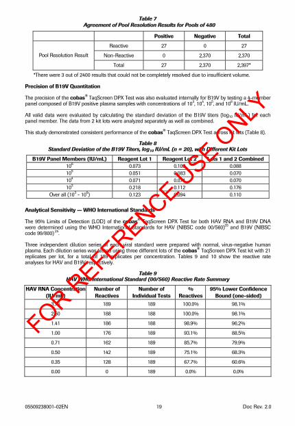
Table 7 Agreement of Pool Resolution Results for Pools of 480
Positive Negative Total
Reactive 27 0 27
Non-Reactive 0 2,370 2,370 Pool Resolution Result
Total 27 2,370 2,397*
*There were 3 out of 2400 results that could not be completely resolved due to insufficient volume.
Precision of B19V Quantitation
The precision of the cobas® TaqScreen DPX Test was also evaluated internally for B19V by testing a 4-member panel composed of B19V positive plasma samples with concentrations of 103, 104, 105, and 106 IU/mL.
All valid data were evaluated by calculating the standard deviation of the B19V titers (log10 IU/mL) for each panel member. The data from 2 kit lots were analyzed separately as well as combined.
This study demonstrated consistent performance of the cobas® TaqScreen DPX Test across kit lots (Table 8).
Table 8 Standard Deviation of the B19V Titers, log10 IU/mL (n = 20), with Different Kit Lots
B19V Panel Members (IU/mL) Reagent Lot 1 Reagent Lot 2 Lots 1 and 2 Combined 106 0.073 0.103 0.088 105 0.051 0.083 0.070 104 0.071 0.071 0.070 103 0.218 0.112 0.176
Over all (103 - 106) 0.123 0.094 0.110 Analytical Sensitivity — WHO International Standards
The 95% Limits of Detection (LOD) of the cobas® TaqScreen DPX Test for both HAV RNA and B19V DNA were determined using the WHO International Standards for HAV (NIBSC code 00/560)33 and B19V (NIBSC code 99/800) 24.
Three independent dilution series of each viral standard were prepared with normal, virus-negative human plasma. Each dilution series was tested using three different lots of the cobas® TaqScreen DPX Test kit with 21 replicates per lot, for a total of 189 replicates per concentration. Tables 9 and 10 show the reactive rate analyses for HAV and B19V respectively.
Table 9 HAV WHO International Standard (00/560) Reactive Rate Summary
HAV RNA Concentration (IU/mL)
Number of Reactives
Number of Individual Tests
% Reactives
95% Lower Confidence Bound (one-sided)
3.75 189 189 100.0% 98.1%
2.50 188 188 100.0% 98.1%
1.41 186 188 98.9% 96.2%
1.00 176 189 93.1% 88.5%
0.71 162 189 85.7% 79.9%
0.50 142 189 75.1% 68.3%
0.35 128 189 67.7% 60.6%
0.00 0 189 0.0% 0.0%
FOR REFERENCE U
SE ONLY
05509238001-02EN 20 Doc Rev. 2.0
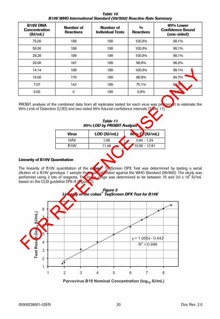
Table 10 B19V WHO International Standard (99/800) Reactive Rate Summary
B19V DNA Concentration
(IU/mL)
Number of Reactives
Number of Individual Tests
% Reactives
95% Lower Confidence Bound
(one-sided)
75.00 189 189 100.0% 98.1%
50.00 188 188 100.0% 98.1%
28.28 189 189 100.0% 98.1%
20.00 187 189 98.9% 96.2%
14.14 189 189 100.0% 98.1%
10.00 170 189 89.9% 84.7%
7.07 142 189 75.1% 68.3%
0.00 0 189 0.0% 0.0% PROBIT analysis of the combined data from all replicates tested for each virus was performed to estimate the 95% Limit of Detection (LOD) and two-sided 95% fiducial confidence intervals (Table 11).
Table 11 95% LOD by PROBIT Analysis
Virus LOD (IU/mL) 95% CI (IU/mL) HAV 1.06 0.94 - 1.24 B19V 11.48 10.56 - 12.91
Linearity of B19V Quantitation
The linearity of B19V quantitation of the cobas® TaqScreen DPX Test was determined by testing a serial dilution of a B19V genotype 1 sample that was calibrated against the WHO Standard (99/800). The study was performed using 2 lots of reagents. The linear range was determined to be between 75 and 3.0 x 10⁸ IU/mL based on the CLSI guideline EP6-A (Figure 2).
Figure 2 Linearity of the cobas® TaqScreen DPX Test for B19V
1
2
3
4
5
6
7
8
1 2 3 4 5 6 7 8
Parvovirus B19 Nominal Concentration (log10 IU/mL)
Te
st
Re
su
lt (
log
10 IU
/mL
)
y = 1.060x - 0.442
R2 = 0.996FOR REFERENCE U
SE ONLY
05509238001-02EN 21 Doc Rev. 2.0
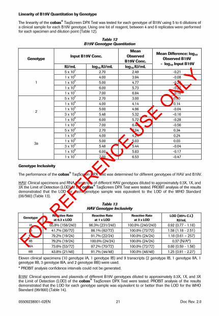
Linearity of B19V Quantitation by Genotype
The linearity of the cobas® TaqScreen DPX Test was tested for each genotype of B19V using 5 to 6 dilutions of a clinical sample for each B19V genotype. Using one lot of reagent, between 4 and 6 replicates were performed for each specimen and dilution point (Table 12).
Table 12 B19V Genotype Quantitation
Input B19V Conc. Mean
Observed B19V Conc. Genotype
IU/mL log10 IU/mL log10 IU/mL
Mean Difference: log10 Observed B19V
- log10 Input B19V
5 x 10² 2.70 2.49 -0.21 1 x 10⁴ 4.00 3.94 -0.08 1 x 10⁵ 5.00 4.77 -0.23 1 x 10⁶ 6.00 5.73 -0.27
1
1 x 10⁷ 7.00 6.84 -0.16 5 x 10² 2.70 3.00 0.30 1 x 10⁴ 4.00 4.14 0.14 1 x 10⁵ 5.00 4.96 -0.04 3 x 10⁵ 5.48 5.32 -0.16 1 x 10⁶ 6.00 5.72 -0.28
2
1 x 10⁷ 7.00 6.44 -0.56 5 x 10² 2.70 3.04 0.34 1 x 10⁴ 4.00 4.24 0.24 1 x 10⁵ 5.00 5.03 0.03 3 x 10⁵ 5.48 5.44 -0.04 1 x 10⁶ 6.00 5.83 -0.17
3a
1 x 10⁷ 7.00 6.53 -0.47
Genotype Inclusivity
The performance of the cobas® TaqScreen DPX Test was determined for different genotypes of HAV and B19V.
HAV: Clinical specimens and RNA transcripts of different HAV genotypes diluted to approximately 0.3X, 1X, and 3X the Limit of Detection (LOD) of the cobas® TaqScreen DPX Test were tested. PROBIT analysis of the results demonstrated that the LOD for each genotype sample was equivalent to the LOD of the WHO Standard (00/560) (Table 13).
Table 13 HAV Genotype Inclusivity
Genotype Reactive Rate at 0.3 x LOD
Reactive Rate at 1 x LOD
Reactive Rate at 3 x LOD
LOD (95% C.I.) IU/mL
IA 65.8% (158/240) 96.3% (231/240) 100.0% (240/240) 0.92 (0.77 - 1.18) IB 41.7% (30/72) 86.1% (62/72) 100.0% (72/72) 1.58 (1.18 - 2.51) IIA 79.2% (19/24) 91.7% (22/24) 100.0% (24/24) 1.18 (0.61 – 257) IIB 79.2% (19/24) 100.0% (24/24) 100.0% (24/24) 0.37 (N/A*) IIIA 73.6% (53/72) 97.2% (70/72) 100.0% (72/72) 0.80 (0.59 - 1.56) IIIB 43.8% (21/48) 91.7% (44/48) 100.0% (48/48) 1.25 (0.91 - 2.27)
Eleven clinical specimens (10 genotype IA, 1 genotype IB) and 9 transcripts (2 genotype IB, 1 genotype IIA, 1 genotype IIB, 3 genotype IIIA, and 2 genotype IIIB) were used. * PROBIT analysis confidence intervals could not be generated.
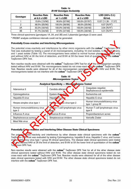
B19V: Clinical specimens and plasmids of different B19V genotypes diluted to approximately 0.3X, 1X, and 3X the Limit of Detection (LOD) of the cobas® TaqScreen DPX Test were tested. PROBIT analysis of the results demonstrated that the LOD for each genotype sample was equivalent to or better than the LOD for the WHO Standard (99/800) (Table 14).
FOR REFERENCE U
SE ONLY
05509238001-02EN 22 Doc Rev. 2.0
Table 14 B19V Genotype Inclusivity
Genotype Reactive Rate at 0.3 x LOD
Reactive Rate at 1 x LOD
Reactive Rate at 3 x LOD
LOD (95% C.I.) IU/mL
1 15.6% (15/96) 90.6% (87/96) 100.0% (97/97) 13.57 (11.39 - 17.27) 2 88.5% (85/96) 100.0% (96/96) 99.0% (95/96) 6.07 (N/A*)
3A 83.3% (40/48) 100.0% (48/48) 100.0% (48/48) 3.94 (N/A*) 3B 91.7% (44/48) 97.9% (47/48) 100.0% (48/48) 5.31 (N/A*)
Three clinical specimens (genotypes 1A, 3A, and 3B) and 2 plasmids (genotype 2) were used. * PROBIT analysis confidence intervals could not be generated.
Potentially Cross-reactive and Interfering Microorganisms
The potential cross-reactivity and interference by other micro organisms with the cobas® TaqScreen DPX Test was evaluated by testing a panel of 22 microorganisms, including 16 viral isolates, 5 bacterial strains, and 1 yeast isolate (Table 15). The microorganisms were added to normal human plasma and tested with and without HAV at 3X the limit of detection, and B19V at 5X the lower limit of quantitation of the cobas® TaqScreen DPX Test.
Non-reactive results were obtained with the cobas® TaqScreen DPX Test for all of the microorganism samples tested without HAV and B19V. The microorganisms tested do not cross-react with the cobas® TaqScreen DPX Test. Reactive results were obtained for all of the microorganism samples tested with HAV and B19V. The microorganisms tested do not interfere with the cobas® TaqScreen DPX Test.
Table 15 Microorganisms Tested
Analytical Specificity — Microorganisms Tested
Adenovirus 5 Candida albicans Coagulase-negative Staphylococcus epidermidis
Cytomegalovirus Epstein-Barr virus Escherichia coli Hepatitis B virus Hepatitis C virus Hepatitis G virus
Herpes simplex virus type 1 Herpes simplex virus type 2 Human immunodeficiency virus type 1 group M
Human immunodeficiency virus type 2
Human T-cell lymphotropic virus type I
Human T-cell lymphotropic virus type II
Influenza A virus Influenza B virus Propionibacterium acnes Staphylococcus aureus Streptococcus viridans Varicella Zoster West Nile virus
Potentially Cross-reactive and Interfering Other Disease State Clinical Specimens
The potential cross-reactivity and interference by other disease state clinical specimens with the cobas® TaqScreen DPX Test was evaluated by testing Cytomegalovirus, hepatitis B virus, Hepatitis C virus, and human immunodeficiency virus type 1 group M clinical specimens,. The disease state clinical specimens were tested with and without HAV at 3X the limit of detection, and B19V at 5X the lower limit of quantitation of the cobas® TaqScreen DPX Test.
Non-reactive results were obtained with the cobas® TaqScreen DPX Test for all of the other disease state clinical specimens tested without HAV and B19V. The other disease state clinical specimens tested do not cross-react with the cobas® TaqScreen DPX Test. Reactive results were obtained for all of the other disease state clinical specimens tested with HAV and B19V. The other disease state clinical specimens tested do not interfere with the cobas® TaqScreen DPX Test.
FOR REFERENCE U
SE ONLY
05509238001-02EN 23 Doc Rev. 2.0
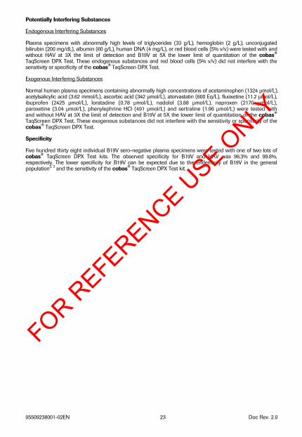
Potentially Interfering Substances
Endogenous Interfering Substances
Plasma specimens with abnormally high levels of triglycerides (33 g/L), hemoglobin (2 g/L), unconjugated bilirubin (200 mg/dL), albumin (60 g/L), human DNA (4 mg/L), or red blood cells (5% v/v) were tested with and without HAV at 3X the limit of detection and B19V at 5X the lower limit of quantitation of the cobas® TaqScreen DPX Test. These endogenous substances and red blood cells (5% v/v) did not interfere with the sensitivity or specificity of the cobas® TaqScreen DPX Test.
Exogenous Interfering Substances
Normal human plasma specimens containing abnormally high concentrations of acetaminophen (1324 μmol/L), acetylsalicylic acid (3.62 mmol/L), ascorbic acid (342 μmol/L), atorvastatin (600 Eq/L), fluoxetine (11.2 μmol/L), ibuprofen (2425 μmol/L), loratadine (0.78 μmol/L), nadolol (3.88 μmol/L), naproxen (2170 μmol/L), paroxetine (3.04 μmol/L), phenylephrine HCl (491 μmol/L) and sertraline (1.96 μmol/L) were tested with and without HAV at 3X the limit of detection and B19V at 5X the lower limit of quantitation of the cobas® TaqScreen DPX Test. These exogenous substances did not interfere with the sensitivity or specificity of the cobas® TaqScreen DPX Test.
Specificity
Five hundred thirty eight individual B19V sero-negative plasma specimens were tested with one of two lots of cobas® TaqScreen DPX Test kits. The observed specificity for B19V and HAV was 96.3% and 99.8%, respectively. The lower specificity for B19V can be expected due to the endemicity of B19V in the general population2, 3 and the sensitivity of the cobas® TaqScreen DPX Test kit.
FOR REFERENCE U
SE ONLY
05509238001-02EN 24 Doc Rev. 2.0
REFERENCES
1. Servant A, Laperche S, Lallemand F, Marinho V, De Saint Maur G, Meritet JF, Garbarg-Chenon A. Genetic diversity within human Erythroviruses: identification of three genotypes. Journal of Virology 2002;76:9124–34.
2. Anderson MJ, Tsou C, Parker RA, Chorba TL, Wolfe H, Tattersal P, Mortimer PP. Detection of antibodies and antigen of human parvovirus B18 by enzyme-linked immunosorbent assay. Journal of Clinical Microbiology 1986;24:522 526.
3. Cohen BJ, Buckley MM. The prevalence of antibody to human parvovirus B19 in England and Wales. Journal of Medical Microbiology 1988;25:151-153.
4. Young NS, Brown KE. Mechanisms of disease: parvovirus B19. New England Journal of Medicine 2004;350:586-597.
5. Yoto Y, Kudoh T, Haseyama K, Suzuki N, Oda T, Katoh T, Takahashi T, Sekiguchi S, Chiba S. Incidence of human parvovirus B19 DNA detection in blood donors. British Journal of Haematology 1995;91:1017-1018.
6. McOmish F, Yap PL, Jordan A, Hart H, Cohen BJ, Simmonds P. Detection of parvovirus B19 in donated blood: a model system for screening by polymerase chain reaction. Journal of Clinical Microbiology 1993;31:323-328.
7. Wu CG, Mason B, Jong J, Erdman D, McKernan L, Oakley M, Soucie M, Evatt B, Yu MY. Parvovirus B19 transmission by a high-purity factor VIII concentrate. Transfusion 2005;45:1003-1010.
8. Saldanha J, Minor P. Detection of human parvovirus B19 in plasma pools and blood products derived from these pools: implications for efficiency and consistency of removal of B19 DNA during manufacture> British Journal of Haematology 1996;93:714-719.
9. Eis-Hubinger AM, Sasowski U, Brackmann HH. Parvovirus B19 DNA contamination in coagulation factor VIII products. Thrombosis and Haemostasis 1999;81:476-477.
10. Schmidt I, Blumel J, Seitz H, Wilkommen H, Lower J. Parvovirus B19 DNA in plasma pools and plasma derivatives. Vox Sanguinis 2001;81:228-235.
11. Azzi A, Moefini M, Mannucci PM. The transfusion-associated transmission of parvovirus B19. Transfusion Medicine Reviews 1999;13:194-204.
12. Yee TT, Cohen BJ, Pasi KL, Lee CA. Transmission of symptomatic parvovirus B19 infection by clotting factor concentrate. British Journal of Haematology 1996;93:457-459.
13. Blümel J., Schmid, I., Effenberger W., Seitz H., Willkommen H., Brackmann H. H., Löwer J. Eis-Hübinger AM. Parvovirus B19 transmission by heat treated clotting factor concentrates. Transfusion 2002;42:1473-1481.
14. Francki RIB, Fauquet CM, Knudson DL, Brown F. Classification and nomenclature of viruses. Archives of Virology 1991;Suppl. 2:320-326.
15. Gust ID. Epidemiology patterns of hepatitis A in different parts of the world. Vaccine 1992;10:856-862.
16. Shapiro CN, Margolis HS. Worldwide epidemiology of hepatitis A virus infection. Journal of Hepatology 1993;18:11-14.
17. Hadler SC. Global impact of hepatitis A virus infection changing patterns. In: Hollinger FB, Lemon SM, and Margolis HS, eds. Viral Hepatitis and Liver Disease. Baltimore, Williams & Wilkins, 1991: 14-20.
18. Robertson BH, Jansen RE, Khanna B, Totsuka A, Nainan OV, Siegl G, Widell A, Margolis H, Isomura S, Ito K, Ishizu T, Moritsugu Y, Lemon, SM. Genetic relatedness of hepatitis A virus strains recovered from different geographical regions. Journal of General Virology 1992;73:1365-1377.
19. Costa-Mattioli M, Cristina J, Romero H, Perez-Bercof R, Casane D, Colina R, Garcia L, Vega I, Glikman G, Romanowsky V, Castello A, Nicand E, Gassin M, Billaudel S, Ferre V. Molecular evolution of hepatitis A virus: a new classification based on the complete VP1 protein. Journal of Virology 2002;76:9516-9525.
20. Bower WA, Nainan OV, Han X, Margolis HS. Duration of viremia in hepatitis A virus infection. Journal of Infectious Diseases 2000;182:12-17.
FOR REFERENCE U
SE ONLY
05509238001-02EN 25 Doc Rev. 2.0
21. Gowland P, Fontana S, Niederhauser C, Manouri Taleghani B. Molecular and serologic tracing of a transfusion-transmitted hepatitis A virus. Transfusion 2004;44:1555-1561.
22. Soucie JM, Robertson BH, Bell BP, McCaustland KA, Evatt BL. Hepatitis A virus infections associated with clotting factor concentrate in the United States. Transfusion 1998;38:573-579.
23. Chudy M, Budek I, Keller-Stanislawski B, McCaustland KA, Neidhold S, Robertson BH, Nübling CM, Seitz R, Löwer J. A new cluster of hepatitis A infection in haemophiliacs traced to a contaminated plasma pool. Journal of Medical Virology 1999;57:91-99.
24. Saldanha J, Lelie N, Yu M-Y, Heath A and the B19 Collaborative Study group. Establishment of the first World Health Organization international standard for human parvovirus B19 DNA nucleic acid amplification techniques. Vox Sanguinis 2002;82:24-31.
25. Longo MC, Berninger MS, Hartley JL. Use of uracil DNA glycosylase to control carry-over contamination in polymerase chain reactions. Gene. 1990;93:125-128.
26. Savva R, McAuley-Hecht K, Brown T, Pearl L. The structural basis of specific base-excision repair by uracil-DNA glycosylase. Nature. 1995;373:487-493.
27. Mol CD, Arvai AS, Slupphaug G, Kavli B, Krokan HE, Mosbaugh DW, Tainer JA. Crystal structure and mutational analysis of human uracil-DNA glycosylase: structural basis for specificity and catalysis. Cell. 1995;80:869-878.
28. Higuchi R, Dollinger G, Walsh PS, Griffith, R. Simultaneous amplification and detection of specific DNA sequences. Biotechnology (NY). 1992;10:413-417.
29. Heid CA, Stevens J, Livak JK, Williams PM. Real time quantitative PCR. Genome Res. 1996;6:986-994.
30. Richmond, J.Y. and McKinney, R.W. (eds.). 1999. Biosafety in Microbiological and Biomedical Laboratories HHS Publication Number (CDC) 93-8395.
31. Clinical and Laboratory Standards Institute (CLSI). Protection of Laboratory Workers from Occupationally Acquired Infections. Approved Guideline-Third Edition. CLSI Document M29-A3 Wayne, PA:CLSI, 2005.
32. International Air Transport Association. Dangerous Goods Regulations. 41st ed. Quebec, Canada. 2000.
33. Saldanha J, Heath A, Lelie N, Pisani G, Yu M-Y, and the Collaborative Study Group. A World Health Organization International Standard for hepatitis A virus RNA nucleic acid amplification technology assays. Vox Sanguinis 2005;89:52-58.
FOR REFERENCE U
SE ONLY
05509238001-02EN 26 Doc Rev. 2.0
FOR REFERENCE U
SE ONLY
05509238001-02EN 27 Doc Rev. 2.0
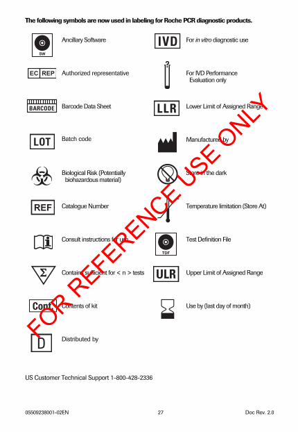
The following symbols are now used in labeling for Roche PCR diagnostic products. Ancillary Software
For in vitro diagnostic use
Authorized representative
For IVD Performance Evaluation only
Barcode Data Sheet
Lower Limit of Assigned Range
Batch code
Manufactured by
Biological Risk (Potentially biohazardous material)
Store in the dark
Catalogue Number
Temperature limitation (Store At)
Consult instructions for use
Test Definition File
Contains sufficient for < n > tests
Upper Limit of Assigned Range
Contents of kit
Use by (last day of month)
Distributed by
US Customer Technical Support 1-800-428-2336
SW
TDF
FOR REFERENCE U
SE ONLY
05509238001-02EN 28 Doc Rev. 2.0
Document Revision Information
Doc Rev. 2.0 07/2011
AMPLILINK Software v3.3.2 and PDM Software v3.0.27 were added to the Instrumentation and Software for cobas s 201 system section. The INSTRUCTIONS FOR USE section was revised to indicate that SPU racks should be loaded into COBAS® AmpliPrep Instrument in positions J, K or L. Please contact your local Roche Representative if you have any questions.
Roche Molecular Systems, Inc., Branchburg, NJ 08876 USA A Member of the Roche Group
U.S. Device Master File No. 14454
Roche Diagnostics 9115 Hague Road Indianapolis, IN 46250-0457 USA
Distributed by (For Technical Assistance call the
Roche Response Center toll-free: 1-800 526-1247)
ROCHE, COBAS, COBAS S, TAQMAN, TAQSCREEN, AMPLILINK, AMPERASE, and AMPLIPREP are trademarks
of Roche. ROCHE RESPONSE CENTER is a service mark of Roche.
All other product names and trademarks are the property of their respective owners.
Armored RNA is a patented technology developed by Ambion, Inc. and Cenetron Diagnostics LLC. U.S. patents #5,677,124, #5,919,625, and #5,939,262 and patents pending. ARMORED RNA is a trademark of Ambion and Cenetron.
The cyanine dyes in this product are subject to patent rights of GE Healthcare Bio-Sciences Corp. and Carnegie Mellon University and are licensed to Roche solely for incorporation into components of research and in-vitro diagnostic kits. Any use of such kits for purposes other than research or in-vitro diagnostics requires sublicenses from GE Healthcare Bio-Sciences Corp., Piscataway, New Jersey, U.S.A. and Carnegie Mellon University, Pittsburgh, Pennsylvania, U.S.A.
© 2011 Roche Molecular Systems, Inc. All rights reserved.
07/2011 05509238001–02 Doc Rev. 2.0
FOR REFERENCE U
SE ONLY


![[TIPE] DPX-3000U - KENWOOD](https://static.fdocuments.net/doc/165x107/6198e54a1ae42335a4313f28/tipe-dpx-3000u-kenwood.jpg)