cobas 4800 KRAS Mutation Test · PDF fileThe cobas® KRAS Mutation Test, for use with the...
26
1 cobas ® KRAS Mutation Test FOR IN VITRO DIAGNOSTIC USE. cobas ® DNA Sample Preparation Kit 24 Tests P/N: 05985536190 cobas ® KRAS Mutation Test 24 Tests P/N: 05852170190 NOTICE: The purchase of this product allows the purchaser to use it for amplification and detection of nucleic acid sequences by polymerase chain reaction (PCR) and related processes for human in vitro diagnostics. No general patent or other license of any kind other than this specific right of use from purchase is granted hereby. INTENDED USE The cobas ® KRAS Mutation Test, for use with the cobas ® 4800 System, is a real-time PCR test for the detection of seven somatic mutations in codons 12 and 13 of the KRAS gene in DNA derived from formalin-fixed paraffin-embedded human colorectal cancer (CRC) tumor tissue. The test is intended to be used as an aid in the identification of CRC patients for whom treatment with Erbitux® (cetuximab) or with Vectibix® (panitumumab) may be indicated based on a no mutation detected result. Specimens are processed using the cobas ® DNA Sample Preparation Kit for manual sample preparation and the cobas z 480 analyzer for automated amplification and detection. SUMMARY AND EXPLANATION OF THE TEST The KRAS protein is a member of the superfamily of small G proteins. KRAS acts as a GDP/GTP-regulated switch to convey extracellular signals that influence cell proliferation, apoptosis and remodeling of the actin cytoskeleton. Mutations affecting amino acids 12 or 13, which occur in a variety of human malignancies, including colorectal cancer (CRC), lock the enzyme in the GTP-bound, activated form, resulting in constitutive signaling and thereby contributing to the oncogenic process. 1 KRAS mutations are observed in 24%-43% of colorectal tumors. 2-3 Although over 3000 point mutations of the KRAS gene have been identified, most occur in codons 12 or 13 (~82% in codon 12 and ~17% in codon 13). 4 These mutations have been shown to result in constitutive activation of KRAS and predict non-response to anti-EGFR monoclonal antibody therapy. 4 Cetuximab and panitumumab are monoclonal antibodies which target the epidermal growth factor receptor (EGFR) and are approved for use in patients with metastatic colorectal cancer. Although 50% to 80% of colorectal tumors overexpress EGFR, EGFR protein expression and gene amplification have only limited predictive value in determining the likelihood of response to cetuximab or panitumumab. 5 However, there is now strong evidence to show that the presence of KRAS mutations correlates with lack of response to EGFR- targeted antibody therapy in patients with metastatic colorectal cancer and that, in some situations, the use of EGFR-targeted antibody therapy in this patient subgroup may be detrimental. 6-7 The supporting evidence for these findings comes from: • Retrospective analyses of single-arm studies 8-9 • Retrospective analyses of randomized studies 10-11 • Prospective randomized studies 12 As a consequence of these studies, KRAS mutation testing is recommended for the selection of patients to receive anti-EGFR antibody therapy by major oncology organizations in the US (ASCO, NCCN) 13-14 and Europe (ESMO). 15 Furthermore, US and European regulatory authorities have restricted the use of these agents to patients with KRAS wild-type tumors. 16-17 The cobas ® KRAS Mutation Test is a PCR-based assay designed to identify the presence of seven somatic mutations involving codons 12 and 13 of the proto-oncogene KRAS Based on data in the COSMIC database (2012 v59), the seven mutations detected by the cobas ® KRAS Mutation Test account for >97% of all reported KRAS mutations in CRC patients. Thus, the test is intended to be used as an aid in the identification of CRC patients for whom treatment with Erbitux® (cetuximab) or with Vectibix® (panitumumab) may be indicated based on a no mutation detected result. PRINCIPLES OF THE PROCEDURE The cobas ® KRAS Mutation Test is based on two major processes: (1) manual specimen preparation to obtain genomic DNA from formalin-fixed, paraffin-embedded tissue (FFPET); and (2) PCR amplification of target DNA using complementary primer pairs and an oligonucleotide probe labeled with fluorescent dye. Mutation detection is achieved by melting curve analysis by the cobas z 480 analyzer. A mutant control, negative control, and calibrator are included in each run to confirm the validity of the run. KRAS DNA SP
Transcript of cobas 4800 KRAS Mutation Test · PDF fileThe cobas® KRAS Mutation Test, for use with the...

1
cobas® KRAS Mutation Test
FOR IN VITRO DIAGNOSTIC USE.
cobas® DNA Sample Preparation Kit 24 Tests P/N: 05985536190
cobas® KRAS Mutation Test 24 Tests P/N: 05852170190
NOTICE: The purchase of this product allows the purchaser to use it for amplification and detection of nucleic acid sequences by polymerase chain reaction (PCR) and related processes for human in vitro diagnostics. No general patent or other license of any kind other than this specific right of use from purchase is granted hereby.
INTENDED USE
The cobas® KRAS Mutation Test, for use with the cobas® 4800 System, is a real-time PCR test for the detection of seven somatic mutations in codons 12 and 13 of the KRAS gene in DNA derived from formalin-fixed paraffin-embedded human colorectal cancer (CRC) tumor tissue. The test is intended to be used as an aid in the identification of CRC patients for whom treatment with Erbitux® (cetuximab) or with Vectibix® (panitumumab) may be indicated based on a no mutation detected result.
Specimens are processed using the cobas® DNA Sample Preparation Kit for manual sample preparation and the cobas z 480 analyzer for automated amplification and detection.
SUMMARY AND EXPLANATION OF THE TEST
The KRAS protein is a member of the superfamily of small G proteins. KRAS acts as a GDP/GTP-regulated switch to convey extracellular signals that influence cell proliferation, apoptosis and remodeling of the actin cytoskeleton. Mutations affecting amino acids 12 or 13, which occur in a variety of human malignancies, including colorectal cancer (CRC), lock the enzyme in the GTP-bound, activated form, resulting in constitutive signaling and thereby contributing to the oncogenic process.1
KRAS mutations are observed in 24%-43% of colorectal tumors.2-3 Although over 3000 point mutations of the KRAS gene have been identified, most occur in codons 12 or 13 (~82% in codon 12 and ~17% in codon 13).4 These mutations have been shown to result in constitutive activation of KRAS and predict non-response to anti-EGFR monoclonal antibody therapy.4
Cetuximab and panitumumab are monoclonal antibodies which target the epidermal growth factor receptor (EGFR) and are approved for use in patients with metastatic colorectal cancer. Although 50% to 80% of colorectal tumors overexpress EGFR, EGFR protein expression and gene amplification have only limited predictive value in determining the likelihood of response to cetuximab or panitumumab.5
However, there is now strong evidence to show that the presence of KRAS mutations correlates with lack of response to EGFR-targeted antibody therapy in patients with metastatic colorectal cancer and that, in some situations, the use of EGFR-targeted antibody therapy in this patient subgroup may be detrimental.6-7 The supporting evidence for these findings comes from:
• Retrospective analyses of single-arm studies8-9
• Retrospective analyses of randomized studies10-11
• Prospective randomized studies12
As a consequence of these studies, KRAS mutation testing is recommended for the selection of patients to receive anti-EGFR antibody therapy by major oncology organizations in the US (ASCO, NCCN)13-14 and Europe (ESMO).15 Furthermore, US and European regulatory authorities have restricted the use of these agents to patients with KRAS wild-type tumors.16-17
The cobas® KRAS Mutation Test is a PCR-based assay designed to identify the presence of seven somatic mutations involving codons 12 and 13 of the proto-oncogene KRAS Based on data in the COSMIC database (2012 v59), the seven mutations detected by the cobas® KRAS Mutation Test account for >97% of all reported KRAS mutations in CRC patients. Thus, the test is intended to be used as an aid in the identification of CRC patients for whom treatment with Erbitux® (cetuximab) or with Vectibix® (panitumumab) may be indicated based on a no mutation detected result.
PRINCIPLES OF THE PROCEDURE
The cobas® KRAS Mutation Test is based on two major processes: (1) manual specimen preparation to obtain genomic DNA from formalin-fixed, paraffin-embedded tissue (FFPET); and (2) PCR amplification of target DNA using complementary primer pairs and an oligonucleotide probe labeled with fluorescent dye. Mutation detection is achieved by melting curve analysis by the cobas z 480 analyzer. A mutant control, negative control, and calibrator are included in each run to confirm the validity of the run.
KRAS
DNA SP

2
Specimen Preparation
FFPET specimens are processed and genomic DNA isolated using the cobas® DNA Sample Preparation Kit, a generic manual specimen preparation based on nucleic acid binding to glass fibers. A deparaffinized 5 µm section of an FFPET specimen is lysed by incubation at an elevated temperature with a protease and chaotropic lysis/binding buffer that releases nucleic acids and protects the released genomic DNA from DNases. Subsequently, isopropanol is added to the lysis mixture that is then centrifuged through a column with a glass fiber filter insert. During centrifugation, the genomic DNA is bound to the surface of the glass fiber filter. Unbound substances, such as salts, proteins and other cellular impurities, are removed by centrifugation. The adsorbed nucleic acids are washed and then eluted with an aqueous solution. The amount of genomic DNA is spectrophotometrically determined and adjusted to a fixed concentration to be added to the amplification/detection mixture. The target DNA is then amplified and detected on the cobas z 480 analyzer using the amplification and detection reagents provided in the cobas® KRAS Mutation Test kit.
PCR Amplification
Target Selection
The cobas® KRAS Mutation Test kit uses primers that define an 85 base-pair sequence for exon 2 containing KRAS codons 12 and 13 in human genomic DNA. Amplification occurs only in the regions of the KRAS gene between the primers; the entire KRAS gene is not amplified.
Target Amplification
A derivative of Thermus species Z05 DNA polymerase is utilized for target amplification. First, the PCR reaction mixture is heated to denature the genomic DNA and expose the primer target sequences. As the mixture cools, the upstream and downstream primers anneal to the target DNA sequences. The Z05 DNA polymerase, in the presence of a divalent metal ion and excess dNTPs, extends each annealed primer, thus synthesizing a second DNA strand. This completes the first cycle of PCR, yielding a double-stranded DNA copy which includes the targeted 85 base-pair region of the KRAS gene. This process is repeated for a number of cycles, with each cycle effectively doubling the amount of amplicon DNA.
Automated Real-time Mutation Detection
The cobas z 480 analyzer is capable of measuring, in real-time, the amount of fluorescence generated by specific PCR products. After amplification, each amplicon generated using the cobas® KRAS Mutation Test is subjected to a melting program in which the temperature is ramped from 40°C to 95°C (TaqMelt). The wild-type specific probe is bound to both wild-type and mutant amplicon at low temperatures. In the bound state, the fluorescein reporter dye on the 5’ end of the probe is sufficiently far away from the 3’ end quencher dye, allowing the fluorescent dye to emit a specific wave length of light. As the temperature rises, the probe dissociates from the amplicon, allowing the quencher dye to come into close proximity to the fluorescent dye, decreasing the amount of measurable fluorescence. Amplicons with a perfect match to the probe (wild-type) melt at a higher temperature than amplicons with one or more mismatches (mutant). The amount of fluorescence at each temperature increment is measured and the melting temperature(s) are calculated. The presence of a mutant KRAS sequence in codons 12 and 13 can be detected when the melting temperatures are within specified ranges. To avoid detection of codon 12 and codon 13 silent mutations (no amino acid change), a modified base serves as a universal base and produces a melting temperature within the wild-type range.
Selective Amplification
Selective amplification of target nucleic acid from the specimen is achieved in the cobas® KRAS Mutation Test by the use of AmpErase (uracil-N-glycosylase) enzyme and deoxyuridine triphosphate (dUTP).18 The AmpErase enzyme recognizes and catalyzes the destruction of DNA strands containing deoxyuridine but not DNA containing thymidine. Deoxyuridine is not present in naturally occurring DNA but is always present in amplicon due to the use of dUTP in place of thymidine triphosphate as one of the nucleotide triphosphates in the Reaction Mix reagent; therefore, only amplicons contain deoxyuridine. Deoxyuridine renders contaminating amplicons susceptible to destruction by AmpErase enzyme prior to amplification of the target DNA. The AmpErase enzyme, which is included in the Reaction Mix reagent, catalyzes the cleavage of deoxyuridine-containing DNA at the deoxyuridine residues by opening the deoxyribose chain at the C1-position. When heated in the first thermal cycling step at alkaline pH, the amplicon DNA chain breaks at the position of the deoxyuridine, thereby rendering the DNA non-amplifiable. The AmpErase enzyme is inactive at temperatures above 55ºC, i.e., throughout the thermal cycling steps, and therefore does not destroy the target amplicon.

3
REAGENTS
cobas® DNA Sample Preparation Kit 24 Tests (P/N: 05985536190)
DNA TLB 1 x 10 mL (DNA Tissue Lysis Buffer)
Tris-HCl buffer Potassium chloride 0.04% EDTA 0.1% Triton X-100 0.09% Sodium azide
PK 1 x 100 mg (Proteinase K)
Proteinase K (lyophilized) Xn Proteinase K
Harmful
DNA PBB 1 x 10 mL (DNA Paraffin Binding Buffer)
Tris-HCl buffer 49.6% Guanidine hydrochloride 0.05% Urea 17.3% Triton X-100 Xn 49.6% (w/w) Guanidine HCl
Harmful
WB I 1 x 25 mL (DNA Wash Buffer I)
Tris-HCl buffer 64% Guanidine hydrochloride Xn 64% (w/w) Guanidine HCl
Harmful
WB II 1 x 12.5 mL (DNA Wash Buffer II)
Tris-HCl buffer Sodium chloride
DNA EB 1 x 6 mL (DNA Elution Buffer)
Tris-HCl buffer 0.09% Sodium azide
FT 1 x 25 pcs (Filter tubes with caps)
CT 3 x 25 pcs (Collection Tubes)
DNA SP

4
cobas® KRAS Mutation Test 24 Tests (P/N: 05852170190)
KRAS MIX 4 x 0.3 mL (KRAS Reaction Mix)
Tricine buffer Potassium acetate Potassium hydroxide Glycerol 4.76% Dimethyl sulfoxide 0.1% ProClin 300 preservative <0.9% dNTPs <0.1% Z05 DNA polymerase (microbial) <0.1% AmpErase (uracil-N-glycosylase) enzyme (microbial)
MGAC 4 x 0.2 mL (Magnesium acetate)
Magnesium acetate 0.09% Sodium azide
KRAS OM1 2 x 0.3 mL (KRAS Oligo Mix 1)
Tris-HCl buffer EDTA Poly-rA RNA (synthetic) 0.1% ProClin 300 preservative <0.01% Upstream and downstream KRAS Primers <0.01% Fluorescent labeled KRAS probe
KRAS OM2 2 x 0.3 mL (KRAS Oligo Mix 2)
Tris-HCl buffer EDTA Poly-rA RNA (synthetic) 0.1% ProClin 300 preservative <0.01% Upstream and downstream KRAS Primers <0.01% Fluorescent labeled KRAS probe
KRAS MC 4 x 0.1 mL (KRAS Mutant Control)
Tris-HCl buffer EDTA Poly-rA RNA (synthetic) 0.05% Sodium azide <0.001% plasmid DNA containing KRAS exon 2 and 3 sequences (microbial) <0.001% KRAS wild-type DNA (cell culture)
KRAS CAL 4 x 0.1 mL (KRAS Calibrator)
Tris-HCl buffer EDTA Poly-rA RNA (synthetic) 0.05% Sodium azide <0.001% KRAS wild-type DNA (cell culture)
DNA SD 2 x 3.5 mL (DNA Specimen Diluent)
Tris-HCl buffer 0.09% Sodium azide
KRAS

5
WARNINGS AND PRECAUTIONS
A. FOR IN VITRO DIAGNOSTIC USE.
B. This test is for use with formalin-fixed paraffin-embedded colorectal cancer tissue specimens.
C. Do not pipette by mouth.
D. Do not eat, drink or smoke in laboratory work areas.
E. Avoid microbial and DNA contamination of reagents.
F. Dispose of unused reagents and waste in accordance with country, federal, state and local regulations.
G. Do not use kits after their expiration dates.
H. Do not pool reagents from different kits or lots.
I. Gloves must be worn and must be changed between handling specimens and reagents to prevent contamination.
J. To avoid contamination of the working Master Mix (working MMX) with DNA specimens, Amplification and Detection should be performed in an area separated from DNA Isolation. The amplification and detection work area should be thoroughly cleaned before working MMX preparation. For proper cleaning, all surfaces including racks and pipettors should be thoroughly wiped with 0.5% sodium hypochlorite* solution followed by wiping with a 70% ethanol solution.
K. DNA PBB and WB I contain guanidine hydrochloride. If liquid containing this buffer is spilled, clean with suitable laboratory detergent and water. If a spill occurs with potentially infectious agents, clean the affected area first with laboratory detergent and water, and then with 0.5% sodium hypochlorite. If spills occur on the cobas z 480 analyzer, follow the instructions in the cobas z 480 analyzer instrument manual.
*NOTE: Commercial liquid household bleach typically contains sodium hypochlorite at a concentration of 5.25%. A 1:10 dilution of household bleach will produce a 0.5% sodium hypochlorite solution.
L. Specimens should be handled as infectious using safe laboratory procedures such as those outlined in Biosafety in Microbiological and Biomedical Laboratories19 and in the CLSI Document M29-A3.20
M. DNA PBB contains Triton X-100, an irritant to mucous membranes. Avoid contact with eyes, skin, and mucous membranes.
N. DNA TLB, DNA EB, MGAC, KRAS MC, KRAS CAL, and DNA SD contain sodium azide. Sodium azide may react with lead and copper plumbing to form highly explosive metal azides. While disposing of sodium azide containing solutions down laboratory sinks, flush the drains with a large volume of cold water to prevent azide buildup.
O. Xylene is a hazardous chemical and should be used in a chemical hood. Discard into chemical waste in accordance with local, state, and federal regulations.
P. Wear eye protection, laboratory coats, and disposable gloves when handling any reagents. Avoid contact of these materials with the skin, eyes, or mucous membranes. If contact does occur, immediately wash with large amounts of water. Burns can occur if left untreated. If spills occur, dilute with water before wiping dry.
Q. All disposable items are for one time use. Do not reuse.
R. Do not use disposable items beyond their expiration date.
S. Do not use sodium hypochlorite solution (bleach) for cleaning the cobas z 480 analyzer. Clean the cobas z 480 analyzer according to procedures described in the cobas z 480 analyzer instrument manual.
T. For additional warnings, precautions and procedures to reduce the risk of contamination for the cobas z 480 analyzer, consult the cobas z 480 analyzer instrument manual.
U. The use of sterile disposable pipettes and DNase-free pipettor tips is recommended.

6
STORAGE AND HANDLING REQUIREMENTS
A. Store DNA TLB, DNA PBB, WB I, WB II, DNA EB, PK, FT and CT at 15ºC to 30ºC. Once opened, DNA TLB, DNA PBB, WB I, WB II, DNA EB, and PK are stable for up to 8 uses over 90 days or until the expiration date, whichever comes first.
B. After addition of sterile, nuclease free water to PK, store unused reconstituted PK in 450 µL aliquots at -20ºC. Once reconstituted, PK must be used within 90 days or until the expiration date, whichever comes first.
C. After addition of absolute ethanol, store WB I and WB II at 15ºC to 30ºC. These working solutions are stable for 90 days or until the expiration date, whichever comes first.
D. Store KRAS MIX, MGAC, KRAS OM1, KRAS OM2, KRAS MC, KRAS CAL, and DNA SD at -25ºC to -15°C. Once opened, these reagents are stable for 4 uses over 60 days or until the expiration date, whichever comes first.
E. Allow all reagents to thaw at 15ºC to 30ºC for at least 1 hour prior to use. Once thawed, use the reagents within 1 hour, and return any unused reagent to -25ºC to -15°C storage within 1 hour. Once opened, each reagent vial, except DNA SD, may be used for pipetting up to 4 aliquots over 60 days or until the expiration date, whichever comes first.
F. KRAS OM1, KRAS OM2, and working MMX (prepared by the addition of KRAS OM1 or KRAS OM2 and MGAC to KRAS MIX) should be protected from prolonged exposure to light.
G. Once prepared, working MMX must be stored at 2ºC to 8ºC in the dark. The prepared specimens and controls must be added within 1 hour of preparation of the working MMX.
H. Processed specimens (extracted DNA) are stable for up to 24 hours at 15ºC to 30°C or up to 14 days at 2ºC to 8°C or up to 60 days at -15ºC to -25°C or after undergoing 3 freeze thaws when stored at -15ºC to -25°C. Extracted DNA should be amplified within the recommended storage periods or before the expiration date of the cobas® DNA Sample Preparation Kit used to extract the DNA, whichever comes first.
I. Amplification must be started within 1 hour from the time that the processed specimens and controls are added to the working MMX (prepared by the addition of KRAS OM1 or KRAS OM2 and MGAC to KRAS MIX).
MATERIALS PROVIDED
A. cobas® DNA Sample Preparation Kit 24 Tests (P/N: 05985536190)
DNA TLB (DNA Tissue Lysis Buffer)
PK (Proteinase K)
DNA PBB (DNA Paraffin Binding Buffer)
WB I (DNA Wash Buffer I)
WB II (DNA Wash Buffer II)
DNA EB (DNA Elution Buffer)
FT (Filter tubes with caps)
CT (Collection Tubes)
B. cobas® KRAS Mutation Test 24 Tests (P/N: 05852170190)
KRAS MIX (Reaction Mix) (Cap with Natural Button)
MGAC (Magnesium acetate) (Cap with Yellow Button)
KRAS OM1 (KRAS Oligo Mix 1) (Cap with White Button)
KRAS
DNA SP

7
KRAS OM2 (KRAS Oligo Mix 2) (Cap with Gold Button)
KRAS MC (KRAS Mutant Control) (Cap with Red Button)
KRAS CAL (KRAS Calibrator) (Cap with Purple Button)
DNA SD (DNA Specimen Diluent)
MATERIALS REQUIRED BUT NOT PROVIDED
• Xylene (Sigma, Cat.# 247642 or Fisher Scientific, Cat.# X5-4)
• Absolute ethanol (Sigma, Cat.# E7023 or Fisher Scientific, Cat.# BP2818-500)
• Isopropanol (Sigma, Cat.# 190764 or Fisher Scientific, Cat.# A451-1)
• Sterile water, nuclease-free (Applied Biosystems PCR Grade Water, Cat.# AM9937 or Thermo Scientific Molecular Biology Grade Water, Cat.# SH-3053801)
• Sterile disposable, serological pipettes: 5 and 25 mL
• cobas® 4800 System Microwell Plate (AD-Plate) and Sealing Foil (Roche P/N 05232724001)
• cobas® 4800 Sealing Foil Applicator (Roche P/N 04900383001)
• Adjustable Pipettors* (capacity 10 µL, 20 µL, 200 µL, and 1000 µL) with aerosol barrier or positive displacement DNase-free tips
• Pipette aid (Drummond P/N: 4-000-100 or equivalent)
• Bench top microcentrifuge capable of 8,000 x g and 16,000 to 20,000 x g (Eppendorf 5417C or equivalent)**
• Two (2) dry heat blocks capable of heating microcentrifuge tubes to 56ºC and 90ºC**
• 1.5 mL Safe-Lock microcentrifuge tubes, sterile, RNase/DNase free, PCR grade (Eppendorf, Cat# 022363212)
• UV-Vis Spectrophotometer (Thermo Scientific Nanodrop ND-1000 or ND-2000) or equivalent**
• Vortex mixer**
• Microcentrifuge tube racks
• Disposable gloves, powder-free
• Calibrated thermometers for dry heat block**
• Waterbath** capable of maintaining 37ºC
• Single edged blade or similar
• Freezer capable of -25°C to-15°C storage
* Pipettors should be maintained according to the manufacturer’s instructions and accurate within 3% of stated volume. Aerosol barrier or positive displacement DNase-free tips must be used where specified to prevent specimen degradation and cross-contamination.
** All equipment should be properly maintained according to manufacturer’s instructions
Instrumentation and Software
• cobas z 480 analyzer
• cobas® 4800 SR2 System Control Unit with OSXP image
• cobas® 4800 SR2 System Software Version 2.1 or higher
• KRAS Analysis Package Software Version 1.2.0.1330 or higher
• Barcode Reader ext USB
• Printer

8
SPECIMEN COLLECTION, TRANSPORT, AND STORAGE
NOTE: Handle all specimens as if they are capable of transmitting infectious agents.
A. Specimen Collection
Colorectal cancer FFPET specimens have been validated for use with the cobas® KRAS Mutation Test.
B. Specimen Transport
FFPET specimens can be transported at 15ºC to 30ºC. Transportation of FFPET specimens must comply with country, federal, state, and local regulations for the transport of etiologic agents. 21
C. Specimen Storage
FFPET specimens may be stored at 15ºC to 30°C for up to 12 months after the date of tissue collection. 5 micron sections mounted on slides may be stored at 15-30°C for up to 60 days.
INSTRUCTIONS FOR USE
NOTE: Only FFPET sections of 5 µm thickness containing at least 10% tumor content are to be used in the cobas® KRAS Mutation Test. Any specimen containing less than 10% tumor content should be macro-dissected prior to DNA extraction.
NOTE: Refer to the cobas z 480 analyzer instrument manual for detailed operating instructions for the cobas z 480 analyzer.
NOTE: Dry heat blocks capable of heating microcentrifuge tubes should be turned on and set at 56°C and 90°C.
Run Size
A single run can include from 1 to 45 specimens (plus controls and calibrator) per 96 well Microwell plate. When running more than 24 specimens, multiple cobas® KRAS Mutation Test kits of the same lot will be required.
The cobas® KRAS Mutation Test contains sufficient reagents for 8 runs of 3 specimens (plus controls and calibrator) for a maximum of 24 specimens per kit.
Workflow
The cobas® KRAS Mutation Test consists of manual specimen preparation using the cobas® DNA Sample Preparation Kit followed by amplification/detection on the cobas z 480 analyzer using the cobas® KRAS Mutation Test kit.
Reagent Preparation
1. Reconstitute Proteinase K (PK) by adding 4.5 mL of sterile, nuclease-free (PCR grade) water to the vial using a sterile, disposable 5 mL serological pipette. Mix by inverting the vial 5 to 10 times. Aliquot 450 µL of reconstituted PK into 1.5 mL Safe-Lock microcentrifuge tubes and store at -20ºC. If the Proteinase K has already been reconstituted and frozen, thaw sufficient number of aliquots to process the number of specimens to be run prior to deparaffinization (70 µL of reconstituted PK is required for each specimen).
2. All solutions stored at 15-30°C should be clear. If precipitate is present in any reagent, warm the solution in a 37°C water bath until the precipitate dissolves. Do not use until all precipitate has been dissolved.
3. Prepare working DNA Wash Buffer I (WB I) by adding 15 mL of absolute ethanol to the bottle of WB I. Mix by inverting the bottle 5 to 10 times. Make a note on the bottle that ethanol has been added and the date. Store working WB I at 15ºC to 30ºC.
4. Prepare working DNA Wash Buffer II (WB II) by adding 50 mL of absolute ethanol to the bottle of WB II. Mix by inverting the bottle 5 to 10 times. Make a note on the bottle that ethanol has been added and the date. Store working WB II at 15ºC to 30ºC.
9
Deparaffinization of FFPET Sections Mounted on Slides
NOTE: If the specimen contains less than 10% tumor content, the section must be mounted on a slide for macro-dissection.
NOTE: Xylene is a hazardous chemical. All steps for deparaffinization should be performed under a chemical hood. See Warnings and Precautions.
A. Add a slide with a mounted 5 µm FFPET section to a container with sufficient xylene to cover the tissue; soak for 5 minutes.
B. Transfer the slide to a container with sufficient absolute ethanol to cover the tissue; soak for 5 minutes.
C. Remove the slide from the ethanol and allow the section to air dry completely (5 to 10 minutes).
D. Perform macro-dissection if the specimen contains <10% tumor content.
E. Label one 1.5 mL Safe-Lock microcentrifuge tube for each specimen with the specimen identification information.
F. Add 180 µL DNA TLB to the 1.5-mL Safe-Lock microcentrifuge tube.
G. Add 70 µL of reconstituted PK to the Safe-Lock tube containing DNA TLB.
H. Scrape the tissue off the slide and into the Safe-Lock tube. Immerse the tissue in the DNA TLB/PK mixture.
I. Continue with Step A of the DNA Isolation procedure.
Deparaffinization of FFPET Sections not Mounted on Slides
NOTE: Xylene is a hazardous chemical. All steps for deparaffinization should be performed under a chemical hood. See Warnings and Precautions.
A. Place one 5-micron FFPET section into a 1.5 mL Safe-Lock microcentrifuge tube labeled with the specimen identification information for each specimen.
B. Add 500 µL Xylene to the Safe-Lock tube containing the FFPET section.
C. Mix well by vortexing for 10 seconds.
D. Let the tube stand for 5 minutes at 15°C to 30°C.
E. Add 500 µL absolute ethanol and mix by vortexing for 10 seconds.
F. Let the tube stand for 5 minutes at 15°C to 30°C.
G. Centrifuge at 16,000 x g to 20,000 x g for 2 minutes. Remove the supernatant without disturbing the pellet. Discard the supernatant into chemical waste.
H. Add 1 mL absolute ethanol and vortex for 10 seconds.
I. Centrifuge at 16,000 x g to 20,000 x g for 2 minutes. Remove the supernatant without disturbing the pellet. Discard the supernatant into chemical waste.
NOTE: If the pellet is floating in the remaining supernatant, spin again for 1 minute at 16,000 x g to 20,000 x g. Remove any remaining supernatant.
J. Dry the tissue pellet for 10 minutes at 56°C in a heating block with the tube open.
NOTE: Make sure the ethanol is completely evaporated and the pellet is dry before proceeding to the next step.
NOTE: If needed, dry pellets can be stored up to 24 hours at 2°C to 8°C.
K. Resuspend the tissue pellet in 180 µL DNA Tissue Lysis Buffer (DNA TLB).
L. Add 70 µL of reconstituted PK.
M. Continue with Step A of the DNA Isolation procedure.

10
SPECIMEN PREPARATION
DNA Isolation Procedure
NOTE: Process a negative control concurrently with the specimen(s). Prepare the negative control by combining 180 µL DNA Tissue Lysis Buffer (DNA TLB) and 70 µL PK solution in a 1.5 mL Safe-Lock microcentrifuge tube labeled as NEG CT. The negative control should be processed following the same procedure as the specimens.
A. Vortex the tubes containing the specimen/DNA TLB/PK mixture and the negative control mixture (NEG CT) for 30 seconds.
NOTE: The tissue must be fully immersed in the DNA TLB/PK mixture.
B. Place tubes in the 56ºC dry heat block and incubate for 60 minutes.
C. Vortex the tubes for 10 seconds.
NOTE: The tissue must be fully immersed in the DNA TLB/PK mixture.
D. Place tubes in the 90ºC dry heat block and incubate for 60 minutes.
NOTE: During the incubation, prepare the required number of filter tubes (FTs) with hinged caps by placing the FT onto a collection tube (CT) and labeling each FT cap with the proper specimen or control identification.
NOTE: Each specimen will need 1 FT, 3 CTs and 1 elution tube (1.5 mL microcentrifuge tube).
NOTE: During the incubation, label the required number of elution tubes (1.5 mL microcentrifuge tube) with the proper specimen or control identification information.
E. Allow the tubes to cool to 15°C to 30°C. After cooling, pulse-centrifuge the tubes to collect liquid from the caps.
F. Add 200 µL DNA PBB to each tube; mix by pipetting up and down 3 times.
G. Incubate the tubes at 15°C to 30°C for 10 minutes.
H. Add 100 µL isopropanol to each tube; mix lysate by pipetting up and down 3 times.
I. Transfer each lysate into the appropriately labeled FT/CT unit.
J. Centrifuge the FT/CT units at 8,000 x g for 1 minute.
K. Place each FT onto a new CT. Discard the flow-through from the old CT into chemical waste, and properly dispose of the used CT.
L. Add 500 µL working WB I to each FT.
NOTE: Preparation of working WB I is described in the Reagent Preparation section.
M. Centrifuge the FT/CT units at 8,000 x g for 1 minute.
N. Discard the flow-through in each CT into chemical waste. Place the FT back into the same CT.
O. Add 500 µL working WB II to each FT.
NOTE: Preparation of working WB II is described in the Reagent Preparation section.
P. Centrifuge the FT/CT units at 8,000 x g for 1 minute.
Q. Place each FT onto a new CT. Discard the flow-through from the old CT into chemical waste, and properly dispose of the used CT.
R. Centrifuge the FT/CT units at 16,000 to 20,000 x g for 1 minute to dry the filter membranes.
S. Place each FT into an elution tube (1.5 mL microcentrifuge tube) pre-labeled with specimen or control identification. Discard the flow-through from the old CT into chemical waste, and properly dispose of the used CT.
T. Add 100 µL DNA EB to the center of each FT membrane without touching the FT membrane.
U. Incubate the FT with elution tube at 15°C to 30°C for 5 minutes.
11
V. Centrifuge the FT with elution tube at 8,000 x g for 1 minute to collect eluate into the elution tube. Properly dispose of the used FT.
W. Close the cap on the elution tube. The elution tube contains the DNA Stock. Proceed to Step A in the DNA Quantitation section.
NOTE: Measurement of DNA concentration should be performed immediately after the DNA Isolation procedure and prior to storage.
DNA Quantitation
A. Mix each DNA Stock by vortexing for 5 seconds.
B. Quantify DNA using a Nanodrop UV-Vis Spectrophotometer (ND-1000 or ND-2000) according to the manufacturer’s protocol. Use DNA EB as the blank for the instrument. An average of two consistent readings is necessary. The two measurements should be within ±10% of each other when the DNA concentration readings are ≥ 20.0 ng/µL. For DNA concentration readings < 20.0 ng/µL, the two measurements should be within ± 2 ng/µL. If the two measurements are not within +/- 10% of each other when the DNA concentration readings are ≥ 20.0 ng/µL or within +/- 2 ng/µL when the DNA concentration readings are < 20.0 ng/µL, an additional 2 readings must be taken until the requirements are met. The average of these two new measurements should then be calculated.
NOTE: The DNA Stock from the processed negative control (NEG CT) does not need to be measured.
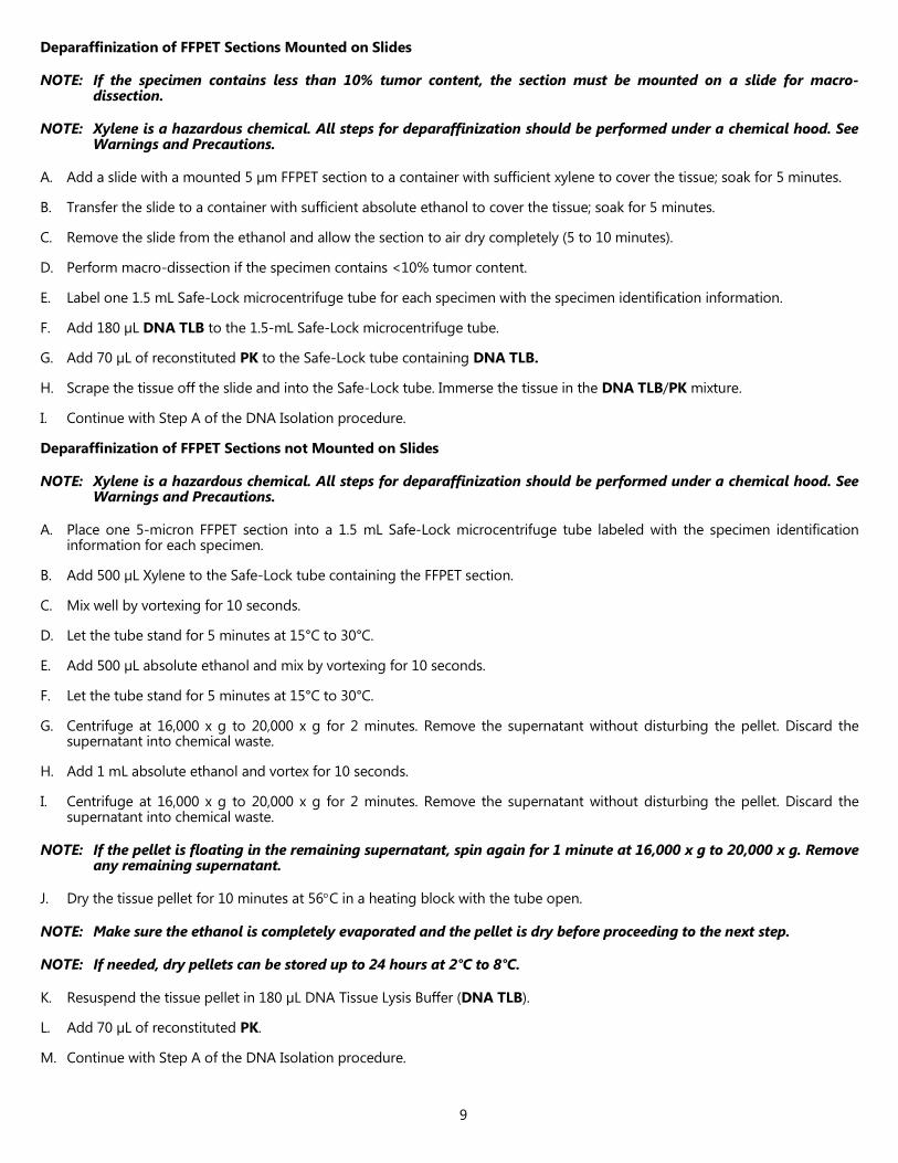
C. The DNA Stock concentration from the specimens must be ≥ 4 ng/µL to perform the cobas® KRAS Mutation Test. Two amplification/detections are run per specimen, using 25 µL of a 2 ng/µL dilution of DNA Stock (total of 50 ng DNA) for each amplification/detection.
NOTE: Each DNA Stock must have a minimum concentration of 4 ng/µL to perform the cobas® KRAS Mutation Test. If the concentration of a DNA Stock is <4 ng/µL, repeat the deparaffinization, DNA Isolation, and DNA Quantitation procedures for that specimen using two 5 µm FFPET sections. For mounted specimens, after deparaffinization, combine the tissue from both sections into one tube, immerse the tissue in DNA TLB + PK, and perform DNA Isolation and Quantitation as described above. For unmounted specimens, combine two sections into one tube and immerse the tissue in DNA TLB + PK, and perform DNA Isolation and Quantitation as described above. If the DNA Stock is still <4 ng/µL, then request another FFPET specimen.
NOTE: Processed specimens (extracted DNA) are stable for up to 24 hours at 15ºC to 30°C or up to 14 days at 2ºC to 8°C or up to 60 days at -15ºC to -25°C or after undergoing 3 freeze thaws when stored at -15ºC to -25°C. Extracted DNA should be amplified within the recommended storage periods or before the expiration date of the cobas® DNA Sample Preparation Kit used to extract the DNA, whichever comes first.
AMPLIFICATION AND DETECTION
NOTE: To avoid contamination of working MMX with DNA specimens, Amplification and Detection should be performed in an area separated from DNA Isolation. The amplification and detection work area should be thoroughly cleaned before working MMX preparation. For proper cleaning, all surfaces including racks and pipettors should be thoroughly wiped with 0.5% sodium hypochlorite solution followed by wiping with a 70% ethanol solution. Commercial liquid household bleach typically contains sodium hypochlorite at a concentration of 5.25%. A 1:10 dilution of household bleach will produce a 0.5% sodium hypochlorite solution.
Instrument Set-Up
Refer to the cobas z 480 analyzer instrument manual for detailed instruction for the cobas z 480 set up.
Test Order Set-up Refer to the cobas® 4800 system Operator’s Manual for cobas® KRAS Mutation Test (cobas® KRAS Operator’s Manual) for detailed instructions on the KRAS workflow steps.
Dilution Calculation of Specimen DNA Stock
Dilution Calculation for DNA Stock Concentrations from 4 ng/µL to 28 ng/µL
NOTE: DNA stocks from specimens should be diluted immediately prior to amplification and detection.
NOTE: Two (2) amplification/detections are run for each specimen requiring a total volume of 50 µL (25 µL each for MMX1 and for MMX2) of a 2 ng/µL dilution of DNA Stock (total of 100 ng DNA).
A. For each specimen, calculate the volume (µL) of DNA stock needed:
µL of DNA stock = (70 µL x 2 ng/µL) ÷ DNA Stock concentration [ng/µL]

12
B. For each specimen, calculate the volume (µL) of DNA Specimen Diluent (DNA SD) needed:
µL of DNA SD = 70 µL – µL of DNA Stock
Example:
DNA stock concentration = 6.5 ng/µL
A. µL of DNA Stock = (70 µL x 2 ng/µL) ÷ 6.5 ng/µL = 21.5 µL
B. µL of DNA SD = (70 µL – 21.5 µL) = 48.5 µL
Dilution Calculation for DNA Stock Concentrations >28 ng/µL
NOTE: DNA Stocks from specimens should be diluted immediately prior to amplification and detection.
NOTE: Two (2) amplification/detections are run for each specimen requiring a total volume of 50 µL (25 µL each for MMX1 and for MMX2) of a 2 ng/µL dilution of DNA stock (total of 100 ng DNA).
A. At DNA Stock concentrations > 28 ng/µL, use the following formula to calculate the amount of DNA Specimen Diluent (DNA SD) required to prepare at least 70 µL of diluted DNA stock. This is to ensure that each specimen uses a minimum of 5 µL of DNA stock.
B. For each specimen, calculate the volume (µL) of DNA SD needed to dilute 5 µL of DNA Stock to 2 ng/µL:
Vol. of DNA SD required in µL = [(5 µL of DNA stock x DNA stock concentration in ng/µL) / 2 ng/µL] – 5 µL
Example:
DNA stock concentration = 31.7 ng/µL
A. Vol. of DNA SD required in µL = [(5 µL x 31.7 ng/µL) / 2 ng/µL] – 5 µL = 74.3 µL
B. Use the calculated volume of DNA SD to dilute 5 µL of DNA stock.
Specimen Dilution
NOTE: Remove the specimen diluent (DNA SD) from -15°C to -25ºC storage and thaw at 15°C to 30°C for at least 1 hour before DNA dilution. Vortex each reagent for 5 seconds and collect liquid at the bottom of the tube before use.
A. Prepare the appropriate number of 1.5 mL Safe-Lock microcentrifuge tubes for DNA Dilutions by labeling them with the proper specimen identification.
B. Using a pipettor with an aerosol-resistant tip, pipette the calculated volumes of DNA SD into the respectively labeled tubes. Pipette 35 µL of DNA SD into a Safe-Lock tube labeled as NEG CT.
C. Vortex each DNA stock and the negative control for 5 to 10 seconds.
D. Using a pipettor with an aerosol-resistant pipette tip (new tip for each pipetting), gently pipette the calculated volume of each DNA stock into the respective tube containing DNA SD. Pipette 35 µL of negative control (extracted eluate) into the NEG CT tube.
E. Cap the tubes and vortex each for 5 to 10 seconds.
F. Change gloves.
Preparation of Working Master Mixes (MMX 1 and MMX 2)
NOTE: KRAS OM1, KRAS OM2, and working MMX are light-sensitive and must be protected from prolonged exposure to light.
NOTE: Due to the viscosity of the KRAS MIX and working MMX, pipette slowly to ensure all mix is completely dispensed from the tip.
NOTE: The KRAS MIX, KRAS OM1 and KRAS OM2 may appear clear to yellow. This does not affect the performance of the reagent.
Prepare two bulk working MMX, one containing KRAS OM1 and the other containing KRAS OM2 in separate 1.5 mL Safe-Lock microcentrifuge tubes.
13
A. Calculate the volume of KRAS MIX required for each working MMX using the following formula:
Volume of KRAS MIX required = (Number of Specimens + 2 Controls + 1 Calibrator +1) x 10 µL
B. Calculate the volume of KRAS OM1 or KRAS OM2 required for each working MMX using the following formula:
Volume of KRAS OM1 or KRAS OM2 required = (Number of Specimens + 2 Controls + 1 Calibrator +1) x 10 µL
C. Calculate the volume of MGAC required for each working MMX using the following formula:
Volume of MGAC required = (Number of Specimens + 2 Controls + 1 Calibrator +1) x 6 µL
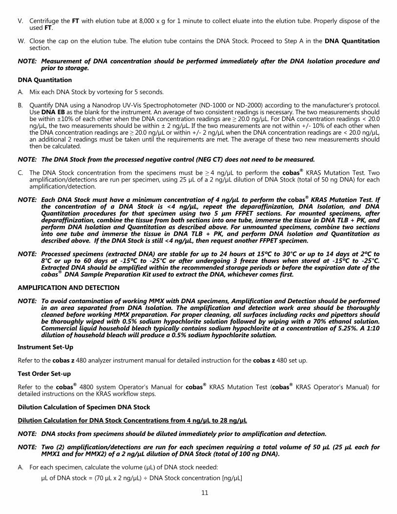
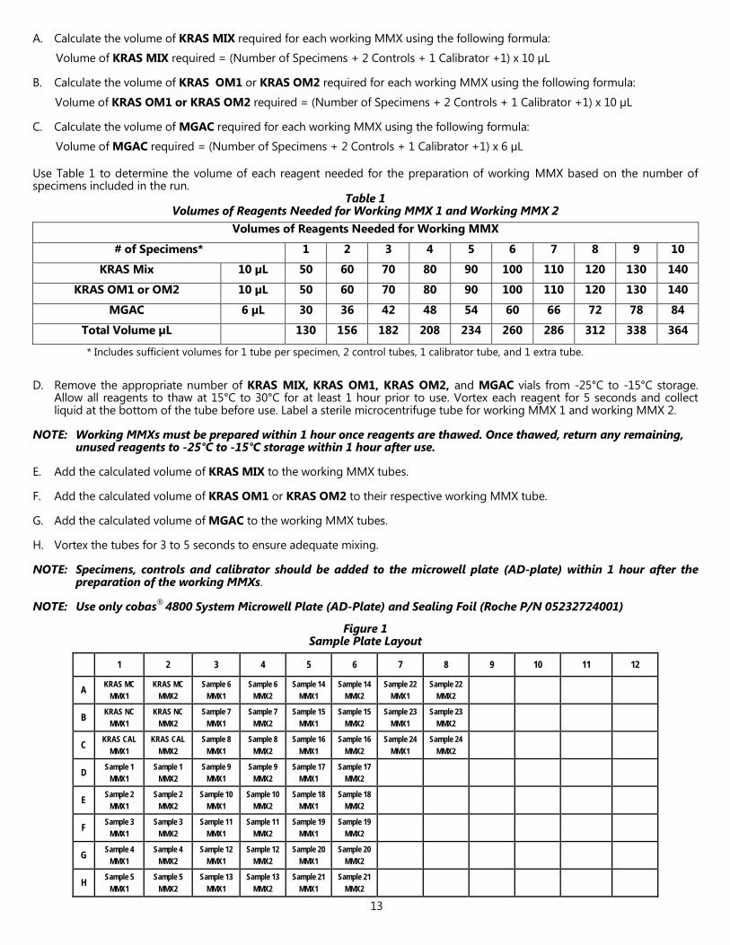
Use Table 1 to determine the volume of each reagent needed for the preparation of working MMX based on the number of specimens included in the run.
Table 1 Volumes of Reagents Needed for Working MMX 1 and Working MMX 2
Volumes of Reagents Needed for Working MMX
# of Specimens* 1 2 3 4 5 6 7 8 9 10
KRAS Mix 10 µL 50 60 70 80 90 100 110 120 130 140
KRAS OM1 or OM2 10 µL 50 60 70 80 90 100 110 120 130 140
MGAC 6 µL 30 36 42 48 54 60 66 72 78 84
Total Volume µL 130 156 182 208 234 260 286 312 338 364
* Includes sufficient volumes for 1 tube per specimen, 2 control tubes, 1 calibrator tube, and 1 extra tube.
D. Remove the appropriate number of KRAS MIX, KRAS OM1, KRAS OM2, and MGAC vials from -25°C to -15°C storage. Allow all reagents to thaw at 15°C to 30°C for at least 1 hour prior to use. Vortex each reagent for 5 seconds and collect liquid at the bottom of the tube before use. Label a sterile microcentrifuge tube for working MMX 1 and working MMX 2.
NOTE: Working MMXs must be prepared within 1 hour once reagents are thawed. Once thawed, return any remaining, unused reagents to -25°C to -15°C storage within 1 hour after use.
E. Add the calculated volume of KRAS MIX to the working MMX tubes.
F. Add the calculated volume of KRAS OM1 or KRAS OM2 to their respective working MMX tube.
G. Add the calculated volume of MGAC to the working MMX tubes.
H. Vortex the tubes for 3 to 5 seconds to ensure adequate mixing.
NOTE: Specimens, controls and calibrator should be added to the microwell plate (AD-plate) within 1 hour after the preparation of the working MMXs.
NOTE: Use only cobas® 4800 System Microwell Plate (AD-Plate) and Sealing Foil (Roche P/N 05232724001)
Figure 1 Sample Plate Layout
1 2 3 4 5 6 7 8 9 10 11 12
A KRAS MC MMX1
KRAS MC MMX2
Sample 6 MMX1
Sample 6 MMX2
Sample 14 MMX1
Sample 14 MMX2
Sample 22 MMX1
Sample 22 MMX2
B KRAS NC MMX1
KRAS NC MMX2
Sample 7 MMX1
Sample 7 MMX2
Sample 15 MMX1
Sample 15 MMX2
Sample 23 MMX1
Sample 23 MMX2
C KRAS CAL MMX1
KRAS CAL MMX2
Sample 8 MMX1
Sample 8 MMX2
Sample 16 MMX1
Sample 16 MMX2
Sample 24 MMX1
Sample 24 MMX2
D Sample 1 MMX1
Sample 1 MMX2
Sample 9 MMX1
Sample 9 MMX2
Sample 17 MMX1
Sample 17 MMX2
E Sample 2 MMX1
Sample 2 MMX2
Sample 10 MMX1
Sample 10 MMX2
Sample 18 MMX1
Sample 18 MMX2
F Sample 3 MMX1
Sample 3 MMX2
Sample 11 MMX1
Sample 11 MMX2
Sample 19 MMX1
Sample 19 MMX2
G Sample 4 MMX1
Sample 4 MMX2
Sample 12 MMX1
Sample 12 MMX2
Sample 20 MMX1
Sample 20 MMX2
H Sample 5 MMX1
Sample 5 MMX2
Sample 13 MMX1
Sample 13 MMX2
Sample 21 MMX1
Sample 21 MMX2

14
PCR Set-up
A. Pipette 25 µL of working MMX into each reaction well of the microwell plate (AD-plate) that is needed for the run. Do not allow the pipettor tip to touch the plate outside the well.
• Add working MMX1 (containing KRAS OM1) to the microwell plate (AD-plate) wells in the odd-numbered columns (1, 3, 5, etc.)
• Add working MMX2 (containing KRAS OM2) to the microwell plate (AD-plate) wells in the even-numbered columns (2, 4, 6, etc.)
B. Pipette 25 µL of KRAS MC into wells A1 and A2 of the microwell plate (AD-plate); mix well using pipette to aspirate and dispense within the well a minimum of two times.
C. Using a new pipettor tip, pipette 25 µL of NEG CT into wells B1 and B2 of the microwell plate (AD-plate); mix well using pipette to aspirate and dispense within the well a minimum of two times.
D. Using a new pipettor tip, pipette 25 µL of KRAS CAL into wells C1 and C2 of the microwell plate (AD-plate); mix well using pipette to aspirate and dispense within the well a minimum of two times.
NOTE: Each run must contain positive control (KRAS MC) in wells A1 and A2, negative control (NEG CT) in wells B1 and B2, and calibrator (KRAS CAL) in wells C1 and C2 or the run will be invalidated.
NOTE: Change gloves as needed to protect against specimen-to-specimen contamination and external PCR reaction tube contamination.
E. Using new pipettor tips for each diluted specimen DNA, add 25 µL of the first specimen DNA to wells D1 and D2 of the microwell plate (AD-plate); mix well using pipette to aspirate and dispense within the well a minimum of two times. Repeat this procedure for the diluted DNA from the second specimen (wells E1 and E2). Follow the template in Figure 1 until all specimens’ DNA Dilutions are loaded onto the microwell plate (AD-plate). Ensure that all liquid is collected at the bottom of the wells.
F. Cover the microwell plate (AD-plate) with sealing foil (supplied with the plates). Use the sealing foil applicator to seal the foil firmly to the microwell plate (AD-plate).
G. Confirm that all liquid is collected at the bottom of each well before starting PCR.
NOTE: Amplification and Detection should be started within 1 hour after the addition of the first specimen DNA dilution to the working MMX.
Starting PCR
Refer to the cobas® KRAS Operator’s Manual for detailed instructions on the KRAS workflow steps.
INTERPRETATION OF RESULTS
NOTE: All run and specimen validation is performed by the cobas®4800 software.
NOTE: A valid test run may include both valid and invalid sample results.
For a valid run, specimen results are interpreted as shown in Table 2.
Table 2 Result Interpretation of cobas® KRAS Mutation Test
Test Result Mutation Result Interpretation
Mutation Detected Codon 12/13 Mutation detected in KRAS codon 12/13.
No Mutation Detected N/A Mutation not detected in KRAS codon 12/13.
Invalid N/A Specimen result is invalid. Repeat the testing of specimens with invalid results following the instructions outlined in the “Retesting of Specimens with Invalid Results” section below.
Failed N/A Failed run due to hardware or software failure. Contact your local Roche office for technical assistance
* A “No Mutation Detected” result does not preclude the presence of a mutation in the KRAS 12/13 codon sites because results depend on percent mutant sequences, adequate specimen integrity, absence of inhibitors, and sufficient DNA to be detected.

15
Retesting of Specimens with Invalid Results
A. Repeat dilution of the invalid specimen DNA stock starting from “Dilution Calculation of Specimen DNA Stock” and “Specimen Dilution” procedures in the “AMPLIFICATION and DETECTION” section.
B. After performing the DNA stock dilution to 2 ng/µL described in “Specimen Dilution” continue with “Preparation of Working Master Mixes (MMX1 and MMX2)” and the remainder of the amplification and detection procedure.
NOTE: If the specimen remains invalid after retesting or there was not enough DNA stock to prepare another dilution in Retesting of Specimens with Invalid Results, step A, repeat the entire test procedure for that specimen, starting with Deparaffinization and DNA Isolation using a new 5-micron FFPET section.
QUALITY CONTROL
One set of cobas® KRAS Test Mutant Control (KRAS MC), negative control (NEG CT) and KRAS Calibrator (KRAS CAL) for working MMX1 and working MMX2 are included in each run. A run is valid if the KRAS Mutant Control (KRAS MC) wells (A1 and A2), the negative control (NEG CT) wells (B1 and B2), and the KRAS Calibrator (KRAS CAL) wells (C1 and C2) are valid. If the KRAS Mutant Control (KRAS MC), negative control (NEG CT) or KRAS Calibrator (KRAS CAL) for working MMX1 or working MMX2 are invalid, the entire run is invalid and must be repeated. Prepare a fresh dilution of the previously isolated specimen DNA Stock to set up a new microwell plate (AD-plate) with controls for amplification and detection.
Positive Control
The KRAS Mutant Control (KRAS MC) result must be ‘Valid’ for both working MMX1 and working MMX2. If the KRAS MC results are consistently invalid, contact your local Roche office for technical assistance.
Negative Control
The negative control (NEG CT) result must be ‘Valid’ for both working MMX1 and working MMX2. If the NEG CT results are consistently invalid, contact your local Roche office for technical assistance.
Calibrator
The KRAS Calibrator (KRAS CAL) result must be ‘Valid’ for both working MMX1 and working MMX2. If the KRAS CAL results are consistently invalid, contact your local Roche office for technical assistance.
PROCEDURAL PRECAUTIONS
As with any test procedure, good laboratory technique is essential to the proper performance of this assay. Due to the high analytical sensitivity of this test, care should be taken to keep reagents and amplification mixtures free of contamination.
PROCEDURAL LIMITATIONS
1. The cobas® KRAS Mutation Test has only been validated for use with Colorectal Cancer FFPET Specimens.
2. The cobas® KRAS Mutation Test has only been validated using the cobas® DNA Sample Preparation Kit (Roche P/N: 05985536190).
3. Detection of a mutation is dependent on the number of copies present in the specimen and may be affected by specimen integrity, amount of isolated DNA, and the presence of interfering substances.
4. Reliable results are dependent on adequate specimen fixation, transport, storage and processing. Follow the procedures in this Package Insert and in the cobas® KRAS Operator’s Manual.
5. The addition of AmpErase enzyme into the cobas® KRAS Mutation Test Master Mix enables selective amplification of target DNA; however, good laboratory practices and careful adherence to the procedures specified in this Package Insert are necessary to avoid contamination of reagents.
6. Use of this product must be limited to personnel trained in the techniques of PCR and the use of the cobas® 4800 system.
7. Only the cobas z 480 analyzer has been validated for use with this product. No other thermal cycler with real-time optical detection can be used with this product.
9. The effects of other potential variables such as specimen fixation variables have not been evaluated.
10. Though rare, mutations within the regions of the genomic DNA of the KRAS gene covered by the cobas® KRAS Mutation Test’s primers and/or probes may result in failure to detect the presence of a mutation. Samples with results reported as "No Mutation Detected" may harbor KRAS mutations not detected by the assay.
11. The presence of PCR inhibitors may cause false negative or invalid results.

16
12. Though rare (< 0.2%22), some complex and multiple mutations of codon 12/13 may result in failure to detect the presence of a mutation (results of “No Mutation Detected”) whereas mutations flanking codon 12/13 on exon 2 may cross-react with the cobas® KRAS Mutation Test (results of “Mutation Detected”).
13 The cobas® KRAS Mutation Test was verified for use with 50 ng of DNA per reaction well. DNA input amounts lower than 50 ng per reaction well are not recommended.
14. The cobas® KRAS Mutation Test is a qualitative test. The test is not for quantitative measurements of percent mutation.
15. The procedure described above must be followed to detect ≥ 5% mutant sequences in a background of wild-type DNA for the KRAS mutations 22 in Table 3.
Table 3 Mutations Detected by the cobas® KRAS Mutation Test
Mutation AA Change COSMIC ID
c.34G>T 12C 516
c.34G>A 12S 517
c.34G>C 12R 518
c.35G>T 12V 520
c.35G>A 12D 521
c.35G>C 12A 522
c.38G>A 13D 532
NON-CLINICAL PERFORMANCE EVALUATION
Analytical Sensitivity – Limit of Blank
To assess performance of the cobas® KRAS Mutation Test in the absence of template and to ensure that a blank sample does not generate an analytical signal that might indicate a low concentration of mutation, no DNA template samples and DNA extracted from CRC FFPET KRAS wild type specimens were evaluated. Only “No Mutation Detected” results were observed in the no DNA template samples and in the presence of KRAS wild type DNA.
Analytical Sensitivity Using FFPET Specimen Blends
Multiple CRC FFPET specimen DNA extracts for specific codon 12 mutants (G12A, G12C, G12D, G12S, G12V, G12R) and a codon 13 mutant (G13D) were blended with KRAS wild-type FFPET specimen extracts to achieve mutant sequences at approximately 10%, 5%, 2.5%, and 1.25% mutation level. The final mutation levels for all specimens were verified by a massively parallel sequencing (MPS) method that was validated for detecting the specific codon 12 mutants (G12A, G12C, G12D, G12S, G12V, G12R) and codon 13 mutant (G13D). Each specimen blend was diluted to 2ng/µL at the time of testing (50.0ng/25 µL). Serial dilutions of each specimen blend were prepared and eight (8) replicates of each panel member were run using each of 3 cobas® KRAS Mutation Test kit lots (n=24 per panel member). The sensitivity of each sample was determined by the lowest amount of DNA that gave a KRAS “Mutation Detected” rate in at least 95% of the replicates, shown in Table 4. This study demonstrates that the cobas® KRAS Mutation Test can detect KRAS codon 12 and 13 mutations at approximately 5% mutant sequences using the standard input of 2ng/µL.
Table 4 Sensitivity of the cobas® KRAS Mutation Test using CRC Specimen FFPET Blends
KRAS Mutation
Codon 12 Codon 13
G12A G12C G12D G12R G12S G12V G13D
Targeted Level 2.5% 2.5% 1.25% 5.0% 2.5% 2.5% 1.25%
LOD 2.93% 2.61% 1.64% 5.78% 2.55% 2.48% 1.67%

17
Correlation to Reference Method
A study was conducted to compare the results of the cobas® KRAS Mutation Test to Sanger sequencing (reference method) using 94 procured CRC FFPET specimens. A 5-micron section was processed to isolate DNA from each tumor specimen using one cobas® DNA Sample Preparation kit lot and the extracted DNA was tested with each of two reagent lots of the cobas®
KRAS Mutation Test. Tumor stage information, results of bi-directional Sanger sequencing and results of cobas® KRAS Mutation Test for the 94 specimens are summarized in the tables below.
Table 5 Tumor Stage vs. Sanger Sequencing
Tumor Stage 2X Bi-directional Sanger Sequencing Results*
Total % of Total codon 12 codon 13 wild-type
Stage I 3 1 4 8 8.5%
Stage II 20 2 8 30 31.9%
Stage III 23 5 6 34 36.2%
Stage IV 10 7 5 22 23.4%
Total 56 15 23 94 100.0%
* 2x Bi-directional Sanger sequencing refer to two reads of bi-directional Sanger sequencing, i.e., a total of 4 reads including 2 forward and 2 reverse reads.
Table 6 Comparison of the cobas® KRAS Mutation Test with Bi-Directional Sanger Sequencing for
Detection of KRAS Mutations in Codon 12/13
cobas® KRAS Mutation Test 2x Bi-directional Sanger Sequencing
Mutation Detected
No Mutation Detected
Total
Mutation Detected 37 3 40
No Mutation Detected 1 53 54
Total 38 56 94
PPA (95% CI) 97.4% (86.5%, 99.5%)
NPA (95% CI) 94.6% (85.4%, 98.2%)
OPA (95% CI) 95.7% (89.6%, 98.3%) PPA: positive percent agreement; NPA: negative percent agreement
OPA: overall percent agreement; CI: confidence intervals
Another study tested 188 colorectal cancer FFPET specimens with both the cobas® KRAS Mutation Test and Sanger sequencing using two lots of the cobas® KRAS Mutation Test kits stored at 2-8°C. Comparable results were obtained with this procured sample set, with a PPA of 97.5 % (CI: 91.4-99.3%), a NPA of 94.4% (CI: 88.3-97.4%), and an OPA of 95.7% (CI: 91.8-97.8%).
Cross-Reactivity to other KRAS Mutations on Codon 13 The cobas® KRAS Mutation Test has been shown to cross-react with the following mutations shown in Table 7. Plasmid constructs (n=4) and CRC FFPET (n=1) containing the rare mutations for codon 13 were blended with wild type genomic DNA to create approximately 5% mutant samples. Results demonstrated that the cobas® KRAS Mutation Test cross-reacts to the following mutations at a 100% hit rate. Analytical performance of the cobas® KRAS Mutation Test in detecting these mutations has not been evaluated.

18
Table 7 Mutations Determined to Cross-React with the cobas® KRAS Mutation Test
Mutation AA Change COSMIC ID
c.37G>T 13C 527
c.37G>A 13S 528
c.37G>C 13R 529
c.38G>C 13A 533
c.38G>T 13V 534
Bold = Tested with plasmids
Specificity – KRAS silent mutation, KRAS Homologs and Microorganisms
Specificity of the cobas® KRAS Mutation Test was evaluated by the following studies;
• Testing KRAS silent mutation plasmids,
• Testing KRAS homolog plasmids,
• Testing colon-related microorganisms.
Cross-reactivity was also evaluated by determining whether or not the presence of KRAS silent mutation plasmids or KRAS homolog plasmids or colon-related microorganisms interfered with detection of KRAS codon 12 and 13 mutations.
KRAS Silent Mutation Plasmids
Plasmid samples in a background of wild-type cell line DNA were prepared and tested for the following three KRAS codon 12 silent mutations: GGA, GGC, and GGG; three KRAS codon 13 silent mutations: GGA, GGT, and GGG. No cross-reactivity was observed with plasmids for KRAS codon 12 silent mutations or codon 13 silent mutations.
Plasmid blends of KRAS codon 12 or codon 13 at 5% mutation in a background of wild-type cell line DNA were prepared and tested in the presence of their respective silent mutation plasmids and no interference from the silent mutation plasmids was detected.
KRAS Homolog Plasmids
Samples containing each of the three KRAS Homolog plasmids (KRAS codon 12/13 pseudogene, NRAS exon 2, and HRAS exon 2) in a background of wild-type cell line DNA were prepared and tested in triplicate using the cobas® KRAS Mutation Test. No cross reactivity was observed with any of the plasmid samples.
Plasmid blends of KRAS codon 12 and codon 13 at 5% mutation in a background of wild-type cell line DNA were prepared and tested in the presence of their respective homolog plasmids and no interference from the homolog plasmids was detected.
Colon-related Microorganisms
The following colon-related microorganisms were found not to cross react in the cobas® KRAS Mutation Test when added to six KRAS codon 12, one codon 13, and one wild-type specimen at 1 x 10⁶ colony forming units during the tissue lysis step:
1. Bacteroides caccae
2. Prevotella intermedia
3. Escherichia coli (E. coli)
The tested microorganisms also did not interfere with the detection of KRAS codon 12 or codon 13 mutations when 1 x 10⁶ colony forming units were added during the tissue lysis step of a specimen containing a KRAS mutation at levels ranging from 14-40%.
Interference Triglycerides (≤ 37mM, CLSI recommended high concentration23) and hemoglobin (≤ 2 mg/mL, CLSI recommended high concentration23) have been shown not to interfere with the cobas® KRAS Mutation Test when the potential interfering substance was added to the lysis step during the specimen preparation procedure.

19
Necrotic Tissue
CRC FFPET specimens with necrotic tissue content up to 50% for KRAS mutant and 70% in KRAS wild-type specimens have been shown not interfere with the call results for the cobas® KRAS Mutation Test.
Repeatability
Repeatability of the cobas® KRAS Mutation Test was assessed using eight colorectal cancer FFPET specimens, including 2 codon 12, 2 codon 13 KRAS mutant specimens, and 4 KRAS wild type specimens. The specimens were tested in duplicate by two operators, using two different reagent lots and four cobas z 480 analyzers over 4 days (n=32/specimen) at one site. The cobas® KRAS Mutation Test had a correct call accuracy of 100% (256/256) for all days, specimens, replicates, operators and reagent lots combined.
Specimen Handling Reproducibility
The reproducibility of the DNA Sample Preparation Kit was examined using sections taken from three CRC FFPET specimen blocks, one containing a codon 12 mutation (G12D, GGT>GAT), one containing a codon 13 mutation (G13D, GGC>GAC), and one that is wild-type for KRAS mutation. From each of the three specimens, thirty-six (36) 5-μm sections were obtained. Each of three external sites tested twelve (12) sections for each specimen over six (6) non-consecutive days. On each test day, one operator from each site isolated, quantified and tested the DNA from two FFPET curl sections for each specimen using one lot of cobas®
DNA Sample Preparation Kit and one lot of the cobas® KRAS Mutation Test kit. All testing was performed on the cobas z 480 analyzer with the KRAS Analysis Package. One lot of the cobas® KRAS Mutation Test kit reagents was used in this study, in combination with three lots of the DNA Sample Preparation kit at each site. All runs performed at the 3 sites were valid. All mutant and wild-type specimen results were valid and yielded the expected call result (correct call =100%, 36/36 for each specimen), supporting the reproducibility for the cobas® KRAS Mutation Test at the pre-analytical step of DNA isolation.
CLINICAL PERFORMANCE EVALUATION
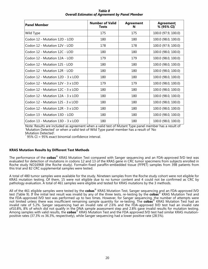
Clinical Reproducibility
An external study was performed to assess the reproducibility of the cobas® KRAS Mutation Test with a 15 member panel of DNA samples extracted from CRC FFPET sections of KRAS wild type (WT) and mutant type (MT) tumor specimens, tested in duplicate, across 3 testing sites with 2 operators per site, 3 reagent lots and 5 non-consecutive testing days. The panel included six codon 12 mutations and one codon 13 mutation along with a WT DNA sample. Each mutation was represented in duplicate at the limit of detection (LOD) and 3x LOD. Two replicates of the panel at desired concentrations were used in each run. Panels were provided to each of the 3 testing sites in a blinded fashion. Operators performed amplification and detection with the cobas z 480 instrument using the cobas® KRAS Mutation Test kits provided by RMS. One panel tested by a given operator at a given site was considered one run. Each run included 2 replicates of each of the 15 specimens in the panel. Each operator completed 5 valid runs with each of 3 reagent lots for a total of 15 days of testing, performed runs on non-consecutive days, and performed runs separately from the other operator at the site. Testing was completed with one lot before testing began with another lot.
Overall 97.8% of runs (90/92) were valid. A total of 3,060 tests were performed in 90 valid runs. From the valid runs, 99.6% of test results (3,048/3,060) were valid. There were ‘No Mutation Detected’ results in 175 valid tests for the Wild Type panel member with 100% agreement. For the Codon 12 and 13 panel members at both 1 x LOD and 3 x LOD, agreement was 100%. Across all variance components (i.e., lot, site/instrument, operator, day, and within run), the overall coefficient of variation ranged from 5.9% to 13.3% across all panel members.
20
Table 8 Overall Estimates of Agreement by Panel Member
Panel Member Number of Valid Tests
Agreement N
Agreement % (95% CI)*
Wild Type 175 175 100.0 (97.9, 100.0)
Codon 12 - Mutation 12D - LOD 180 180 100.0 (98.0, 100.0)
Codon 12 - Mutation 12V - LOD 178 178 100.0 (97.9, 100.0)
Codon 12 - Mutation 12C - LOD 180 180 100.0 (98.0, 100.0)
Codon 12 - Mutation 12A - LOD 179 179 100.0 (98.0, 100.0)
Codon 12 - Mutation 12S - LOD 180 180 100.0 (98.0, 100.0)
Codon 12 - Mutation 12R - LOD 180 180 100.0 (98.0, 100.0)
Codon 12 - Mutation 12D - 3 x LOD 180 180 100.0 (98.0, 100.0)
Codon 12 - Mutation 12V - 3 x LOD 179 179 100.0 (98.0, 100.0)
Codon 12 - Mutation 12C - 3 x LOD 180 180 100.0 (98.0, 100.0)
Codon 12 - Mutation 12A - 3 x LOD 180 180 100.0 (98.0, 100.0)
Codon 12 - Mutation 12S - 3 x LOD 180 180 100.0 (98.0, 100.0)
Codon 12 - Mutation 12R - 3 x LOD 180 180 100.0 (98.0, 100.0)
Codon 13 - Mutation 13D - LOD 180 180 100.0 (98.0, 100.0)
Codon 13 - Mutation 13D - 3 x LOD 180 180 100.0 (98.0, 100.0) Note: Results are included as agreement when a valid test of Mutant Type panel member has a result of 'Mutation Detected' or when a valid test of Wild Type panel member has a result of 'No Mutation Detected'. * 95% CI = 95% exact binomial confidence interval.
KRAS Mutation Results by Different Test Methods
The performance of the cobas® KRAS Mutation Test compared with Sanger sequencing and an FDA-approved IVD test was evaluated for detection of mutations in codons 12 and 13 of the KRAS gene in CRC tumor specimens from subjects enrolled in Roche study NO16968 (the Roche study). Formalin-fixed paraffin-embedded tissue (FFPET) samples from 398 patients from this trial and 82 CRC supplemental samples were tested.
A total of 480 tumor samples were available for the study. Nineteen samples from the Roche study cohort were not eligible for KRAS mutation testing. Of them, 15 were not eligible due to no tumor content and 4 could not be confirmed as CRC by pathology evaluation. A total of 461 samples were eligible and tested for KRAS mutations by the 3 methods.
All of the 461 eligible samples were tested by the cobas® KRAS Mutation Test, Sanger sequencing and an FDA-approved IVD test (Table 9). If the initial test result was invalid by any of the three tests, re-testing by the cobas® KRAS Mutation Test and the FDA-approved IVD test was performed up to two times. However, for Sanger sequencing, the number of attempts were not limited unless there was insufficient remaining sample quantity for re-testing. The cobas® KRAS Mutation Test had an invalid rate of 5.2%. Sanger sequencing had an invalid rate of 2.6% and the FDA-approved IVD test had an invalid rate of10.8%, 8% of which did not qualify in the DNA sample assessment step and 2.8% gave invalid results for mutation testing. Among samples with valid results, the cobas® KRAS Mutation Test and the FDA approved IVD test had similar KRAS mutation-positive rates (37.3% vs 36.3%, respectively), while Sanger sequencing had a lower positive rate (28.5%).

21
Table 9 KRAS Mutation Result by Different Testing Methods
cobas® KRAS Mutation Test
FDA-approved IVD test
Sanger Sequencing
Number of Samples Tested 461 461 461
Invalid result 24 (5.2 %) 50 (10.8 %) 12 (2.6 %)
Valid result 437 (94.8 %) 411 (89.2 %) 449 (97.4 %)
No Mutation Detected 274 (62.7 %) 262 (63.7 %) 321 (71.5 %)
Mutation Detected 163 (37.3 %) 149 (36.3 %) 128 (28.5 %) Note: 37 samples with tumor content ≥20% failed sample assessment for the FDA-approved IVD test. These samples did not go through the next step of mutation detection and were counted as invalid results.
Agreement between the Three Test Methods: 3-way Method Comparison
The agreement of the cobas® KRAS Mutation Test using Sanger sequencing or the FDA-approved IVD test as the reference method for detection of mutations in codon 12/13 is presented in Table 10. The PPA between the cobas® KRAS Mutation Test and Sanger sequencing was 96.9% (95% CI: 92.2% to 98.8%), and the NPA was 88.7% (95% CI: 84.7% to 91.8 %). The PPA between the cobas® KRAS Mutation Test and the FDA-approved IVD test was 93.3% (95% CI: 88.1% to 96.3%), and the NPA was 96.5% (95% CI: 93.5% to 98.1%).
Table 10 Comparison of the cobas® KRAS Mutation Test with Reference Methods for
Detection of KRAS Mutation in Codon 12/13
cobas® KRAS Mutation Test
Reference Method Sanger Sequencing FDA-approved IVD test
Mutation Detected
No Mutation Detected
Invalid Total Mutation
Detected No
Mutation Detected
Invalid Total
Mutation Detected 124 34 5 163 139 9 15 163
No Mutation Detected 4 268 2 274 10 248 16 274
Invalid 0 19 5 24 0 5 19 24
Total 128 321 12 461 149 262 50 461
PPA (95% CI) 96.9% (92.2%, 98.8%) 93.3% (88.1%, 96.3%)
NPA (95% CI) 88.7% (84.7%, 91.8%) 96.5% (93.5%, 98.1%)
OPA (95% CI) 91.2% (88.1%, 93.5%) 95.3% (92.8%, 97.0%) PPA: positive percent agreement; NPA: negative percent agreement
OPA: overall percent agreement; CI: confidence intervals
Predictive Values of the cobas® KRAS Mutation Test
The predictive values of the cobas® KRAS Mutation Test were calculated by combining the PPA and NPA of cobas® KRAS Mutation Test relative to a comparator method together with the prevalence of a KRAS “Mutation Detected” result by the comparator method in published clinical studies for cetuximab or panitumumab. 24-27 The positive and negative predictive values (PPV and NPV) of cobas® KRAS Mutation Test refer to the predictive values of cobas “Mutation Detected” and “No Mutation Detected” results for the comparator method, respectively. 28 The clinical performance was summarized by the quantity of PPV+NPV–1; this quantity has been called the attenuation factor. 28

22
Table11 Attenuation Factors for cobas® KRAS Mutation Test
Data Source Ref Comparator
Method PPV
(95% CI) NPV
(95% CI) Attenuation Factor
(95% CI)
Cetuximab 24 Sanger Sequencing 0.858
(0.811, 0.902) 0.975
(0.946, 0.994) 83.3%
(77.7, 88.3)
Cetuximab 29 FDA-approved IVD test
0.957 (0.927, 0.981)
0.945 (0.909, 0.978)
90.2% (85.6, 94.4)
Panitumumab 27, 30 FDA-approved IVD test
0.949 (0.914, 0.977)
0.956 (0.927, 0.981)
90.4.% (86.1, 94.4)
23
REFERENCES 1. Shankaran V, Obel J, Benson III AB. Predicting response to EGFR inhibitors in metastatic colorectal cancer: current practice
and future directions. The Oncologist 2010 15:157-67.
2. Samowitz WS, Curtin K, Schaffer D, et al. Relationship of Ki-ras mutations in colon cancers to tumor location, stage, and survival: a population-based study. Cancer Epidemiol Biomarkers Prev 2000 Nov 9(11):1193-7.
3. Andreyev HJ, Norman AR, Cunningham D, et al. Kirsten ras mutations in patients with colorectal cancer: the 'RASCAL II' study. Br J Cancer 2001 Sep 1;85(5):692-6.
4. De Roock W, Claes B, Bernasconi D. et al. Effects of KRAS, BRAF, NRAS, and PIK3CA mutations on the efficacy of cetuximab plus chemotherapy in chemotherapy-refractory metastatic colorectal cancer: a retrospective consortium analysis. Lancet Oncology, 2010 Aug 11(8):753-62.
5. Siena S, Sartore-Bianchi A, Di Nicolantonio F, et al. Biomarkers predicting clinical outcome of epidermal growth factor receptor-targeted therapy in metastatic colorectal cancer. J Natl Cancer Inst 2009 Oct 7;101(19):1308-24.
6. Bokemeyer C, Bondarenko I, Makhson A, et al. Fluorouracil, leucovorin, and oxaliplatin with and without cetuximab in the first-line treatment of metastatic colorectal cancer. J Clin Oncol 2009 Feb 10;27(5):663-71.
7. Tol J, Koopman M, Cats A, et al. Chemotherapy, bevacizumab, and cetuximab in metastatic colorectal cancer. N Engl J Med 2009 Feb 5;360(6):563-72.
8. Lièvre A, Bachet JB, Le Corre D, et al. KRAS mutation status is predictive of response to cetuximab therapy in colorectal cancer. Cancer Res 2006 Apr 15;66(8):3992-5.
9. Benvenuti S, Sartore-Bianchi A, Di Nicolantonio F, et al. Oncogenic activation of the RAS/RAF signaling pathway impairs the response of metastatic colorectal cancers to anti-epidermal growth factor receptor antibody therapies. Cancer Res 2007 Mar 15;67(6):2643-8.
10. Amado RG, Wolf M, Peeters M, et al. Wild-type KRAS is required for panitumumab efficacy in patients with metastatic colorectal cancer. J Clin Oncol 2008 Apr 1;26(10):1626-34.
11. Karapetis CS, Khambata-Ford S, Jonker DJ, et al. K-ras mutations and benefit from cetuximab in advanced colorectal cancer. N Engl J Med 2008 Oct 23;359(17):1757-65.
12. Douillard J-Y, Siena S, Cassidy J et al. randomized, Phase III trial of panitumumab with infusional fluorouracil, leucovin, and Oxaliplatin (FOXFOX4) versus FOLFOX4 alone as first-line treatment in patients with previously untreated metastatic colorectal cancer: The PRIME Study. J Clin Oncol 2010 28:4697-4705.
13. Allegra CJ, Jessup JM, Somerfield MR, et al. American Society of Clinical Oncology provisional clinical opinion: testing for KRAS gene mutations in patients with metastatic colorectal carcinoma to predict response to anti-epidermal growth factor receptor monoclonal antibody therapy. J Clin Oncol 2009 Apr 20;27(12):2091-6.
14. National Comprehensive Cancer Network. NCCN Clinical Practice Guidelines in Oncology. Colon cancer, 2010, v.2.
15. Van Cutsem E, Oliveira J; ESMO Guidelines Working Group. Advanced colorectal cancer: ESMO clinical recommendations for diagnosis, treatment and follow-up. Ann Oncol 2009 May;20 Suppl 4:61-3.
16. Food and Drug Administration. Class labeling changes to anti-EGFR monoclonal antibodies, cetuximab (Erbitux) and panitumumab (Vectibix): KRAS mutations. http://www.fda.gov/AboutFDA/CentersOffices/ CDER/ucml172905.htm.
17. European Medicines Agency: Committee for Medicinal Products for Human Use post-authorisation summary of positive opinion for Erbitux. http://www.emea.europa.eu/pdfs/human Erbitux_28040208en.pdf.
18 Longo, M.C., Berninger, M.S. and Hartley, J.L. 1990. Use of uracil DNA glycosylase to control carry-over contamination in polymerase chain reactions. Gene. 93:125-128.
19. Chosewood, L.C. and Wilson, D.E. Biosafety and Microbiological and Biomedical Laboratories. HHS Publication Fifth # edition.(CDC) 21-1112. 2009.
20. Clinical and Laboratory Standards Institute (CLSI). Protection of Laboratory Workers from Occupationally Acquired Infections. Approved Guideline-Third Edition. CLSI Document M29-A3 Villanova, PA:CLSI, 2005.
21. International Air Transport Association. Dangerous Goods Regulations, 52nd Edition. 2011.
22. Catalogue of Somatic Mutations in Cancer (COSMIC), 2011, v.51, http://www.sanger.ac.uk/genetics/CGP/cosmic/.
23. Clinical and Laboratory Standards Institute (CLSI) EP7-A2, Interference Testing in Clinical Chemistry; Approved Guidelines – Second Edition, Appendix D 2005.
24. Karapetis CS, et al. Kras mutations and benefit from cetuximab in advanced colorectal cancer. N Engl J Med. 2008 Oct 23;359(17):1757-65.
25. Van Cutsem E, et al. Open label Phase III trial of panitumumab plus best supportive care compared with best supportive care alone in patients with chemotherapy-refractory metastatic colorectal cancer. J Clin Oncol 25: 1658 -1664, 2007.
26. Amado RG, et al: Wild type KRAS is required for Panitumumab efficacy in patients with metastatic colorectal cancer. J Clin Oncol 26: 1626-1634, 2008.

24
27. Douillard J, et al. Randomized, phase II trial of panitumumab with infusional fluorouracil, leucovorin, and oxaliplatin (FOLFOX4) versus FOLFOX4 alone as first-line treatment in patients with previously untreated metastatic colorectal cancer: The PRIME Study. J Clin Oncol 28: 4697-4705, 2010.
28. Pennello GA. Analytical and clinical evaluation of biomarkers assays: when are biomarkers ready for prime time? Clin Trials 10(5): 666-76, 2013.
29. Summary Of Safety And Effectiveness Data for PMA P110030 http://www.accessdata.fda.gov/cdrh_docs/pdf11/P110030b.pdf
30. Summary Of Safety And Effectiveness Data for PMA P110027 http://www.accessdata.fda.gov/cdrh_docs/pdf11/P110027b.pdf

25
Manufactured in the United States Roche Diagnostics GmbH Sandhofer Straße 116 68305 Mannheim, Germany
Distributed by
Roche Diagnostics 9115 Hague Road Indianapolis, IN 46250-0457 USA (For Technical Assistance call the Roche Response Center toll-free: 1-800 526-1247)
COBAS, COBAS Z, TAQMELT, and AMPERASE are trademarks of Roche.
Carryover prevention technology in the AmpErase enzyme is covered by U.S. Patent 5,035,996 and foreign counterparts owned by Invitrogen Corporation and licensed to Roche Molecular Systems, Inc.
EPPENDORF is a trademark of Eppendorf AG.
PIPET-AID is a trademark of Drummond Scientific.
NANODROP is a trademark of Thermo Scientific.
© 2015 Roche Molecular Systems, Inc.

26
The following symbols are used in labeling for Roche PCR diagnostic products.
Ancillary Software In Vitro Diagnostic Medical Device
Authorized Representative in the European Community
For IVD Performance Evaluation Only
Barcode Data Sheet Lower Limit of Assigned Range
Batch code Manufacturer
Biological Risks Store in the dark
Catalogue Number Temperature Limitation
Consult instructions for use Test Definition File
Sufficient For Upper Limit of Assigned Range
Contents of kit Use By
Distributed by This product fulfills the requirements of the European Directive 98/79 EC for in vitro diagnostic medical devices.
US Customer Technical Support 1-800-526-1247