Clinical Program for Cerebrovascular Disorders Mount Sinai Medical Center
Upload
everett-stokesCategory
view
218download
0
Clinical Program for Cerebrovascular Disorders
Mount Sinai Medical Center
Cerebellar Infarction
Clinical Case PresentationClara Raquel Epstein, MD Fellow

Cerebellar Infarction Clinical Case Presentation

Cerebellar Infarction Clinical Case Presentation
• A 69 year old right handed female presented to the Mount Sinai Medical Center Emergency Department after being found on the floor by her brother. One week prior to admission, the patient complained of back pain after moving furniture at home and was placed on Flexaril. On physical exam the vitals were T=39, HR 128, BP 122/98, RR 24. The patient was lethargic, but arrousable and followed simple commands. No other neurologic deficits were appreciated. However coordination and gait testing was deferred secondary to altered mental status.

Cerebellar Infarction Hospital Course
The patient was transferred to the MICU and an initial evaluation was pursued for possible meningitis vs. sepsis. A head CT without contrast was obtained.
On hospital day #2, the patient was re-evaluated and pertinent findings included that the patient was unresponsive with minimally reactive pupils.

Cerebellar Infarction Hospital Course
Further studies included repeat head CTs, as well as a TEE which revealed a large mobile plaque in the ascending aorta.
The patient subsequently underwent an open thoracotomy with clot removal.

Diagnostic Studies

CT Head 12/29/99

CT Head 12/29/99

Cerebellar InfarctionDiscussion

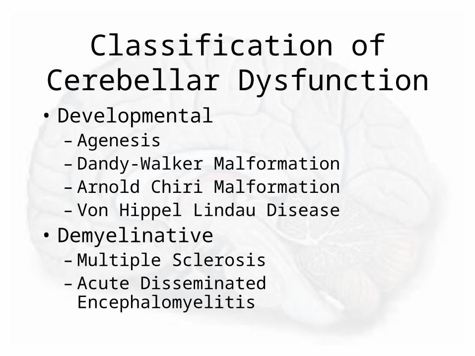
Classification of Cerebellar Dysfunction
• Developmental– Agenesis– Dandy-Walker Malformation– Arnold Chiri Malformation– Von Hippel Lindau Disease
• Demyelinative– Multiple Sclerosis– Acute Disseminated Encephalomyelitis

Classification of Cerebellar Dysfunction
• Degenerative– Cerebellar Degeneration– Multi-system Atrophy– Olivopontocerebellar Atrophy
• Neoplastic– Astrocytoma, Medulloblastoma,
Hemangioblastoma, metastasis

Classification of Cerebellar Dysfunction
• Paraneoplastic– Subacute Cerebellar Degeneration
• Infectious– Abscess Formation– Acute Cerebellitis (viral)– Creutzfeldt-Jacob Disease

Classification of Cerebellar Dysfunction
• Metabolic– Myxoedema– Hypoxia, Hypoglycemia– Alcohol (Vit B1Deficiency)
• Vascular– Cerebellar Hemorrhage– Cerebellar Infarction
• Drugs/toxins– Alcohol– Phenytoin

Structural Lesions of the Cerebellum
• Infarcts, Hemorrhages, or tumors – may produce mass effect with enlargement
• Due to CSF outflow obstruction it can cause:– Hydrocephalus– Increased ICP with papilledema

Phylogenetic Subdivisions of the Cerebellum
• Paleocerebellum (Anterior Lobe) – Receives afferent fibers form the spinal cord
(spinocerebellar pathways)– Function: maintenance of gait
• Neocerebellum (Posterior Lobe)– Receives afferent fibers and projects efferent fibers
from and to the motor cortex/vestibular nuclei, basal ganglia and pons
– Function: maintenance of postural tone and modulation of motor skills
• Archicerebellum (Flocculonodular Lobe)– Receives afferent fibers from vestibular system– Function: maintenance of balance

Signs and Symptoms of Cerebellar Disorders
• Ataxia - reeling, wide-based gait
• Dysmetria - inability to control range of movement
• Disdiadochokinesia – inability to perform rapid alternating movements
• Hypotonia – decreased muscle tone

Signs and Symptoms of Cerebellar Disorders
• Decomposition of movement – inability to sequence properly fine, coordinated acts
• Tremor - intention• Dysarthria – with slurring, inappropriate phrasing,
and lack of modulation of speech volume (scanning speech)
• Nystagmus - with the fast component maximal toward the side of the cerebellar lesion

Clinical Features Suggestive of Cardiogenic Brain Embolism
• Primary Features– Abrupt onset of maximal deficit.– Presence of a potential embolic source.– Multiple brain infarcts involving the cortex or
cerebellum in multiple vascular territories.

Clinical Features Suggestive of Cardiogenic Brain Embolism
• Secondary Features– Hemorrhagic infarct by CT.– Absence of atherosclerotic vascular disease by
angiography– Angiographic evidence of “vanishing
occlusions”– Evidence of embolism to other organs.– Cardiac thrombi demonstrated by echo, cardiac
CT or MRI.

Cardiac Sources of Embolic Stroke

Distribution of Associated Infarcts

Endocarditis Stroke Risk and Hemorrhage

Early Recurrent Embolism after Embolic Stroke

Superior Cerebellar Artery Infarction Risk Factors

SCA Infarction Symptoms and Signs

Anticoagulant Therapy for Embolic Stroke
• Controversial – Early vs. Delayed Anticoagulant Therapy– Aggregate Data suggests 12% of patients will have a
second embolic stroke within 2 weeks.– Immediate heparin reduces the rate of early recurrent
embolism• Aggregate studies reported a reduction of early recurrent
embolism (within 14 days)• Risk of symptomatic brain hemorrhage associated with
immediate anticoagulation exists• Large infarcts appear to be over-represented in hemorrhagic
worsening data from the studies reviewed

Anticoagulant Therapy for Embolic Stroke
• Controversial – Early vs. Delayed Anticoagulant Therapy– While iatrogenically exacerbated brain hemorrhage
may not be entirely avoidable the following guidelines may be helpful:
• Immediate anticoagulation of small to moderate-sized embolic strokes may be of overall benefit if a CT 24-48 hours post stroke shows no hemorrhage
• Patients with large embolic infarcts– Seem to be at special risk for delayed hemorrhagic
transformation– Postponing anticoagulation for 5-7 days may be judicious